Embed Size (px)
Citation preview

Gestational trophoblastic disease: ESMOClinical PracticeGuidelines for diagnosis, treatment and follow-up†
M. J. Seckl1, N. J. Sebire2, R. A. Fisher1, F. Golfier3, L. Massuger4 & C. Sessa5, on behalf of theESMO Guidelines Working Group*1Department of Cancer Medicine; 2Department of Histopathology, Charing Cross Gestational Trophoblastic Disease Centre, Charing Cross Hospital Campus of ImperialCollege London, London, UK; 3Centre de Référence des Maladie Trophoblastiques, Hospices Civils de Lyon, Lyon, France; 4Department of Obstetrics and Gynaecology,Radboud University Nijmegen Medical Centre, Nijmegen, The Netherlands; 5Oncology Institute of Southern Switzerland, Ospedale San Giovanni, Bellinzona, Switzerland;
These Clinical Practice Guidelines are endorsed by the Japanese Society of Medical Oncology (JSMO).
incidence and epidemiologyGestational trophoblastic disease (GTD) comprises a spectrumof disorders from the pre-malignant conditions of complete(CHM) and partial (PHM) hydatidiform moles through to themalignant invasive mole, choriocarcinoma (CC) and very rareplacental site trophoblastic tumour/epithelioid trophoblastictumour (PSTT/ETT). The malignant forms of the disease arealso collectively known as gestational trophoblastic tumours orneoplasia (GTN). In the UK, all GTD cases are nationallyregistered, with central pathology review. The incidence isestimated at 1-3: 1000 pregnancies for CHM and 3: 1000pregnancies for PHM, respectively, with other western countriesreporting similar data [1]. GTD appears to be more frequent inAsia than in North America or Europe. This may be because ofdiscrepancies between hospital- and population-based data,availability of central pathological review or may reflect dietaryand genetic influences. An increased risk of molar pregnancy isseen in the very young (<16 years), but is most associated withadvanced maternal age (>45 years) [1]. Following a molarpregnancy, the risk of a further CHM or PHM increases to ∼1%.After two molar gestations, the risk of a third mole is 15%–20%and is not decreased by changing partners.The frequency of CC and PSTT is less clear, since these can
arise after any type of pregnancy. CC develops after around1:50 000 deliveries, while recent data suggest that PSTTrepresents 0.2% of UK GTD cases [2]. GTN risk may also relateto hormonal factors since women with menarche after 12 yearsof age, light menstrual flow and prior use of oral contraceptivesare at increased risk. Additionally, the subsequent risk ofmalignancy following a hydatidiform mole (HM) has beenlinked in some but not all series to oral contraceptives, if startedwhile the human chorionic gonadotrophin (hCG) is stillelevated [1]. This hormone is essential for the diagnosis,management and subsequent surveillance of GTD and detailsregarding hCG and its measurement are provided in Box 1.
Box 1. hCGmeasurementHCG comprises an alpha subunit common to allglycoprotein hormones including lutenising hormone (LH)and thyroid-stimulating hormone (TSH) and a specific betasubunit. Consequently, assays to detect hCG use antibodiesdirected against the beta subunit. In pregnancy, this subunitis usually intact and becomes hyperglycosylated particularlyduring the first trimester. In contrast, cancer-related betahCG can exist in several different forms/fragments includingnicked free beta, c-terminal peptide, hyperglycosylated andso it is essential that the hCG assay used to detect hCG incancer patients can measure all forms of beta hCG equallywell. There are currently many commercial hCG assaysavailable that are very good for assessing hCG in pregnancy,but their ability to work well in cancer is less clear. Severalreports indicate that some assays either fail to detect all thehCG isoforms/fragments or significantly under or over-readcertain isoforms. This can lead to false-negative results andthere are also several assays that appear to have particularproblems with false-positive results. Clinicians need to beaware of these potential problems and when hCG results donot fit the clinical picture, they should measure the hCG ona different assay. When a false positive is suspected,assessment of the urine hCG can also be helpful as cross-reactive molecules in the blood that cause false positivesrarely get into the urine. Consequently, a positive urine hCGexcludes a false-positive serum result. Further details onhCG assays and monitoring in GTN are available in ref. [1].
diagnosis, genetics/molecular biologyand pathology
diagnosisCHMs and PHMs most commonly present with vaginalbleeding in the first trimester of pregnancy. Previously reportedfeatures such as anaemia, uterine enlargement, pre-eclampsia,hyperemesis, hyperthyroidism and respiratory distress are now†Approved by the ESMO Guidelines Working Group: July 2013.
*Correspondence to: ESMO Guidelines Working Group, ESMO Head Office, ViaL. Taddei 4, CH-6962 Viganello-Lugano, Switzerland.E-mail: [email protected]
clinicalpractice
guidelines
clinical practice guidelines Annals of Oncology : 1–12, 2013doi:10.1093/annonc/mdt345
© The Author 2013. Published by Oxford University Press on behalf of the European Society for Medical Oncology.All rights reserved. For permissions, please email: [email protected].
00
Annals of Oncology Advance Access published September 1, 2013 by guest on Septem
ber 3, 2013http://annonc.oxfordjournals.org/
Dow
nloaded from

rare [3], reflecting the introduction of routine ultrasonographyin early pregnancy. Characteristic sonographic findings forCHM in the second trimester, of a heterogeneous mass(‘snowstorm’), without foetal development and with theca-lutein ovarian cysts, are not seen in the first trimester, andultrasonography is not diagnostically reliable [4]. Indeed, falsepositive and negative rates are high with ultrasound, especiallyfor PHM, and histological examination is essential to achieve acorrect diagnosis [4]. All products of conception from non-viable pregnancies must undergo histological examinationregardless of ultrasound findings [5].The safest method of evacuation is suction dilation and
curettage (D&C) under ultrasound control to ensure adequateemptying of uterine contents and to avoid uterine perforation[1]. A proportion of women who miscarry or who undergomedical terminations will have unsuspected molar pregnancies.As histological examination is not routinely requested, thediagnosis of GTN can be delayed resulting in significantlygreater morbidity [6]. Histological examination of everytermination is impractical, and perhaps a simple measurementof the urine or serum hCG level 3–4 weeks post-treatment toensure return to normal is indicated [6]. All women with adiagnosis of molar pregnancy require careful hCG monitoringto look for the recurrence of disease, suggesting malignantchange indicated by a plateaued or rising hCG on three and twoconsecutive samples, respectively (see Box 1 for details abouthCG testing) [1]. Re-biopsy to confirm malignant change is notadvised because of the risk of triggering life-threateninghaemorrhage.The other malignant forms of GTD, CC and PSTT/ETT can
be much more tricky to diagnose as the disease can developmonths or many years after a prior pregnancy with proteanpresentations possible. Although change in menstruation isfrequent, it does not always occur. It is therefore essential tomeasure the hCG in any woman of childbearing age who hasunexplained metastatic disease. Biopsy of lesions without theability to control bleeding is highly risky in this very vasculardisease and is not essential before commencing chemotherapy.However, where complete excision is possible this can provideuseful histological confirmation of the diagnosis and materialfor genetic analysis (see below).
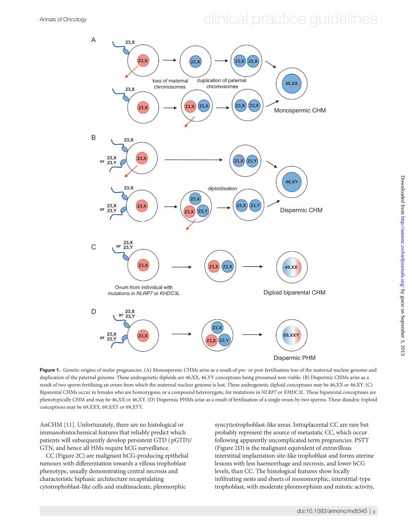
genetics/molecular biologyCHMs are usually diploid and androgenetic in origin, ∼80%resulting from duplication of the haploid genome of a single spermwhile 20% arise by dispermic fertilisation of an ovum (Figure 1Aand B). In either case maternal chromosomes are lost before, orshortly after, fertilisation. However, while nuclear DNA isentirely paternal in CHM, mitochondrial DNA remainsmaternal in origin [1].Recent evidence indicates that some patients with recurrent
CHM have diploid biparental CHM (BiCHM) rather than thetypical androgenetic CHM (AnCHM) (Figure 1C). In thesecases, the molar phenotype is due to an autosomal recessivecondition, familial recurrent HM (FRHM) that predisposeswomen to recurrent pregnancy loss, most usually CHM.Mutations in two genes have now been associated with thiscondition: NLRP7 and, more rarely, KHDC3L. While women
with recurrent AnCHM are likely to have normal live births insubsequent pregnancies and benefit from conventional in vitrofertilisation, women with FRHM are unlikely to achieve anormal pregnancy except through ovum donation from anunaffected individual [7].PHMs are almost always triploid, usually as a result of
fertilisation of an apparently normal ovum by two sperm oroccasionally a diploid sperm (Figure 1D). The existence ofdiploid PHM is unlikely, most reported cases representingmisdiagnosed complete moles, hydropic abortions or twinpregnancies.While most molar pregnancies are diploid CHM or triploid
PHM, numerical and structural abnormalities have beenreported in both CHM and PHM. In addition, CHM, andoccasionally PHM, can be associated with a twin pregnancywith a coexistent normal twin [8]. The continuance of such twinpregnancies results in healthy babies in ∼40% of cases, withoutan obvious increase in the risk of malignant change [8].Since post-molar GTN is treated on a clinical, rather than
pathological, diagnosis tumour tissue is rarely available forgenetic analysis. However, where tissue is available from GTN,the genotype will reflect that of the causative pregnancy, havingboth maternal and paternal chromosomes if the tumouroriginated in a term pregnancy, hydropic abortion or PHM butonly paternal genes if the causative pregnancy was a CHM.Since the interval from the causative pregnancy to the time ofGTN diagnosis carries prognostic information, genotyping canbe helpful particularly in patients with multiple pregnancies [1].Genetics can also be important in the differential diagnosisbetween gestational and non-gestational tumours, such as lungand gastric cancers, that can occasionally present as CC, but willhave a genotype reflecting that of the patient [9]. These non-gestational CC often initially respond to GTN-based therapies,but their outcome is invariably poor, reflecting the originatingtissue [1].
pathologyAll forms of GTD are derived from components of the normalhuman placenta; HM plus CC, and PSTT/ETT, representingabnormal counterparts of the villous and extravillous(interstitial) trophoblast, respectively. Most CHM and PHMhave distinctive morphological characteristics, but it isrecommended that cases of suspected GTD be reported byspecialist histopathologists. CHMs show a characteristic villousarchitecture, associated with abnormal trophoblast hyperplasia,stromal hypercellularity, stromal karyorrhectic debris andcollapsed villous blood vessels (Figure 2A). In contrast, PHMsshow patchy villous hydropic change with scattered abnormallyshaped irregular villi with trophoblastic pseudoinclusions andpatchy trophoblast hyperplasia (Figure 2B) [10]. Themorphological distinction between non-molar miscarriage,especially when associated with chromosomal abnormality, andPHM can sometimes be difficult, and ancillary techniques maybe required including immunostaining with p57KIP2 (negative inCHM), ploidy analysis by in situ hybridisation or flowcytometry or molecular genotyping. Genotyping can also beuseful in the identification of BiCHM, associated with FRHM,since most are pathologically indistinguishable from typical
clinical practice guidelines Annals of Oncology
| Seckl et al.
by guest on September 3, 2013
http://annonc.oxfordjournals.org/D
ownloaded from
AnCHM [11]. Unfortunately, there are no histological orimmunohistochemical features that reliably predict whichpatients will subsequently develop persistent GTD (pGTD)/GTN, and hence all HMs require hCG surveillance.CC (Figure 2C) are malignant hCG-producing epithelial
tumours with differentiation towards a villous trophoblastphenotype, usually demonstrating central necrosis andcharacteristic biphasic architecture recapitulatingcytotrophoblast-like cells and multinucleate, pleomorphic
syncytiotrophoblast-like areas. Intraplacental CC are rare butprobably represent the source of metastatic CC, which occurfollowing apparently uncomplicated term pregnancies. PSTT(Figure 2D) is the malignant equivalent of extravillousinterstitial implantation site-like trophoblast and forms uterinelesions with less haemorrhage and necrosis, and lower hCGlevels, than CC. The histological features show locallyinfiltrating nests and sheets of monomorphic, interstitial-typetrophoblast, with moderate pleomorphism and mitotic activity,
Figure 1. Genetic origins of molar pregnancies. (A) Monospermic CHMs arise as a result of pre- or post-fertilisation loss of the maternal nuclear genome andduplication of the paternal genome. These androgenetic diploids are 46,XX, 46,YY conceptuses being presumed non-viable. (B) Dispermic CHMs arise as aresult of two sperm fertilising an ovum from which the maternal nuclear genome is lost. These androgenetic diploid conceptuses may be 46,XX or 46,XY. (C)Biparental CHMs occur in females who are homozygous, or a compound heterozygote, for mutations in NLRP7 or KHDC3L. These biparental conceptuses arephenotypically CHM and may be 46,XX or 46,XY. (D) Dispermic PHMs arise as a result of fertilisation of a single ovum by two sperms. These diandric triploidconceptions may be 69,XXX, 69,XXY or 69,XYY.
Annals of Oncology clinical practice guidelines
doi:10.1093/annonc/mdt345 |
by guest on September 3, 2013
http://annonc.oxfordjournals.org/D
ownloaded from

and expression of human placental lactogen (hPL) and otherextravillous trophoblast markers. A specific variant of PSST withdistinctive hyalinisation and a slightly differentimmunohistochemical profile has been reported, ETT which isclinically thought to behave like PSTT [12].
staging and risk assessment
indications for treatmentFollowing suction curretage of a PHM, patients should haveanti-Rhesus D prophylaxis. After any HM, the onset ofmalignant change, referred to as pGTD or post-mole GTN, isnearly always indicated by a plateaued or rising hCG (Table 1).In the UK, this occurs after 15% and 0.5%–1% of CHM andPHM, respectively [1]. In other countries, these rates may behigher, possibly reflecting differences in hCG assays, hCGcriteria for the diagnosis of GTN, lack of whole populationdemographics or, less likely, a genuine difference in diseasebiology. The precise hCG surveillance protocol varies bycountry, but principles are similar. In the UK, serum and urinehCG is measured two weekly until normal and then monthly inurine [1]. The durations of monitoring once the hCG is normalalso vary between countries, reflecting uncertainty around theimportance of a very low risk of disease recurrence once thehCG is normal. Women completing the UK scheme have anestimated 1:2000 chance of missed disease [13], but the risk isalready very low with the first normal hCG value even for CHM.The UK indications for commencing chemotherapy are listed inTable 1 and are broadly similar to those of the InternationalFederation of Gynecology and Obstetrics (FIGO) [14]. The
commonest is a plateaued or rising hCG, but others include atissue diagnosis of CC and spread to other organs. However, ourUK experience indicates that the disease is also unlikely tospontaneously remit if the hCG is >20 000 IU/l 1 month afterHM evacuation (also associated with an increased risk of uterineperforation) or there are lung or vaginal metastasis of >2 cm(smaller lesions may spontaneously regress) [1]. In addition, inthe UK, chemotherapy is started to help stop heavy bleedingthat requires transfusion even if the hCG is falling. Interestingly,recent data have overturned the previous UK and FIGOguidance that women who continue to have a falling hCG 6months after uterine evacuation automatically needchemotherapy. Indeed, the hCG spontaneously normalised in
Figure 2. Photomicrographs demonstrating the various histopathological forms of GTD. (A) Complete hydatidiform mole, (B) partial hydatidiform mole, (C)choriocarcinoma and (D) placental site trophoblastic tumour. All are characterised by abnormal forms of trophoblast proliferation, associated with dysmorphicchorionic villi in CHM and PHM, but no villi and abnormal trophoblast invasion in CC and PSTT. (Original magnifications ×40, ×20, ×200 and ×100,respectively.)
Table 1. UK indications for chemotherapy following the diagnosis of GTD
Indications for chemotherapyPlateaued or rising hCG after evacuationa
Heavy vaginal bleeding or evidence of gastrointestinal or intraperitonealhaemorrhageHistological evidence of choriocarcinomaEvidence of metastases in the brain, liver or gastrointestinal tract, orradiological opacities of >2 cm on chest X-raySerum hCG of ≥20 000 IU/l >4 weeks after evacuation, because of the riskof uterine perforationRaised hCG 6 months after evacuation even if still falling (now omitted[15])
aPlateaued or rising is defined as four or more equivalent values of hCGover at least 3 weeks (days 1, 7, 14 and 21) and two consecutive rises in hCGof 10% or greater over at least 2 weeks (days 1, 7 and 14), respectively.
clinical practice guidelines Annals of Oncology
| Seckl et al.
by guest on September 3, 2013
http://annonc.oxfordjournals.org/D
ownloaded from

all such individuals left on surveillance [15]. Thus, thisindication for chemotherapy has now been removed from UKguidelines.
staging investigations and treatment stratificationafter a molar pregnancyMost patients developing GTN post-HM are detected early viahCG monitoring and so extensive investigation is rarelyrequired. Information to determine therapy can be obtainedfrom the clinical history, examination, measurement of serumhCG and a Doppler pelvic ultrasound to confirm the absence ofa pregnancy, to measure the uterine size/volume, spread ofdisease within the pelvis and its vascularity (Figure 3). The latterassessed by the Doppler pulsatility index is an independentprognostic factor for resistance to single-agent methotrexate
(MTX) therapy [16] and is now being evaluated in a prospectivetrial. Pulmonary metastases are most common, so a chestradiograph is essential [17]. Computed tomography (CT) of thechest is not required if the chest X-ray (CXR) findings arenormal, since discovery of micrometastases, which may be seenin ∼40% of patients, does not influence outcome [18]. However,if lesions are noted on CXR, magnetic resonance imaging (MRI)of the brain and CT body are indicated (Figure 4) to excludemore widespread disease involving, for example, the brain orliver, which would significantly alter management.FIGO reports data on GTN using prognostic scoring and
anatomic staging systems (Table 2) [19]. Since 2002, allphysicians treating GTN should use this system to enable thecomparison of data. The prognostic score predicts the potentialfor developing resistance to single-drug chemotherapy withMTX or actinomycin D (ActD). A score of 0–6 and ≥7 indicates
Figure 3. Pelvic Doppler ultrasonography of persisting GTN following a HM. (A) Pre-chemotherapy. (B) Post-chemotherapy. (Reprinted from ref. [1],Copyright 2010, with permission from Elsevier.)
Figure 4. Algorithm of imaging investigations for patients with GTN following a HM on hCG surveillance (left-hand panel) or after any other type ofpregnancy (right-hand panel). USS, ultrasound scan; CT, computerised tomography; MRI, magnetic resonance imaging; PET, positron emission tomography;−ve, negative; +ve, positive; mets, metastases.
Annals of Oncology clinical practice guidelines
doi:10.1093/annonc/mdt345 |
by guest on September 3, 2013
http://annonc.oxfordjournals.org/D
ownloaded from

a low and high risk of resistance, respectively. The latter hasalmost no chance of being cured with single-drug therapy andrequires multi-agent treatment. The anatomical staging does nothelp with determining therapy, but provides additionalinformation to help clinicians who compare results betweencentres. The variables that are assessed in the prognostic scoreinclude: (i) tumour volume (hCG level, size of metastases andnumber of metastases), (ii) site of involvement, (iii) priorchemotherapy resistance and (iv) duration of disease fromantecedent pregnancy (Table 2) [19].
staging investigations for CC and PSTT/ETTWomen who present with an elevated hCG and suspected GTN(CC or PSTT/ETT) following a prior pregnancy require muchmore extensive staging investigations, which include a contrastenhanced CT of the chest and abdomen, MRI of the brain andpelvis, a Doppler ultrasound of the pelvis and may benefit froma lumbar puncture to assess the cerebrospinal fluid to serumhCG ratio. The latter if more than 1:60 suggests occult centralnervous system disease [1]. In addition, where there is doubtover the clinical diagnosis, tissue should be obtained and geneticanalysis undertaken to confirm the gestational origin of thetumour through the presence of paternal genes. For CC, theFIGO scoring/staging system is the same as described above.However, PSTT/ETT has a discrete biological behaviour withless hCG production, slower growth, late metastasis and slightlyless chemosensitivity. Consequently, the scoring system is notvalid for PSTT/ETT, but FIGO staging is used to help adapttreatment intensity (see below). Some investigators haverecently started using positron emission tomography (PET)/CTimaging, but experience is still quite limited. It appears that thisimaging modality is more helpful in relapsed disease to identifysites for resection and, as with other cancers, is prone to bothfalse-positive and false-negative results [1].
management of low-risk diseaseAbout 95% of patients with HM who develop GTN are low risk(score 0–6). In women with stage I disease apparently confinedto the uterine cavity, the role of second D&C in reducing the
need for chemotherapy remains controversial. UK resultsindicate that this procedure is only valuable if the hCG is <5000IU/l with disease in the cavity rather than myometrium. Indeed,the low efficacy of a second D&C, small risks of introducinginfection, causing haemorrhage and uterine perforation shouldbe balanced against the almost 100% cure rate and relative safetyof chemotherapy (reviewed in [1]). Sometimes patients withstage I GTN who have completed their families requesthysterectomy, which, although possible, may not completelyobviate the need for chemotherapy.Consequently, for nearly all low-risk GTN patients, single-
agent chemotherapy with either MTX or ActD is the preferredtreatment. A variety of regimens have been developed, which innon-randomised, mostly retrospective, studies demonstrate a50%–90% chance of inducing remission [20]. This variabilityreflects differences in dose, frequency and route ofadministration as well as criteria used to select patients fortherapy [17]. Some investigators have argued that more intensetherapies given daily over 5–8 days every 2 weeks are superior totreatments given once every 2 weeks [21]. Others have suggestedthat ActD is more likely to induce remission than MTX. Thefew randomised studies to address some of these issues [22]have been underpowered and compared regimens that are notfrequently used internationally [20]. Consequently, a new largerinternational randomised trial has recently commencedcomparing the more commonly used MTX regimens in Europe/many parts of the world (Table 3) and some centres elsewhere[MTX 0.4 mg/kg (maximum 25 mg) IV d1–5 every 2 weeks][23] with ActD 1.25 mg/m2 IV every 2 weeks. Importantly,patients failing first-line therapy, usually because of resistance,can be easily salvaged with second and occasionally third-linechemotherapy so that the overall survival (OS) is ∼100%[23–25]. As survival is so high, it seems sensible to start withthe least toxic therapy first to minimise the exposure of patientsto more harmful treatments.The MTX with folinic acid rescue (MTX/FA) regimen
developed at Charing Cross Hospital (Table 3) is effective, well-tolerated and unlike ActD, does not induce hair loss, so MTX/FA has been widely adopted [24]. After a short stay in hospitalto monitor for bleeding complications, most of the patients can
Table 2. FIGO 2000 scoring system for GTN
Prognostic factor Score
0 1 2 4
Age (years) <40 ≥40 – –
Antecedent pregnancy (AP) Mole Abortion Term –
Interval (end of AP to chemotherapy in months) <4 4–6 7–12 >12hCG (IU/l) <103 103–104 104–105 >105
Number of metastases 0 1–4 5–8 >8Site of metastases Lung Spleen and kidney GI tract Brain and liverLargest tumour mass – 3–5 cm >5 cmPrior chemotherapy – – Single drug >2 drugs
The total score for a patient is obtained by adding the individual scores for each prognostic factor. Low risk, 0–6; high risk, ≥7. PSTT should not be scored andinstead requires staging. Stage I, disease confined to the uterus; stage II, disease extending into the pelvis; stage III, disease spread to lungs and/or vagina; stageIV, all other metastatic sites including liver, kidney, spleen and brain. (Reprinted [19] Copyright 2002, with permission from Elsevier for the InternationalFederation of Gynecology and Obstetrics.)
clinical practice guidelines Annals of Oncology
| Seckl et al.
by guest on September 3, 2013
http://annonc.oxfordjournals.org/D
ownloaded from

be treated at home, with their general practitioner, or in theirnearest hospital depending on local health servicearrangements. About 2% of women suffer mouth ulcers, soreeyes or rarely pleuritic or peritoneal pains from serositis [24].During chemotherapy, the hCG should ideally be measured atleast once per week, so that at least two samples with a plateauor rise are available to enable an early decision regarding theonset of resistance indicating a need for a change in therapy. Inthose developing resistance to MTX/FA, a switch to ActD orcombination agent chemotherapy depending on whether thehCG was <300 or >300 IU/l, respectively, will cure nearly allremaining women [25]. Chemotherapy should be continueduntil the hCG is normal and then for a further 6 weeks(Figure 5). The latter helps to eliminate any residual tumourcells and to minimise the chances of relapse [26]. Indeed, non-randomised data suggest that reducing the consolidationtherapy by just one cycle doubles the risk of relapse [26]. In viewof these data, the Dutch have recently moved from giving two tonow using three consolidation cycles. Only 30% of patientsscoring 5–6 can expect to be cured with low-risk therapy [1].Consequently, it would be helpful to refine the FIGO scoringsystem, so that the 70% of women in this group who developMTX/FA resistance could be identified initially for moreintensive therapy. It is possible that the vascularity seen on
Doppler ultrasound may help [16]. Moreover, recent dataindicate that women in this category with an hCG of >400 000IU/l are unlikely to be cured by MTX/FA and so multi-agenttreatment should be given from the outset [27]. Otherpromising strategies to identify patients with drug resistance atan early time-point during initial therapy have employednormograms and hCG kinetic analyses [28, 29].
management of high-risk GTNPatients scoring of ≥7 (Table 2) are at high risk of developingdrug resistance and so are very unlikely to be cured with single-agent chemotherapy. Consequently, several different multi-agent therapies have been developed including: MTX, FA andActD (MFA); MTX, ActD, cyclophosphamide, doxorubicin,melphalan, hydroxyurea and vincristine (CHAMOCA); MTX,ActD and cyclophosphamide (MAC); etoposide, MTX andActD (EMA) and others [30]. At Charing Cross Hospital, aftermany years of progressive experience, a regimen was developedconsisting of EMA alternating weekly with cyclophosphamideand vincristine (CO; see Table 4). This has been widely adoptedworldwide [30], because it is effective with predictable and easilymanaged short-term toxicity. Indeed, a retrospectivecomparison from the Korean GTD centre’s experience of MFA,MAC, CHAMOCAwith EMA-CO demonstrated a remissionrate of 63.3% (31 of 49), 67.5% (27 of 40), 76.2% (32 of 45) and90.6% (87 of 96), respectively [31]. The EMA/CO regimenrequires one overnight stay every 2 weeks and causes reversiblealopecia. It is myelosuppressive but granulocyte colonystimulating factor (G-CSF) support helps to maintainneutrophil count, treatment intensity and avoid neutropenicfebrile episodes [1].Five-year OS of patients treated with this schedule has been
reported to vary between 75% and 90% [31–33]. In the 272cases at Charing Cross Hospital treated between 1980 and 1994,
Table 3. Methotrexate and folinic acid chemotherapy regimen for low-riskpatients
Methotrexate (MTX) 50 mg by intramuscular injection repeated every48 h for a total of four doses
Calcium folinate(folinic acid)
15 mg orally 30 h after each injection of MTX
Courses repeated every 2 weeks, i.e. days 1, 15, 29, etc.
Figure 5. HCG tumour marker treatment graph demonstrating a patient responding to low-risk chemotherapy. Following uterine evacuation of a CHM, thehCG remained plateaued indicating persisting GTD/GTN, so the patient was commenced on methotrexate and folinic acid (MTX/FA). Therapy was continuedfor 6 weeks after the hCG was normal (<5 IU/l) as indicated. (Reprinted from ref. [1], Copyright 2010, with permission from Elsevier.)
Annals of Oncology clinical practice guidelines
doi:10.1093/annonc/mdt345 |
by guest on September 3, 2013
http://annonc.oxfordjournals.org/D
ownloaded from

OS was 86.2% [95% confidence interval (CI) 81.9% to 90.5%][32]. While these results were good, the presence of liver orbrain metastases correlated with only 27% or 70% long-termsurvival, respectively, and was just 10% with both liver and brainmetastases (reviewed in [1]). Most of the patients with adverseoutcomes did not have a prior HM, were not registered for hCGfollow-up and consequently presented with extensive disease.This was associated with death from haemorrhage or metaboliccomplications of overwhelming disease within 4 weeks ofadmission and/or before adequate chemotherapy could begiven. If such patients are excluded, survival of patients withbrain metastasis is similar to other patients [34]. The situationwith liver metastasis may be similar; of 37 patients with livermetastasis treated between 1977 and 2005 at Charing CrossHospital, OS had increased to ∼50% at 5 years but if earlydeaths were excluded, survival was nearly 70% [35]. In additionto disease extent, other factors associated with poor outcomeinclude the type of, and duration from, the antecedentpregnancy and the prior use of chemotherapy (reviewed in [1]).To reduce early deaths in patients with very advanced disease,
we have found that commencing chemotherapy gently withlow-dose etoposide 100 mg/m2 and cisplatin 20 mg/m2 on days1 and 2 repeated weekly for 1–3 weeks has virtually eliminatedthis problem. Indeed, low-dose induction etoposide andcisplatin combined with genetic testing to exclude non-gestational CC has helped to improve long-term OS data to over94% in high-risk patients [36]. Further details on themanagement and modifications of treatment required for theseand other challenging clinical situations such as brainmetastasis and pulmonary failure are beyond the scope of thepresent review, but are contained within the followingreferences [34, 37].
Similar to low-risk disease, therapy is continued for 6 weeksof normal hCG values or 8 weeks if poor prognostic featuressuch as liver or brain metastases are present [1]. Patients arethen re-imaged to document the post-treatment appearance forfuture comparison. Removal of residual masses is unnecessaryas it does not reduce the risk of recurrence which is less than∼3% [1].
management of drug-resistant diseaseAbout 20% of high-risk GTN patients will progress on or afterprimary chemotherapy, but these individuals still have anexcellent outcome with ∼75%–80% still being salvaged [36].This is partly because relapse is detected early due to hCGmonitoring so disease volume is small. Moreover, hCGmonitoring enables the early detection of resistance duringtherapy, which could potentially be more rapidly detectedthrough the use of normograms and kinetic models [28, 29, 38].In relapsed patients, fluorine-18 fluorodeoxyglucose-PET(FDG-PET) scanning may help identify the site of active diseaseto facilitate surgical resection and cure [39]. The T1/2 for hCG is≤48 h after surgery if all the disease has been removed [1].However, if surgery is not possible or the hCG fallsinappropriately, several salvage regimens have been eithercreated or adopted from the germ cell tumour setting [40]. AtCharing Cross Hospital, we developed a regimen combiningetoposide with cisplatin (EP) alternating weekly with EMA thatomitted the second day of etoposide and ActD [41]. Survivalrates are >80% but toxicity is significant [41], and less toxicsalvage therapies are required. Several cases of drug-resistantGTN have been reported to respond and/or be cured bypaclitaxel-based single-agent or combination therapy [42–45],gemcitabine and capecitabine [46, 47]. Of these, an alternatingtwo weekly doublet of paclitaxel/cisplatin and paclitaxel/etoposide (TP/TE; Table 5) appears from non-randomised datato be much better tolerated than EP/EMA and is effective inpatients with relapsed and/or refractory GTN [45]. In view of
Table 4. EMA/CO chemotherapy regimen for high-risk patients
EMADay 1Etoposide 100 mg/m2 by i.v. infusion over
30 minActinomycin D 0.5 mg i.v. bolusMethotrexate 300 mg/m2 by i.v. infusion over 12 h
Day 2Etoposide 100 mg/m2 by i.v. infusion over
30 minActinomycin D 0.5 mg i.v. bolusFolinic acid rescue (starting24 h after commencing the
methotrexate infusion)
15 mg i.v. or orally every 12 h for fourdoses
CODay 8Vincristine 1 mg/m2 i.v. bolus (maximum 2 mg)Cyclophosphamide 600 mg/m2 i.v. infusion over 30 min
EMA alternates with CO every week. To avoid extended intervals betweencourses caused by myelosuppression, it may occasionally be necessary toreduce the EMA by omitting the day 2 doses of etoposide and actinomycinD. i.v., intravenous. (Reprinted from ref. [1], Copyright 2010, withpermission from Elsevier.)
Table 5. TP/TE schedule for relapsed GTN
Regimen Schedule
Day 1Dexamethasone 20 mg oral (12 h pre-paclitaxel)Dexamethasone 20 mg oral (6 h pre-paclitaxel)Cimetidine 30 mg in 100 ml NS over 30 min i.v.Chlorphenamine 10 mg bolus i.v.Paclitaxel 135 mg/m2 in 250 ml NS over 3 h i.v.Mannitol 10% in 500 ml over 1 h i.v.Cisplatin 60 mg/m2 in 1 l NS over 3 h i.v.
Post-hydration 1 l NS + KCl 20 mmol + 1 g MgSO4 over 2 h i.v.Day 15Dexamethasone 20 mg oral (12 h pre-paclitaxel)Dexamethasone 20 mg oral (6 h pre-paclitaxel)Cimetidine 30 mg in 100 ml NS over 30 min i.v.Chlorphenamine 10 mg bolus i.v.Paclitaxel 135 mg/m2 in 250 ml NS over 3 h i.v.Etoposide 150 mg/m2 in 1 l NS over 1 h i.v.
NS, normal saline; i.v., intravenous. (Reprinted from ref. [1], Copyright2010, with permission from Elsevier.)
clinical practice guidelines Annals of Oncology
| Seckl et al.
by guest on September 3, 2013
http://annonc.oxfordjournals.org/D
ownloaded from

these results, the International Society of the Study ofTrophoblastic Diseases (ISSTD) has recently proposed arandomised trial of TE/TP versus EP/EMA to determine theoptimal therapy for patients relapsing after non-cisplatin/paclitaxel-based combination therapies such as EMA/CO.Another approach in patients with refractory disease involves
high-dose chemotherapy with peripheral stem-celltransplantation. However, cures are not common [48], soimproved patient selection may be required to achieve betteroutcomes from this approach.
management of PSTT and ETTPSTT differs from CC, growing more slowly, metastasising later,involving lymph nodes more commonly and producing lesshCG [1]. However, like CC, it can arise after any type ofpregnancy, including PHM, [49] and usually presents withabnormal vaginal bleeding [2]. PSTT may be suspected if thehCG level is low for the volume of disease present on imagingcombined with an elevated free beta form of hCG, but none ofthese features are diagnostic [50, 51]. Consequently histologicalconfirmation is essential.A recent large population-based series of PSTT comprised 62
cases over 30 years, representing 0.2% of UK GTD cases, andexamined prognostic features [2]. On univariate analysis, stage,hCG, mitotic index and a duration of >4 years from thepreceding pregnancy were prognostic, but the FIGO score wasunhelpful. Only the duration from the prior pregnancyremained predictive of survival on multivariate analysis with100% (13 of 13) dying and 98% (48 of 49) surviving for those≥48 and <48 months, respectively. This effect was not explainedby differences in disease stage or hCG levels, but may reflect abiological switch in the tumours after this time [2].The management of PSTT differs from CC. Patients with
metastatic disease require combination chemotherapy with, forexample, EP/EMA continued for 8 weeks of normal hCG levels[2]. Unlike CC, residual masses are removed surgically, includingthe uterus, as this can harbour microscopic disease. This maycause difficulties in the management of stage I disease [52]. Thesafest option is hysterectomy with pelvic lymph node samplingand ovarian conservation unless there is a family history ofovarian cancer or the patient is post-menopausal. In the absenceof sufficient data regarding adjuvant therapy, we currentlyadvocate 8 weeks of EP/EMA or TE/TP when there are poor riskfactors such as disease presenting beyond 4 years of the antecedentpregnancy. Indeed, in the latter group, a case can bemade forincluding high-dose chemotherapy. However, in youngernulliparous women, there is often a strong desire to preservefertility particularly when there appears to be a focal abnormalityin the uterus. While uterine-sparing surgery is possible [1],multifocal microscopic uterine disease can occur [52], whichcould compromise survival and careful counselling is required.Currently, it is thought that ETT behaves very similarly to
PSTT but in reality, little data are available to be sure of this.PSTT and ETT are so rare that it is unlikely that their treatmentwill ever be fully optimised, so that the ISSTD has now launchedan international PSTT/ETT database to pool cases [53].
personalised medicineGTN is one of the rare examples of a group of related cancerswhere novel molecularly targeted agents have not beenemployed, as cure has been achieved through the use ofconventional chemotherapeutic agents. This is because GTN areexquisitely sensitive to these drugs and the serial measurementof hCG, a highly sensitive biomarker of the disease [1], hasenabled early recognition of resistance, so that second- andthird-line therapies can be commenced before significanttumour re-growth has occurred. Very rarely, multi-drug-resistant disease develops that is not amenable to surgicalresection or any other existing treatment, so it is unclearwhether anything can be done in this case. Since GTN is veryvascular it is plausible that vascular targeting agents such asbevacizumab might be active. The tumours can also over-express epidermal growth factor receptor, leading to thequestion whether erlotinib or gefitinib could demonstrateefficacy. Anecdotally, thus far, we have not seen any benefitfrom these agents in several multi-drug-resistant patients. Thepotential for an anti-hCG targeted therapy has not beenexplored and could be of interest in women who havecompleted their families or have run out of other options.
follow-up and long-term implicationsThe risk of relapse after chemotherapy is ∼3% and most occurin the first year of follow-up. Therefore, careful hCG monitoringis required and pregnancy should ideally be delayed untilbeyond this period. Any method of contraception can be usedincluding the oral contraceptive pill, as long as there are noother contraindications to their use. In the UK, the hCG ismonitored weekly for 6 weeks post-chemotherapy, and then inserum and urine two weekly until 6 months, before switching tojust urine assessments, initially monthly, but eventuallydecreasing to just six monthly (Table 6). We continue this forlife as we are currently uncertain when it is safe to stopmonitoring and it enables us to collect long-term dataconcerning late effects of treatment including second cancers.
Table 6. UK follow-up protocol of GTN patients who have been treatedwith chemotherapy
Low-/high-risk post-chemotherapypatients, hCG concentrationsampling
Urine Blood
Year 1Week 1–6 after chemotherapy Weekly WeeklyMonth 2–6 Two weekly Two weeklyMonth 7–12 Two weekly –
Year 2 Four weekly –
Year 3 Eight weekly –
Year 4 Three monthly –
Year 5 Four monthly –
After Year 5 Six monthly –
Reprinted from ref. [1], Copyright 2010, with permission from Elsevier.
Annals of Oncology clinical practice guidelines
doi:10.1093/annonc/mdt345 |
by guest on September 3, 2013
http://annonc.oxfordjournals.org/D
ownloaded from

Fortunately, apart from EMA/CO bringing forward themenopause date by 3 years, fertility is not otherwise affectedwith 83% of women becoming pregnant after either MTX/FA orEMA/CO chemotherapy [1]. Moreover, there is no obviousincrease in the incidence of congenital malformations. When apatient does become pregnant, it is important to confirm byultrasound and other appropriate means that the pregnancy isnormal. Follow-up is then discontinued, but the hCG should berechecked at 6 and 10 weeks after the pregnancy to ensure norecurrence or new disease.Late sequelae from chemotherapy have been remarkably rare.
In 15 279 patient-years of follow-up, there was no significantincrease in the incidence of second tumours [54] followingMTX therapy. In contrast, 26 patients receiving combinationchemotherapy for GTN developed another cancer when theexpected rate was only 16.45, a significant difference [54]. Mostof this risk appears to occur if combination chemotherapy iscontinued beyond 6 months. Interestingly, new data in over30 000 patient-years of follow-up now show that, for EMA/CO,there is no overall increased risk of second cancers with a slightbut significant excess of leukaemias but reduction in othercancers including breast cancer risk (data submitted). Thisemphasises the continued importance of long-term monitoringof our treated patient populations.
summary of recommendationsRecommendations are largely based on non-randomisedretrospective cohort studies from single centres and/or nationalexperiences where the level of evidence (LOE) is IV. However,because of the measurable large benefit to patients, the grade ofrecommendation (GOR) is generally very high at A. LOE andGOR are given in brackets.
• Management of GTN is optimised by the centralisation ofcare, pathology review and hCG monitoring [IV, A].
• Women with singleton molar pregnancies should, in general,have these terminated by suction D&C [IV, A]. Second D&Cfor recurrence does not usually prevent the subsequent needfor chemotherapy and should only be attempted afterdiscussion with a GTD reference centre [IV, A].
• Anti-D prophylaxis is recommended following suction D&Cof PHM [IV, A].
• The FIGO scoring system should be used to determine therisk of GTN becoming resistant to single-agentchemotherapy, but is not of value in PSTT/ETT [IV, A].
• Patients with a FIGO score of 0–6 can be treated with eithersingle-agent MTX with or without FA, or ActD [II–IV, A]. Inmost European centres, MTX/FA (Table 3) is preferredbecause it is less toxic than MTX alone or single-agent ActD,and all patients can expect to be cured even if first-linetherapy fails [II–IV, A]. A randomised trial comparing themost frequently used MTX/FA and ActD regimens iscurrently underway.
• Chemotherapy for low-risk disease should be continued for6 weeks of maintenance treatment after hCG normalisation[IV, A].
• Patients with a FIGO score of ≥7 should receive multi-agentchemotherapy and most centres now use EMA/CO (Table 4),
as it is highly effective, simple to administer and relativelynon-toxic [IV, A].
• Patients with high-risk disease should have maintenancetherapy for 6 weeks extended to 8 weeks with poor prognosticfeatures such as liver with or without brain metastasis [IV, A].
• Early deaths in ultrahigh-risk GTN can be reduced byinduction of low-dose etoposide and cisplatin [IV, A]. Suchpatients may also benefit from substitution of EMA/CO withEP/EMA [IV, A].
• Residual lung or uterine masses following chemotherapy forlow-risk or high-risk diseases are not predictive of recurrenceand do not require surgical excision [IV, A].
• High-risk failures can be frequently salvaged with furtherchemotherapy and most centres use either EP/EMA or TE/TP(Table 5) [IV, A]. A randomised trial comparing theseregimens is being developed.
• Surgery alone can effectively salvage some patients withisolated foci of chemoresistant disease [IV, A].
• PSTT/ETT is managed according to its stage and risk factorsfor poor outcome, the most dominant of which is the intervalfrom last known pregnancy. Hysterectomy with pelvic lymphnode sampling is recommended for stage I disease presentingwithin 4 years of the last known pregnancy [IV, A]. Multi-agent chemotherapy with, for example, EP/EMA isrecommended for metastatic disease [IV, A]. Patientspresenting beyond 4 years may benefit from multi-agent andsubsequent high-dose chemotherapy [IV, B].
search strategy and selection criteriaAll authors performed a detailed review of published work andcontributed to the writing, review and editing of the manuscript.MJS had access to all the data used to write the report and hadfinal responsibility for submission. All authors saw andapproved the final version. Our search strategy was formulatedto identify any meta-analyses and previous systematic reviews inall aspects of GTD, in addition to all published cohort studies(and where appropriate, comparison groups) and case-controlstudies. We searched the Cochrane Library, Medline (viaPubMed, Internet Grateful Med, OVID and Knowledgefinder),with a combination of keywords including: ‘trophoblasticdisease’, ‘GTD’, ‘GTN’, ‘choriocarcinoma’, ‘molar pregnancy’,‘hydatidiform mole’, ‘placental site trophoblastic tumor’,‘genetics’, ‘epidemiology’, ‘pathology’, ‘treatment’,‘chemotherapy’, ‘methotrexate’, ‘actinomycin D’,‘dactinomycin’, ‘cisplatin’, ‘paclitaxel’, ‘high-dose’,‘management’, ‘risk factors’, ‘hCG’, ‘imaging’, ‘ultrasound’,‘PET’, ‘CT’, ‘MRI’, ‘prognosis’, and ‘staging’. The reference listsand bibliographies of all previous publications were scanned tofind any publications not already identified by our electronicsearch strategy.
noteLevels of evidence and grades of recommendation have beenapplied using the system shown in Table 7. Statements withoutgrading were considered justified standard clinical practice bythe experts and the ESMO faculty.
clinical practice guidelines Annals of Oncology
| Seckl et al.
by guest on September 3, 2013
http://annonc.oxfordjournals.org/D
ownloaded from

acknowledgementsMJS, NS and RAF wish to thank the Department of Health,National Commissioning Group and the Cancer Treatment andResearch Trust for their continued support. MJS alsoacknowledges support from the Imperial College ExperimentalCancer Medicine Centre and Biomedical Research Centregrants.
conflict of interestThe authors have declared no potential conflicts of interest.
references1. Seckl MJ, Sebire NJ, Berkowitz RS. Gestational trophoblastic disease. Lancet
2010; 376: 717–729.2. Schmid P, Nagai Y, Agarwal R et al. Prognostic markers and long-term outcome of
placental-site trophoblastic tumours: a retrospective observational study. Lancet2009; 374: 48–55.
3. Hou JL, Wan XR, Xiang Y et al. Changes of clinical features in hydatidiform mole:analysis of 113 cases. J Reprod Med 2008; 53: 629–633.
4. Fowler DJ, Lindsay I, Seckl MJ, Sebire NJ. Histomorphometric features ofhydatidiform moles in early pregnancy: relationship to detectability by ultrasoundexamination. Ultrasound Obstet Gynecol 2007; 29: 76–80.
5. Hinshaw K, Fayyad A, Munjuluri P. The management of early pregnancy loss. InGreen-top Guideline. London: Royal College of Obstetricians and Gynaecologists,2006.
6. Seckl MJ, Gillmore R, Foskett M et al. Routine terminations of pregnancy-shouldwe screen for gestational trophoblastic neoplasia. Lancet 2004; 364: 705–707.
7. Fisher RA, Lavery SA, Carby A et al. What a difference an egg makes. Lancet2011; 378: 1974.
8. Sebire NJ, Foskett M, Paradinas FJ et al. Outcome of twin pregnancies withcomplete hydatidiform mole and healthy co-twin. Lancet 2002; 359: 2165–2166.
9. Fisher RA, Savage PM, MacDermott C et al. The impact of molecular geneticdiagnosis on the management of women with hCG-producing malignancies.Gynecol Oncol 2007; 107: 413–419.
10. Sebire NJ, Seckl MJ. Immunohistochemical staining for diagnosis and prognosticassessment of hydatidiform moles: current evidence and future directions.J Reprod Med 2010; 55: 236–246.
11. Sebire NJ, Savage PM, Seckl MJ, Fisher RA. Histopathological features ofbiparental complete hydatidiform moles in women with NLRP7 mutations. Placenta2013; 34: 50–56.
12. Shih IM, Kurman RJ. Epithelioid trophoblastic tumor: a neoplasm distinct fromchoriocarcinoma and placental site trophoblastic tumor simulating carcinoma. AmJ Surg Pathol 1998; 22: 1393–1403.
13. Sebire NJ, Foskett M, Short D et al. Shortened duration of human chorionicgonadotrophin surveillance following complete or partial hydatidiform mole:evidence for revised protocol of a UK regional trophoblastic disease unit. BJOG2007; 114: 760–762.
14. Kohorn EI. Negotiating a staging and risk factor scoring system for gestationaltrophoblastic neoplasia. A progress report. J Reprod Med 2002; 47: 445–450.
15. Agarwal R, Teoh S, Short D et al. Chemotherapy and human chorionicgonadotropin concentrations 6 months after uterine evacuation of molarpregnancy: a retrospective cohort study. Lancet 2012; 379: 130–135.
16. Agarwal R, Harding V, Short D et al. Uterine artery pulsatility index: a predictor ofmethotrexate resistance in gestational trophoblastic neoplasia. Br J Cancer 2012;106: 1089–1094.
17. Berkowitz RS, Goldstein DP. Current management of gestational trophoblasticdiseases. Gynecol Oncol 2009; 112: 654–662.
18. Darby S, Jolley I, Pennington S, Hancock BW. Does chest CT matter in the stagingof GTN? Gynecol Oncol 2009; 112: 155–160.
19. FIGO Oncology Committee, FIGO staging for gestational trophoblastic neoplasia2000. International Journal of Gynecology & Obstetrics 77: 285–287.
20. Alazzam M, Tidy J, Hancock BW et al. First line chemotherapy in low riskgestational trophoblastic neoplasia. Cochrane Database Syst Rev 2009 Jan 21; (1):CD007102.
21. Kohorn EI. Is lack of response to single-agent chemotherapy in gestationaltrophoblastic disease associated with dose scheduling or chemotherapyresistance? Gynecol Oncol 2002; 85: 36–39.
22. Osborne RJ, Filiaci V, Schink JC et al. Phase III trial of weekly methotrexate orpulsed dactinomycin for low-risk gestational trophoblastic neoplasia: a gynecologiconcology group study. J Clin Oncol 2011; 29: 825–831.
23. Lurain JR, Chapman-Davis E, Hoekstra AV, Schink JC. Actinomycin D formethotrexate-failed low-risk gestational trophoblastic neoplasia. J Reprod Med2012; 57: 283–287.
24. McNeish IA, Strickland S, Holden L et al. Low risk persistent gestationaltrophoblastic disease: outcome after initial treatment with low-dose methotrexateand folinic acid, 1992 to 2000. J Clin Oncol 2002; 20: 1838–1844.
25. Sita-Lumsden A, Short D, Lindsay I et al. Treatment outcomes for 618 womenwith gestational trophoblastic tumours following a molar pregnancy at the CharingCross Hospital, 2000–2009. Br J Cancer 2012; 107: 1810–1814.
Table 7. Levels of evidence and grades of recommendation (adapted from the Infectious Diseases Society of America-United States Public Health ServiceGrading Systema)
Levels of evidence
I Evidence from at least one large randomised, controlled trial of good methodological quality (low potential for bias) or meta-analyses of well-conductedrandomised trials without heterogeneity
II Small randomised trials or large randomised trials with a suspicion of bias (lower methodological quality) or meta-analyses of such trials or of trials withdemonstrated heterogeneity
III Prospective cohort studiesIV Retrospective cohort studies or case–control studiesV Studies without control group, case reports, experts opinions
Grades of recommendation
A Strong evidence for efficacy with a substantial clinical benefit, strongly recommendedB Strong or moderate evidence for efficacy but with a limited clinical benefit, generally recommendedC Insufficient evidence for efficacy or benefit does not outweigh the risk or the disadvantages (adverse events, costs,…), optionalD Moderate evidence against efficacy or for adverse outcome, generally not recommendedE Strong evidence against efficacy or for adverse outcome, never recommended
aDykewicz CA. Summary of the guidelines for preventing opportunistic infections among hematopoietic stem cell transplant recipients. Clin Infect Dis 2001;33: 139–144. By permission of the Infectious Diseases Society of America.
Annals of Oncology clinical practice guidelines
doi:10.1093/annonc/mdt345 |
by guest on September 3, 2013
http://annonc.oxfordjournals.org/D
ownloaded from

26. Lybol C, Sweep FC, Harvey R et al. Relapse rates after two versus threeconsolidation courses of methotrexate in the treatment of low-risk gestationaltrophoblastic neoplasia. Gynecol Oncol 2012; 125: 576–579.
27. McGrath S, Short D, Harvey R et al. The management and outcome of women withpost-hydatidiform mole ‘low-risk’ gestational trophoblastic neoplasia, but hCGlevels in excess of 100 000 IU l(-1). Br J Cancer 2010; 102: 810–814.
28. van Trommel NE, Massuger LF, Schijf CP et al. Early identification of resistance tofirst-line single-agent methotrexate in patients with persistent trophoblasticdisease. J Clin Oncol 2006; 24: 52–58.
29. You B, Harvey R, Henin H et al. Early prediction of treatment resistance in low-riskgestational trophoblastic neoplasia using population kinetic modelling of hCGmeasurements. Br J Cancer 2013; 108: 1810–1816.
30. Deng L, Yan X, Zhang J et al. Combination chemotherapy for high-risk gestationaltrophoblastic tumour. Cochrane Database Syst Rev 2009 April 15; (2): CD005196.
31. Kim SJ, Bae SN, Kim JH et al. Effects of multiagent chemotherapy andindependent risk factors in the treatment of high-risk GTT—25 years experiencesof KRI-TRD. Int J Gynaecol Obstet 1998; 60 (Suppl 1): S85–S96.
32. Bower M, Newlands ES, Holden L et al. EMA/CO for high-risk gestationaltrophoblastic tumours: results from a cohort of 272 patients. J Clin Oncol 1997;15: 2636–2643.
33. Turan T, Karacay O, Tulunay G et al. Results with EMA/CO (etoposide,methotrexate, actinomycin D, cyclophosphamide, vincristine) chemotherapy ingestational trophoblastic neoplasia. Int J Gynecol Cancer 2006; 16: 1432–1438.
34. Newlands ES, Holden L, Seckl MJ et al. Management of brain metastases inpatients with high-risk gestational trophoblastic tumors. J Reprod Med 2002; 47:465–471.
35. Ahamed E, Short D, North B et al. Survival of women with gestational trophoblasticneoplasia and liver metastases: is it improving? J Reprod Med 2012; 57: 262–269.
36. Alifrangis C, Agarwal R, Short D et al. EMA/CO for high-risk gestationaltrophoblastic neoplasia: good outcomes with induction low-dose etoposide-cisplatin and genetic analysis. J Clin Oncol 2013; 31: 280–286.
37. Seckl MJ, Newlands ES. Investigation and treatment of patients with persistentgestational trophoblastic disease and gestational trophoblastic tumours/neoplasiain the United Kingdom. In: Hancock BW, Seckl MJ, Berkowitz RS, Cole LA (eds)Gestational Trophoblastic Disease, 3rd Edition, 2009; 335–365; ISSTD.org.ISSTD, London.
38. Lybol C, Westerdijk K, Sweep FC et al. Human chorionic gonadotropin (hCG)regression normograms for patients with high-risk gestational trophoblasticneoplasia treated with EMA/CO (etoposide, methotrexate, actinomycin D,cyclophosphamide and vincristine) chemotherapy. Ann Oncol 2012; 23:2903–2906.
39. Dhillon T, Palmieri C, Sebire NJ et al. Value of whole body 18FDG-PET to identifythe active site of gestational trophoblastic neoplasia. J Reprod Med 2006; 51:879–887.
40. Lurain JR, Nejad B. Secondary chemotherapy for high-risk gestationaltrophoblastic neoplasia. Gynecol Oncol 2005; 97: 618–623.
41. Newlands ES, Mulholland PJ, Holden L et al. Etoposide and cisplatin/etoposide,methotrexate, and actinomycin D (EMA) chemotherapy for patients with high-riskgestational trophoblastic tumors refractory to EMA/cyclophosphamide andvincristine chemotherapy and patients presenting with metastatic placental sitetrophoblastic tumors. J Clin Oncol 2000; 18: 854–859.
42. Jones WB, Schneider J, Shapiro F, Lewis JL, Jr. Treatment of resistant gestationalchoriocarcinoma with taxol: a report of two cases. Gynecol Oncol 1996; 61:126–130.
43. Osborne R, Covens A, Mirchandani D, Gerulath A. Successful salvage of relapsedhigh-risk gestational trophoblastic neoplasia patients using a novel paclitaxel-containing doublet. J Reprod Med 2004; 49: 655–661.
44. Termrungruanglert W, Kudelka AP, Piamsomboon S et al. Remission of refractorygestational trophoblastic disease with high-dose paclitaxel. Anticancer Drugs1996; 7: 503–506.
45. Wang J, Short D, Sebire NJ et al. Salvage chemotherapy of relapsed or high-riskgestational trophoblastic neoplasia (GTN) with paclitaxel/cisplatin alternating withpaclitaxel/etoposide (TP/TE). Ann Oncol 2008; 19:1578–1583.
46. Pandian Z, Seckl MJ, Smith R, Lees DA. Gestational choriocarcinoma: an unusualpresentation with response to gemcitabine and surgery. BJOG 2004; 111:382–384.
47. Bianconi M, Jankilevich G, Otero S et al. Successful salvage of a relapsed highrisk gestational trophoblastic neoplasia patient using capecitabine. Gynecol Oncol2007; 106: 268–271.
48. El-Helw LM, Seckl MJ, Haynes R et al. High-dose chemotherapy and peripheralblood stem cell support in refractory gestational trophoblastic neoplasia. Br JCancer 2005; 93: 620–621.
49. Palmieri C, Fisher RA, Sebire NJ et al. Placental site trophoblastic tumour arisingfrom a partial hydatidiform mole. Lancet 2005; 366: 688.
50. Cole LA, Khanlian SA, Muller CY et al. Gestational trophoblastic diseases: 3.Human chorionic gonadotropin-free beta-subunit, a reliable marker of placentalsite trophoblastic tumors. Gynecol Oncol 2006; 102: 160–164.
51. Harvey RA, Pursglove HD, Schmid P et al. Human chorionic gonadotropin freebeta-subunit measurement as a marker of placental site trophoblastic tumors. JReprod Med 2008; 53: 643–648.
52. Pfeffer PE, Sebire N, Lindsay I et al. Fertility-sparing partial hysterectomy forplacental-site trophoblastic tumour. Lancet Oncol 2007; 8: 744–746.
53. The ISSTD global Placental Site Trophoblastic Tumour database. InternationalSociety of the Study of Trophoblastic Diseases. https://pstt.shef.ac.uk.
54. Rustin GJ, Newlands ES, Lutz JM et al. Combination but not single agentmethotrexate chemotherapy for gestational trophoblastic tumours increases theincidence of second tumours. J Clin Oncol 1996; 14: 2769–2773.
clinical practice guidelines Annals of Oncology
| Seckl et al.
by guest on September 3, 2013
http://annonc.oxfordjournals.org/D
ownloaded from