Embed Size (px)
Citation preview
Incidence of optic canal fracture in the traumaticoptic neuropathy and its effect on the visualoutcomeWentao Yan,2 Yingbai Chen,3 Zhenbin Qian,2 Dinesh Selva,4 Daniel Pelaez,5
Yunhai Tu,2 Wencan Wu1
1Department of Orbital andOculoplastic Surgery, EyeHospital of Wenzhou MedicalUniversity, Wenzhou, Zhejiang,PR China2Department of Orbital andOculoplastic Surgery, EyeHospital of Wenzhou MedicalUniversity, Wenzhou, PR China3Department ofOphthalmology, Shanghai JiaoTong University School ofMedicine Affiliated RenjiHospital, Shanghai, China4Discipline of Ophthalmologyand Visual Sciences, Universityof Adelaide and SouthAustralian Institute ofOphthalmology, Adelaide,Australia5Bascom Palmer Eye Institute,University of Miami MillerSchool of Medicine, Miami,Florida, USA
Correspondence toProfessor Wencan Wu,Department of Orbital andOculoplastic Surgery, EyeHospital of Wenzhou MedicalUniversity, No. 270 Xueyuan XiRoad, Wenzhou, Zhejiang325027, P. R. China;[email protected]
WY and YC are co-first authorsand contributed equally.
Received 28 October 2015Revised 29 April 2016Accepted 12 May 2016Published Online First6 June 2016
To cite: Yan W, Chen Y,Qian Z, et al. Br JOphthalmol 2017;101:261–267.
ABSTRACTObjective To correlate the intraoperative endoscopicfindings with high-resolution CT (HRCT) for the diagnosisof optic canal fractures (OCF). To compare the visualoutcome of patients with different types of OCF andwithout.Design A retrospective, comparative case series.Participants 1275 consecutive patients (1275 eyes)with indirect traumatic optic neuropathy (TON).Methods Altogether, 1275 patients who underwentendoscopic transethmoid optic canal decompression(ETOCD) for unilateral indirect TON were reviewed from1 October 2006 to 30 September 2014. HRCTperformed prior to surgery were compared with findingsduring surgery. The visual outcomes before and aftersurgery were also compared.Main outcome measures The presence and type ofOCF detected by HRCT and during surgery; visual acuityprior to surgery and at 3 months after surgery.Results A total of 1275 patients (1275 eyes) wereincluded, with 708 patients that had OCF visible onHRCT image. During surgery, an additional 187 (20.9%)patients with OCF were noticed. Among these, 136 hadundisplaced fractures, most of which were linearintracanalicular fractures. The initial visual acuity ofpatients with OCF was worse than that of patientswithout OCF (p<0.01). However, no statistical differenceexisted in the final visual acuity at 3 months aftersurgery (>0.05). Significant statistical difference ofsurgical efficacy existed between all the patients withOCF and without OCF (p<0.001).Conclusions Among patients with OCF, 20.9% werenot detected by HRCT. Patients with OCF had a worseinitial visual acuity than those without OCF. There wasno significant difference in final visual acuity afterETOCD.
INTRODUCTIONThe most common form of traumatic optic neur-opathy (TON) is indirect and results from concus-sive force to the head, especially the forehead. Thisimpact is thought to transmit a shock wave to theoptic canal, damaging the optic nerve.1–3
Therefore, the surrounding soft tissue and bonyinjuries of the optic canal are believed to be helpfulto indirectly assess the degree of impact force onthe optic nerve and may be used as a prognosis pre-dictor of TON.4–12 There is inadequate evidencethat the presence of optic canal fractures (OCF) issufficient to shift the risk/benefit equation in favourof canal decompression, but early canal decompres-sion is generally recommended when there is a
displaced fragment impinging on the opticnerve.3 11 13–15
The role of high-resolution CT (HRCT) intesting TON has been well established and is gener-ally considered essential for detecting possiblecontributors to optic nerve injury, including retro-bulbar haemorrhage, optic nerve oedema, intraor-bital emphysema and OCF.4–11 13 However, theactual incidence, position and orientation of OCFin indirect TON are uncertain. The reported inci-dence of OCF on CT scans varies considerablyfrom 5% to 92%.1 7 16 Based on direct endoscopicobservation at the time of surgery in a few previouscase series, OCF may exist even if it cannot bedetected radiologically.14–15 17–19
The effect of OCF on visual outcome is debat-able, with a number of papers reporting orbitalfractures as a poor predictor.20–23 Here, wecompare the results of HRCT scans prior tosurgery with the findings during surgery in a largegroup of patients with indirect TON. Our intentionwas to verify the reliability of HRCT in OCF diag-nosis. Meanwhile, we reported the visual outcomeof patients with different types of OCF.
MATERIALS AND METHODSPatient selectionAll patients were admitted and underwent endo-scopic transethmoid optic canal decompression(ETOCD) due to unilateral indirect TON from 1October 2006 to 30 September 2014. They wereidentified from the hospital clinical databases at theEye Hospital of Wenzhou Medical University, theLishui Central Hospital, the Jinhua CentralHospital, Lihuili Hospital in Ningbo, QuzhouPeople’s Hospital in Zhjiang province, Xiamen EyeHospital in Fujian province, The Fourth Hospitalof Shengyang in Liaoning province, and TheSecond People’s Hospital of Zhengzhou city inHenan province. TON was diagnosed based onsevere reduction or loss in visual acuity (VA)accompanied by a relatively afferent papillarydefect after a closed head injury. TON could not beexplained by any other causes. If necessary, visualfield examination and visual evoked potential(VEP) were performed for diagnosis. ETOCD wasperformed on patients whose VA was no more than20/100 with no improvement after 4–5 days ofconservative treatment. The conservative treatmentincluded intravenous administration of methylpred-nisolone of 500–1000 mg for the first 2 days andhalf dosage for the following days, intramuscularinjection of mouse-derived nerve growth factor
Yan W, et al. Br J Ophthalmol 2017;101:261–267. doi:10.1136/bjophthalmol-2015-308043 261
Clinical science on 5 M
ay 2019 by guest. Protected by copyright.
http://bjo.bmj.com
/B
r J Ophthalm
ol: first published as 10.1136/bjophthalmol-2015-308043 on 6 June 2016. D
ownloaded from
(NGF) (Staidson (Beijing) Biopharmaceuticals Co.) of 30 μg/dayfor 4–5 days, etc. In extreme cases that patients had no lightperception (NLP) immediately after trauma without anyimprovement after methylprednisolone administration for2 days, ETOCD was recommended to be performed immedi-ately. All patients were examined by neurologists to make surethe safety for ETOCD. Patients with bilateral TON wereexcluded, because they were usually so badly injured that theywere unable to cooperate with the ophthalmic examinations ornot suitable for ETOCD. Patients with impaired consciousnessand patients under the age of 16 were ruled out because of lackof cooperation and unavailable informed consent by themselves.Patients without detected VEP were not admitted for this pro-cedure, considering poor therapeutic outcome for severelyimpaired retinofugal pathway. In order to avoid the interferenceof bone healing on the results, patients who had ETOCD laterthan the 14th day after the injury were also excluded. Writtenconsent was obtained from all patients before surgery in accord-ance with the institutional review board’s policies. The researchfollowed the principles of the Declaration of Helsinki. Thisresearch was approved by the ethics committees of all eight hos-pitals involved.
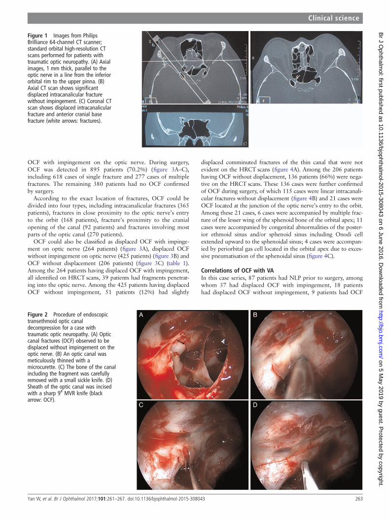
Image interpretationOrbital HRCT was performed for each patient at the time ofadmission with a 40-section or 64-section CT system (GE,Milwaukee, Wisconsin, USA; or Philips, Amsterdam, China)(figure 1). A standard protocol was used similar to thatdescribed by Lee et al.9 Axial images, 1 mm thick, were madeparallel to the optic nerve in a line from the inferior orbital rimto the upper pinna. Coronal images, 3 mm thick, were madefrom the globes to the dorsum sella, with the sections perpen-dicular to the canthomeatal line. In patients unable to hyper-extend the neck, coronal reformations from the axial imageswere performed. For the patients at the Eye Hospital ofWenzhou Medical University, the bone and soft tissue windowsof all of the axial and coronal CT scans were carefully reviewedand analysed together by an ophthalmological clinician (DrYunhai Tu) and two experienced radiologists with expertise inhead and neck imaging for consensus prior to surgery. Forpatients at the other seven institutes, all clinical data were sentto Dr Yunhai Tu by email for consultation, and the CT scan ofeach patient was prospectively reviewed by Dr Yunhai Tu andthe two experienced radiologists for consensus beforeoperation.
We recorded the age of the patient, Snellen’s VA prior tosurgery and the final VA at 3 months after surgery, time intervalbetween injury and surgery, HRCT findings and the surgicalfindings. Colour vision, visual field defects and VEP were notconsistently documented in the medical records and were notincluded in the study. Usually, patients were followed up to3 months at these eight institutions. For a few patients unable tocome, we recommended them to local community hospitals forbasic post-op examinations and collected outcome from themby telephone at 3 months after surgery. For the convenience ofdata analysis and comparison, Snellen’s VA was transferred tologMAR units.21 24–26 We applied the formula of Chen et al25
to define the percentage of improvement degree of VA (IDVA):
postoperative log MAR� preoperative log MAR=ð0:12� �preoperative log MARÞ
*where log MAR 0.12 (20/15) is considered perfect vision.
If the IDVA decreased or remained the same after surgery, thesurgery was considered to be ineffective. If the IDVA increasedno more than 50%, the surgery was considered to be moderateeffective, and if the IDVA increased more than 50%, the surgerywas considered to be very effective.
Surgical procedure of ETOCDAll of the procedures were performed under general anaesthesiaby the surgeon (Dr Wencan Wu) as previously described.27–29
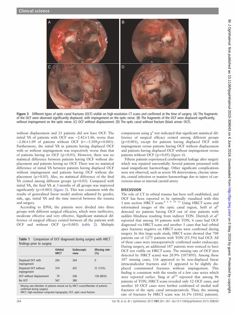
The orbital lamina papyracea was exposed via a standardisedethmoidectomy under direct visualisation, with a 4 mm 45°transnasal endoscope (KARL STORZ, Tuttlingen, Germany),prior to opening the sphenoid sinus. The bulge caused by theoptic canal and internal carotid artery was identified in thelateral wall of the sphenoidal sinus. After meticulous removal ofthe sinus mucosa, the area was inspected for OCF, fractures ofthe lesser wing of the sphenoid bone, anterior skull base frac-tures and/or cerebrospinal rhinorrhoea (figure 2A). The lesserwing of the sphenoidal bone and the medial wall of the opticcanal, which spans from the orbital aperture to the cranialcavity, were thinned with a microdrill and removed with amicrocurette (figure 2B, C). Then, the periorbita of the orbitalapex, annulus of Zinn and the optic nerve sheath were incisedwith a sharp 9# MVR knife (figure 2D). Finally, at the end ofthe surgery, the operating field of the optic canal was covered bya piece of sterile gelatine sponge that was immersed in dexa-methasone (5 mg/2 mL) and mouse-derived NGF (30 mg/mL)(Staidson (Beijing) Biopharmaceuticals Co.).
Postoperative care included intravenous methylprednisolone,30 mg/kg/day for 3 days and intravenous ceftriaxone for 5 days.At the third postoperative day, the dexamethasone and mouse-derived NGF were locally administered after removal of the gel-atine, and this procedure was done every 2 days for a total offive times.
Statistical analysisStatistical analyses were performed using SPSS V.20.0 software(SPSS, Chicago, Illinois, USA). Univariate analysis was per-formed by Pearson’s χ2 tests for discrete variables, and pairedsample t test or independent samples t test and one-way analysisof variance for continuous variables. Results were consideredsignificant at p<0.05 and reported as mean±SD.
RESULTSBaseline characteristics and periorbital injuryAltogether, 1275 consecutive patients (1275 eyes) wereincluded, with a male to female ratio of 3.7 to 1. The age of thepatients ranged from 16 to 50 years old, with a mean of 29.8±7.3 years old. The time interval from injury to surgical inter-vention ranged from 1 to 14 days, with a mean of 10±2 days.Road traffic accident was the most common cause of injury (659patients), followed by blunt assault (261 patients), work-relatedinjuries (226 patients) and fall from a height (131 patients).Multiple facial fractures were present in 517 patients.Intracerebral haemorrhage was noticed in 217 patients.Concurrent HRCT findings related to intraorbital soft tissueinjuries included extraconal haematoma (255 patients), intraco-nal haematoma (149 patients), intraconal emphysema (132patients) and intrasheath haematoma along the optic nerve (53patients).
Differences between images and surgical findingsHRCT images prior to surgery showed that 708 patients(55.5%) had visible OCF, of which 264 (20.7%) were displaced
262 Yan W, et al. Br J Ophthalmol 2017;101:261–267. doi:10.1136/bjophthalmol-2015-308043
Clinical science on 5 M
ay 2019 by guest. Protected by copyright.
http://bjo.bmj.com
/B
r J Ophthalm
ol: first published as 10.1136/bjophthalmol-2015-308043 on 6 June 2016. D
ownloaded from
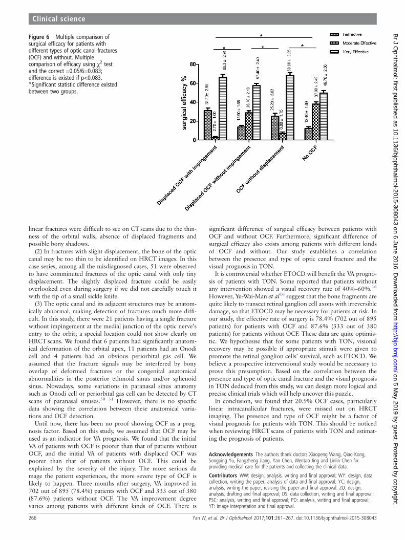
OCF with impingement on the optic nerve. During surgery,OCF was detected in 895 patients (70.2%) (figure 3A–C),including 618 cases of single fracture and 277 cases of multiplefractures. The remaining 380 patients had no OCF confirmedby surgery.
According to the exact location of fractures, OCF could bedivided into four types, including intracanalicular fractures (365patients), fractures in close proximity to the optic nerve’s entryto the orbit (168 patients), fracture’s proximity to the cranialopening of the canal (92 patients) and fractures involving mostparts of the optic canal (270 patients).
OCF could also be classified as displaced OCF with impinge-ment on optic nerve (264 patients) (figure 3A), displaced OCFwithout impingement on optic nerve (425 patients) (figure 3B) andOCF without displacement (206 patients) (figure 3C) (table 1).Among the 264 patients having displaced OCF with impingement,all identified on HRCT scans, 39 patients had fragments penetrat-ing into the optic nerve. Among the 425 patients having displacedOCF without impingement, 51 patients (12%) had slightly
displaced comminuted fractures of the thin canal that were notevident on the HRCT scans (figure 4A). Among the 206 patientshaving OCF without displacement, 136 patients (66%) were nega-tive on the HRCT scans. These 136 cases were further confirmedof OCF during surgery, of which 115 cases were linear intracanali-cular fractures without displacement (figure 4B) and 21 cases wereOCF located at the junction of the optic nerve’s entry to the orbit.Among these 21 cases, 6 cases were accompanied by multiple frac-ture of the lesser wing of the sphenoid bone of the orbital apex; 11cases were accompanied by congenital abnormalities of the poster-ior ethmoid sinus and/or sphenoid sinus including Onodi cellextended upward to the sphenoidal sinus; 4 cases were accompan-ied by periorbital gas cell located in the orbital apex due to exces-sive pneumatisation of the sphenoidal sinus (figure 4C).
Correlations of OCF with VAIn this case series, 87 patients had NLP prior to surgery, amongwhom 37 had displaced OCF with impingement, 18 patientshad displaced OCF without impingement, 9 patients had OCF
Figure 1 Images from PhilipsBrilliance 64-channel CT scanner;standard orbital high-resolution CTscans performed for patients withtraumatic optic neuropathy. (A) Axialimages, 1 mm thick, parallel to theoptic nerve in a line from the inferiororbital rim to the upper pinna. (B)Axial CT scan shows significantdisplaced intracanalicular fracturewithout impingement. (C) Coronal CTscan shows displaced intracanalicularfracture and anterior cranial basefracture (white arrows: fractures).
Figure 2 Procedure of endoscopictransethmoid optic canaldecompression for a case withtraumatic optic neuropathy. (A) Opticcanal fractures (OCF) observed to bedisplaced without impingement on theoptic nerve. (B) An optic canal wasmeticulously thinned with amicrocurette. (C) The bone of the canalincluding the fragment was carefullyremoved with a small sickle knife. (D)Sheath of the optic canal was incisedwith a sharp 9# MVR knife (blackarrow: OCF).
Yan W, et al. Br J Ophthalmol 2017;101:261–267. doi:10.1136/bjophthalmol-2015-308043 263
Clinical science on 5 M
ay 2019 by guest. Protected by copyright.
http://bjo.bmj.com
/B
r J Ophthalm
ol: first published as 10.1136/bjophthalmol-2015-308043 on 6 June 2016. D
ownloaded from
without displacement and 23 patients did not have OCF. Theinitial VA of patients with OCF was −2.42±1.06, worse than−2.06±1.09 of patients without OCF (t=−5.509,p<0.001).Furthermore, the initial VA in patients having displaced OCFwith or without impingement was respectively worse than thatof patients having no OCF (p<0.01). However, there was nostatistical difference between patients having OCF without dis-placement and patients having no OCF. There was no statisticaldifference of initial VA between patients having displaced OCFwithout impingement and patients having OCF without dis-placement (p>0.05). Also, no statistical difference of the finalVA existed among different groups (p>0.05). Compared withinitial VA, the final VA at 3 months of all groups was improvedsignificantly (p<0.001) (figure 5). This was consistent with theresults of generalised linear model analysis adjusted by gender,side, age, initial VA and the time interval between the traumaand surgery.
According to IDVA, the patients were divided into threegroups with different surgical efficacies, which were ineffective,moderate effective and very effective. Significant statistical dif-ference of surgical efficacy existed between all the patients withOCF and without OCF (p<0.001) (table 2). Multiple
comparisons using χ2 test indicated that significant statistical dif-ference of surgical efficacy existed among different groups(p<0.001), except for patients having displaced OCF withimpingement versus patients having OCF without displacementand patients having displaced OCF without impingement versuspatients without OCF (p>0.05) (figure 6).
Fifteen patients experienced cerebrospinal leakage after surgerywhich was repaired uneventfully. Several patients presented withnasal insignificant haemorrhage. Other significant complicationswere not observed, such as severe VA deterioration, chronic sinus-itis, cranial infection or massive haemorrhage due to injury of car-vernous sinus or internal carotid artery.
DISCUSSIONThe role of CT in orbital trauma has been well established, andOCF has been reported to be optimally visualised with thin1 mm section HRCT scans.4 6 8 10 12 Using HRCT scans andreformatted images of the optic canal region, Seiff et al6
detected six patients having OCF out of nine patients withsudden blindness resulting from indirect TON. Dietrich et al5
reported that among 10 patients with TON, 6 cases had OCFdiagnosed via HRCT scans and another 3 cases that had orbitalapex fractures negative on HRCT scans were confirmed duringsurgery. In this large-scale study, HRCT scans showed that 708patients out of 1275 patients with TON (55.5%) had OCF. Allof these cases were intraoperatively confirmed under endoscopy.During surgery, an additional 187 patients were noticed to haveOCF not visible on HRCT scans. The missing rate of OCF (notdetected by HRCT scans) was 20.9% (187/895). Among these187 missing cases, 136 appeared to be non-displaced linearintracanalicular fractures and 51 appeared to be slightly dis-placed comminuted fractures without impingement. Thisfinding is consistent with the results of a few case series whichwere reported earlier. Yang et al14 reported that among 96patients of TON, HRCT scans revealed only 52 OCF cases, andanother 10 OCF cases were further confirmed of medial wallfractures of the optic canal intraoperatively. Thus, the missingrate of fractures by HRCT scans was 16.1% (10/62 patients).
Figure 3 Different types of optic canal fractures (OCF) visible on high-resolution CT scans and confirmed at the time of surgery. (A) The fragmentsof the OCF were observed significantly displaced, with impingement on the optic nerve. (B) The fragments of the OCF were displaced significantly,without impingement on the optic nerve. (C) OCF without displacement. (D) The optic canal without fracture (black arrow: OCF).
Table 1 Comparison of OCF diagnosed during surgery with HRCTfindings prior to surgery
OrbitalHRCT
Endoscopicview
Missing rate(%)
Displaced OCF withimpingement
264 264 0
Displaced OCF withoutimpingement
374 425 51 (12%)
OCF without displacement 70 206 136 (66%)No OCF 567 380
Missing rate=(Number of patients missed out by HRCT scans)/(Number of patientsconfirmed during surgery).HRCT, high-resolution computed topography; OCF, optic canal fracture.
264 Yan W, et al. Br J Ophthalmol 2017;101:261–267. doi:10.1136/bjophthalmol-2015-308043
Clinical science on 5 M
ay 2019 by guest. Protected by copyright.
http://bjo.bmj.com
/B
r J Ophthalm
ol: first published as 10.1136/bjophthalmol-2015-308043 on 6 June 2016. D
ownloaded from

Imachi15 also reported 16 HRCT-undetected OCF cases out of80 cases with TON, making missing rate 20%. Gupta et al17
reported an even poor correlation between CT and operativefindings, showing 7 OCF cases visible on CT and 13 OCF casesdetected during surgery. Wohlrab et al18 reported a high false-negative rate that only one OCF was detected by CTwhile eightwere found intraoperatively. Along with our large-scale study,these reports clearly demonstrate that OCF may still exist evenif it comes clear on CT image. So, attention should be paid tothe reliability of radiographic evidence for diagnosis of OCFwhen assessing patients with sudden vision loss after headtrauma.
The sensitivity of CT scans in OCF diagnosis relies on severalinfluential factors, including the scanner machine, thickness ofthe slices taken, position and angle of the head, anatomicalcharacteristics and experience of the clinician who interprets thescans. Based on this study, we believe that the causes of misdiag-nosed OCF on HRCT scans may be attributed to the followingthree factors:
(1) Linear fractures are difficult to be detected. HRCT scan-ning is definitely accurate and reliable when the OCF is signifi-cantly displaced. However, for 206 patients withoutdisplacement, only 70 patients (34%) were detected, and 136patients (66%) were missed out. The linear fractures seem to bethe most difficult to detect with HRCT. As we can see, amongthe undetected 136 patients, 115 patients (84.6%, 115/136)were intraoperatively observed to have linear intracanalicularfractures. In 1984, Unger10 also proposed that undisplaced
Figure 4 Optic canal fractures invisible on the high-resolution CT scans. (A) Comminuted fracture of the thin optic canal, though minimallydisplaced but without impingement. (B) Undisplaced linear intracanalicular fracture. (C) A minimally displaced fracture of the optic canal that passedthrough an abnormal small Onodi cell between the posterior ethmoidal sinus and the sphenoid sinus; its surrounding bones were thick andabnormal (black arrow: fractures; white arrow: Onodi cell).
Figure 5 Visual acuity (VA) of different types of optic canal fractures(OCF) before and after surgery. *p<0.01 versus patients without OCFusing one-way analysis of variance. #p<0.001 versus the final VA at3 months after the surgery using paired-samples t test.
Table 2 Surgical efficacy for patients with different types of OCFand without OCF
IneffectiveModerateeffective
Veryefficient
Group N % N % N %
Kinds of OCFDisplaced OCF impingement 82 31.1 7 2.7 175 66.3Displaced OCF with noimpingement
59 13.9 122 28.7 244 57.4
OCF without displacement 52 25.2 14 6.8 140 68.0No OCF 47 12.4 144 37.9 189 49.7χ2 166.416P <0.001
Moderate effective=IDVA no more than 50%; very efficient=IDVA≥50%;ineffective=no improvement of VA after surgery.P=difference of efficacy between patients with OCF and no OCF, using χ2 test.IDVA, improvement degree of VA; No OCF, patients who did not have OCF; OCF, opticcanal fracture; VA, visual acuity.
Yan W, et al. Br J Ophthalmol 2017;101:261–267. doi:10.1136/bjophthalmol-2015-308043 265
Clinical science on 5 M
ay 2019 by guest. Protected by copyright.
http://bjo.bmj.com
/B
r J Ophthalm
ol: first published as 10.1136/bjophthalmol-2015-308043 on 6 June 2016. D
ownloaded from
linear fractures were difficult to see on CT scans due to the thin-ness of the orbital walls, absence of displaced fragments andpossible bony shadows.
(2) In fractures with slight displacement, the bone of the opticcanal may be too thin to be identified on HRCT images. In thiscase series, among all the misdiagnosed cases, 51 were observedto have comminuted fractures of the optic canal with only tinydisplacement. The slightly displaced fracture could be easilyoverlooked even during surgery if we did not carefully touch itwith the tip of a small sickle knife.
(3) The optic canal and its adjacent structures may be anatom-ically abnormal, making detection of fractures much more diffi-cult. In this study, there were 21 patients having a single fracturewithout impingement at the medial junction of the optic nerve’sentry to the orbit; a special location could not show clearly onHRCT scans. We found that 6 patients had significantly anatom-ical deformation of the orbital apex, 11 patients had an Onodicell and 4 patients had an obvious periorbital gas cell. Weassumed that the fracture signals may be interfered by bonyoverlap of deformed fractures or the congenital anatomicalabnormalities in the posterior ethmoid sinus and/or sphenoidsinus. Nowadays, some variations in paranasal sinus anatomysuch as Onodi cell or periorbital gas cell can be detected by CTscans of paranasal sinuses.30 31 However, there is no specificdata showing the correlation between these anatomical varia-tions and OCF detection.
Until now, there has been no proof showing OCF as a prog-nosis factor. Based on this study, we assumed that OCF may beused as an indicator for VA prognosis. We found that the initialVA of patients with OCF is poorer than that of patients withoutOCF, and the initial VA of patients with displaced OCF waspoorer than that of patients without OCF. This could beexplained by the severity of the injury. The more serious damage the patient experiences, the more severe type of OCF islikely to happen. Three months after surgery, VA improved in702 out of 895 (78.4%) patients with OCF and 333 out of 380(87.6%) patients without OCF. The VA improvement degreevaries among patients with different kinds of OCF. There is
significant difference of surgical efficacy between patients withOCF and without OCF. Furthermore, significant difference ofsurgical efficacy also exists among patients with different kindsof OCF and without. Our study establishes a correlationbetween the presence and type of optic canal fracture and thevisual prognosis in TON.
It is controversial whether ETOCD will benefit the VA progno-sis of patients with TON. Some reported that patients withoutany intervention showed a visual recovery rate of 40%–60%.16
However, Yu-Wai-Man et al16 suggest that the bone fragments arequite likely to transect retinal ganglion cell axons with irreversibledamage, so that ETOCD may be necessary for patients at risk. Inour study, the effective rate of surgery is 78.4% (702 out of 895patients) for patients with OCF and 87.6% (333 out of 380patients) for patients without OCF. These data are quite optimis-tic. We hypothesise that for some patients with TON, visionalrecovery may be possible if appropriate stimuli were given topromote the retinal ganglion cells’ survival, such as ETOCD. Webelieve a prospective interventional study would be necessary toprove this presumption. Based on the correlation between thepresence and type of optic canal fracture and the visual prognosisin TON deduced from this study, we can design more logical andprecise clinical trials which will help uncover this puzzle.
In conclusion, we found that 20.9% OCF cases, particularlylinear intracanalicular fractures, were missed out on HRCTimaging. The presence and type of OCF might be a factor ofvisual prognosis for patients with TON. This should be noticedwhen reviewing HRCT scans of patients with TON and estimat-ing the prognosis of patients.
Acknowledgements The authors thank doctors Xiaopeng Wang, Qiao Kong,Songping Yu, Fangzheng Jiang, Yan Chen, Wentao Jing and Linlin Chen forproviding medical care for the patients and collecting the clinical data.
Contributors WW: design, analysis, writing and final approval; WY: design, datacollection, writing the paper, analysis of data and final approval; YC: design,analysis, writing the paper, revising the paper and final approval. ZQ: design,analysis, drafting and final approval; DS: data collection, writing and final approval;PSC: analysis, writing and final approval; PD: analysis, writing and final approval;YT: image interpretation and final approval.
Figure 6 Multiple comparison ofsurgical efficacy for patients withdifferent types of optic canal fractures(OCF) and without. Multiplecomparison of efficacy using χ2 testand the correct =0.05/6=0.083;difference is existed if p<0.083.*Significant statistic difference existedbetween two groups.
266 Yan W, et al. Br J Ophthalmol 2017;101:261–267. doi:10.1136/bjophthalmol-2015-308043
Clinical science on 5 M
ay 2019 by guest. Protected by copyright.
http://bjo.bmj.com
/B
r J Ophthalm
ol: first published as 10.1136/bjophthalmol-2015-308043 on 6 June 2016. D
ownloaded from

Funding This study was supported by the Natural Science Foundation of China(81371028), Natural Science Foundation of Zhejiang Province, PR China(LY12H12006) and the Guided Innovation Project of the Eye Hospital of WenzhouMedical University (YNCX201104).
Competing interests None declared.
Patient consent Obtained.
Ethics approval The tenets of the Declaration of Helsinki were observed for thisretrospective comparative case series, and the study was approved by the eighthospitals’ ethics committees, which include the Eye Hospital of Wenzhou MedicalUniversity, the Central Hospital of Lishui, the Central Hospital of Jinhua, LihuiliHospital in Ningbo, Quzhou People’s Hospital in Zhjiang province, Xiamen EyeHospital in Fujian province, The Fourth Hospital of Shengyang in Liaoning province,and The Second People’s Hospital of Zhengzhou city in Henan province.
Provenance and peer review Not commissioned; externally peer reviewed.
REFERENCES1 Anderson RL, Panje WR, Gross CE. Optic nerve blindness following blunt forehead
trauma. Ophthalmology 1982;89:445–55.2 Lee AG. Traumatic optic neuropathy. Ophthalmology 2000;107:814.3 Steinsapir KD, Goldberg RA. Traumatic optic neuropathy: an evolving
understanding. Am J Ophthalmol 2011;151:928–33.4 Mauriello JA Jr, Lee HJ, Nguyen L. CT of soft tissue injury and orbital fractures.
Radiol Clin North Am 1999;37:241–52.5 Dietrich U, Feldges A, Nau HE, et al. Computed tomographic assessment of orbital
fractures in traumatic damage of the optic nerve. Rofo 1990;152:185–90.6 Seiff SR, Berger MS, Guyon J, et al. Computed tomographic evaluation of the optic
canal in sudden traumatic blindness. Am J Ophthalmol 1984;98:751–5.7 Guyon JJ, Brant-Zawadzki M, Seiff SR. CT demonstration of optic canal fractures.
Am J Roentgenol 1984;143:1031–4.8 Kubal WS. Imaging of the orbital trauma. Radiographics 2008;28:1729–39.9 Lee HJ, Jilani M, Frohman L, et al. CT of orbital trauma. Emerg Radiol
2004;10:168–72.10 Unger JM. Orbital apex fractures: the contribution of computed tomography.
Radiology 1984;150:713–17.11 Cole P, Kaufman Y, Hollier L. Principles of facial trauma: orbital fracture
management. J Craniofac Surg 2009;20:101–4.12 Bodanapally UK, Van der Byl G, Shanmuganathan K, et al. Traumatic optic
neuropathy prediction after blunt facial trauma: derivation of a risk score based onfacial CT findings at admission. Radiology 2014;272:824–31.
13 Ramsay JH. Optic nerve injury in fracture of the canal. Br J Ophthalmol1979;63:607–10.
14 Yang QT, Zhang GH, Liu X, et al. The therapeutic efficacy of endoscopic optic nervedecompression and its effects on the prognoses of 96 cases of traumatic opticneuropathy. J Trauma Acute Care Surg 2012;72:1350–5.
15 Imachi Y. Clinical and pathohistological investigations of the optic nerve lesionscaused by head injuries. Nippon Ganka Gakkai Zasshi 1967;71:1874–908.
16 Yu-Wai-Man P, Griffiths PG. Surgery for traumatic optic neuropathy. CochraneDatabase Syst Rev 2005;19:CD005024.
17 Gupta AK, Gupta AK, Gupta A, et al. Traumatic optic neuropathy in pediatricpopulation: early intervention or delayed intervention? Int J Ped Otorhinolaryngol2007;71:559–62.
18 Wohlrab TM, Maas S, de Carpentier JP. Surgical decompression in traumatic opticneuropathy. Acta Ophthalmol Scand 2002;80:287–93.
19 Mine S, Yamakami I, Yamaura A, et al. Outcome of traumatic optic neuropathy.Comparsion between surgical and nonsurgical treatment. Acta Neurochir1999;141:27–30.
20 Wang BH, Robertson BC, Girotto JA, et al. Traumatic optic neuropathy: a review of61 patients. Plast Reconstr Surg 2001;107:1655–64.
21 Cook MW, Levin LA, Joseph MP, et al. Traumatic optic neuropathy: a meta-analysis.Arch Otolaryngol Head Neck Surg 1996;122:389–92.
22 Chou PI, Sadam AA, Chen YC, et al. Clinical experiences in the management oftraumatic optic neuropathy. Neuro-ophthalmology 1996;16:325–36.
23 Tandon D, Thakar A, Mahapatra AK, et al. Trans-ethmoidal optic nervedecompression. Clin Otolaryngol Allied Sci 1994;19:98–104.
24 Li KK, Teknos TN, Lai A, et al. Traumatic optic neuropathy: result in 45 consecutivesurgically treated patients. Otolaryngol Head Neck Surg 1999;120:5–11.
25 Chen CT, Huang F, Tsay PK, et al. Endoscopically assistedtransconjunctival decompression of traumatic optic neuropathy. J Craniofac Surg2007;18:19–26.
26 Levin LA, Beck RW, Joseph MP, et al. The treatment of traumatic opticneuropathy: the International Optic Nerve Trauma Study. Ophthalmology1999;106:1268–77.
27 Chen C, Selva D, Floreani S, et al. Endoscopic optic nerve decompression fortraumatic optic neuropathy: an alternative. Otolaryngol Head Neck Surg2006;135:155–7.
28 Chen F, Zuo K, Feng S, et al. A modified surgical procedure for endoscopic opticnerve decompression for the treatment of traumatic optic neuropathy. N Am J MedSci 2014;6:270–3.
29 Levin LA, Joseph MP, Rizzo JF, et al. Optic canal decompression in indirect opticnerve trauma. Ophthalmology 1994;101:566–9.
30 Lu Y, Pan J, Qi S, et al. Pneumatization of the sphenoid sinus in Chinese:the differences from Caucasian and its application in the extended transsphenoidalapproach. J Anat 2011;219:132–42.
31 Kantarci M, Karasen RM, Alper F, et al. Remarkable anatomic variations inparanasal sinus region and their clinical importance. Eur J Radiol2004;50:296–302.
Yan W, et al. Br J Ophthalmol 2017;101:261–267. doi:10.1136/bjophthalmol-2015-308043 267
Clinical science on 5 M
ay 2019 by guest. Protected by copyright.
http://bjo.bmj.com
/B
r J Ophthalm
ol: first published as 10.1136/bjophthalmol-2015-308043 on 6 June 2016. D
ownloaded from