Embed Size (px)
Citation preview
Topographical relationship between the choroidalwatershed zone and submacular idiopathic choroidalneovascularisationJi Eun Lee,1,2 Min Kyu Shin,1,2 In Young Chung,3 Joo Eun Lee,4 Hyun Woong Kim,5
Sang Joon Lee,6,7 Sung Who Park,1,2 Ik Soo Byon1,8
1Department ofOphthalmology, Collegeof Medicine, Pusan NationalUniversity, Yangsan,Republic of Korea2Medical Research Institute,Pusan National UniversityHospital, Busan,Republic of Korea3Department ofOphthalmology, GyeongsangNational University, JinJu,Republic of Korea4Department ofOphthalmology, Haeundae PaikHospital, College of Medicine,Inje University, Busan,Republic of Korea5Department ofOphthalmology, Busan PaikHospital, College of Medicine,Inje University, Busan,Republic of Korea6Department ofOphthalmology, Gospelhospital, College of Medicine,Kosin University, Busan,Republic of Korea7Institute for medicine, KosinUniversity, Busan, Republic ofKorea8Research Institute forConvergence of BiomedicalScience and Technology,Pusan National UniversityYangsan Hospital, Yangsan,Republic of Korea
Correspondence toProfessor Ji Eun Lee,Department of Ophthalmology,Pusan National UniversityHospital, 179 Gudeok-ro,Seo-gu, Busan 602-739,Republic of Korea;[email protected]
Received 24 January 2015Revised 4 August 2015Accepted 6 August 2015Published Online First31 August 2015
To cite: Lee JE, Shin MK,Chung IY, et al. Br JOphthalmol 2016;100:652–659.
ABSTRACTAims To investigate the relationship between idiopathicchoroidal neovascularisation (CNV) and choroidalwatershed zones (CWZs) using indocyanine greenangiography (ICGA).Design Multicentre, retrospective, interventional caseseries.Methods The medical records and ICGA findings of44 patients (44 eyes) diagnosed with idiopathic CNVwere reviewed. CWZs, defined as hypofluorescence thatdisappeared during the early phase of ICGA, wereclassified, and the findings were compared with thoseof a control group of 30 eyes. The topographicalrelationship between CWZs and CNV was evaluated.Visual acuity and recurrence were analysed accordingto the CWZ classification.Results The CNV lesion was subfoveal in 16 eyes,juxtafoveal in 12 eyes and extrafoveal in 16 eyes. Themost common types of CWZs were stellate (23 eyes,52.3%) and vertical (19 eyes, 43.2%). CWZs involvingthe fovea were seen in more patients with idiopathicCNV (37 eyes, 84.1%) than in the control group(11 eyes, 36.7%, p<0.001). The topographicalrelationship between CWZs and CNV was determined in42 eyes (95.5%), with the CNV located within the CWZin 39 eyes and at the margin in 3 eyes. Extrafoveal CNVwas within the CWZ in all 16 affected eyes. At6 months, visual acuity was significantly worse inpatients with subfoveal CNV (p=0.028) or stellate CWZs(p=0.039).Conclusions The findings of a CWZ were related tothe location and functional outcome of idiopathic CNV.Our results suggest that choroidal circulation is apredisposing factor for the development of CNV inyoung patients.
INTRODUCTIONChoroidal neovascularisation (CNV) may be com-plicated by various conditions, includingage-related macular degeneration (AMD), whitedot syndromes, high myopia, presumed ocularhistoplasmosis syndrome, angioid streak and trau-matic choroidal rupture. Idiopathic CNV refers toCNV that develops without any predisposingabnormalities,1–3 and few studies have suggestedmechanisms for idiopathic CNV. Yang et al4
reported that inflammatory cytokines such as vascu-lar endothelial growth factor (VEGF) and immuno-globulin E were detected at high levels in the serumof patients with idiopathic CNV. Favourable out-comes of anti-VEGF treatments5–8 support this
theory. However, no clinical findings have beenreported suggesting a possible source of VEGF.The results of several studies have demonstrated
that the human choroid has endarterial systemssupplied by various posterior ciliary arteries (PCAs)and that multiple watershed zones may be locatedin the macula (figure 1).9 10 A choroidal watershedzone (CWZ) was demonstrated by fluorescein angi-ography (FA) to course vertically around the opticnerve head (ONH) typically in normal subjects,11
and shows various patterns with indocyanine greenangiography (ICGA).12 The CWZ has a topograph-ical relationship with CNV in exudative AMD andwas proposed to be related to the development ofCNV with an ischaemic mechanism.13–15
The purpose of this study was to investigate thetopographical relationship of CWZs and idiopathicCNV with the objective of suggesting a mechanismfor idiopathic CNV development.
METHODSThe medical records of patients who were diag-nosed with idiopathic CNV in five hospitals (PusanNational University Hospital, Gyeongsang NationalUniversity Hospital, Haeundae Paik Hospital,Busan Paik Hospital and Gospel Hospital) from2008 to 2013 were reviewed retrospectively. Thecontrol cohort, using the same inclusion criteriaexcluding the presence of idiopathic CNV, wascollected in Pusan National University Hospital in2014. The study conducted adhered to the tenetsof the Declaration of Helsinki.The presence of CNV was confirmed using FA
and optical coherence tomography (OCT). Theinclusion criteria were as follows: age ≤45 years, anaxial length of <26 mm and a spherical equivalentrefractive error of not more than −5.0 dioptres (D).Patients with polypoidal choroidal vasculopathy,white dot syndromes, presumed ocular histoplasmo-sis syndrome, high myopia, hereditary dystrophy,angioid streak and other choroidal abnormalitiesrelated to CNV were excluded. Eyes with possiblepredisposing factors such as a choroidal scar, softdrusen, and a history of uveitis or central serouschorioretinopathy were also excluded.Patients were evaluated using slit-lamp biomicro-
scopy with a 90 D lens, fundus photography andOCT (Cirrus HD-OCT; Carl Zeiss Meditec,Dublin, California, USA). Visual acuity was mea-sured using the Snellen chart and converted to thelogarithm of the minimum angle of resolution. FAand ICGA were performed using a confocal scan-ning laser ophthalmoscope (Heidelberg Retina
Editor’s choiceScan to access more
free content
652 Lee JE, et al. Br J Ophthalmol 2016;100:652–659. doi:10.1136/bjophthalmol-2015-306678
Clinical science on 9 N
ovember 2018 by guest. P
rotected by copyright.http://bjo.bm
j.com/
Br J O
phthalmol: first published as 10.1136/bjophthalm
ol-2015-306678 on 31 August 2015. D
ownloaded from
Angiograph 2; Heidelberg Engineering, Heidelberg, Germany).For the early phase, the images were immediately obtained afterinjection of fluorescein or indocyanine green dye at a rate of atleast two to three images per second for 30 s.
A CWZ was defined as delayed filling of the choriocapillariswhich appeared as a hypofluorescence and then disappearedduring the early phase of FA and ICGA.10 12–15 However, sinceassessment in FA was unreliable due to the pigmentation in
Asians, CWZ was evaluated in ICGA only. ICGA findings wereanalysed independently by two readers ( JEL and MKS), and theextent of their agreement was calculated. For discrepant assess-ments, the final decision was made on the basis of a consensusamong the two readers.
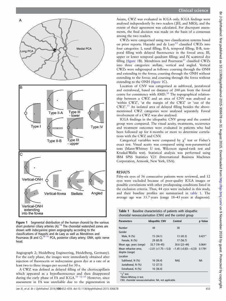
CWZs were categorised using two classification systems basedon prior reports. Hayashi and de Laey12 classified CWZs intofour categories: I, nasal filling; II-A, temporal filling; II-B, tem-poral filling with delayed fluorescence in the foveal area; III,upper or lower temporal quadrant filling; and IV, scattered dyefilling (figure 1B). Mendrinos and Pournaras13 classified CWZsinto three categories: stellate, vertical and angled. VerticalCWZs were subgrouped as follows: coursing through the ONHand extending to the fovea; coursing through the ONH withoutextending to the fovea; and coursing through the fovea withoutextending to the ONH (figure 1C).
Location of CNV was categorised as subfoveal, juxtafovealand extrafoveal, based on distance of 200 μm from the fovealcentre for consistency with AMD.16 The topographical relation-ship between a CWZ and an area of CNV was analysed as‘within CWZ’, ‘at the margin of the CWZ’ or ‘out of theCWZ’.13 An isolated area of delayed filling besides the above-mentioned CWZ categories were analysed separately. Fovealinvolvement of a CWZ was also analysed.
ICGA findings in the idiopathic CNV group and the controlgroup were compared. The visual acuity, treatments, recurrenceand treatment outcomes were evaluated in patients who hadbeen followed up for 6 months or more to determine correla-tions with the CWZ and CNV.
Categorical variables were compared by χ2 test or Fisher’sexact test. Visual acuity was compared using non-parametricaltests (Mann-Whitney U test, Wilcoxon signed-rank test andKruskal-Wallis test). Statistical analysis was performed usingIBM SPSS Statistics V.21 (International Business MachinesCorporation, Armonk, New York, USA).
RESULTSFifty-six eyes of 56 consecutive patients were reviewed, and 12eyes were excluded because of poor-quality ICGA images orpossible correlations with other predisposing conditions listed inthe exclusion criteria. Thus, 44 eyes were included in this study,and their baseline profiles are summarised in table 1. Theaverage age was 33.7 years (range 18–45 years at diagnosis).
Figure 1 Segmental distribution of the human choroid by the variousshort posterior ciliary arteries (A).14 The choroidal watershed zones areshown with indocyanine green angiography according to theclassifications of Hayashi and de Laey as well as Mendrinos andPournaras (B and C).10 11 PCA, posterior ciliary artery; ONH, optic nervehead.
Table 1 Baseline characteristics of patients with idiopathicchoroidal neovascularisation (CNV) and the control group
Parameters Idiopathic CNV Control p Value
Number 44 30GenderMale, N (%) 15 (34.1) 13 (43.3) 0.421*Female, N (%) 29 (65.9) 17 (56.7)
Mean age, years (range) 33.7 (18–45) 30.6 (22–44) 0.064†Mean refractive error,dioptre (range)
−2.01 (+1.75–−5.0) −1.45 (+0.83–−4.33) 0.179†
LocationSubfoveal, N (%) 16 (36.4) NA§ NAJuxtafoveal, N (%) 12 (27.3)Extrafoveal, N (%) 16 (36.4)
*χ2 test.†Mann-Whitney U test.CNV, choroidal neovascularisation; NA, not applicable.
Lee JE, et al. Br J Ophthalmol 2016;100:652–659. doi:10.1136/bjophthalmol-2015-306678 653
Clinical science on 9 N
ovember 2018 by guest. P
rotected by copyright.http://bjo.bm
j.com/
Br J O
phthalmol: first published as 10.1136/bjophthalm
ol-2015-306678 on 31 August 2015. D
ownloaded from
The control group was composed of a cohort of 30 normal con-trols (20–44 years old), who showed no significant differencefrom the idiopathic CNV group in sex, age and refractive error.
Classification of CWZsA CWZ began to show just after appearance of choroidal arterybetween 6 s and 16 s after injection of indocyanine green solu-tion, depending on the injection site. A CWZ was discerniblefor 5.9 s (±1.8, range 3–12 s) in idiopathic CNV group, and5.1 s (±1.3, range 3–8 s) in control group. The difference wasnot statistically significant (p=0.071, Mann-Whitney U test).
The agreement was 77.3% for Mendrinos’ classification,73.3% for Hayashi’s classification and 90.7% for foveal involve-ment of a CWZ. Table 2 shows the results of the CWZ classifi-cation in ICGA. According to Hayashi’s classification, type IVwas most common (23 eyes, 52.3%). Type II-A was seen inseven eyes (15.9%), type II-B in five eyes (11.4%) and type IIIin nine eyes (20.5%). Using Mendrinos’ classification, stellateCWZs were most common and were found in 23 eyes (52.3%,figure 2). Vertically oriented CWZs (figures 3–6) were seen in19 eyes (43.2%) and angled CWZs in 2 eyes (4.5%). In thecontrol group, the distribution did not differ from that of thepatients with idiopathic CNV in both CWZ classifications(p=0.150 and 0.261, χ2 test).
CWZs involving the fovea were seen in more patients withidiopathic CNV (37 eyes, 84.1%) than in the control group (11eyes, 36.7%, p<0.001). The foveal area in the control groupwas commonly spared even in eyes with stellate CWZs (7 of 13eyes, figure 7). In addition to the abovementioned CWZ classifi-cations, 11 eyes (25.0%) had isolated areas of delayed filling(figures 5 and 6). Some of these areas appeared to correlatewith the distribution of short PCAs rather than with the CWZ(figure 5).
Table 2 Classification of a choroidal watershed zone in patientswith idiopathic choroidal neovascularisation (CNV) and normalcontrols
IdiopathicCNV Control p Value*
Hayashi’s classificationType II-A 7 (15.9%) 7 (23.3%) 0.261Type II-B 5 (11.4%) 0 (0%)Type III 9 (20.5%) 7 (23.3%)Type IV 23 (52.3%) 16 (53.3%)
Mendrinos’ classificationStellate 23 (52.3%) 15 (50.0%) 0.150Vertical 19 (43.2%) 15 (50.0%)
Extending to the fovea 11 (57.9%) 3 (10.0%)Without extending to the fovea 7 (36.8%) 11 (36.7%)Through the fovea 1 (5.3%) 1 (3.3%)
Angled 2 (4.5%) 0 (0%)Foveal involvementFoveal 37 (84.1%) 11 (36.7%) <0.0001Non-foveal 7 (15.9%) 19 (63.3%)
*χ2 test.CNV, choroidal neovascularisation.
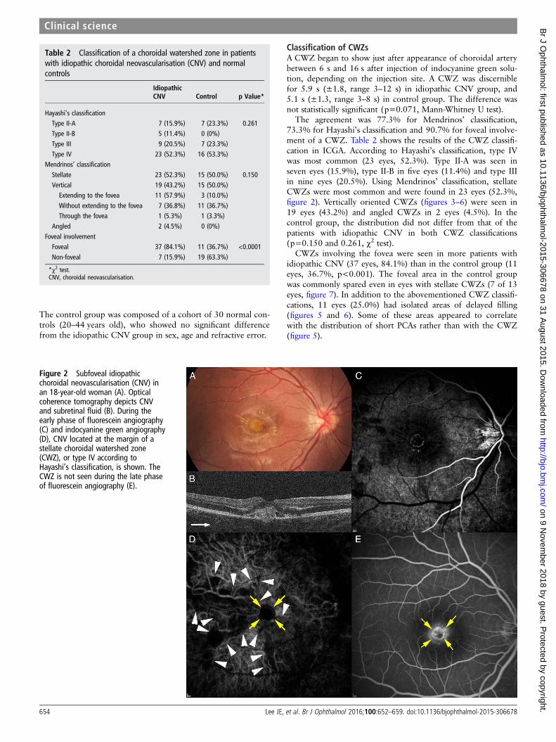
Figure 2 Subfoveal idiopathicchoroidal neovascularisation (CNV) inan 18-year-old woman (A). Opticalcoherence tomography depicts CNVand subretinal fluid (B). During theearly phase of fluorescein angiography(C) and indocyanine green angiography(D), CNV located at the margin of astellate choroidal watershed zone(CWZ), or type IV according toHayashi’s classification, is shown. TheCWZ is not seen during the late phaseof fluorescein angiography (E).
654 Lee JE, et al. Br J Ophthalmol 2016;100:652–659. doi:10.1136/bjophthalmol-2015-306678
Clinical science on 9 N
ovember 2018 by guest. P
rotected by copyright.http://bjo.bm
j.com/
Br J O
phthalmol: first published as 10.1136/bjophthalm
ol-2015-306678 on 31 August 2015. D
ownloaded from
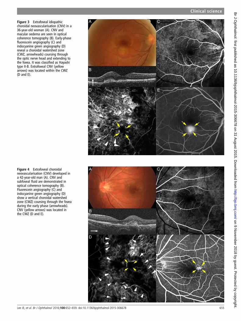
Figure 3 Extrafoveal idiopathicchoroidal neovascularisation (CNV) in a36-year-old woman (A). CNV andmacular oedema are seen in opticalcoherence tomography (B). Early-phasefluorescein angiography (C) andindocyanine green angiography (D)reveal a choroidal watershed zone(CWZ, arrowheads) coursing throughthe optic nerve head and extending tothe fovea. It was classified as Hayashitype II-B. Extrafoveal CNV (yellowarrows) was located within the CWZ(D and E).
Figure 4 Extrafoveal choroidalneovascularisation (CNV) developed ina 42-year-old man (A). CNV andsubfoveal fluid are demonstrated inoptical coherence tomography (B).Fluorescein angiography (C) andindocyanine green angiography (D)show a vertical choroidal watershedzone (CWZ) coursing through the foveaduring the early phase (arrowheads).CNV (yellow arrows) was located inthe CWZ (D and E).
Lee JE, et al. Br J Ophthalmol 2016;100:652–659. doi:10.1136/bjophthalmol-2015-306678 655
Clinical science on 9 N
ovember 2018 by guest. P
rotected by copyright.http://bjo.bm
j.com/
Br J O
phthalmol: first published as 10.1136/bjophthalm
ol-2015-306678 on 31 August 2015. D
ownloaded from
Topographical relationship between CWZ and CNVCNV location was subfoveal in 16 eyes (36.4%), juxtafoveal in12 eyes (27.3%) and extrafoveal in 16 eyes (36.4%, table 1).CNV was located within an area of delayed filling in 39 eyes(88.6%, figures 3–5) and at the margin in 3 eyes (6.8%,figure 2). Two eyes (4.5%), one with subfoveal CNV and theother with juxtafoveal CNV, had CNVoutside the areas of a ver-tical CWZ coursing through the disc (figure 6). ExtrafovealCNV was located within an area of delayed filling in all eyes(figures 3–5). Six eyes (13.6%) had a CNV located within theisolated areas of delayed filling (figure 5).
No relationship was found between CNV location and CWZclassification (p=0.342–0.510, χ2 test, table 3).
Treatment outcomesThe primary treatment was intravitreal ranibizumab for 10patients, intravitreal bevacizumab for 31 patients and photo-dynamic therapy for 1 patient. Two patients received no treat-ment. Thirty-six patients were followed up for ≥6 months; 19patients were followed up for ≥12 months. The mean follow-upduration was 16.0 months.
In patients followed up for ≥6 months, the median visualacuity significantly improved from 20/60 at baseline to 20/20 at6 months (p=0.002) and 20/25 at 12 months (p=0.003,Wilcoxon signed-rank test). Patients received photodynamictherapy or an intravitreal injection 2.7 times by 6 months and3.1 times by 1 year in average. Better baseline visual acuity wasrelated to better visual outcomes (p<0.001). At 6 months,visual acuity was better for the eyes with non-subfoveal CNV
than for the eyes with subfoveal CNV (p=0.028,Mann-Whitney U test, table 4). A stellate CWZ or type IVCWZ was related to significantly worse visual acuity than theother types (p=0.039, Mann-Whitney U test, table 4). No othercorrelation was found (p=0.058–0.746, Mann-Whitney U testand Kruskal-Wallis test).
There was a recurrence of CNV activity in 8 of the 36patients (22.2%) by 6 months and in 7 of the 19 patients(36.8%) by 12 months. A vertical CWZ extending to the foveawas the most common type among the patients showing recur-rence by 6 months (4 eyes, 50.0%) and 12 months (5 eyes,71.4%), but recurrence was not statistically significant withregards to classification of CWZ (p=0.334–0.694, Fisher’sexact test and χ2 test).
DISCUSSIONOur study revealed that idiopathic CNV had topographical rela-tionships with CWZs, suggesting that delayed perfusion of thechoriocapillaris may predispose patients to idiopathic CNVdevelopment. Hayreh suggested a pathological role of a CWZin development of CNV for the first time in 1974.17 However,because of the unique features of the choroidal vasculature thedebate regarding a hypoxic role for CWZs continues. Forexample, angiography in vivo shows that the choroidal lobule issupplied by a terminal arteriole not anastomosing with the adja-cent lobule,18 but a corrosion cast study demonstrated that thesubmacular choroidal vessels anastomose at various levels ofcapillaries, arterioles and venules.19
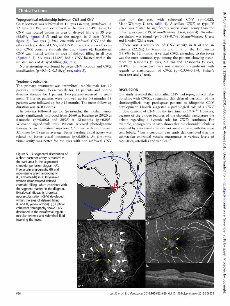
Figure 5 A segmental distribution ofa short posterior artery is marked asthe dark area in the segmentedchoroidal perfusion diagram (A).Fluorescein angiography (B) andindocyanine green angiography(C, arrowheads) in a 30-year-oldwoman demonstrated delayedchoroidal filling, which correlates withthe segment marked in the diagram.Extrafoveal idiopathic choroidalneovascularisation (CNV) developedwithin the area of delayed filling(C and D, yellow arrows). (E) Opticalcoherence tomography shows CNVdeveloped in the extrafoveal region,macular oedema and subretinal fluidinvolving the fovea.
656 Lee JE, et al. Br J Ophthalmol 2016;100:652–659. doi:10.1136/bjophthalmol-2015-306678
Clinical science on 9 N
ovember 2018 by guest. P
rotected by copyright.http://bjo.bm
j.com/
Br J O
phthalmol: first published as 10.1136/bjophthalm
ol-2015-306678 on 31 August 2015. D
ownloaded from
Many studies have substantiated that CWZs play a patho-logical role. In vivo studies using FA have indicated that thechoroid has an endarterial system along with segmental bloodflow from the PCAs.9 17 Laser-targeted angiography has demon-strated that cross-flow from lobule to lobule does not occur inthe normal choroid.20 The segmental distribution of choroidalperfusion from the various PCAs has been studied in eyes withischaemic optic neuropathy or choroidal infarction.21 22 CWZs,representing boundaries situated between the segmental perfu-sions,10 are important in various clinical situations because oftheir relatively poor blood flow.23
Several studies have shown that AMD has a relationshipwith choroidal filling patterns.13 15 24 In these studies, themost common CWZ was the stellate type, with a topograph-ical relationship rate from 71% to 100%. Choroidal thin-ning and elevated intraocular VEGF levels in patients withAMD also suggest that choroidal ischaemia may lead toCNV.25–27 Decreased choroidal circulatory parameters wereassociated with AMD development and progression.28
Moreover, eyes with delayed patchy choroidal filling seen onFA had worse functional outcomes following anti-VEGFtreatment.29
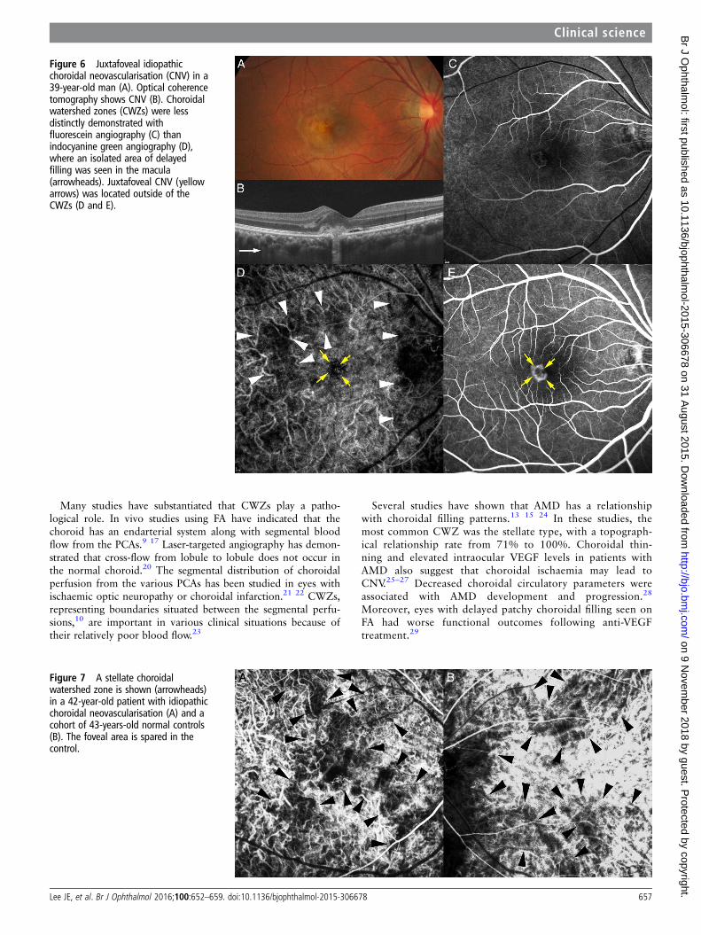
Figure 6 Juxtafoveal idiopathicchoroidal neovascularisation (CNV) in a39-year-old man (A). Optical coherencetomography shows CNV (B). Choroidalwatershed zones (CWZs) were lessdistinctly demonstrated withfluorescein angiography (C) thanindocyanine green angiography (D),where an isolated area of delayedfilling was seen in the macula(arrowheads). Juxtafoveal CNV (yellowarrows) was located outside of theCWZs (D and E).
Figure 7 A stellate choroidalwatershed zone is shown (arrowheads)in a 42-year-old patient with idiopathicchoroidal neovascularisation (A) and acohort of 43-years-old normal controls(B). The foveal area is spared in thecontrol.
Lee JE, et al. Br J Ophthalmol 2016;100:652–659. doi:10.1136/bjophthalmol-2015-306678 657
Clinical science on 9 N
ovember 2018 by guest. P
rotected by copyright.http://bjo.bm
j.com/
Br J O
phthalmol: first published as 10.1136/bjophthalm
ol-2015-306678 on 31 August 2015. D
ownloaded from
In our study, idiopathic CNV was related to the presence of aCWZ. A stellate or type IV CWZ was found most frequently,concordant with previous reports involving patients withAMD.13 14 The topographical relationship between idiopathicCNV and CWZs was found in 42 eyes (95.5%). We also found
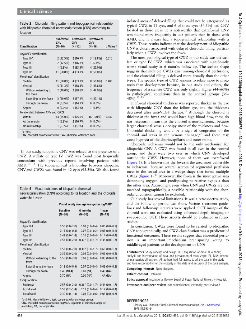
isolated areas of delayed filling that could not be categorised astypical CWZ in 11 eyes, and 6 of these eyes (54.5%) had CNVlocated in those areas. It is noteworthy that extrafoveal CNVwas found more frequently in our patients than in those withAMD, and it always had a topographical relationship with aCWZ. These results indicate that the development of idiopathicCNV is closely associated with delayed choroidal filling, particu-larly when a CWZ involves the fovea.
The most pathological type of CWZ in our study was the stel-late or type IV CWZ, which was associated with significantlyworse visual acuity at 6 months follow-up. The stellate shapesuggests that multiple CWZs exist among choroidal perfusions,and the choroidal filling is delayed more broadly than the othertypes. The specific type of CWZ appears to relate more to prog-nosis than development because, in our study and others, thefrequency of a stellate CWZ was only slightly higher (44–60%)in pathological conditions than in the control groups (35–48%).12 13
Subfoveal choroidal thickness was reported thicker in the eyewith idiopathic CNV than the fellow eye, and the thicknessdecreased after anti-VEGF therapy.30 Although the choroid isthickest at the fovea and would have high blood flow, these donot necessarily mean that the choroid is non-ischaemic, becauselarger choroidal vessels occupy most of the thickness and flow.Choroidal thickening would be a sign of congestion of thechoroid and stasis in the venous drainage,27 and these maycause hypoxia of the choriocapillaris and outer retina.
Choroidal ischaemia would not be the only mechanism foridiopathic CNV. A CWZ was found in all eyes in the controlgroup, and there were two eyes in which CNV developedoutside the CWZ. However, none of them was extrafoveal(figure 6). It is known that the fovea is the area most vulnerableto ischaemia, because several sectors of segmental perfusionmeet in the foveal area in a wedge shape that forms multipleCWZs (figure 1).17 Moreover, the fovea is the most active areademanding oxygen, and predisposing to relative hypoxia thanthe other area. Accordingly, even when CNV and CWZs are notmatched topographically, a possible relationship with the chor-oidal circulation cannot be excluded.
Our study has several limitations. It was a retrospective study,and the follow-up period was short. Various treatment guide-lines and follow-up intervals were applied. OCT images of thechoroid were not evaluated using enhanced depth imaging orswept-source OCT. These aspects should be evaluated in futurestudies.
In conclusion, CWZs were found to be related to idiopathicCNV topographically, and CWZ classification was a predictor offunctional outcomes. These results suggest that choroidal perfu-sion is an important mechanism predisposing young tomiddle-aged patients to the development of CNV.
Contributors Study concept and design: JEL; acquisition of data: all authors;analysis and interpretation of data, and preparation of manuscript: JEL, MKS; reviewof manuscript: all authors. All authors had full access to all the data in the studyand take responsibility for the integrity of the data and accuracy of the data analysis.
Competing interests None declared.
Patient consent Obtained.
Ethics approval Institutional Review Board of Pusan National University Hospital.
Provenance and peer review Not commissioned; externally peer reviewed.
REFERENCES1 Cleasby GW. Idiopathic focal subretinal neovascularization. Am J Ophthalmol
1976;81:590–9.
Table 3 Choroidal filling pattern and topographical relationshipwith idiopathic choroidal neovascularisation (CNV) according tolocation
Classification
SubfovealCNV(N=16)
JuxtafovealCNV(N=12)
ExtrafovealCNV(N=16) p Value*
Hayashi’s classificationType II-A 2 (12.5%) 2 (16.7%) 3 (18.8%) 0.510Type II-B 2 (12.5%) 2 (16.7%) 1 (6.3%)Type III 1 (6.3%) 4 (33.3%) 4 (25.0%)Type IV 11 (68.8%) 4 (33.3%) 8 (50.0%)
Mendrinos’ classification
Stellate 11 (68.8%) 4 (33.3%) 8 (50.0%) 0.400Vertical 5 (31.3%) 7 (58.3%) 7 (43.8%)
Without extending tothe fovea
2 (40.0%) 2 (28.6%) 3 (42.9%)
Extending to the fovea 3 (60.0%) 4 (57.1%) 4 (57.1%)Through the fovea 0 (0.0%) 1 (14.3%) 0 (0.0%)
Angled 0 (0.0%) 1 (8.3%) 1 (6.3%)Relationship between CNV and CWZ‡Within 14 (75.0%) 9 (75.0%) 16 (100%) 0.342At the margin 1 (6.3%) 2 (16.7%) 0 (0.0%)
No relationship 1 (6.3%) 1 (8.3%) 0 (0.0%)
*χ2 test.CNV, choroidal neovascularisation; CWZ, choroidal watershed zone.
Table 4 Visual outcomes of idiopathic choroidalneovascularisation (CNV) according to its location and the choroidalwatershed zone
Visual acuity average (range) in logMAR*
Baseline(N=36)
6 months(N=36)
1 year(N=19)
Hayashi’s classificationType II-A 0.56 (0.0–2.0) 0.08 (0.0–0.4) 0.05 (0.0–0.1)Type II-B 0.13 (0.0–0.3) 0.07 (0.0–0.2) 0.03 (0.0–0.1)Type III 0.41 (0.0–1.4) 0.10 (0.0–0.4) 0.14 (0.0–0.4)Type IV 0.53 (0.0–2.0) 0.30* (0.0–1.7) 0.38 (0.0–1.7)
Mendrinos’ classificationStellate 0.53 (0.0–2.0) 0.30* (0.0–1.7) 0.63 (0.0–1.7)Vertical 0.39 (0.0–2.0) 0.09 (0.0–0.4) 0.09 (0.0–0.4)
Without extending to thefovea
0.56 (0.0–2.0) 0.08 (0.0–0.4) 0.05 (0.0–0.1)
Extending to the fovea 0.18 (0.0–0.5) 0.07 (0.0–0.4) 0.06 (0.0–0.2)Through the fovea 1.40 (NA‡) 0.40 (NA) 0.40 (NA)
Angled 0.70 (NA) 0.00 (NA) NA (NA)CNV§ locationSubfoveal 0.57 (0.0–2.0) 0.36* (0.0–1.7) 0.44 (0.0–1.7)Juxtafoveal 0.58 (0.2–1.4) 0.11 (0.0–0.4) 0.17 (0.0–0.4)Extrafoveal 0.30 (0.0–1.4) 0.08 (0.0–0.6) 0.03 (0.0–0.2)
*p<0.05, Mann-Whitney U test, compared with the other groups.CNV, choroidal neovascularisation; logMAR, logarithm of minimum angle ofresolution; NA, not applicable.
658 Lee JE, et al. Br J Ophthalmol 2016;100:652–659. doi:10.1136/bjophthalmol-2015-306678
Clinical science on 9 N
ovember 2018 by guest. P
rotected by copyright.http://bjo.bm
j.com/
Br J O
phthalmol: first published as 10.1136/bjophthalm
ol-2015-306678 on 31 August 2015. D
ownloaded from
2 Ho AC, Yannuzzi LA, Pisicano K, et al. The natural history of idiopathic subfovealchoroidal neovascularization. Ophthalmology 1995;102:782–9.
3 Toju R, Iida T, Sekiryu T, et al. Near-infrared autofluorescence in patients withidiopathic submacular choroidal neovascularization. Am J Ophthalmol2012;153:314–19.
4 Yang F, Dou HL, Ma Z, et al. Serum inflammatory factors in patients with idiopathicchoroidal neovascularization. Ocul Immunol Inflamm 2010;18:390–4.
5 Zhang H, Liu ZL, Sun P, et al. Intravitreal bevacizumab for treatment of subfovealidiopathic choroidal neovascularization: results of a 1-year prospective trial. Am JOphthalmol 2012;153:300–6.
6 Mandal S, Garg S, Venkatesh P, et al. Intravitreal bevacizumab for subfovealidiopathic choroidal neovascularization. Arch Ophthalmol 2007;125:1487–92.
7 Kang HM, Koh HJ. Intravitreal anti-vascular endothelial growth factor therapy versusphotodynamic therapy for idiopathic choroidal neovascularization. Am J Ophthalmol2013;155:713–19.
8 Inoue M, Kadonosono K, Watanabe Y, et al. Results of 1-year follow-upexaminations after intravitreal bevacizumab administration for idiopathic choroidalneovascularization. Retina 2010;30:733–8.
9 Hayreh SS. Segmental nature of the choroidal vasculature. Br J Ophthalmol1975;59:631–48.
10 Hayreh SS. In vivo choroidal circulation and its watershed zones. Eye 1990;4(Pt 2):273–89.
11 Giuffre G. Main posterior watershed zone of the choroid. Variations of its positionin normal subjects. Doc Ophthalmol 1989;72:175–80.
12 Hayashi K, de Laey JJ. Indocyanine green angiography of submacular choroidalvessels in the human eye. Ophthalmologica 1985;190:20–9.
13 Mendrinos E, Pournaras CJ. Topographic variation of the choroidal watershed zoneand its relationship to neovascularization in patients with age-related maculardegeneration. Acta Ophthalmol 2009;87:290–6.
14 Hayashi K, de Laey JJ. Indocyanine green angiography of choroidal neovascularmembranes. Ophthalmologica 1985;190:30–9.
15 Ross RD, Barofsky JM, Cohen G, et al. Presumed macular choroidal watershedvascular filling, choroidal neovascularization, and systemic vascular disease inpatients with age-related macular degeneration. Am J Ophthalmol1998;125:71–80.
16 Macular Photocoagulation Study Group. Argon laser photocoagulation for senilemacular degeneration: results of a randomized clinical trial. Arch Ophthalmol1982;100:912–18.
17 Hayreh SS. Submacular choroidal vascular pattern. Experimental fluorescein fundusangiographic studies. Graefes Arch Clin Exp Ophthalmol 1974;192:181–96.
18 Hayreh SS. The choriocapillaris. Graefes Arch Clin Exp Ophthalmol 1974;192:165–79.19 Shimizu K, Ujiie K. Morphology of the submacular choroid: vascular structure.
Ophthalmologica 1981;183:5–10.20 Hirata Y, Nishiwaki H, Miura S, et al. Analysis of choriocapillaris flow patterns by
continuous laser-targetd angiography in monkeys. Invest Ophthalmol Vis Sci2004;45:1954–62.
21 Hayreh SS, Baines JA. Occlusion of the posterior ciliary artery. 3. Effects on the opticnerve head. Br J Ophthalmol 1972;56:754–64.
22 Hayreh SS, Baines JA. Occlusion of the posterior ciliary artery. II. Chorio-retinallesions. Br J Ophthalmol 1972;56:736–53.
23 Hayreh SS. Posterior ciliary artery circulation in health and disease: the Weisenfeldlecture. Invest Ophthalmol Vis Sci 2004;45:749–57;748.
24 Giovannini A, Mariotti C, Ripa E, et al. Choroidal filling in age-related maculardegeneration: indocyanine green angiographic findings. Ophthalmologica1994;208:185–91.
25 Tong JP, Chan WM, Liu DT, et al. Aqueous humor levels of vascular endothelialgrowth factor and pigment epithelium-derived factor in polypoidal choroidalvasculopathy and choroidal neovascularization. Am J Ophthalmol 2006;141:456–62.
26 Spaide RF. Age-related choroidal atrophy. Am J Ophthalmol 2009;147:801–10.27 Chung SE, Kang SW, Lee JH, et al. Choroidal thickness in polypoidal choroidal
vasculopathy and exudative age-related macular degeneration. Ophthalmology2011;118:840–5.
28 Xu W, Grunwald JE, Metelitsina TI, et al. Association of risk factors for choroidalneovascularization in age-related macular degeneration with decreased foveolarchoroidal circulation. Am J Ophthalmol 2010;150:40–7.
29 Gewaily DY, Grunwald JE, Pistilli M, et al. Delayed patchy choroidal filling in theComparison of Age-Related Macular Degeneration Treatments Trials (CATT). Am JOphthalmol 2014;158:525–31.
30 Ahn SJ, Kim TW, Ahn J, et al. Subfoveal choroidal thickness in idiopathic choroidalneovascularization. Ophthalmology 2014:34;1554–9.
Lee JE, et al. Br J Ophthalmol 2016;100:652–659. doi:10.1136/bjophthalmol-2015-306678 659
Clinical science on 9 N
ovember 2018 by guest. P
rotected by copyright.http://bjo.bm
j.com/
Br J O
phthalmol: first published as 10.1136/bjophthalm
ol-2015-306678 on 31 August 2015. D
ownloaded from


![Y [dYo [Wioäjeä dijWbbäWdZäi[hl Y[ cWZ[äm j^äm[ bCcybW … · 2014-01-10 · D { C y w Dy ^_]^ä[\\_Y_[dYo [Wioäjeä_dijWbbäWdZäi[hl_Y[cWZ[äm_j^äm[_bCcybW_dägkWb_jo]läCä]](https://img.pdfslide.net/doc/110x75/5f02433e7e708231d40363a9/y-dyo-wioje-dijwbbwdzihl-y-cwzm-jm-bccybw-2014-01-10-d-c.jpg)