Embed Size (px)
Citation preview
Clinical StudyHealing of the Acutely Injured AnteriorCruciate Ligament: Functional Treatment with the ACL-Jack,a Dynamic Posterior Drawer Brace
Matthias Jacobi,1,2 Nikolaus Reischl,2,3 Karolin Rönn,2,4 Robert A. Magnusson,5
Emanuel Gautier,2 and Roland P. Jakob2,6
1Orthopadie Rosenberg, Rosenbergstrasse 150, St. Gallen, Switzerland2Department of Orthopedic Surgery, HFR Hopital Cantonal, Fribourg, Switzerland3Private Clinic Hansa Graz, Korblergasse 42, 8010 Graz, Austria4Schulthess Klinik, Lengghalde 2, Zurich, Switzerland5Department of Orthopaedic Surgery, Sports Health and Performance Institute, The Ohio State University, Columbus, OH, USA6En Chambaz, Motier, Switzerland
Correspondence should be addressed to Matthias Jacobi; [email protected]
Received 7 September 2016; Accepted 11 October 2016
Academic Editor: Elizaveta Kon
Copyright © 2016 Matthias Jacobi et al.This is an open access article distributed under the Creative Commons Attribution License,which permits unrestricted use, distribution, and reproduction in any medium, provided the original work is properly cited.
Background. The injured anterior cruciate ligament (ACL) has a limited healing capacity leading to persisting instability.Hypothesis/Purpose. To study if the application of a brace, producing a dynamic posterior drawer force, after acute ACL injuryreduces initial instability. Study Design. Cohort study.Methods. Patients treated with the ACL-Jack brace were compared to controlstreatedwith primaryACL reconstruction und controls treated nonsurgically with functional rehabilitation.Measurements includedanterior laxity (Rolimeter), clinical scores (Lysholm, Tegner, and IKDC), and MRI evaluation. Patients were followed up to 24months. Results. Patients treated with the ACL-Jack brace showed a significant improvement of anterior knee laxity comparableto patients treated with ACL reconstruction, whereas laxity persisted after nonsurgical functional rehabilitation. The failure risk(secondary reconstruction necessary) of the ACL-Jack group was however 21% (18 of 86) within 24 months. Clinical scores weresimilar in all treatment groups. Conclusion. Treatment of acute ACL tears with the ACL-Jack brace leads to improved anterior kneelaxity compared to nonsurgical treatment with functional rehabilitation.
1. Introduction
The acutely injured anterior cruciate ligament (ACL) hasa poor healing capacity, resulting regularly in persistentinstability of the knee [1, 2]. Surgical reconstruction hasbecome an accepted treatment to restore ACL stability in theyounger and more active patient [3]. The reason that ACLhealing is uncommon is not fully understood, but biological,biomechanical, and anatomical factors all likely contribute[4, 5]. The ACL, in contrast to extra-articular ligaments, doesnot form a fibrin-platelet clot to initiate tissue healing. Clotformation is likely inhibited by factors in the surroundingsynovial fluid [4, 6]. Further, during rehabilitation and
normal daily activities following ACL injury, the quadriceps-induced anterior drawer and other movements of the kneecan pull the ligament stumps apart [7], potentially resultingin a lengthened ligament even in cases in which healingdoes occur. Finally, the positions of the ligament stumps maybe such that there is no contact between them after injury,effectively preventing healing.
The ACL does have characteristics that could promotehealing. For example, the ligament is well vascularized, whichis required for tissues healing [8, 9]. Different methods havebeen undertaken to enhance healing of the ACL, includingprimary suture repair, healing response techniques, immobi-lization, bracing, and supplementation with scaffolds, growth
Hindawi Publishing CorporationAdvances in OrthopedicsVolume 2016, Article ID 1609067, 7 pageshttp://dx.doi.org/10.1155/2016/1609067
2 Advances in Orthopedics
factors, and collagen-platelet composites [5, 10–14]. Althoughprimary ACL suture has been shown to improve laxity inthe short term, it has shown a high failure rate with longerfollow-up [5, 11, 14]. Several functional knee braces have beenevaluated and noted not to affect knee anterior laxity [10, 15,16]. Fujimoto et al., however, showed in a group of 31 patientswith low athletic demands that bracing with an extensionblock improved stability in 74% of patients, but 26% of thepatients went on to require ACL reconstruction [17]. Biologicstrategies to enhance ACL healing are quite promising, butonly in vitro and animal studies are available currently [4, 5].Internal stabilization techniques report promising results.They also rely on the self-healing of the injured ACL [18–22].
The purpose of the present study was to assess whetherACL healing and final knee laxity can be improved in patientswith acute ACL injuries by altering the biomechanical con-ditions during healing through the use of a brace producinga dynamic posterior drawer force. We hypothesized that (1)ACL healing and anterior laxity of the knee are improvedthrough the use of the ACL-Jack brace relative to a controlgroup with no brace and (2) that in patients in whom useof the ACL-Jack brace results in satisfactory knee functionanterior laxity is equal to that achieved with primary ACLreconstruction.
2. Patients and Method
2.1. Inclusion. From March 2004 to February, 2009, 86patients with acute ACL injury were enrolled in a prospectivestudy at our institution to evaluate the effectiveness the ACL-Jack brace for management of acute ACL injuries. Addition-ally 40 patients were enrolled to compare the treatment withthe ACL-Jack brace to two standard treatments (20 patientseach). Patients were recruited and enrolled with the followinginclusion criteria:
(i) acute injury (<3 weeks),(ii) complete or subtotal ACL tear confirmed clinically
and with MRI,(iii) informed consent of the patient about the planned
therapy including possible complications and draw-backs,
(iv) patients with associated grade I or II MCL injuryincluded.
Additional treatment was provided in the following situa-tions:
(i) In case of meniscal tears, either a partial menis-cectomy or meniscal repair was performed prior tobracing.
(ii) If the ruptured ACL showed anteriorly displacedfibers on MRI, these fibers were reduced arthroscop-ically.
Exclusion criteria were
(i) patients unwilling to follow the treatment protocol orinability to comprehend it,
(ii) ACL injuries older than 3 weeks,
(iii) associated injury of the PCL, LCL, MCL (grade III),or any other lesion requiring surgery.
Allocation to the groups relied on patient’s choice afterinformed consent.
2.2. Study Groups
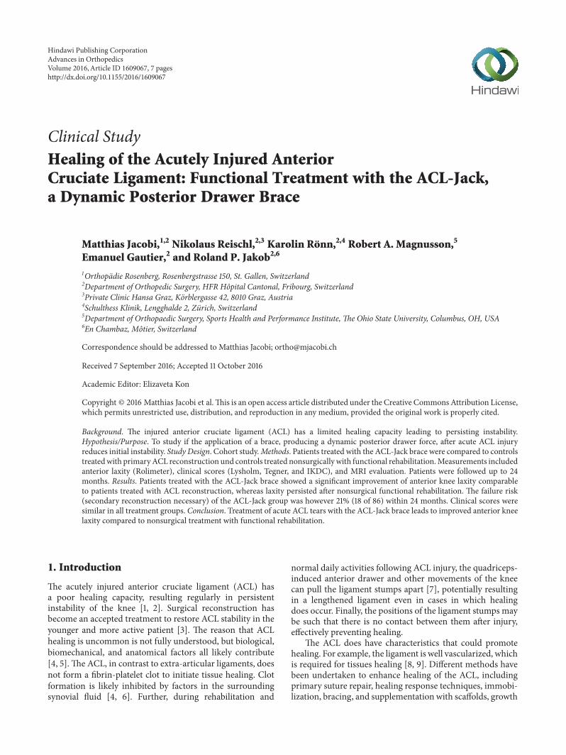
2.2.1. ACL-Jack Group. Patients in this main study groupwere treatedwith theACL-Jack brace.Theprefabricated bracewas adjusted by an orthopaedic technician and worn forthree months day and night and for an additional monthduring daytime only. Full weight bearing was allowed fromthe start of the treatment. Range of movement to the extentpossible in the brace was allowed, giving patients a rangeof flexion of about 0∘ to 100∘. Removal of the brace wasallowed in 90∘ of knee flexion (sitting position) withoutquadriceps contraction. With the knee in flexion it wasalso the recommended position to take a shower. Specialattention was given to the instruction to patients with writteninformation and regular assessment of compliance in theinitial phase of treatment. After four months, the brace wasremoved and exercises and physiotherapy were started to aidthe recovery of muscle strength and full mobility. Sportingactivity, including cutting and pivoting, was allowed aftersix months. Patients received thromboprophylaxis duringthe first four weeks of treatment with low-molecular-weightheparin due to the compressive nature of the brace.
2.2.2. Functional Treatment Group. This group underwent afunctional rehabilitation protocol in physiotherapy (musclestrengthening coordination and proprioception program)without any brace for 2 to 4 months.
2.2.3. Primary ACL Reconstruction Group. This group under-went primary reconstruction with an anatomic single bundle(patellar tendon) technique. Tunnels were drilled on thefemoral side with an outside-in drill guide. Femoral fixationwas performed with a press fit technique (conical bone blockin a conical tunnel). Tibial fixation was accomplished with aninterference screw.
For subgroup analysis groupswere divided in successfullytreated and failed patients if necessary.
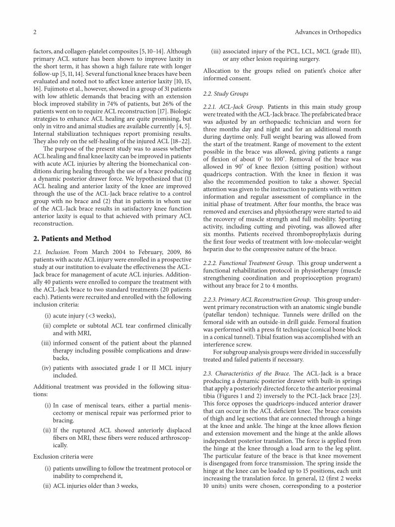
2.3. Characteristics of the Brace. The ACL-Jack is a braceproducing a dynamic posterior drawer with built-in springsthat apply a posteriorly directed force to the anterior proximaltibia (Figures 1 and 2) inversely to the PCL-Jack brace [23].This force opposes the quadriceps-induced anterior drawerthat can occur in the ACL deficient knee. The brace consistsof thigh and leg sections that are connected through a hingeat the knee and ankle. The hinge at the knee allows flexionand extension movement and the hinge at the ankle allowsindependent posterior translation. The force is applied fromthe hinge at the knee through a load arm to the leg splint.The particular feature of the brace is that knee movementis disengaged from force transmission. The spring inside thehinge at the knee can be loaded up to 15 positions, each unitincreasing the translation force. In general, 12 (first 2 weeks10 units) units were chosen, corresponding to a posterior
Advances in Orthopedics 3
Figure 1: Photograph of the ACL-Jack brace.
Spring
1
42
5 3
F
Figure 2: Diagram showing that the brace consists of an upper thigh(1) and a leg part (2) connected through a hinge at the ankle (3) andknee (4).The load is applied through a relocatable load arm (5) fromthe hinge to the leg part, which rotates around the distal hinge (3).𝐹 = force.
force of 6 kg to 7 kg. The force is maintained throughout fullrange of movement [24]. To reduce the direct pressure to theanterior rim of the tibia the prefabricated brace was adaptedin a way that themain pressure was appliedmedial and lateralto the tibial metaphysis. Additionally the anterior skin andsoft tissues were protected by pads.
2.4. Initial Assessment. All patients underwent routine clini-cal examination of the knee. Anterior knee laxity was assessedusing the Rolimeter arthrometer (Aircast; DJO, Vista, Cali-fornia) in 20∘ of knee flexion in comparisonwith the oppositeside. Every knee was evaluated with MRI with a special focuson anteriorly displaced fibers of the injured ACL. Examina-tions were done by the first or the senior author. Preinjurypatient-reported knee scoring was done using the Lysholmknee function scoring scale [25], the Tegner activity levelrating scale [26], and the International Knee DocumentationCommittee (IKDC) knee scoring system [27].
2.5. Follow-Up Assessment. For all studied groups, scheduledfollow-up appointments took place at six, 12, and 24 months.They involved clinical examination of the knee, bilateral com-parative Rolimeter arthrometry. Examinations were done bythe first or the senior author. An MRI was performed aftersixmonths and evaluated by an independent radiologist.MRIwas not performed in the primaryACL reconstruction group.Patients completed the follow-up by evaluation using theLysholm scale, the Tegner scale, and the IKDC Score at 12 and24 months.
2.6. Statistical Analysis. Data are presented asmean, standarddeviation, and range. Due to data distribution, nonparamet-ric analysis techniques were utilized, including the Wilcoxonsigned-rank test, Kruskal-Wallis test, and Friedman test. A 𝑝value ≤ 0.05 was considered to be significant. Calculationsand graphs were performed using MedCalc Software version10.4.8.0 (MedCalc Software Buba, Mariakerke, Belgium).
3. Results
3.1. Baseline Data. The ACL-Jack group consisted of 86patients, of which 84 (98%) had complete follow-up. Onepatient moved abroad and one other was lost to follow-up.Of the 84 remaining patients 18 (21%) required a secondaryACL reconstruction due to persistent and disabling instability(𝑛 = 13) or repeat injury (𝑛 = 5) within 24 months (Table 1).
The functional treatment and the primary ACL recon-struction group consisted both of 20 patients and had 100%follow-up. Six patients (30%) in the functional treatmentgroup required secondary ACL reconstruction due to dis-abling instability within 24 months. No recurrent instabilityepisodes or revision ACL reconstructions occurred in theprimary ACL reconstruction group (Table 1).
As allocation to the groups was based on patients choice,patients treated with the ACL-Jack and the functional treat-ment group were both significantly older (𝑝 = 0.00002)and had a higher female to male ratio than the primaryreconstruction group (Table 1).
The highest failure risk was observed among young menwith higher level sport activities on the Tegner scale (Table 2).
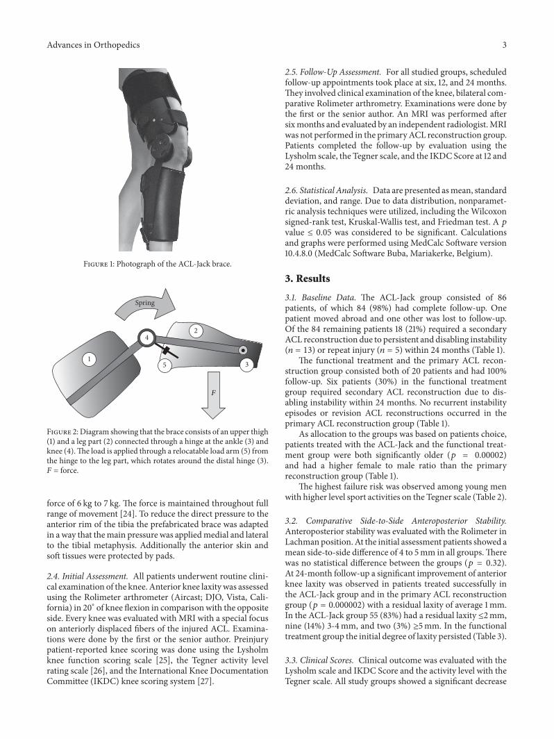
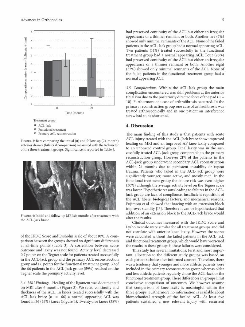
3.2. Comparative Side-to-Side Anteroposterior Stability.Anteroposterior stability was evaluated with the Rolimeter inLachmanposition. At the initial assessment patients showed amean side-to-side difference of 4 to 5mm in all groups.Therewas no statistical difference between the groups (𝑝 = 0.32).At 24-month follow-up a significant improvement of anteriorknee laxity was observed in patients treated successfully inthe ACL-Jack group and in the primary ACL reconstructiongroup (𝑝 = 0.000002) with a residual laxity of average 1mm.In the ACL-Jack group 55 (83%) had a residual laxity ≤2mm,nine (14%) 3-4mm, and two (3%) ≥5mm. In the functionaltreatment group the initial degree of laxity persisted (Table 3).
3.3. Clinical Scores. Clinical outcome was evaluated with theLysholm scale and IKDC Score and the activity level with theTegner scale. All study groups showed a significant decrease
4 Advances in Orthopedics
Table 1: Baseline data of the ACL-Jack group, the functional treatment group, and the primary ACL reconstruction group.
ACL-Jack Functional treatment Primary ACL reconstruction 𝑝
Included patientsPatients 𝑛 86 20 20Dropouts (total) 𝑛 (%) 20 (23%) 6 (30%) 0
(i) Failures 𝑛 (%) 18 (20.9%) 6 (30%) 0(ii) Lost to follow up 𝑛 (%) 2 (2%) 0 0
Age years 32 ± 14 (14–74) 35 ± 10 (21–48) 23 ± 7 (15–40) 0.00002Sex M/F 52/33 13/7 14/6Side R/L 48/37 8/12 12/8Meniscus tear 𝑛 (%) 11 (12%) 0 6 (30%)ACL displaced 𝑛 (%) 28 (33%) 6 (30%) —Injury to treatment days 14 ± 10 (10–21) — 37 ± 26 (10–89) 0.0001
Analyzed patientsPatients 𝑛 66 14 20Sex M/F 36/30 8/6 14/6Side R/L 38/28 5/9 12/8Meniscus tear 𝑛 (%) 9 (14%) 0 (0%) 6 (30%)ACL displaced 𝑛 (%) 23 (27%) 4 (29%) —Injury to treatment days 14 ± 8 (10–21) — 37 ± 26 (10–89) 0.0001
Table 2: Comparative data of successfully treated and failed patients within the ACL-Jack group.
ACL-Jack groupSuccessful Failures 𝑝
Patients 𝑛 66 18Age Years 34 ± 15 (14–74) 24 ± 12 (15–57) 0.00002Sex M/F 36/31 16/2Meniscus tear 𝑛 (%) 9 (14%) 2 (11%) n.s.ACL displaced 𝑛 (%) 23 (27%) 5 (28%) n.s.Injury to treatment Days 13 ± 5 (3–21) 14 ± 7 (5–21) n.s.
Table 3: Clinical outcome and side-to-side ACL stability (Rolimeter) of the ACL-Jack (successful), functional treatment (successful), andprimary ACL reconstruction group.
ACL-Jack 𝑝 Functional treatment 𝑝 Primary ACL reconstruction 𝑝 𝑝 intergroupTegner preinjury 6.6 ± 2 (4–10)
<0.000015.1 ± 1.4 (2–6)
<0.000018.6 ± 1.3 (5–10)
0.000260.00002
Tegner 12 months 5.6 ± 2.1 (3–10) 3.4 ± 0.9 (2–5) 7.7 ± 1.8 (4–10) <0.000001Tegner 24 months 5.9 ± 2 (3–10) 3.5 ± 1 (2–5) 7.9 ± 1.7 (4–10) <0.000001Lysholm preinjury 99.7 ± 1.2 (95–100)
<0.00001100 ± 0 (100-100)
<0.0000198.6 ± 2.4 (94–100)
<0.000010.047
Lysholm 12 months 92.8 ± 8.6 (67–100) 93.7 ± 6.3 (79–100) 88.4 ± 6.9 (79–100) 0.055Lysholm 24 months 93.3 ± 8.3 (67–100) 92.7 ± 7.4 (67–100) 89.1 ± 7.7 (74–100) 0.034IKDC preinjury 96.5 ± 5.2 (72–100)
<0.0000197 ± 3.5 (91–100)
<0.0000198.4 ± 3 (90–100)
<0.000010.17
IKDC 12 months 88.7 ± 9.4 (58–100) 85.2 ± 9.1 (66–100) 88.1 ± 8.4 (72–100) 0.72IKDC 24 months 90 ± 8.7 (69–100) 86.4 ± 11 (66–100) 88.3 ± 8.6 (74–100) 0.37Diff injury (mm) 4.3 ± 2 (3–11)
<0.00001
4.5 ± 2.5 (2–10)
0.71
4.6 ± 0.8 (4–6)
<0.0001
0.32Diff 6 months (mm) 0.9 ± 1.8 (0–4.5) — — —Diff 12 months (mm) 1 ± 1.4 (0–4) — — —Diff 24 months (mm) 1.1 ± 2 (0–5) 4.8 ± 2.4 (2–8) 0.9 ± 1.1 (0–3) 0.000002
Advances in Orthopedics 5
−1
0
0Time (month)
24
Treatment group ACL-JackFunctional treatment Primary ACL reconstruction
Com
para
tive a
nter
ior d
raw
er (R
olim
eter
) (m
m)
1
2
3
4
5
6
7
8
Figure 3: Bars comparing the initial (0) and follow-up (24-month)anterior drawer (bilateral comparison)measuredwith the Rolimeterof the three treatment groups. Significance is reported in Table 3.
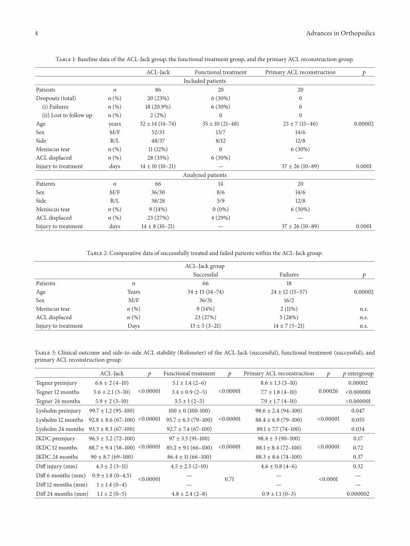
Figure 4: Initial and follow-upMRI sixmonths after treatment withthe ACL-Jack brace.
of the IKDC Score and Lysholm scale of about 10%. A com-parison between the groups showed no significant differencesat all-time points (Table 3). A correlation between scoreoutcome and laxity was not found. Activity level decreased0.7 points on the Tegner scale for patients treated successfullyin the ACL-Jack group and the primary ACL reconstructiongroup and 1.6 points for the functional treatment group. 39 ofthe 66 patients in the ACL-Jack group (59%) reached on theTegner scale the preinjury activity level.
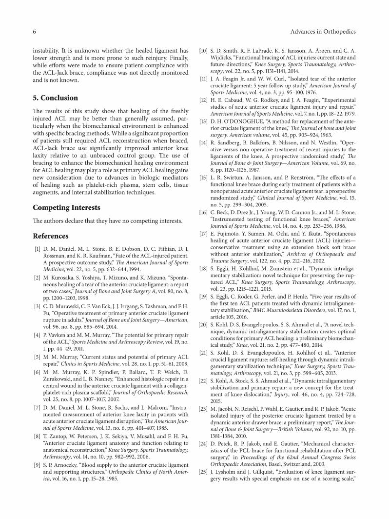
3.4. MRI Findings. Healing of the ligament was documentedon MRI after 6 months (Figure 3). We rated continuity andthickness of the ACL. In knees treated successfully with theACL-Jack brace (𝑛 = 66) a normal appearing ACL wasfound in 36 (55%) knees (Figure 4). Twenty-five knees (38%)
had preserved continuity of the ACL but either an irregularappearance or a thinner remnant or both. Another five (7%)showedonlyminimal remnants of theACL.None of the failedpatients in the ACL-Jack group had a normal appearing ACL.Two patients (14%) treated successfully in the functionaltreatment group had a normal appearing ACL. Four (28%)had preserved continuity of the ACL but either an irregularappearance or a thinner remnant or both. Another eight(57%) showed only minimal remnants of the ACL. None ofthe failed patients in the functional treatment group had anormal appearing ACL.
3.5. Complications. Within the ACL-Jack group the maincomplication encountered was skin problems at the anteriortibial rim due to the posteriorly directed force of the pad (𝑛 =10). Furthermore one case of arthrofibrosis occurred. In theprimary reconstruction group one case of arthrofibrosis wastreated arthroscopically and in one patient an interferencescrew had to be shortened.
4. Discussion
The main finding of this study is that patients with acuteACL injury treated with the ACL-Jack brace show improvedhealing on MRI and an improved AP knee laxity comparedto an unbraced control group. Final laxity was in the suc-cessfully treated ACL-Jack group comparable to the primaryreconstruction group. However 21% of the patients in theACL-Jack group underwent secondary ACL reconstructionwithin 24 months due to persistent instability or repeattrauma. Patients who failed in the ACL-Jack group weresignificantly younger, more active, and mostly men. In thefunctional treatment group the failure risk was even higher(30%) although the average activity level on the Tegner scalewas lower. Hypothetic reasons leading to failures in the ACL-Jack group are lack of compliance, insufficient reposition ofthe ACL fibers, biological factors, and mechanical reasons.Fujimoto et al. showed that bracing with an extension blockimproves stability [17]. Therefore it can be hypothesized thataddition of an extension block to the ACL-Jack brace wouldalter the results.
Clinical outcomes measured with the IKDC Score andLysholm scale were similar for all treatment groups and didnot correlate with anterior knee laxity. However the scoreswere calculated without the failed patients in the ACL-Jackand functional treatment group, which would have worsenedthe results in these groups if these failures were considered.
This study has several limitations. First and most impor-tant, allocation to the different study groups was based oneach patient’s choice after informed consent. Therefore, therewas a tendency that younger and more athletic patients wereincluded in the primary reconstruction group whereas olderand less athletic patients regularly chose the ACL-Jack or thefunctional treatment group.These differences in groups limitconclusive comparison of outcomes. We however assumethat comparison of knee laxity is meaningful within thethree groups. Furthermore no information is available aboutbiomechanical strength of the healed ACL. At least fivepatients sustained a new relevant injury with recurrent
6 Advances in Orthopedics
instability. It is unknown whether the healed ligament haslower strength and is more prone to such reinjury. Finally,while efforts were made to ensure patient compliance withthe ACL-Jack brace, compliance was not directly monitoredand is not known.
5. Conclusion
The results of this study show that healing of the freshlyinjured ACL may be better than generally assumed, par-ticularly when the biomechanical environment is enhancedwith specific bracingmethods.While a significant proportionof patients still required ACL reconstruction when braced,ACL-Jack brace use significantly improved anterior kneelaxity relative to an unbraced control group. The use ofbracing to enhance the biomechanical healing environmentfor ACL healingmay play a role as primaryACL healing gainsnew consideration due to advances in biologic mediatorsof healing such as platelet-rich plasma, stem cells, tissueaugments, and internal stabilization techniques.
Competing Interests
The authors declare that they have no competing interests.
References
[1] D. M. Daniel, M. L. Stone, B. E. Dobson, D. C. Fithian, D. J.Rossman, and K. R. Kaufman, “Fate of the ACL-injured patient.A prospective outcome study,” The American Journal of SportsMedicine, vol. 22, no. 5, pp. 632–644, 1994.
[2] M. Kurosaka, S. Yoshiya, T. Mizuno, and K. Mizuno, “Sponta-neous healing of a tear of the anterior cruciate ligament: a reportof two cases,” Journal of Bone and Joint Surgery A, vol. 80, no. 8,pp. 1200–1203, 1998.
[3] C.D.Murawski, C. F.VanEck, J. J. Irrgang, S. Tashman, andF.H.Fu, “Operative treatment of primary anterior cruciate ligamentrupture in adults,” Journal of Bone and Joint Surgery—American,vol. 96, no. 8, pp. 685–694, 2014.
[4] P. Vavken and M. M. Murray, “The potential for primary repairof theACL,” SportsMedicine andArthroscopy Review, vol. 19, no.1, pp. 44–49, 2011.
[5] M. M. Murray, “Current status and potential of primary ACLrepair,” Clinics in Sports Medicine, vol. 28, no. 1, pp. 51–61, 2009.
[6] M. M. Murray, K. P. Spindler, P. Ballard, T. P. Welch, D.Zurakowski, and L. B. Nanney, “Enhanced histologic repair in acentral wound in the anterior cruciate ligament with a collagen-platelet-rich plasma scaffold,” Journal of Orthopaedic Research,vol. 25, no. 8, pp. 1007–1017, 2007.
[7] D. M. Daniel, M. L. Stone, R. Sachs, and L. Malcom, “Instru-mented measurement of anterior knee laxity in patients withacute anterior cruciate ligament disruption,”TheAmerican Jour-nal of Sports Medicine, vol. 13, no. 6, pp. 401–407, 1985.
[8] T. Zantop, W. Petersen, J. K. Sekiya, V. Musahl, and F. H. Fu,“Anterior cruciate ligament anatomy and function relating toanatomical reconstruction,” Knee Surgery, Sports Traumatology,Arthroscopy, vol. 14, no. 10, pp. 982–992, 2006.
[9] S. P. Arnoczky, “Blood supply to the anterior cruciate ligamentand supporting structures,” Orthopedic Clinics of North Amer-ica, vol. 16, no. 1, pp. 15–28, 1985.
[10] S. D. Smith, R. F. LaPrade, K. S. Jansson, A. Arøen, and C. A.Wijdicks, “Functional bracing of ACL injuries: current state andfuture directions,” Knee Surgery, Sports Traumatology, Arthro-scopy, vol. 22, no. 5, pp. 1131–1141, 2014.
[11] J. A. Feagin Jr. and W. W. Curl, “Isolated tear of the anteriorcruciate ligament: 5 year follow up study,” American Journal ofSports Medicine, vol. 4, no. 3, pp. 95–100, 1976.
[12] H. E. Cabaud, W. G. Rodkey, and J. A. Feagin, “Experimentalstudies of acute anterior cruciate ligament injury and repair,”American Journal of SportsMedicine, vol. 7, no. 1, pp. 18–22, 1979.
[13] D. H. O’DONOGHUE, “A method for replacement of the ante-rior cruciate ligament of the knee,”The Journal of bone and jointsurgery. American volume, vol. 45, pp. 905–924, 1963.
[14] R. Sandberg, B. Balkfors, B. Nilsson, and N. Westlin, “Oper-ative versus non-operative treatment of recent injuries to theligaments of the knee. A prospective randomized study,” TheJournal of Bone & Joint Surgery—American Volume, vol. 69, no.8, pp. 1120–1126, 1987.
[15] L. R. Swirtun, A. Jansson, and P. Renstrom, “The effects of afunctional knee brace during early treatment of patients with anonoperated acute anterior cruciate ligament tear: a prospectiverandomized study,” Clinical Journal of Sport Medicine, vol. 15,no. 5, pp. 299–304, 2005.
[16] C. Beck, D. Drez Jr., J. Young,W. D. Cannon Jr., andM. L. Stone,“Instrumented testing of functional knee braces,” AmericanJournal of Sports Medicine, vol. 14, no. 4, pp. 253–256, 1986.
[17] E. Fujimoto, Y. Sumen, M. Ochi, and Y. Ikuta, “Spontaneoushealing of acute anterior cruciate ligament (ACL) injuries—conservative treatment using an extension block soft bracewithout anterior stabilization,” Archives of Orthopaedic andTrauma Surgery, vol. 122, no. 4, pp. 212–216, 2002.
[18] S. Eggli, H. Kohlhof, M. Zumstein et al., “Dynamic intraliga-mentary stabilization: novel technique for preserving the rup-tured ACL,” Knee Surgery, Sports Traumatology, Arthroscopy,vol. 23, pp. 1215–1221, 2015.
[19] S. Eggli, C. Roder, G. Perler, and P. Henle, “Five year results ofthe first ten ACL patients treated with dynamic intraligamen-tary stabilisation,” BMCMusculoskeletal Disorders, vol. 17, no. 1,article 105, 2016.
[20] S. Kohl, D. S. Evangelopoulos, S. S. Ahmad et al., “A novel tech-nique, dynamic intraligamentary stabilization creates optimalconditions for primary ACL healing: a preliminary biomechan-ical study,” Knee, vol. 21, no. 2, pp. 477–480, 2014.
[21] S. Kohl, D. S. Evangelopoulos, H. Kohlhof et al., “Anteriorcrucial ligament rupture: self-healing through dynamic intrali-gamentary stabilization technique,” Knee Surgery, Sports Trau-matology, Arthroscopy, vol. 21, no. 3, pp. 599–605, 2013.
[22] S. Kohl, A. Stock, S. S. Ahmad et al., “Dynamic intraligamentarystabilization and primary repair: a new concept for the treat-ment of knee dislocation,” Injury, vol. 46, no. 4, pp. 724–728,2015.
[23] M. Jacobi, N. Reischl, P.Wahl, E. Gautier, and R. P. Jakob, “Acuteisolated injury of the posterior cruciate ligament treated by adynamic anterior drawer brace: a preliminary report,”The Jour-nal of Bone & Joint Surgery—British Volume, vol. 92, no. 10, pp.1381–1384, 2010.
[24] D. Petek, R. P. Jakob, and E. Gautier, “Mechanical character-istics of the PCL-brace for functional rehabilitation after PCLsurgery,” in Proceedings of the 62nd Annual Congress SwissOrthopaedic Association, Basel, Switzerland, 2003.
[25] J. Lysholm and J. Gillquist, “Evaluation of knee ligament sur-gery results with special emphasis on use of a scoring scale,”
Advances in Orthopedics 7
American Journal of Sports Medicine, vol. 10, no. 3, pp. 150–154,1982.
[26] Y. Tegner and J. Lysholm, “Rating systems in the evaluationof knee ligament injuries,” Clinical Orthopaedics and RelatedResearch, vol. 198, pp. 43–49, 1985.
[27] E. Hefti, W. Muller, R. P. Jakob, and H.-U. Staubli, “Evaluationof knee ligament injuries with the IKDC form,” Knee Surgery,Sports Traumatology, Arthroscopy, vol. 1, no. 3-4, pp. 226–234,1993.

Submit your manuscripts athttp://www.hindawi.com
Stem CellsInternational
Hindawi Publishing Corporationhttp://www.hindawi.com Volume 2014
Hindawi Publishing Corporationhttp://www.hindawi.com Volume 2014
MEDIATORSINFLAMMATION
of
Hindawi Publishing Corporationhttp://www.hindawi.com Volume 2014
Behavioural Neurology
EndocrinologyInternational Journal of
Hindawi Publishing Corporationhttp://www.hindawi.com Volume 2014
Hindawi Publishing Corporationhttp://www.hindawi.com Volume 2014
Disease Markers
Hindawi Publishing Corporationhttp://www.hindawi.com Volume 2014
BioMed Research International
OncologyJournal of
Hindawi Publishing Corporationhttp://www.hindawi.com Volume 2014
Hindawi Publishing Corporationhttp://www.hindawi.com Volume 2014
Oxidative Medicine and Cellular Longevity
Hindawi Publishing Corporationhttp://www.hindawi.com Volume 2014
PPAR Research
The Scientific World JournalHindawi Publishing Corporation http://www.hindawi.com Volume 2014
Immunology ResearchHindawi Publishing Corporationhttp://www.hindawi.com Volume 2014
Journal of
ObesityJournal of
Hindawi Publishing Corporationhttp://www.hindawi.com Volume 2014
Hindawi Publishing Corporationhttp://www.hindawi.com Volume 2014
Computational and Mathematical Methods in Medicine
OphthalmologyJournal of
Hindawi Publishing Corporationhttp://www.hindawi.com Volume 2014
Diabetes ResearchJournal of
Hindawi Publishing Corporationhttp://www.hindawi.com Volume 2014
Hindawi Publishing Corporationhttp://www.hindawi.com Volume 2014
Research and TreatmentAIDS
Hindawi Publishing Corporationhttp://www.hindawi.com Volume 2014
Gastroenterology Research and Practice
Hindawi Publishing Corporationhttp://www.hindawi.com Volume 2014
Parkinson’s Disease
Evidence-Based Complementary and Alternative Medicine
Volume 2014Hindawi Publishing Corporationhttp://www.hindawi.com