Embed Size (px)
Citation preview
Clinical StudyProspective Observational Study of Single-SiteMultiport Per-umbilical Laparoscopic Endosurgery versusConventional Multiport Laparoscopic Cholecystectomy:Critical Appraisal of a Unique Umbilical Approach
Priyadarshan Anand Jategaonkar1 and Sudeep Pradeep Yadav2
1 Department of General & Laparoscopic Surgery, Mahatma Gandhi Institute of Medical Sciences, Sevagram, Wardha,Maharashtra 442102, India
2Department of General & Laparoscopic Surgery, JagjivanramWestern Railway Hospital, Mumbai Central, Mumbai,Maharashtra 400008, India
Correspondence should be addressed to Priyadarshan Anand Jategaonkar; [email protected]
Received 18 January 2014; Accepted 7 April 2014; Published 30 April 2014
Academic Editor: Stephen Kavic
Copyright © 2014 P. A. Jategaonkar and S. P. Yadav. This is an open access article distributed under the Creative CommonsAttribution License, which permits unrestricted use, distribution, and reproduction in any medium, provided the original work isproperly cited.
Purpose. This prospective observational study compares an innovative approach of Single-Site Multi-Port Per-umbilicalLaparoscopic Endo-surgery (SSMPPLE) cholecystectomy with the gold standard—Conventional Multi-port LaparoscopicCholecystectomy (CMLC)—to assess the feasibility and efficacy of the former.Methods. In all, 646 patients were studied. SSMPPLEcholecystectomy utilized three ports inserted through three independent mini-incisions at the umbilicus. Only the day-to-dayrigid laparoscopic instruments were used in all cases.The SSMPPLE cholecystectomy group had 320 patients and the CMLC grouphad 326 patients. The outcomes were statistically compared. Results. SSMPPLE cholecystectomy had average operative time of43.8min and blood loss of 9.4mL.Their duration of hospitalization was 1.3 days (range, 1–5). Six patients (1.9%) of this group wereconverted to CMLC. Eleven patients had controlled gallbladder perforations at dissection. The Visual Analogue Scores for pain onpostoperative days 0 and 7, the operative time, and the scar grades were significantly better for SSMPPLE than CMLC. However,umbilical sepsis and seroma outcomes were similar. We had no bile-duct injuries or port-site hernias in this study. Conclusion.SSMPPLE cholecystectomy approach complies with the principles of laparoscopic triangulation; it seems feasible and safe methodof minimally invasive cholecystectomy. Overall, it has a potential to emerge as an economically viable alternative to single-portsurgery.
1. Introduction
Conventional Multi-port Laparoscopic Cholecystectomy(CMLC) is the gold-standard for tackling benign gallbladderdiseases; it generally requires 4 (sometimes even 5 ormore) ports spread across different quadrants of abdomen.Recently, the surgeons’ quest for reducing the access-traumaby reducing the number of ports has led to several technicalmodifications regarding minimally invasive cholecystectomy[1, 2]. And the natural-orifice transluminal endoscopicsurgery (NOTES) with its potential to achieve completely
scarless abdomen, though the most sought for, seems tohave fallen out of favor owing to the technical complexity,the prolonged learning curve, and the questionable safetydue to the issues regarding closure of mucosal breach.Logically, the per-umbilical approach, with its potentialto produce almost the similar results, has been warmlywelcomed by the surgeons and the industry. However, this“third generation” surgery is far from being accepted as thestandardized approach due to the lack of ease and uniformityin instrumentation/technique apart from the paucity ofconvincing data. In this paper, we present an investigational
Hindawi Publishing CorporationMinimally Invasive SurgeryVolume 2014, Article ID 909321, 9 pageshttp://dx.doi.org/10.1155/2014/909321
2 Minimally Invasive Surgery
technique—what we called it as the Single-Site Multi-PortPer-umbilical Laparoscopic Endo-surgery (SSMPPLE).We further compare it prospectively with CMLC for itscritical appraisal. The encouraging results of our first 15patients (10 straightforward cases and 5 acute cholecystitiscases) prompted us to undertake this comparative analysis.These patients have been excluded from this study. As such,SSMPPLE should be considered distinct methodology fromthe conventional single-incision technique.
2. Materials and Methods
2.1. Informed Consent. One-to-one discussion sessions werearranged between each of the 646 patients and the surgeonto converse about the technical details of procedure. Theinvestigative nature of the procedure along with its likelyadvantages and disadvantages/complications at the backdropof the well-established technique of CMLC were clearlyexplained to all; subsequently, they were allowed to chooseone of the operative techniques. Accordingly, a writteninformed consent was obtained from everybody.
2.2. Study Population. Following criteria were designed forincluding or excluding the subjects for this study.
2.3. Inclusion Criteria. The inclusion criteria of this studycomprised of: (1) biliary colic, (2) chronic calculus cholecys-titis, (3) acute calculus cholecystitis, (4) gallbladder polypswith cholelithiasis, (5) gallbladder mucocele, (6) gallbladderempyema, and (7) biliary pancreatitis.
2.4. Exclusion Criteria. Anticipating the technical difficulty,we offered upfront CMLC or open cholecystectomy to thefollowing patients. Hence, these patients were excluded fromthe study: (1) patients with choledocholithiasis, (2) perforatedgallbladder, (3) remnant calculus cholecystitis, (4) Mirizzisyndrome, (5) suspected carcinoma gallbladder, (6) obesepatients with the body mass index (BMI) >35 kg/m2, and (7)patients unfit for laparoscopy.
2.5. Patient Information
2.5.1. SSMPPLE Group. Three hundred and twenty patientsunderwent SSMPPLE cholecystectomy from March 2007 toMarch 2011. Out of them, 221 were females and 99weremales.The mean BMI for the males was 27.7 kg/m2 (range, 17–31.5)and that for the females was 28.4 kg/m2 (range, 19–33.7). Themean age of the males was 42.5 years (range, 17–64) andthat for the females was 45.3 years (range, 22–68). Eightypatients in this series had some form of medical comorbidity.Indications of surgery included both “simple” as well as“difficult” gallbladder pathologies. We could also successfullyapply SSMPPLE technique to patients with abdominal scarsof prior surgeries like laparoscopic tubal ligation (𝑛 = 22)with umbilical scar, laparoscopic appendectomy (𝑛 = 6), andmidline laparotomy (𝑛 = 6) (Table 1).
2.5.2. CMLC Group. Out of a total of 326 patients operatedduring the same time frame, 95 were males and 231 werefemales. The mean BMI for males was 25.3 kg/m2 (range,18–30) and that for the females was 27.5 kg/m2 (range,18–32.3). Eighty eight patients had some form of medicalcomorbidity. As with SSMPPLE group, this arm also includedsimilar varieties of straightforward as well as technicallydifficult cases. Five patients had midline scar of exploratorylaparotomy, 25 patients had umbilical scar of laparoscopictubal ligation, and 4 patients had port scars of laparoscopicappendectomy (Table 1).
2.6. Preoperative Assessment and Preparation. We evaluatedall these 646 patients preoperatively by the same biochemical(complete blood count, liver, and renal function tests) and theradiological (abdominal ultrasonography) tests.The decisionto offer contrast-enhanced abdominal computed tomographyscans or magnetic resonance cholangiography for studyingbiliary system in detail was taken on case-to-case basis.However, none of the patients from either group neededthese special tests. Preanesthesia check was obtained fortheir fitness to withstand general anesthesia. Preoperativeoptimization was ensured for all patients from both groups,especially for smokers (by abstinence from smoking) andcardiac patients (by enhancing the exercise tolerance). Wedo not perform per-operative cholangiography routinely. Inan attempt to keep a check on the rate of umbilical sepsis,all patients were subjected to meticulous umbilical cleaningpreoperatively (twice the previous evening and once on theday of surgery) with chlorhexidine.
2.7. Instrumentation. Only “day-to-day” autoclavable laparo-scopic instruments were used in this study. For SSMPPLEcholecystectomy, we used one 10mm trocar (for 30∘ 10mmlaparoscope) and two 5mm valved threaded plastic trocars(for right and left hand working instruments). Monopolarelectrosurgery was used for majority of the cases. Except fortheHarmonic scalpel (Ethicon Endosurgery, Cincinnati, OH,USA) engaged selectively for the technically difficult cases(17 from SSMPPLE group and 15 from CMLC group), noother specialized equipment was used. Standard port-closureneedle was used for the gallbladder traction when required.Though not used in this series, it would be advisable to useextralong instruments if available.
2.8. Surgical Team. To avoid bias, all the patients of bothgroupswere operated on by the same surgeon and the surgicalteam.
2.9. Anesthesia and Patient Position. All the patients of bothgroups were operated under general anesthesia. They wereplaced in supine position with 30∘ head-up and 20∘ right-up position. A nasogastric tube was inserted and single-doseof broad-spectrum antibiotic was administered at inductionin all. The monitor was placed at the right shoulder of thepatient. The surgeon stood on the left of the patient and thecamera assistant stood on the left of the surgeon.
Minimally Invasive Surgery 3
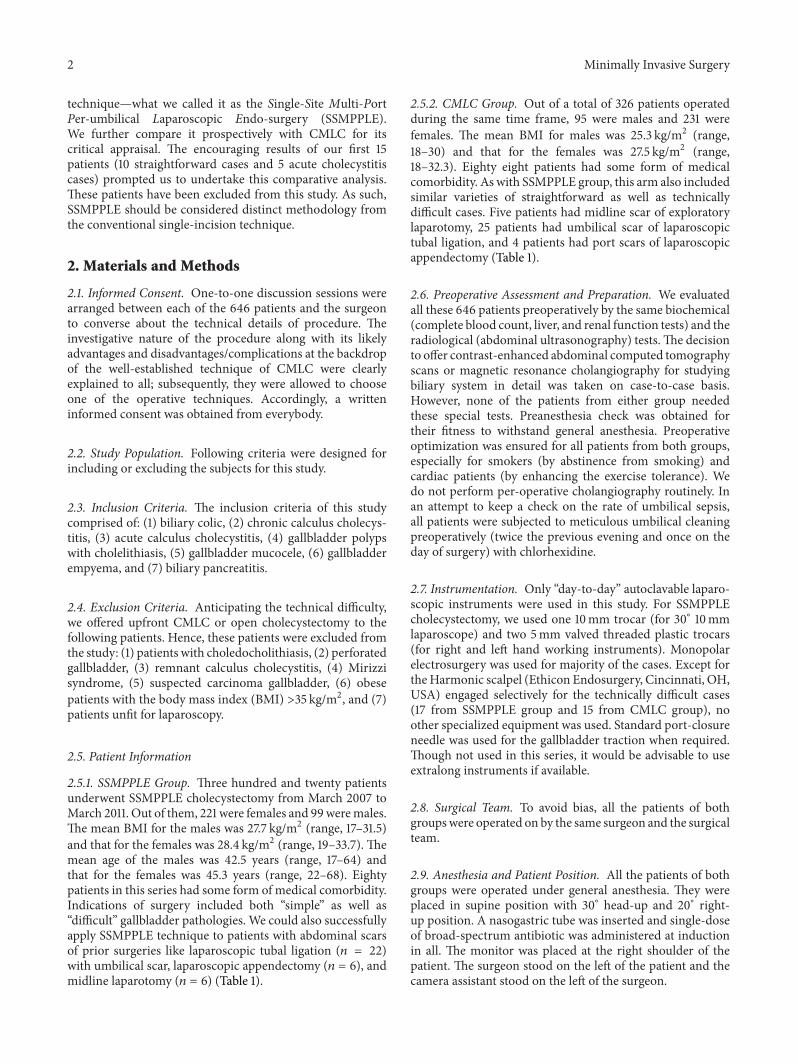
Table 1: Patient demographics.
Patient variables SSMPPLE CMLCNumber of patients 320 326Sex (male : female) 99 : 221 95 : 231Mean age (years)
Male 42.5 (range, 17–64) 43.8 (range, 15–67)Female 45.3 (range, 22–68) 44.9 (range, 16–70)
Mean BMI (Kg/m2)Male 27.7 (range, 17–31.5) 25.3 (range, 18–30)Female 28.4 (range, 19–33.7) 27.5 (range, 18–32.3)
Indications for cholecystectomyBiliary colic 161 160Acute calculus cholecystitis 20 15Chronic calculus cholecystitis 95 110Gallbladder polyp with cholelithiasis 7 8
Mucocele of gallbladder 7 15Empyema of gallbladder 10 4Biliary pancreatitis 20 14
Medical comorbiditiesHTN 16 15DM 17 19HTN + DM 14 15Heart disease
Old healed MI 6 5Left ventricular hypertrophy 5 6
Pulmonary diseaseOld healed tuberculosis 18 21COPD (controlled) 4 7
Previous abdominal surgery (scar)LTL (umbilical) 22 25LA (umbilical + right iliac fossa + suprapubic) 6 4Laparotomy (midline) 6 5
HTN: hypertension, DM: diabetes,MI: myocardial infarction, LTL: laparoscopic tubal ligation, OA: open appendectomy, LA: laparoscopic appendectomy, SPC:suprapubic cystostomy, and DL: diagnostic laparoscopy.
2.10. Clinical Parameters Studied. Postoperative outcomesstudied for both groups were operative time (defined as thetime interval between the first port entry till the last portclosure), blood loss, bile duct injury, viscus injury, gallbladderperforation during dissection, conversion to either CMLCor open, postoperative pain, stages of recovery, durationof hospitalization, umbilical seroma/sepsis, cosmetic results,and the rate of port-site hernia. The Visual Analogue Scale(VAS, 0–10) was used for assessing the postoperative painon days 0, 1, 7, and 30. Considering the suboptimal educa-tional and socioeconomic background of our rural patients,we developed an easy-to-use scar grading scale (I-Thrilled,II-Happy, III-Not bothered, and IV-Unhappy) as per thesubjective feeling about the scar they have received. Wefelt this scale was just the handy method of judging thescar outcomes in our part of the world. Although thissystem lacked the detailed questionnaire (and hence detailedobjective evaluation) regarding the cosmetic outcomes, itassessed the cosmetic results on a gross scale.
2.11. Statistical Analysis. Using SPSS 10.0 software (SPSS Inc.,Chicago, IL, USA), Pearson’s Chi-square test was appliedto assess the statistically significant difference between thevariables. This difference was considered significant if the 𝑃value was <0.05.
2.12. Surgical Techniques
2.12.1. SSMPPLE Cholecystectomy. The technique of creationof pneumoperitoneum by Veress needle was subject to theshape of umbilicus and the presence of abdominal scar (ifany) of previous surgery. In the patient with wide umbilicus(defined as ≥2.5 cm diameter) and without any abdominalscar, a 2mm stab incision was placed at the 12 O’clockposition on/just inside the umbilical mound for insertingthe Veress needle before creating the pneumoperitoneum. Inthese patients, we set the intra-abdominal pressure (IAP) at14mmHg. For patients with cardiac and pulmonary comor-bidities, we lowered the IAP to 10–12mmHg to minimize the
4 Minimally Invasive Surgery
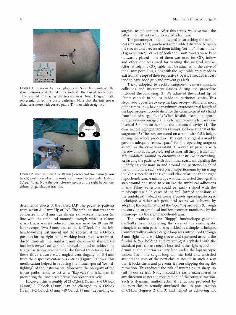
Figure 1: Incisions for port placement. Solid lines indicate theskin incisions and dotted lines indicate the fascial trajectories.This resulted in spacing the trocars away. Inset. Diagrammaticrepresentation of the ports pathways. Note that the intertrocardistance is more with curved paths (D) than with straight (d).
A
B C
Triangulation
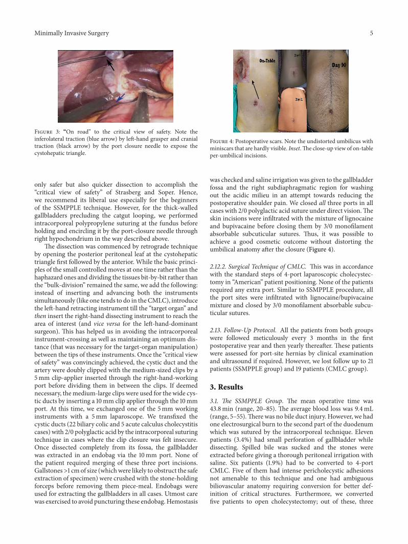
Figure 2: Port position. One 10mm (arrow) and two 5mm (arrowheads) ports placed on the umbilical mound in triangular fashion(Upper inset). Note the port-closure needle at the right hypochon-drium for gallbladder traction.
detrimental effects of the raised IAP. The pediatric patientswere set on 8–10mmHg of IAP. The stab incision was thenconverted into 11mm curvilinear skin-crease incision (inline with the umbilical mound) through which a 10mmsharp trocar was introduced. This was used for 10mm 30∘laparoscope. Two 5mm, one at the 8 O’clock for the left-hand-working instrument and the another at the 4 O’clockposition for the right-hand-working instrument were intro-duced through the similar 5mm curvilinear skin-creaseincisions on/just inside the umbilical mound to achieve thetriangular trocar ergonomics. The fascial trajectories for allthese three trocars were angled centrifugally by 3-4mmfrom the respective cutaneous entries (Figures 1 and 2). Thismodification helped in reducing the intracorporeal “sword-fighting” of the instruments. Moreover, the obliquity of thetrocar paths tends to act as a “flap-valve” mechanism inpreventing the trocar-site herniation postoperatively.
However, this assembly of 12 O’clock (10mm)–4 O’clock(5mm)–8 O’clock (5mm) can be changed to 6 O’clock(10mm)–2 O’clock (5mm)–10 O’clock (5mm) depending on
surgical team’s comfort. After this series, we have used thelatter in 17 patients with no added advantage.
The pneumoperitoneum helped in stretching the umbil-ical ring and, thus, purchased some added distance betweenthe trocars and prevented them falling “on-top” of each other(Figure 2, inset). Valves of both the 5mm trocars were keptoutwardly placed—one of them was used for CO
2inflow
and other one was used for venting the surgical smoke.Alternatively, the CO
2cable may be attached to the valve of
the 10mm port.This, along with the light cable, were made toexit from the tops of their respective trocars.Threaded trocarstend to have good grip and prevent gas leak.
Tricks adopted to rectify surgeon-to-camera-assistantcollisions and instrument-clashes during the procedureincluded the following. (1) We adjusted the distant tip of10mm cannula to be just inside the peritoneal cavity. Thisstepmade it possible to keep the laparoscopewithdrawnmostof the times, thus, having maximum extracorporeal length ofthe laparoscope. It could distance the camera-assistant’s handfrom that of surgeon’s. (2) When feasible, extralong laparo-scopes were encouraged. (3) Both 5mmworking trocars wereinserted 3-4mm farther into the peritoneal cavity. (4) Thecamera holding right handwas always laid beneath that of thesurgeon’s. (5) The surgeon stood on a stool with 0.5 ft heightduring the whole procedure. This entire surgical assemblygave an adequate “elbow-space” for the operating surgeonas well as the camera-assistant. However, in patients withnarrow umbilicus, we preferred to insert all the ports just out-side umbilical mound to circumvent instrument crowding.Regarding the patients with abdominal scars, anticipating theunderlying adhesions in and around the peritoneal side ofthe umbilicus, we achieved pneumoperitoneum by insertingthe Veress needle at the right mid-clavicular line in the righthypochondrium. Aminiscope was then inserted through thisstab wound and used to visualize the umbilical adhesionsif any. Filmy adhesions could be easily swiped with theminiscope itself. In cases of the well-formed adhesions atthe umbilicus, instead of using a purely open-laparoscopictechnique, a rather safe peritoneal access was achieved byadopting the combination of the “open” laparoscopy (throughthe curvilinear umbilical incision) counter-monitored by theminiscope via the right hypochondrium.
The problem of the “floppy” fundus/large gallblad-der/bulky liver obliterating the view of the cystohepatictriangle in certain patients was tackled by a simple technique.Commercially available catgut loop was introduced through5mm right-hand-working trocar and tightened around thefundus before holding and retracting it cephalad with thestandard port-closure needle inserted in the right hypochon-drium at the anterior axillary line under the laparoscopicvision. Then, the catgut-loop-tail was held and encircledaround the jaws of the port-closure needle in such a waythat it locks them and prevents it from slipping during theretraction. This reduced the risk of trauma by its sharp tip(nil in our series). Now, it could be easily maneuvered inany direction as per the requirement of the counter traction.Such a dynamic multidirectional retraction provided bythe port-closure actually simulated the 4th port tractionof CMLC (Figures 2 and 3) and helped us achieving not
Minimally Invasive Surgery 5
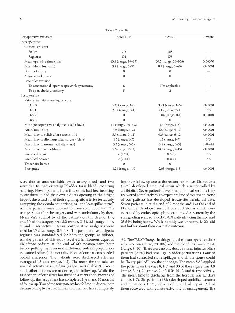
Figure 3: “On road” to the critical view of safety. Note theinferolateral traction (blue arrow) by left-hand grasper and cranialtraction (black arrow) by the port closure needle to expose thecystohepatic triangle.
only safer but also quicker dissection to accomplish the“critical view of safety” of Strasberg and Soper. Hence,we recommend its liberal use especially for the beginnersof the SSMPPLE technique. However, for the thick-walledgallbladders precluding the catgut looping, we performedintracorporeal polypropylene suturing at the fundus beforeholding and encircling it by the port-closure needle throughright hypochondrium in the way described above.
The dissection was commenced by retrograde techniqueby opening the posterior peritoneal leaf at the cystohepatictriangle first followed by the anterior. While the basic princi-ples of the small controlled moves at one time rather than thehaphazard ones and dividing the tissues bit-by-bit rather thanthe “bulk-division” remained the same, we add the following:instead of inserting and advancing both the instrumentssimultaneously (like one tends to do in the CMLC), introducethe left-hand retracting instrument till the “target organ” andthen insert the right-hand dissecting instrument to reach thearea of interest (and vice versa for the left-hand-dominantsurgeon). This has helped us in avoiding the intracorporealinstrument-crossing as well as maintaining an optimum dis-tance (that was necessary for the target-organ manipulation)between the tips of these instruments. Once the “critical viewof safety” was convincingly achieved, the cystic duct and theartery were doubly clipped with the medium-sized clips by a5mm clip-applier inserted through the right-hand-workingport before dividing them in between the clips. If deemednecessary, themedium-large clips were used for the wide cys-tic ducts by inserting a 10mm clip applier through the 10mmport. At this time, we exchanged one of the 5mm workinginstruments with a 5mm laparoscope. We transfixed thecystic ducts (22 biliary colic and 5 acute calculus cholecystitiscases) with 2/0 polyglactic acid by the intracorporeal suturingtechnique in cases where the clip closure was felt insecure.Once dissected completely from its fossa, the gallbladderwas extracted in an endobag via the 10mm port. None ofthe patient required merging of these three port incisions.Gallstones>1 cmof size (whichwere likely to obstruct the safeextraction of specimen) were crushed with the stone-holdingforceps before removing them piece-meal. Endobags wereused for extracting the gallbladders in all cases. Utmost carewas exercised to avoid puncturing these endobag.Hemostasis
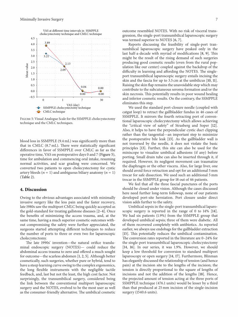
Figure 4: Postoperative scars. Note the undistorted umbilicus withminiscars that are hardly visible. Inset.The close-up view of on-tableper-umbilical incisions.
was checked and saline irrigationwas given to the gallbladderfossa and the right subdiaphragmatic region for washingout the acidic milieu in an attempt towards reducing thepostoperative shoulder pain. We closed all three ports in allcases with 2/0 polyglactic acid suture under direct vision.Theskin incisions were infiltrated with the mixture of lignocaineand bupivacaine before closing them by 3/0 monofilamentabsorbable subcuticular sutures. Thus, it was possible toachieve a good cosmetic outcome without distorting theumbilical anatomy after the closure (Figure 4).
2.12.2. Surgical Technique of CMLC. This was in accordancewith the standard steps of 4-port laparoscopic cholecystec-tomy in “American” patient positioning. None of the patientsrequired any extra port. Similar to SSMPPLE procedure, allthe port sites were infiltrated with lignocaine/bupivacainemixture and closed by 3/0 monofilament absorbable subcu-ticular sutures.
2.13. Follow-Up Protocol. All the patients from both groupswere followed meticulously every 3 months in the firstpostoperative year and then yearly thereafter. These patientswere assessed for port-site hernias by clinical examinationand ultrasound if required. However, we lost follow up to 21patients (SSMPPLE group) and 19 patients (CMLC group).
3. Results
3.1. The SSMPPLE Group. The mean operative time was43.8min (range, 20–85). The average blood loss was 9.4mL(range, 5–55).There was no bile duct injury. However, we hadone electrosurgical burn to the second part of the duodenumwhich was sutured by the intracorporeal technique. Elevenpatients (3.4%) had small perforation of gallbladder whiledissecting. Spilled bile was sucked and the stones wereextracted before giving a thorough peritoneal irrigation withsaline. Six patients (1.9%) had to be converted to 4-portCMLC. Five of them had intense pericholecystic adhesionsnot amenable to this technique and one had ambiguousbiliovascular anatomy requiring conversion for better def-inition of critical structures. Furthermore, we convertedfive patients to open cholecystectomy; out of these, three
6 Minimally Invasive Surgery
Table 2: Results.
Perioperative variables SSMPPLE CMLC P valueIntraoperative
Camera assistantFellow 216 168 —Registrar 104 158 —
Mean operative time (min) 43.8 (range, 20–85) 39.5 (range, 28–106) 0.00370Mean blood loss (mL) 9.4 (range, 5–55) 8.7 (range, 5–40) <0.0001Bile duct injury 0 0 —Major vessel injury 0 0 —Rate of conversion
To conventional laparoscopic cholecystectomy 6 Not applicable —To open cholecystectomy 5 2 —
PostoperativePain (mean visual analogue score)
Day 0 3.21 ( range, 3–5) 3.89 (range, 3–6) <0.0001Day 1 2.09 (range, 1–4) 2.13 (range, 2–4) NSDay 7 0 0.04 (range, 0-1) 0.00018Day 30 0 0 —
Mean postoperative analgesics used (days) 1.7 (range, 0.5–4.8) 3.3 (range, 1–5) <0.0001Ambulation (hr) 4.6 (range, 4–8) 4.8 (range, 4–12) <0.0001Mean time to solids after surgery (hr) 5.7 (range, 5–12) 6.6 (range, 6–12) <0.0001Mean time to discharge after surgery (days) 1.3 (range, 1–5) 1.2 (range, 1–7) NSMean time to normal activity (days) 3.2 (range, 3–7) 3.4 (range, 3–5) 0.00444Mean time to work (days) 9.6 (range, 7–18) 10.5 (range, 7–15) <0.0001Umbilical sepsis 6 (1.9%) 5 (1.5%) NSUmbilical seroma 7 (2.2%) 6 (1.8%) NSTrocar site hernia 0 0 —Scar grade 1.28 (range, 1–3) 2.03 (range, 1–3) <0.0001
were due to uncontrollable cystic artery bleeds and twowere due to inadvertent gallbladder fossa bleeds requiringsuturing. Eleven patients from this series had low-insertingcystic ducts, 8 had their cystic ducts opening in their righthepatic ducts and 4 had their right hepatic arteries tortuouslyoccupying the cystohepatic triangles—the “caterpillar turns”All the patients were allowed to have solid food by 5.7 h(range, 5–12) after the surgery and were ambulatory by then.Mean VAS applied to all the patients on the days 0, 1, 7,and 30 of the surgery was 3.2 (range, 3–5), 2.1 (range, 1–4),0, and 0, respectively. Mean postoperative analgesics wereused for 1.7 days (range, 0.5–4.8).The postoperative analgesiaregimen was standardized for both the groups as follows.All the patient of this study received intravenous aqueousdiclofenac sodium at the end of 6th postoperative hourbefore putting them on oral diclofenac sodium preparation(sustained release) the next day. None of our patients neededopioid analgesics. The patients were discharged after anaverage of 1.3 days (range, 1–5). The mean time to take upnormal activity was 3.2 days (range, 3–7) (Table 2). Except4, all other patients are under regular follow up. While thefirst patient of our series has finished 4 years and 9 months offollow up, the last patient has completed 1 year and 10monthsof followup. Two of the four patents lost followup due to theirdemise owing to cardiac ailments. Other two have completely
lost their follow up due to the reasons unknown. Six patients(1.9%) developed umbilical sepsis which was controlled byantibiotics. Seven patients developed umbilical seroma; theyrecovered completely by an expectant line of treatment. Noneof our patients has developed trocar-site hernia till date.Seven patients (4 at the end of 9 months and 4 at the end of13 months) developed residual bile duct stones which wereextracted by endoscopic sphincterotomy. Assessment by thescar grading scale revealed 73.01% patients being thrilled and25.56% being happy. While nobody was unhappy, 1.42% didnot bother about their cosmetic outcome.
3.2.TheCMLCGroup. In this group, themean operative timewas 39.5min (range, 28–106) and the blood loss was 8.7mL(range, 5–40).There were no bile duct or viscus injuries. Ninepatients (2.8%) had small gallbladder perforations. Four ofthem had controlled stone spillages and all the stones couldbe “berry-picked” into the endobags. The mean VAS appliedthe patients on the days 0, 1, 7, and 30 of the surgery was 3.9(range, 3–6), 2.1 (range, 2–4), 0.04 (0-1), and 0, respectively.The mean time to discharge from the hospital was 1.2 days(range, 1–7). Six patients (1.8%) developed umbilical seromaand 5 patients (1.5%) developed umbilical sepsis. All ofthem recovered with conservative line of management. The
Minimally Invasive Surgery 7
4.5
4.0
3.5
3.0
2.5
2.0
1.5
1.0
0.5
0.0
−0.5
Mea
n va
lue
VAS (day)0 1 7 30
0.04
0.00
0.00
0.00
2.13
2.09
3.89
3.21
SSMPPLE cholecystectomy techniqueCMLC technique
VAS at different time intervals in SSMPPLE cholecystectomy technique and CMLC technique
Figure 5: Visual Analogue Scale for the SSMPPLE cholecystectomytechnique and the CMLC techniques.
blood loss in SSMPPLE (9.4mL) was significantly more thanthat in CMLC (8.7mL). There were statistically significantdifferences in favor of SSMPPLE over CMLC as far as theoperative time, VAS on postoperative days 0 and 7 (Figure 5),time for ambulation and commencing oral intake, resumingnormal activities, and scar grading were concerned. Weconverted two patients to open cholecystectomy for cysticartery bleeds (𝑛 = 1) and ambiguous biliary anatomy (𝑛 = 1)(Table 2).
4. Discussion
Owing to the obvious advantages associated with minimallyinvasive surgery like the less pain and the faster recovery,late 1980s saw the multiport CMLC being quickly accepted asthe gold-standard for treating gallstone diseases [2–4]. Oncethe benefits of minimizing the access trauma, and, at thesame time, having a much superior cosmetic outcomes with-out compromising the safety were further appreciated, thesurgeons started attempting different techniques to reducethe number of ports to three or even two for laparoscopiccholecystectomy.
The late 1990s’ invention—the natural orifice translu-minal endoscopic surgery (NOTES)— could reduce theabdominal access trauma to zero and offered a much soughtfor outcome—the scarless abdomen [1, 2, 5]. Although bettercosmetically, such surgeries, whether pure or hybrid, tend tohave a steep learning curve owing to the complex ergonomics,the long flexible instruments with the negligible tactilefeedback, and, last but not the least, the high cost factor. Notsurprisingly, the transumbilical surgery, considered beingthe link between the conventional multiport laparoscopicsurgery and the NOTES, evolved to be the most user as wellas the consumer-friendly alternative. The umbilical cosmetic
outcome resembled NOTES. With no risk of visceral trans-gression, the single-port transumbilical laparoscopic surgerywas termed superior to NOTES [6, 7].
Reports discussing the feasibility of single-port tran-sumbilical laparoscopic surgery have peaked only in thelast half-a-decade with myriad of modifications [8, 9]. Thismight be the result of the rising demand of such surgeriesproducing good cosmetic results (even from the rural pop-ulation like our center) coupled against the backdrop of thedifficulty in learning and affording the NOTES. The single-port transumbilical laparoscopic surgery entails incising theskin and the fascia for up to 3.5 cm at the umbilicus [10, 11].Raising the skin flap remains the unavoidable step whichmaycontribute to the subcutaneous seroma formation and/or theskin necrosis. This potentially results in poor wound healingand inferior cosmetic results. On the contrary, the SSMPPLEeliminates this step.
We used the standard port-closure needle (coupled withcatgut loop) to retract the gallbladder fundus in 46 cases ofSSMPPLE. It mirrors the fourth retracting port of conven-tional laparoscopic cholecystectomy which allows achievingthe “critical view of safety” of Strasberg and Soper [12].Also, it helps to have the perpendicular cystic duct clippingrather than the tangential—an important step to minimizethe postoperative bile leak [13]. As the gallbladder wall isnot traversed by the needle, it does not violate the basicprinciples [13]. Further, this site can also be used for theminiscope to visualize umbilical adhesions (if any) beforeporting. Small drain tube can also be inserted through it, ifrequired. However, its negligent movement can traumatizethe diaphragm or the other viscera. Also, for large liver, oneshould avoid force retraction and opt for an additional 5mmtrocar for safe dissection. We used such an additional 5mmtrocar in the SSMPPLE group for 18 out of 46 patients.
We feel that all the three fascial punctures of the portsshould be closed under vision. Although the cases discussedhere need further long-term followup, none of our patientsdeveloped port-site herniation. Port closure under directvision adds further to the safety.
Umbilical sepsis in the single-port transumbilical laparo-scopic surgery is reported in the range of 0 to 14% [14].We had six patients (1.9%) from the SSMPPLE group thatdeveloped umbilical sepsis; three of them were diabetic. Allof them recovered completely with antibiotics. As reportedearlier, we always use endobags for the gallbladder extraction[15]. This potentially reduces the umbilical contamination.The conversion rates reported in the literature are 0–24% forthe single-port transumbilical laparoscopic cholecystectomy[14, 16]. In our series, it was 1.9%. However, we shouldkeep a low threshold for conversion to standard multiportlaparoscopy or open surgery [14, 17]. Furthermore, Blinmanhas elegantly discussed the relationship of tension (and hencepain) at the incision site to the lengths of the incision; thetension is directly proportional to the square of lengths ofincisions and not the addition of the lengths [18]. Hence,the projected amount of tension acting at the three ports ofSSMPPLE technique (476.1 units) would be lesser by a thirdthan that produced at 25mm incision of the single-incisionsurgery (1540.6 units).
8 Minimally Invasive Surgery
A recent meta-analysis of 13 randomized trials (includ-ing 923 patients) that studied comparisons between single-incision laparoscopic cholecystectomy and conventionalcholecystectomy reported higher failure rate, operative time,and blood loss with the former [19]. The two approacheswere found comparable in terms of conversion to opensurgery, length of hospital stay, postoperative pain, port-siteinfections, or hernias.The cosmetic outcomes were better forthe former especially when 10mm ports were used in thelatter. However, we feel that, with the technical modificationsdescribed in this paper, we could achieve acceptable results.Further, we need to state at this point that the only sim-ilarity between SSMPPLE and single-incision laparoscopiccholecystectomy is the very site of access (i.e., the umbilicus).Rest all the elements in this technique (like the number, theplacement and the sizes of incisions, the instruments used,the ergonomics, etc.) differ largely. Thus it tends to amal-gamate the operative site (umbilicus) of the single-incisionlaparoscopic cholecystectomy and the instrumentation withoperative techniques of the gold-standard—CMLC. Hence itshould not be considered amodification of the single-incisionlaparoscopic cholecystectomy but should rather be taken as adistinct laparoscopic cholecystectomy technique.
A similar technique described in literature [17] used all5mm ports and joined the two port sites for the specimenextraction.However, we think that 10mm laparoscope shouldalways be used right from the commencement of the surgeryas it gives much brighter, clearer, and wider vision. Also, itcan be used for the 10mm clip applier and the specimenextraction. For initial few cases of our series, the operativetime was longer as our surgical team was under the learningcurve of this technique. As the number of cases and theexperience increased, the operative time went on decreas-ing. Another recently reported method uses three ports atperiumbilical location to carry out cholecystectomy [20].Although the reported technique achieved triangulation, theport placement was away from the umbilical fold. Thus, thescars did not recede within the umbilicus. The SSMPPLEhelps the scars to recede at the umbilicus to produce betteraesthetics.
However, the SSMPPLE has certain limitations. (i) If notprecisely and strategically placed, the ports can lie too close toeach other leading to extracorporeal clashing. (ii) Althoughit may be technically easy in wide umbilicus, a narrow or a“slit-like” umbilicus may pose a real challenge. In fact, weshould keep a very low threshold for conversion to the CMLCin these cases. (iii) If the cutaneous and the fascial portalpunctures lie in vertical line (rather than oblique), one mayend up in having the instruments lying parallel to each otherleading to difficulty in dissecting. Moreover, notable flaws ofthis study are (1) limited cohort, (2) nonrandomized study,(3) limited duration of the followup for drawing definitiveconclusions about rate of port-site hernia, and (4) the VisualAnalogue Scale for incision-related pain and the scar gradingscale assessing the respective parameters in a subjectivemanner rather than the desired objective manner.
Although we have not conducted any cost-analysis com-parisons in this study, given that the routine laparoscopicinstruments were used with better operative timings without
any major complications (Table 2), we feel that the SSMP-PLE may become a valuable option of the per-umbilicallaparoscopy especially for the patients of the developingnations. However, this technique is a modification of min-imally invasive cholecystectomy. We further stress that it isnot a modification of single incision laparoscopic cholecys-tectomy in any way because it includes three separate skinincisions/punctures.
5. Conclusion
The presented SSMPPLE cholecystectomy technique doesnot need any specialized ports or other equipment; it seemssafe, efficient, and potentially economically viable alternativeto the single-incision laparoscopic cholecystectomy usingcommercially available specialized port/instruments.
Conflict of Interests
The authors declare that there is no conflict of interestsregarding the publication of this paper.
References
[1] T. Kagaya, “Laparoscopic cholecystectomy via two ports, usingthe “Twin-Port” system,” Journal of Hepato-Biliary-PancreaticSurgery, vol. 8, no. 1, pp. 76–80, 2001.
[2] S. Trichak, “Three-port versus standard four-port laparoscopiccholecystectomy: a prospective randomized study,” SurgicalEndoscopy andOther Interventional Techniques, vol. 17, no. 9, pp.1434–1436, 2003.
[3] M. Gagner and A. Garcia-Ruiz, “Technical aspects of mini-mally invasive abdominal surgery performed with needlescopicinstruments,” Surgical Laparoscopy, Endoscopy and Percuta-neous Techniques, vol. 8, no. 3, pp. 171–179, 1998.
[4] S. Purkayastha, H. S. Tilney, P. Georgiou, T. Athanasiou, P.P. Tekkis, and A. W. Darzi, “Laparoscopic cholecystectomyversus mini-laparotomy cholecystectomy: a meta-analysis ofrandomised control trials,” Surgical Endoscopy and Other Inter-ventional Techniques, vol. 21, no. 8, pp. 1294–1300, 2007.
[5] A. N. Kalloo, V. K. Singh, S. B. Jagannath et al., “Flexibletransgastric peritoneoscopy: a novel approach to diagnostic andtherapeutic interventions in the peritoneal cavity,”Gastrointesti-nal Endoscopy, vol. 60, no. 1, pp. 114–117, 2004.
[6] M. T. Gettman and M. L. Blute, “Transvesical peritoneoscopy:initial clinical evaluation of the bladder as a portal for naturalorifice translumenal endoscopic surgery,”Mayo Clinic Proceed-ings, vol. 82, no. 7, pp. 843–845, 2007.
[7] J. D. Raman, J. A. Cadeddu, P. Rao, and A. Rane, “Single-incision laparoscopic surgery: initial urological experienceand comparison with natural-orifice transluminal endoscopicsurgery,” BJU International, vol. 101, no. 12, pp. 1493–1496, 2008.
[8] J. Erbella Jr. and G. M. Bunch, “Single-incision laparoscopiccholecystectomy: the first 100 outpatients,” Surgical Endoscopyand Other Interventional Techniques, vol. 24, no. 8, pp. 1958–1961, 2010.
[9] B. Bokobza, A. Valverde, E. Magne et al., “Single umbilicalincision laparoscopic cholecystectomy: initial experience of theCoelio Club,” Journal of visceral surgery, vol. 147, no. 4, pp. e253–e257, 2010.
Minimally Invasive Surgery 9
[10] R. Sinha, “Transumbilical single-incision laparoscopic chole-cystectomy with conventional instruments and ports: the wayforward?” Journal of Laparoendoscopic and Advanced SurgicalTechniques, vol. 21, no. 6, pp. 497–503, 2011.
[11] T. Adachi, T. Okamoto, S. Ono, T. Kanematsu, and T. Kuroki,“Technical progress in single-incision laparoscopic cholecystec-tomy in our initial experience,”Minimally Invasive Surgery, vol.2011, Article ID 972647, 4 pages, 2011.
[12] P. A. Jategaonkar and S. P. Yadav, “Mirroring dynamic gallblad-der retraction of conventional laparoscopic cholecystectomy atthe transumbilical approach,” Annals of the Royal College ofSurgeons of England, vol. 96, no. 2, pp. 167–168, 2014.
[13] E. R. Podolsky and P. G. Curcillo II, “Reduced-port surgery:preservation of the critical view in single-port-access chole-cystectomy,” Surgical Endoscopy and Other Interventional Tech-niques, vol. 24, no. 12, pp. 3038–3043, 2010.
[14] C. Palanivelu, P. A. Jategaonkar, M. Rangarajan, and B.Srikanth, “‘Pseudo’ cholelithiasis: sequelae of minimally inva-sive cholecystectomywithmaximum surprise: an unusual case,”Endoscopy, vol. 41, supplement 2, pp. E186–E187, 2009.
[15] P. G. Curcillo II, A. S. Wu, E. R. Podolsky et al., “Single-port-access (SPAŮ) cholecystectomy: a multi-institutional report ofthe first 297 cases,” Surgical Endoscopy and Other InterventionalTechniques, vol. 24, no. 8, pp. 1854–1860, 2010.
[16] H. Massoumi, N. Kiyici, and H. Hertan, “Bile leak after laparo-scopic cholecystectomy,” Journal of Clinical Gastroenterology,vol. 41, no. 3, pp. 301–305, 2007.
[17] T. A. Azeez and K. M. Mahran, “Transumbilical laparo-scopic cholecystectomy: towards a scarless abdominal surgery,”Hepato-Gastroenterology, vol. 58, no. 106, pp. 298–300, 2011.
[18] T. Blinman, “Incisions do not simply sum,” Surgical EndoscopyandOther Interventional Techniques, vol. 24, no. 7, pp. 1746–1751,2010.
[19] S. Trastulli, R. Cirocchi, J. Desiderio et al., “Systematic reviewand meta-analysis of randomized clinical trials comparingsingle-incision versus conventional laparoscopic cholecystec-tomy,” British Journal of Surgery, vol. 100, pp. 191–208, 2012.
[20] J. Y. Ge, L. Wang, H. Zou, and X. W. Zhang, “Periumbilicallaparoscopic surgery through triple channels using commoninstrumentation,” Experimental and Therapeutic Medicine, vol.5, no. 4, pp. 1053–1056, 2013.

Submit your manuscripts athttp://www.hindawi.com
Stem CellsInternational
Hindawi Publishing Corporationhttp://www.hindawi.com Volume 2014
Hindawi Publishing Corporationhttp://www.hindawi.com Volume 2014
MEDIATORSINFLAMMATION
of
Hindawi Publishing Corporationhttp://www.hindawi.com Volume 2014
Behavioural Neurology
EndocrinologyInternational Journal of
Hindawi Publishing Corporationhttp://www.hindawi.com Volume 2014
Hindawi Publishing Corporationhttp://www.hindawi.com Volume 2014
Disease Markers
Hindawi Publishing Corporationhttp://www.hindawi.com Volume 2014
BioMed Research International
OncologyJournal of
Hindawi Publishing Corporationhttp://www.hindawi.com Volume 2014
Hindawi Publishing Corporationhttp://www.hindawi.com Volume 2014
Oxidative Medicine and Cellular Longevity
Hindawi Publishing Corporationhttp://www.hindawi.com Volume 2014
PPAR Research
The Scientific World JournalHindawi Publishing Corporation http://www.hindawi.com Volume 2014
Immunology ResearchHindawi Publishing Corporationhttp://www.hindawi.com Volume 2014
Journal of
ObesityJournal of
Hindawi Publishing Corporationhttp://www.hindawi.com Volume 2014
Hindawi Publishing Corporationhttp://www.hindawi.com Volume 2014
Computational and Mathematical Methods in Medicine
OphthalmologyJournal of
Hindawi Publishing Corporationhttp://www.hindawi.com Volume 2014
Diabetes ResearchJournal of
Hindawi Publishing Corporationhttp://www.hindawi.com Volume 2014
Hindawi Publishing Corporationhttp://www.hindawi.com Volume 2014
Research and TreatmentAIDS
Hindawi Publishing Corporationhttp://www.hindawi.com Volume 2014
Gastroenterology Research and Practice
Hindawi Publishing Corporationhttp://www.hindawi.com Volume 2014
Parkinson’s Disease
Evidence-Based Complementary and Alternative Medicine
Volume 2014Hindawi Publishing Corporationhttp://www.hindawi.com