Embed Size (px)
Citation preview
International Scholarly Research NetworkISRN GastroenterologyVolume 2011, Article ID 914013, 5 pagesdoi:10.5402/2011/914013
Clinical Study
Acute Corrosive Injuries of the Stomach: A Single UnitExperience of Thirty Years
N. Ananthakrishnan, G. Parthasarathy, and Vikram Kate
Department of Surgery, Jawaharlal Institute of Postgraduate Medical Education and Research, Pondicherry 605006, India
Correspondence should be addressed to N. Ananthakrishnan, [email protected]
Received 11 September 2010; Accepted 21 October 2010
Academic Editors: R. P. Bleichrodt, G. H. Kang and T. Takahashi
Copyright © 2011 N. Ananthakrishnan et al. This is an open access article distributed under the Creative Commons AttributionLicense, which permits unrestricted use, distribution, and reproduction in any medium, provided the original work is properlycited.
Introduction. The spectrum of gastric injury due to corrosives can vary. This paper presents a single center experience of over 30years of corrosive gastric injuries of 39 patients with acute gastric injuries from 1977 till 2006. Patients and Methods. Two thirdsof the patients in the acute injury group had a concomitant esophageal injury. The age of the patients ranged from 4 years to 65years with a slight preponderance of males. (M : F ratio 22 : 17). Results. 36 out of 39 acute gastric injuries were due to ingestionof acids. Three patients had history of caustic soda ingestion. Oral hyperemia or ulcers of varying extent were seen in all patients.The stomach showed hyperemia in 10, extensive ulcers in 13, and mucosal necrosis in 10 patients. Fifteen patients (15/39, 38.5%)were managed conservatively. Twenty four patients (24/39, 61.5%) underwent laparotomy: one for frank peritonitis, 10 for gastricmucosal necrosis, and 13 others for extensive gastric ulcerations. Overall the mortality rate was 29.6%. Conclusion. Althoughthe mortality and morbidity of acute corrosive gastric injuries is high, the key to improve the survival is early identification ofperforation, maintenance of nutrition and control of sepsis.
1. Introduction
Corrosive injuries of the stomach are not uncommonin developing countries. In these countries, accidental orsuicidal ingestion of acids is encountered more often thanin developed countries where lye or alkaline corrosives aremore frequent [1]. Both accidental ingestion, particularly inchildren, due to careless storing of chemicals and ingestionwith suicidal intent and due to free availability of the causticagents contribute to their occurrence.
The relative extent of esophageal and gastric involvementlargely depends on the nature of the corrosive ingested.Acids affect the stomach more commonly than alkalis [2],cause mucosal damage by coagulation necrosis, and requirea longer duration of contact [3]. However, alkali damage ofthe stomach has also been reported [4, 5]. Acids are clearedrapidly from the esophagus to the stomach where they poolin the prepyloric area due to corrosive-induced pylorospasm[6–8]. Prolonged contact with the prepyloric mucosa resultsin a prepyloric stricture. Strictures can also occur in the
antrum, body, or in the pyloroduodenal area. When thevolume of the corrosive ingested is large, the entire stomachgets scarred leading to a diffusely contracted stomach. On theother hand, alkalis cause liquefaction necrosis [3], are moreviscous, and tend to adhere to the esophageal mucosa withonly a relatively small amount reaching the stomach. Theextent of esophageal damage is greater with alkalis than acids.
Extensive acute injuries are usually fatal and therefore,the spectrum of acute and chronic gastric injury seen ata tertiary care referral hospital is not reflective of theoverall picture as patients with the most severe gastric andesophageal injuries die at peripheral centers.
The spectrum of gastric injury due to corrosives can varyfrom acute partial or total gastric mucosal or transmuralnecrosis to chronic gastric injuries of different types. Thisarticle presents a single center experience of over 30 yearsof corrosive gastric injuries of 39 patients with acute gastricinjuries emphasizing the spectrum of injuries and the extentof involvement and highlighting the possible modes ofmanagement.

2 ISRN Gastroenterology
Table 1: Nature of corrosive ingested.
Acute Gastric Injury (n = 39)
Acids 36
Aqua regia∗ 15
Bathroom cleaning acid+ 21
Alkalis 3∗A mixture of hydrochloric and nitric acids used by goldsmiths as a solvent,+Concentrated nitric acid
2. Patients and Methods
Thirty nine consecutive patients with injury to the stomachfollowing ingestion of a corrosive were treated in our institutefrom 1977 till 2006. Two thirds of the patients in the acuteinjury group had a concomitant esophageal injury. The ageof the patients ranged from 4 years to 65 years with a slightpreponderance of males over females (male : female ratio of22 : 17).
3. Results
There were a total of 39 acute and 109 chronic gastricinjuries seen at our institute. The ratio 39 : 109 (1 : 2.8)between acute and chronic injuries does not reflect the trueproportion between acute and chronic injuries in real life asseveral patients with acute injuries die at peripheral hospitalswithout reaching a tertiary care centre. Also a number ofchronic gastric injuries were referred to us because of theinterest of the unit in this problem.
36 out of 39 acute gastric injuries were due to ingestion ofacids (Aqua regia : 15 and bathroom cleaning acid : 21).Theseare readily available: the former being used by goldsmithsand the latter being freely available commercially. Threepatients had history of caustic soda ingestion (Table 1).
There were 6 children less than 12 years of age, all ofwhom had ingested the corrosive accidentally. Of the 33adults, 21 were suicidal and 12 accidental ingestions. Allpatients with acute corrosive injury presented in a criticalcondition with abdominal pain, vomiting, and hematemesis.These patients also had severe odynophagia and 24/39(61.5%) in addition, had dysphagia and drooling of saliva.One patient with previous history of a truncal vagotomy andgastrojejunostomy for benign gastric outlet obstruction dueto chronic cicatrizing duodenal ulcer presented with featuresof peritonitis following perforation of the efferent limb ofthe gastrojejunostomy. None of the remaining patients hadfrank features of esophageal or abdominal hollow viscusperforation.
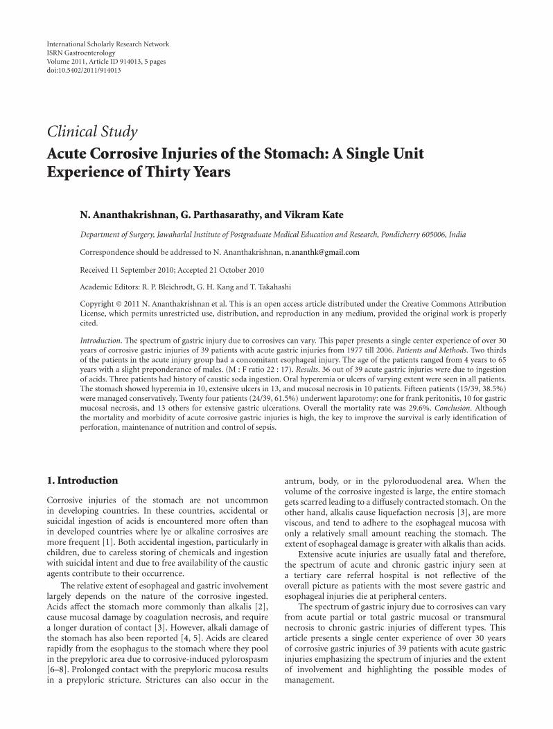
The details of endoscopic appearance and clinical courseare shown in Figure 1. Endoscopic evaluation was performedin 33/39 patients. One patient with overt peritonitis andfive patients who were critically ill and in shock did notundergo endoscopic evaluation. The timing of endoscopyvaried from patient to patient depending on the clinicalcondition. However, where feasible it was the policy of theunit to do endoscopy 3-4 days after the injury to have a true
picture of the extent of the damage. Endoscopy was carefullyperformed with minimal air insufflation.
Oral hyperemia or ulcers of varying extent were seen inall patients. Twenty four patients had grade 2 or 3 esophagealinjury. The stomach showed hyperemia in 10, extensiveulcers in 13, and varying degrees of mucosal necrosis in theremaining 10 patients.
A total of fifteen patients (15/39, 38.5%) were managedconservatively. Of these, five patients were critically ill,while 10 patients had only minor endoscopic evidence ofinjury (hyperemia). The conservative management includednil per oral status (NPO), placement of an indwellingnasogastric tube, and starting of parenteral feeding. Onepatient, who had extensive laryngeal burns and stridor,needed an emergency tracheostomy for airway control. Thefive patients who presented in shock died within 24 hours.In the remaining ten patients, tube feeds were resumedsuccessfully after one week gradually progressing to oral feedsover the next few days.
Twenty four patients (24/39, 61.5%) underwentexploratory laparotomy: one for frank peritonitis, 10 withgastric mucosal necrosis on endoscopy, and 13 otherswith extensive gastric ulcerations considered at high riskfor perforation. In 13 patients, the stomach appearedrelatively normal on the serosal aspect at laparotomy withno evidence of perforation. Feeding jejunostomy werethe sole procedure carried out in these patients. In onepatient, there were perforation of the efferent loop of aprevious gastrojejunostomy. Partial gastrectomy and redogastrojejunostomy was done. In 10 patients, there wasvarying degrees of gastric necrosis. The extent of mucosalnecrosis, in general, was much more as seen at gastrotomyas compared to the extent of transmural necrosis seen fromthe serosal side. In six patients, a distal gastrectomy anda Polya reconstruction (end to side gastrojejunostomy)were performed along with a feeding jejunostomy. In theother four, a total gastrectomy was carried out. Due tothe critical condition of the patients, no reconstructionwas attempted. A cervical esophagostomy was done. Theabdominal esophagus was closed around a drainage tube,the duodenal stump was closed with drainage, and a feedingjejunostomy was done. The details of the management ofpatients with acute corrosive injuries of the stomach areshown in Figure 1.
Overall, the mortality rate was 29.6% (Figure 1). Allpatients who presented in shock, 3 of 4 with total gastrectomyand 2 of 6 with distal gastrectomy, expired due to sepsis andshock.
4. Discussion
Corrosive injuries of the stomach and esophagus are notinfrequent causes of hospitalization in countries like India[1]. Both accidental ingestion, particularly in children,due to careless storing of chemicals and ingestion withsuicidal intent due to free availability of the caustic agentscontribute to their occurrence. In most reported series, thechemicals that are most commonly responsible are alkalis like
ISRN Gastroenterology 3
Acute corrosive gastric injury
(n = 39 )
Severe endoscopic injury
perforation
n = 24, 61.6%
Minor endoscopicinjury (hyperemia)
n = 10, 25.6%
Conservative management
Normal serosaNo perforation
Feeding jejunostomyn = 13, 54.2%
Died Died Died Died Died
Perforation(efferent limb)n = 1, 4.2%
Partial gastrectomyRedo gastrojejunostomy
Total gastrectomy
n = 4
Distal gastrectomyPolya reconstruction
n = 6
Died
Critically ill,shock
n = 5, 12.8%
Exploratory laparotomy
Gastric necrosisn = 10, 41.7%
(extensive ulcers/necrosis)
n = 0, 0% n = 0, 0%n = 0, 0% n = 2, 33% n = 3, 75% n = 5, 100%
Figure 1: Flow chart showing details of management of acute corrosive gastric injuries.
potassium and sodium hydroxide [7, 9–11]. In contrast, themajority of the corrosive injuries in India are due to acids.The most common acids implicated are bathroom cleaningacid (concentrated hydrochloric acid) and aqua regia.
Two thirds of the patients with acute gastric injury inthe present series had a concomitant esophageal injury. Theincidence of coexistent esophageal injury in the literaturevaries from 20% to as high as 62.5% [12–14].
The most common presentation of an acute corro-sive gastric burn is with abdominal pain, vomiting, andhematemesis [12, 15, 16]. Rarely a full thickness burn cancause a gastric perforation. This normally tends to presenta few days after ingestion of the corrosive. Hematemesisfollowing corrosive ingestion is usually self-limiting. How-ever, there are reports of subacute massive bleeding from thestomach or duodenum following corrosive ingestion [17].Massive bleeding typically occurs two weeks after ingestion.Such bleeds may warrant a gastrectomy if the source isthe stomach or a duodenotomy and under running of thebleeding vessel in the duodenum.
The most useful investigation in the evaluation of anacute corrosive gastric injury is an upper gastrointestinalendoscopy. Endoscopic evaluation has been advised as soonas possible after corrosive ingestion, since it is believed thatthe risk of perforation is lowest at this point [3]. Also,it is believed that endoscopic evaluation at this juncturehelps plan early intervention, if required. However, in ourexperience clinical features and radiological examination inthe form of a CT are more useful in assessing threatenedor existing perforation. Early endoscopy carries the risk
of misdiagnosing the extent of transmural damage in thepresence of extensive hyperemia. A repeat study is againrequired a few days subsequently to assess the true damage.It is our policy therefore, to do endoscopy 72–96 hours afterthe corrosive ingestion.
Laparoscopy is also a useful adjunct in assessing a patientwho has a high risk of gastric perforation as seen onendoscopy or in patients with severe esophageal injury inwhom an upper gastrointestinal endoscopy to assess thestomach is not feasible. Some authors have advocated routinelaparoscopic examination in all injuries of second degree orgreater [18]. However, this has not been our practice. Thereis also a report on the use of a Meckel’s scan to assess theseverity of the gastric injury [19].
Following an acute injury to the stomach by corrosiveingestion, the initial management is usually conservative.Adherence to the basic tenet of avoiding a gastric lavagein any corrosive poisoning cannot be overemphasized. Thepatient usually has associated burns to the upper aerodiges-tive tract which needs attention as well, if required, with atracheostomy. Attempts at neutralizing the acids or alkalisare ill-advised and the resulting exothermic reaction fromthe neutralization process may do more harm than good[20]. Similarly, there is not much role for measures to dilutethe corrosive with milk, water and so forth, as the definitiveextent of the injury is determined within minutes afteringestion [18].
Emergency surgical intervention is needed if the patientdevelops any signs of esophageal perforation, peritonitis,or uncontrolled massive hematemesis [14]. In view of
4 ISRN Gastroenterology
the high probability of slow but relentless progression oftransmural necrosis, there should be a low threshold forconsideration of laparotomy at the earliest suspicion. Ifthere is severe esophageal burn with a high likelihood ofstricture formation, a feeding jejunostomy is performed andthe stomach is assessed intraoperatively at this time. If theesophagus is relatively spared with moderate injury to thestomach, the patient is fed through a jejunostomy and kepton regular observation to monitor the progress of the gastricburn. If, on the other hand, the stomach appears soft andnecrotic, a gastrotomy is made and the extent of transmuraland mucosal necrosis is assessed before planning a resection,either a distal gastrectomy or a total gastrectomy.
Patients with extensive gastric injury are often criticallyill and do not withstand lengthy reconstructive procedures.Hence, as a policy for extensive gastric necrosis, we do totalgastrectomy, closure and drainage of the esophageal, andduodenal stump, a cervical esophagostomy along with afeeding jejunostomy, leaving reconstruction (using a jejunalloop) for a more opportune moment should the patientcomes out of the acute phase. For less extensive acute gastricinjury a distal gastrectomy may suffice. The line of sectionshould be decided after gastrotomy since mucosal necrosisis more extensive than what is apparent from the serosalside. There is no role for procedures such as closure ofa perforation since the stomach is like wet blotting paperaround the site of perforation.
All patients with second degree or greater corrosive burnsare given parenteral broad spectrum antibiotics. Intravenousproton pump inhibitors are also widely used with the aimof minimizing the insult to the injured gastric mucosa.However, there are no studies supporting their role in thissetting.
The debate over the use of steroids in corrosive burnsto prevent stricture formation has been put to rest with tworecent meta-analyses [21, 22].The authors found no benefitwith the use of systemic corticosteroids in corrosive ingestionand proscribe their routine use to prevent stricture forma-tion. However, there is a report of the use of intralesionalsteroids in corrosive pyloric strictures [23].
The mandatory need for gastric resection as prophylaxisagainst future malignancy has been overstated in the litera-ture. There have been reports of malignancy developing in ascarred esophagus or stomach following corrosive ingestion[24–26]. However, in our experience this association hasbeen found to be tenuous. In an experience of over 500corrosive injuries seen over a thirty year period, there wasonly one solitary instance of cricopharyngeal carcinomafollowing esophageal burns by caustic ingestion and one caseof peri-gastroenterostomy carcinoma seventeen years afterthe ingestion of acid. In the latter, it is not clear whether thecarcinoma was corrosive induced or secondary to chronicbile reflux through the gastrojejunostomy stoma (stumpcarcinoma or postgastric surgery carcinoma).
The mortality and morbidity of acute corrosive gastricinjuries are high and dependent on the severity of initialdamage caused by the corrosive agent with a significantproportion of patients succumbing to their injuries eitherbefore reaching tertiary care or soon thereafter. The key to
improving the survival of such patients in the acute settingremains in early identification of perforation and supportivecare with maintenance of nutrition and control of sepsis.One has to be pithy in one’s surgical interventions for acutecorrosive burns, limiting the resection to only the grosslyinjured bowel and leaving the reconstruction part for a latterday. On the other hand, the mortality and morbidity ofchronic gastric corrosive injuries can be significantly reducedby adequate preoperative preparation and a planned protocolof approach dependent on the type of injury.
References
[1] N. Ananthakrishnan, K. S. V. K. Subba Rao, and P. Radjendi-ran, “Mid-colon esophagocoloplasty for corrosive esophagealstrictures,” Australian and New Zealand Journal of Surgery, vol.63, pp. 389–395, 1993.
[2] K. S. Subbarao, A. K. Kakar, V. Chandrasekhar, N. Ananthakr-ishnan, and A. Banerjee, “Cicatrical gastric stenosis caused bycorrosive ingestion,” Australian and New Zealand Journal ofSurgery, vol. 58, no. 2, pp. 143–146, 1988.
[3] D. Lahoti and S. L. Broor, “Corrosive injury to the uppergastrointestinal tract,” Indian Journal of Gastroenterology, vol.12, no. 4, pp. 135–141, 1993.
[4] E. G. Bowill, F. A. Bulawa, and R. G. Olivetti, “Severe corrosivegastritis with antral stenosis following ingestion of Sani-Flush,” Gastroenterology, vol. 17, no. 3, pp. 436–441, 1951.
[5] W. S. Boikan and H. A. Singer, “Gastric sequelae of corrosivepoisoning,” Archives of Internal Medicine, vol. 40, pp. 342–357,1930.
[6] A. O. Ciftci, M. E. Senocak, N. Buyukpamukcu, and A.Hicsonmez, “Gastric outlet obstruction due to corrosive inges-tion: incidence and outcome,” Pediatric Surgery International,vol. 15, no. 2, pp. 88–91, 1999.
[7] J. E. Lowe, D. Y. Graham, E. V. Boisaubin Jr., and F. L. Lanza,“Corrosive injury to the stomach: the natural history and roleof fiberoptic endoscopy,” American Journal of Surgery, vol. 137,no. 6, pp. 803–806, 1979.
[8] N. L. Poteshman, “Corrosive gastritis due to hydrochloricacid ingestion. report of a case,” The American Journal ofRoentgenology, Radium Therapy, and Nuclear Medicine, vol. 99,no. 1, pp. 182–185, 1967.
[9] J. Imre and M. Kopp, “Arguments against long-term conser-vative treatment of oesophageal strictures due to corrosiveburns,” Thorax, vol. 27, no. 5, pp. 594–598, 1972.
[10] E. Sarfati, D. Gossot, P. Assens, and M. Celerier, “Managementof caustic ingestion in adults,” British Journal of Surgery, vol.74, no. 2, pp. 146–148, 1987.
[11] L. P. Goldman and J. M. Weigert, “Corrosive substanceingestion: a review,” American Journal of Gastroenterology, vol.79, no. 2, pp. 85–90, 1984.
[12] S. A. Zargar, R. Kochhar, B. Nagi, S. Mehta, and S. K. Mehta,“Ingestion of strong corrosive alkalis: spectrum of injury toupper gastrointestinal tract and natural history,” AmericanJournal of Gastroenterology, vol. 87, no. 3, pp. 337–341, 1992.
[13] A. Chaudhary, A. S. Puri, P. Dhar et al., “Elective surgeryfor corrosive-induced gastric injury,” World Journal of Surgery,vol. 20, no. 6, pp. 703–706, 1996.
[14] R. W. Postlethwait, “Chemical burns of the esophagus,”Surgical Clinics of North America, vol. 63, no. 4, pp. 915–924,1983.
ISRN Gastroenterology 5
[15] A. B. Karon and H. C. Wall, “Pyloric stenosis caused byingestion of a corrosive acid simulating gastric carcinoma:report of a case,” Gastroenterology, vol. 17, no. 3, pp. 445–449,1951.
[16] W. R. Moore, “Caustic ingestions. Pathophysiology, diagnosis,and treatment,” Clinical Pediatrics, vol. 25, no. 4, pp. 192–196,1986.
[17] Y.-L. Tseng, M.-H. Wu, M.-Y. Lin, and W.-W. Lai, “Massiveupper gastrointestinal bleeding after acid-corrosive injury,”World Journal of Surgery, vol. 28, no. 1, pp. 50–54, 2004.
[18] T. B. Hugh and M. D. Kelly, “Corrosive ingestion and thesurgeon,” Journal of the American College of Surgeons, vol. 189,no. 5, pp. 508–522, 1999.
[19] D. K. Chung, M. P. Wines, G. E. Cummins, and R. B.Howman-Giles, “Application of the Meckel’s scan in a case ofgastric corrosive injury,” Pediatric Surgery International, vol.19, no. 1-2, pp. 9–10, 2003.
[20] K. I. Maull, A. P. Osmand, and C. D. Maull, “Liquidcaustic ingestions: an in vitro study of the effects of buffer,neutralization, and dilution,” Annals of Emergency Medicine,vol. 14, no. 12, pp. 1160–1162, 1985.
[21] D. Pelclova and T. Navratil, “Do corticosteroids preventoesophageal stricture after corrosive ingestion?” ToxicologicalReviews, vol. 24, no. 2, pp. 125–129, 2005.
[22] J. M. Howell, W. C. Dalsey, F. W. Hartsell, and C. A. Butzin,“Steroids for the treatment of corrosive esophageal injury:a statistical analysis of past studies,” American Journal ofEmergency Medicine, vol. 10, no. 5, pp. 421–425, 1992.
[23] R. Kochhar, P. V. J. Sriram, J. D. Ray, S. Kumar, B. Nagi, and K.Singh, “Intralesional steroid injections for corrosive inducedpyloric stenosis,” Endoscopy, vol. 30, no. 8, pp. 734–736, 1998.
[24] H. Eaton and G. E. Tennekoon, “Squamous carcinoma of thestomach following corrosive acid burns,” British Journal ofSurgery, vol. 59, no. 5, pp. 382–387, 1972.
[25] L. L. Gonzalez, M. M. Zinninger, and W. A. Altemeier,“Cicatricial gastric stenosis caused by ingestion of corrosivesubstances,” Annals of Surgery, vol. 156, no. 1, pp. 84–89, 1962.
[26] J. F. Nicosia, J. P. Thornton, F. A. Folk, and J. D. Saletta,“Surgical management of corrosive gastric injuries,” Annals ofSurgery, vol. 180, no. 2, pp. 139–143, 1974.

Submit your manuscripts athttp://www.hindawi.com
Stem CellsInternational
Hindawi Publishing Corporationhttp://www.hindawi.com Volume 2014
Hindawi Publishing Corporationhttp://www.hindawi.com Volume 2014
MEDIATORSINFLAMMATION
of
Hindawi Publishing Corporationhttp://www.hindawi.com Volume 2014
Behavioural Neurology
EndocrinologyInternational Journal of
Hindawi Publishing Corporationhttp://www.hindawi.com Volume 2014
Hindawi Publishing Corporationhttp://www.hindawi.com Volume 2014
Disease Markers
Hindawi Publishing Corporationhttp://www.hindawi.com Volume 2014
BioMed Research International
OncologyJournal of
Hindawi Publishing Corporationhttp://www.hindawi.com Volume 2014
Hindawi Publishing Corporationhttp://www.hindawi.com Volume 2014
Oxidative Medicine and Cellular Longevity
Hindawi Publishing Corporationhttp://www.hindawi.com Volume 2014
PPAR Research
The Scientific World JournalHindawi Publishing Corporation http://www.hindawi.com Volume 2014
Immunology ResearchHindawi Publishing Corporationhttp://www.hindawi.com Volume 2014
Journal of
ObesityJournal of
Hindawi Publishing Corporationhttp://www.hindawi.com Volume 2014
Hindawi Publishing Corporationhttp://www.hindawi.com Volume 2014
Computational and Mathematical Methods in Medicine
OphthalmologyJournal of
Hindawi Publishing Corporationhttp://www.hindawi.com Volume 2014
Diabetes ResearchJournal of
Hindawi Publishing Corporationhttp://www.hindawi.com Volume 2014
Hindawi Publishing Corporationhttp://www.hindawi.com Volume 2014
Research and TreatmentAIDS
Hindawi Publishing Corporationhttp://www.hindawi.com Volume 2014
Gastroenterology Research and Practice
Hindawi Publishing Corporationhttp://www.hindawi.com Volume 2014
Parkinson’s Disease
Evidence-Based Complementary and Alternative Medicine
Volume 2014Hindawi Publishing Corporationhttp://www.hindawi.com