Embed Size (px)
Citation preview

CLINICALTHYROIDOLOGY
VOLUME 18 ● ISSUE 3 NOVEMBER 2006
A publication of the American Thyroid Association
Intra-Amniotic Administration of Thyroxine Is Effective Therapy in Fetuses with Goiter and Hypothyroidism . . . . . . . . . . . . . . . . . . . . . . . . . . . . . 52
NODULAR GOITER
No Single Ultrasonographic Finding Reliably Distinguishes Thyroid Carcinomas from Benign Thyroid Nodules . . . . . . . . . . . . . . . . . . . . . . . . . . 53
THYROID CANCER
The Frequency of Carcinoma Is Similar in Patients with One Thyroid Nodule and Those with Multiple Nodules. . . . . . . . . . . . . . . . . . . . . . . . . . . . . . . . . . . . . . . . 54
The Follicular-Variant Subtype of Papillary Thyroid Carcinoma May Be Either Encapsulated or Nonencapsulated . . . . . . . . . . . . . . . . . . . . . . . . . . . . . . 55
Radioiodine Therapy Can Cause Testicular Dysfunction in Men with Thyroid Carcinoma . . . . . . . . . 56
AUTOIMMUNE THYROID DISEASE
Five to 10 Percent of Pregnant Women Have Postpartum Thyroiditis . . . . . . . . . . . . . . . . . . . . . . . . . . . 57
THYROID FUNCTION IN PREGNANCY
Thyroxine Treatment Reduces the Rates of Miscarriage and Preterm Delivery in Pregnant Women with Autoimmune Thyroid Disease. . . . . . . . . . . . . . . . . . . . . . 58
THYROID DIAGNOSIS
Serum Thyroglobulin Concentrations Increase Markedly Soon after Biopsy of a Thyroid Nodule. . . . . . 59
THYROID HORMONE ACTION
Triiodothyronine Has Vasodilatory and Neuroprotective Actions Mediated by a Nongenomic Increase in Endothelial-Cell Nitric Acid Synthase Activity. . . . . . . . . 60
THYROID DISEASE
Subclinical Thyroid Dysfunction Is Not Associated with Anxiety, Depression, or Cognitive Dysfunction. . . . 41
Blood Pressure Is Increased Slightly in Subclinical Hyperthyroidism but Not in Subclinical Hypothyroidism. . . . . . . . . . . . . . . . . . . . . . 42
HYPERTHYROIDISM
More of the Serum Triiodothyronine in Patients with Hyperthyroidism Is of Thyroidal Origin than in Normal Subjects . . . . . . . . . . . . . . . . . . . . . . . . . . 43
Patients Treated with Radioiodine Can Trigger Airport Radiation Sensors for Many Weeks . . . . . . . . . . . 44
Restricting Iodine Intake Does Not Alter the Efficacy of Methimazole . . . . . . . . . . . . . . . . . . . . . . . . . . 45
GRAVES’ OPHTHALMOPATHY
Pioglitazone May Increase Protrusion of the Eyes . . . . . 46
Measurements of Serum Thyrotropin-Receptor Antibodies May Predict the Course of Ophthalmopathy in Graves’ Disease. . . . . . . . . . . . . . . . . 47
HYPOTHYROIDISM
The Risk of Coronary Heart Disease Is Increased in Subclinical Hypothyroidism. . . . . . . . . . . . . . . . . . . . . . 48
Patients with Adrenal Insufficiency May Have Reversible Hypothyroidism . . . . . . . . . . . . . . . . . . . . . . . . 49
Small Changes in Thyroxine Dose Do Not Alter Well-Being or Symptoms in Patients with Hypothyroidism . . . . . . . . . . . . . . . . . . . . . . . . . . . . . . . . . 50
Combined Thyroxine and Triiodothyronine Therapy Is Not More Effective than Thyroxine Alone in Patients with Hypothyroidism . . . . . . . . . . . . . . . . . . . . . . 51
EditorRobert D. Utiger, M.D.Thyroid DivisionDepartment of MedicineBrigham & Women’s Hospital77 Avenue Louis PasteurBoston, MA 02115(617) 525-5171 Telephone(617) 731-4718 [email protected]
PresidentDavid S. Cooper, M.D.
President-ElectRebecca S. Bahn, M.D.
SecretaryGregory A. Brent, M.D.
TreasurerCharles H. Emerson, M.D.
Executive DirectorBarbara R. Smith, C.A.E.American Thyroid Association6066 Leesburg Pike, Suite 550Falls Church, VA 22041Telephone: 703-998-8890Fax: 703-998-8893Email: [email protected]
Designed ByKaren DurlandEmail: [email protected]
Clinical ThyroidologyCopyright © 2006American Thyroid Association, Inc.Printed in the USA. All rights reserved.
CLINICALTHYROIDOLOGYVOLUME 18 ● ISSUE 3 NOVEMBER 2006
www.thyroid.org
Clinical Thyroidology is available on the ATA web site —
www.thyroid.org
Sign up for an online subscription and email notifi cation
of each new issue!

CLINICAL THYROIDOLOGY ● VOLUME 18 ● ISSUE 3 ● 41
THYROID DISEASE
Subclinical Thyroid Dysfunction Is Not Associated with Anxiety, Depression, or Cognitive Dysfunction
Roberts LM, Pattison H, Roalfe A, Franklyn J, Wilson S, Hobbs FD, Parle JV. Is subclinical thyroid dysfunction in the elderly associated with depression or cognitive dysfunction? Ann Intern Med 2006;145:573-81.
SUMMARY
Background Patients with subclinical hypothyroidism or subclinical hyperthyroidism may have an increased frequency of mood disorders or cognitive dysfunction. In this study, the frequency of anxiety, depression, and cognitive dysfunction was determined in a large cohort of normal subjects and subjects with subclinical and also overt thyroid dysfunction.
Methods The study subjects were 5857 subjects aged 65 years or older (2974 women, 2883 men; mean age, 73 years) attending 20 general practices in central England. Subjects with known thyroid disease were excluded. Anxiety and depression were assessed using the Hospital Anxiety and Depression Scale, which consists of seven questions about anxiety and seven about depression, each scored 0 to 3 (maximum, 21 for each component). Anxiety (or depression) is considered mild if the score is 8 to 10, moderate if 11 to 14, and severe if 15 or more. Cognitive function was assessed using the Folstein Mini–Mental State Examination (MMSE) and the Middlesex Elderly Assessment of Mental State (MEAMS) test. These tests assess several aspects of cognitive function, including learning, memory, perception, and attention; the MMSE is scored 0 to 30 and the MEAMS 0 to 12; for both tests higher scores indicate less impairment.
Serum thyrotropin (TSH), free thyroxine (T4), and, in some subjects, free triiodothyronine (T3) were measured at the same time. The subjects were subdivided into five groups: overt hyperthyroidism, low serum TSH and high free T4 or free T3; subclinical hyperthyroidism, low serum TSH, normal free T4 and free T3; normal, serum TSH and free T4 both normal; subclinical hypothyroidism; high serum TSH,
normal free T4; and overt hypothyroidism, high serum TSH, low serum free T4.
Results There were 15 subjects with overt hyperthyroidism (0.2 percent), 127 with subclinical hyperthyroidism (2.2 percent), 5524 with normal thyroid function (94.3 percent), 168 with subclinical hypothyroidism (2.9 percent), and 23 with overt hypothyroidism (0.4 percent). The anxiety score was ≥8 in 370 subjects (6.3 percent) and the depression score was ≥8 in 136 (2.3 percent).
The score for anxiety was lower in the subjects with overt hyperthyroidism (3.4) than in those with subclinical hyperthyroidism (5.4) or the normal subjects (4.8). There were no differences in the scores for anxiety in the subjects with overt hypothyroidism or subclinical hypothyroidism and the normal subjects.
The score for depression also was lower in the subjects with overt hyperthyroidism (2.3) than in the normal subjects (3.4), and it was higher (4.2) in the subjects with subclinical hyperthyroidism. There were no differences in the scores for depression in the subjects with overt hypothyroidism or subclinical hypothyroidism and the normal subjects.
The MMSE test results indicated the presence of cognitive dysfunction in 305 subjects (5.2 percent); the comparable MEAMS test result is not given. There were no differences in the MMSE or MEAMS scores in any of the groups.
Conclusion Among elderly subjects with overt or sub-clinical thyroid dysfunction, the frequency of anxiety, depression, and cognitive dysfunction is similar to that in normal subjects.
COMMENTARY
These were on the whole healthy subjects, at least with respect to mood and cognitive function. There were the curious findings that the subjects with overt hyperthyroidism had lower anxiety and depression scores than the normal subjects, and on the other hand that the subjects with subclinical hyperthyroidism had higher anxiety and depression scores than the normal subjects. However, most
of the scores were not different when the results were adjusted for age, sex, comorbidity, and medications. Other analyses revealed that an increase in serum TSH of 50 mU/L was associated with a 1-point decrease in anxiety score (less anxiety) and a 1-point increase in MMSE (better cognitive function); even if statistically significant these associations seem random and of no clinical importance.
The similarity of the results in the
subjects with subclinical hypothyroidism or subclinical hyperthyroidism and the normal subjects should have an impact on clinical practice, because of the question of whether subjects with either disorder should be treated. The lack of changes in mood or cognitive function in these subjects supports the view that therapy of these subjects in unlikely to be beneficial.
Robert D. Utiger, M.D.

42 ● CLINICAL THYROIDOLOGY ● VOLUME 18 ● ISSUE 3
THYROID DISEASE
Blood pressure is increased slightly in subclinical hyperthyroidism but not in subclinical hypothyroidism
Walsh JP, Bremner AP, Bulsara MK, O’Leary P, Leedman PJ, Feddema P, Michelangeli V. Subclinical thyroid dysfunction and blood pressure: a community-based study. Clin Endocrinol (Oxf) 2006;65:486-91.
SUMMARY
Background Hyperthyroidism and hypothyroidism are associated with increases in systolic and diastolic blood pressure, respectively, but whether blood pressure is altered in subclinical hyperthyroidism and subclinical hypothyroidism is not clear. In this study, the relationships between blood pressure and subclinical thyroid disease were evaluated in a large cohort of subjects.
Methods The study subjects were 2033 predominantly white subjects (982 women, 1051 men; mean age, 50 years [range, 17 to 89]) living in a rural town in Australia (Busselton, West Australia) in whom seated blood pressure and serum thyrotropin (TSH) and free thyroxine (T4) were measured once. They included 299 subjects being treated for hypertension. They were categorized as follows: euthyroid, serum TSH 0.4 to 4.0 mU/L; subclinical hypothyroidism, serum TSH >4.0 mU/L and normal serum free T4; and subclinical hyperthyroidism, serum TSH <0.4 mU/L and normal serum free T4.
Results There were 35 subjects (1.7 percent) with subclinical hyperthyroidism and 82 (4.8 percent) with subclinical hypothyroidism; 1591 (93.7 percent) were euthyroid (subjects with hypertension excluded). Overall, there was no correlation between serum TSH or free T4 and systolic or diastolic blood pressure after adjustment for age and sex.
The mean systolic, but not diastolic, blood pressure was higher in the subjects with subclinical hyperthyroidism than in those who were euthyroid (Table), and among the former the increase was limited to the few subjects (n=8)
with serum TSH values <0.1 mU/L. In contrast, the values in the subjects with subclinical hypothyroidism were similar to those in the euthyroid subjects. Among the subjects with subclinical hypothyroidism, the mean blood pressure was slightly higher in those with serum TSH values of >4.0 to 10.0 mU/L than in those with serum TSH values >10.0 mU/L (132/78 vs. 125/72 mm Hg).
Table. Mean Systolic and Diastolic Blood Pressure in Subjects with Subclinical Thyroid Disease and Euthyroid Subjects.
Subclinical Hyperthyroidism
(n=35)
Euthyroid (n=1591)
Subclinical Hypothyroidism
(n=82)Women/men 17/18 726/865 57/25Mean age (yrs) 48 46 57Systolic pressure (mm Hg) 132* 126 131Diastolic pressure (mm Hg) 78 75 77
*P<0.01, as compared with the euthyroid group, adjusted for age and sex.
The frequency of hypertension, defined as blood pressure ≥140/≥90 mm Hg or treatment for hypertension, was higher in the subjects with subclinical hyperthyroidism (29 percent), as compared with the euthyroid subjects (14 percent) (odds ratio, 2.8), but similar in the euthyroid group and the subjects with subclinical hypothyroidism (22 percent) (odds ratio, 0.9). The results were similar if hypertension was defined as blood pressure ≥160/≥100 mm Hg.
Conclusion Subjects with subclinical hyperthyroidism have slightly higher systolic blood pressure and a slightly higher frequency of hypertension than do euthyroid subjects, whereas blood pressure and frequency of hypertension are similar in subjects with subclinical hypothyroidism.
COMMENTARY
Blood pressure has been studied more often in subjects with subclinical hypothyroidism than in those with sub-clinical hyperthyroidism, for several reasons. One, subclinical hypothyroidism is considerably more common. Two, hyper-tension is more common in patients with overt hypothyroidism than in those with overt hyperthyroidism, so it would be expected to be more common in patients with subclinical hypothyroidism than in those with subclinical hyperthyroidism. Three, hypothyroidism may be associated with increased cardiovascular morbidity, and if so subclinical hypothyroidism may also be, and hypertension is certainly a risk factor for cardiovascular morbidity and mortality.
Walsh et al. found blood pressure to be no higher in subjects with subclinical
hypothyroidism than in euthyroid subjects, as defined by single measurements of blood pressure and serum TSH. Indeed, among the subjects with subclinical hypothyroidism, the mean blood pressure and the frequency of hypertension were lower in the subjects who had serum TSH values >10.0 mU/L than in those with values of >4.0 to 10.0 mU/L. Others, however, have found blood pressure to be slightly higher in subjects with subclinical hypothyroidism. There are no longitudinal studies linking blood pressure and subclinical hypothyroidism at base line with hypertension, overt hypothyroidism, or cardiovascular outcomes.
The finding of slightly higher systolic blood pressure in subjects with subclinical hyperthyroidism is rather tenuous, given the very small number of subjects, but more-or-less in agreement with the notion that hyperthyroidism raises systolic blood
pressure. However, in an even larger recent study (4087 subjects), subjects with subclinical hyperthyroidism had lower systolic blood pressure and similar diastolic pressure, as compared with normal subjects (1).
Some of these differences may be resolved when more is learned about the cardiovascular effects of thyroid hormone, especially its direct effects on blood vessels (see page 60).
Robert D. Utiger, M.D.
Reference
1. Volzke H, Alte D, Dorr M, et al. The association between subclinical hyperthyroidism and blood pressure in a population-based study. J Hypertens 2006;24:1947-53.

CLINICAL THYROIDOLOGY ● VOLUME 18 ● ISSUE 3 ● 43
HYPERTHYROIDISM
More of the serum triiodothyronine in patients with hyperthyroidism is of thyroidal origin than in normal subjects
Woeber KA. Triiodothyronine production in Graves’ hyperthyroidism. Thyroid 2006;16:687-90.
SUMMARY
Background In normal subjects, 20 percent of the triiodothyronine (T3) is produced by the thyroid and 80 percent by extrathyroidal (peripheral) deiodination of thyroxine (T4). The production of both hormones is increased in patients with hyperthyroidism, but the relative contribution of the two sources to the increase in T3 production is not clear. In this study, the thyroidal and extrathyoidal contributions to T3 production were estimated from measurements of serum free T4 and free T3 in patients with hyperthyroidism and patients with hypothyroidism treated with T4.
Methods The study subjects were 31 patients with Graves’ hyperthyroidism and 21 patients with thyroid carcinoma who were taking T4. The 31 patients with hyperthyroidism included 25 patients (23 women, 2 men; median age, 33 years) with high serum free T4 and free T3 concentrations (T4-plus-T3 hyperthyroidism group) and 6 patients (4 women, 2 men; median age, 47 years) with normal serum free T4 and high free T3 concentrations (T3-hyperthyroidism group); all had high serum concentrations of thyrotropin (TSH)-receptor or other antithyroid antibodies or diffuse goiter. The 21 patients with thyroid carcinoma (T4-therapy group) included 14 women and 7 men (median age, 36 years); all had been treated with total thyroidectomy and radioactive iodine and were taking sufficient T4 to lower TSH secretion to below normal.
The contribution of extrathyroidal production of T3 to the serum free T3 concentration in the patients with hyperthyroidism was calculated as follows: serum free T4 (pmol/L) ÷ serum free T4:free T3 molar ratio (pmol/L:pmol/L) in the T4-therapy group.
This calculated value represents the patient’s serum free T3 concentration if all the free T3 was produced by extrathyroidal production, as it is in the T4-therapy group. The difference between the patient’s measured serum free T3
value and this calculated value is the thyroidal contribution to the patient’s serum free T3 concentration.
Results The mean serum free T4 and free T3 concentrations in the T4-plus-T3 hyperthyroidism and the T3-hyperthyroidism groups are shown in the Table; the serum free T4:free T3 molar ratios were 2.7 and 2.6, respectively. The mean serum free T4 and free T3 concentrations in the T4-therapy group were 20 pmol/L (1.6 ng/dl) and 5.1 pmol/L (0.4 ng/dl), respectively, and the serum free T4:free T3 molar ratio was 4.0. The respective values in 20 normal subjects were 14 pmol/L (1.1 ng/dl), 4.2 pmol/L (0.3 ng/dl), and 3.3.
Table. Mean Serum Free T4 and Free T3 Concentrations and Extrathyroidal and Thyroidal (Secreted) Contributions to Serum Free T3 Concentrations in Patients with Hyperthyroidism.
Hyperthyroidism
T4-plus-T3 Group (n=25) T3 Group (n=6)Serum free T4 (pmol/L) 45 23Serum free T3 (pmol/L) 17.3 8.8Extrathyroidal free T3 (pmol/L) 11.3 5.7Secreted free T3 (pmol/L) 6.1 3.1Secreted free T3 (%) 33 34
To convert serum free T4 and free T3 values to ng/dl, divide by 12.9 and 15.4, respectively.
Based on the serum free T4:free T3 molar ratio in the T4-therapy group, the serum free T3 concentrations attributable to extrathyroidal production of T3 in the two groups of patients with hyperthyroidism were 11.3 and 5.7 pmol/L (0.7 and 0.4 ng/dl), respectively. The concentrations attributable to thyroidal secretion were 6.1 and 3.1 pmol/L (0.4 and 0.2 ng/dl), respectively, which amounted to 35 percent of the serum free T3 concentrations in each group.
Conclusion Approximately one third of serum T3 in patients with T4- and T3-hyperthyroidism and T3-hyperthyroidism is produced by the thyroid, as compared with one fifth in normal subjects.
COMMENTARY
The distinction between T4-plus-T3 hyperthyroidism and T3-hyperthyroidism is based on the results of measurements of serum free T4 and free T3. Is the concentration of both T4 and T3, or only that of T3, high? In these patients with hyperthyroidism, those with T3-hyperthyroidism had serum free T4 and free T3 concentrations approximately 50 percent of those in the patients with T4-plus-T3 hyperthyroidism, but the serum free T4:free T3 ratios in the two groups were very similar. The difference between the groups was a difference in the severity of hyperthyroidism, not in a disproportionate
increase in T3 production, of either thyroidal or extrathyroidal origin, in the T3-hyperthyroidism group.
The thyroidal contribution to serum free T3 was increased in the patients with hyperthyroidism, as compared with normal subjects, most likely a result of the increased thyroidal deiodinase activity known to occur in Graves’ hyperthyroidism (1). The thyroidal contribution might also be increased in patients with hyper-thyroidism (of any cause) whose iodine intake was marginal, and in those with T4-hyperthyroidism (high serum free T4 and normal free T3 concentrations), in whom extrathyroidal T3 production was inhibited by illness or drugs. It could also
occur in patients with autonomous thyroid tissue in which there was an abnormality of thyroglobulin or thyroid peroxidase that favored T3 synthesis over that of T4. The simple technique described in this paper should make it relatively easy to investigate these and other conditions that result in unusual changes in serum free T4 and free T3 concentrations.
Robert D. Utiger, M.D.
Reference
1. Salvatore D, Tu H, Harney JW, Larsen PR. Type 2 iodothyronine deiodinase is highly expressed in human thyroid. J Clin Invest 1996;98:962-8.

44 ● CLINICAL THYROIDOLOGY ● VOLUME 18 ● ISSUE 3
SUMMARY
Background Patients who undergo diagnostic studies or are treated with a radionuclide may activate radiation de-tectors in airports or other public facilities. This short paper describes a patient with hyperthyroidism who had been treated with radioiodine (I-131) and whose travel was delayed because he activated a radiation detector at an air terminal.
Case Report The patient was a 46-year-old man referred for evaluation of weight loss, diarrhea, and excessive perspiration. Physical examination revealed tachycardia; his thyroid was not enlarged. His serum free thyroxine concentration was high (7.2 ng/dl [93 pmol/L]) and his serum thyrotropin concentration was low (0.02 mU/L). He was treated with 30 mg of carbimazole daily, and improved. The dose was gradually reduced, but hyperthyroidism recurred when the dose was reduced to 5 mg
daily one year later. The dose was increased to 30 mg daily, again with improvement. Five months later, he was treated with 10.8 mCi (400 MBq) of I-131. He was given a card listing some precautions that he should take to minimize radiation exposure to others, but it did not mention the possibility that he could activate a radiation detector.
Six weeks later he flew to the United States. When he went to board his flight to return to the United Kingdom, he set off a radiation detector at the security station. He was further tested (Figure), strip-searched, sniffed by a dog, and questioned at length before being allowed to board the aircraft.
Conclusion Patients who have received I-131 therapy should be warned that they can activate radiation detectors at airports and other facilities for several months and provided with documentation of their treatment.
HYPERTHYROIDISM
Patients treated with radioiodine can trigger airport radiation sensors for many weeks
Gangopadhyay K, Sundram F, De P. Triggering radiation alarms after radioiodine treatment. BMJ 2006;333:293-4.
COMMENTARY
All patients with hyperthyroidism or thyroid carcinoma who are treated with I-131 are advised to take precautions to minimize radiation exposure to themselves, other family members, and the public at large. These precautions include avoiding close contact with other family members and staying home from school or work for several days. Most of the information underlying these recommendations is based on measurements of radioactivity retained by the patient (or present in the patient’s sweat, saliva, or urine), but there have studies in which the radiation exposure of family members and pets was directly measured continuously for up to 10 days after a patient had been treated (1-3).
Additional often-advised precautions are to limit travel on public transport for a few days post-treatment. Now, for the patient (and indirectly for co-travelers) comes a new hazard—the triggering of radiation detectors that are part of airport and other security systems, leading to inconvenience and delay. The sensitivity and perhaps the use of these detectors vary; the patient described in this case report was able to leave the United Kingdom without triggering an alarm.
Note that the time interval between administration of I-131 and triggering of the detector was more than six weeks (>5 physical half-lives of I-131); the sensitivity of some portable radiation detectors is such that 100 µCi (3.7 MBq) of I-131 can be detected for up to 95 days (Table) (4).
Among the radionuclides in wide use in clinical medicine, I-131 is most likely to be detected, because it has a relatively long physical half-life (8 days) and high doses are often administered. The numbers of days that 100-µCi doses (3.7 MBq) of other radionuclides might activate a detector are shown in the Table.
Table. Physical Half-Life of Clinically Useful Radionuclides and Number of Days a 100-µCi (3.7 MBq) Dose Can Be Detected.
T½
No. of Days*
Fluorine-18 111 min 1Technetium-99m 6 hr 3Iodine-123 13 hr 3Indium-111 67 hr 14Thallium-201 73 hr 30Gallium-67 78 hr 30Iodine-131 8 days 95
*From ref. 4.
It is obvious that patients need to be routinely provided with documentation that they have been given radionuclides,
because the use of radiation detectors is very likely to become more widespread and their sensitivity increased.
Robert D. Utiger, M.D.
References
1. O’Doherty MJ, Kettle AG, Eustance CN, et al. Radiation dose rates from adult patients receiving 131I therapy for thyro-toxicosis. Nucl Med Comm 1993;14:160-8.
2. Grigsby PW, Siegel BA, Baker S, Eichling JO. Radiation exposure from outpatient radioactive iodine (131I) therapy for thyroid carcinoma. JAMA 2000;283:2272-4.
3. Matheoud R, Reschini E, Canzi C, et al. Potential third-party radiation exposure from outpatients treated with 131I for hyperthyroidism. Med Phys 2004;31:3194-200.
4. Zuckier L, Stabin M, Garetano G, et al. Sensitivity of personal homeland security radiation detectors to medical radionuclides and implications forcounseling of nuclear medicinepatients. Accessed 11/23/06 at http://rsna2004.rsna.org/rsna2004/V2004/conference/event_display.cfm?em_id=4407767.
Figure. Image of passenger being scanned at an airport.

CLINICAL THYROIDOLOGY ● VOLUME 18 ● ISSUE 3 ● 45
HYPERTHYROIDISM
Restricting iodine intake does not alter the efficacy of methimazole
Hiraiwa T, Ito M, Imagawa A, Takamatsu J, Kuma K, Miyauchi A, Hanafusa T. Restriction of dietary iodine does not ameliorate the early effect of anti-thyroid drug therapy for Graves’ disease in an area of excessive iodine intake. J Endocrinol Invest 2006;29:380-4.
SUMMARY
Background Antithyroid drug therapy may not be as effective in patients with hyperthyroidism whose iodine intake is relatively high, as compared with those whose iodine intake is lower. In this study, the short-term efficacy of methimazole was compared in patients whose iodine intake was relatively high and those in whom it was restricted.
Methods The study was done in 70 Japanese patients (59 women, 11 men; mean age, 38 years) with hyperthyroidism caused by Graves’ disease, as defined by high serum free thyroxine (T4) and free triiodothyronine (T3) concentrations and high serum thyrotropin (TSH)-receptor antibody values (measured by radioreceptor assay) or high thyroid radioiodine uptake values.
Thirty-one patients agreed to restrict their dietary iodine intake, by excluding popular iodine-rich foods, such as kelp, seaweed, and seafood, and 39 continued to eat their usual diet and therefore served as a control group. Serum free T4, free T3, and TSH-receptor antibodies and urinary iodine and creatinine were measured at base line and after treatment with 15 mg of methimazole daily for 4 and 8 weeks.
Results At base line, the patients in the iodine-restriction group and the control group had similar serum free T4, free T3, and TSH-receptor antibody concentrations and urinary
iodine/creatinine values. During methimazole therapy, serum free T4 and free T3 concentrations decreased similarly in both groups (Table). The median urinary iodine/creatinine ratio decreased from 220 µg/g at base line to 150 µg/g at 8 weeks in the iodine-restricted group and it increased from 195 to 339 µg/g in the control group. The serum concentrations of TSH-receptor antibodies did not change in either group.
Table. Median Serum Free T4 and Free T3 Concentrations and Urinary Iodine/Creatinine Ratios in Iodine-Restricted and Control Patients before and during Methimazole Therapy.
Serum Free T4(ng/dl)
Serum Free T3(ng/dl)
Urinary Iodine/Creatinine (µg/g)
Iodine-restricted group Base line 4.2 1.6 220 4 Weeks 1.8 0.4 207 8 Weeks 1.1 0.3 150Control group Base line 3.9 1.5 195 4 Weeks 1.8 0.5 299 8 Weeks 0.9 0.3 339To convert serum free T4 and free T3 values to pmol/L, multiply by 12.9 and 15.4, respectively
Conclusion In patients with hyperthyroidism caused by Graves’ disease, decreasing dietary iodine intake when methimazole therapy is initiated does not alter the short-term efficacy of the drug.
COMMENTARY
Key steps in thyroid hormone biosynthesis are the formation of a complex between oxidized iodine and thyroid peroxidase and then transfer of the iodine to tyrosine residues of thyroglobulin (iodination). Methimazole inhibits iodination of tyrosyl residues (and their coupling to form T4 and T3) by binding oxidized iodine, and at least in vitro the extent and nature of the inhibition are dependent on the dose of drug and the concentration of iodine. The inhibitory effect of low drug concentrations on iodination is reversible, whereas that of high concentrations is not, and the effect of the drug is reduced in the presence of high iodine concentrations.
Therefore, methimazole should be more effective, or at least more rapidly effective, in patients with hyperthyroidism whose iodine intake is low. That was indeed the case in two previous studies. In one study of 36 patients with Graves’ hyperthyroidism treated with 30 mg of methimazole daily for four weeks, serum
free T4 index values fell by 82 percent in 18 patients who lived in Tehran, a region of mild iodine deficiency, but by only 28 percent in 18 patients who lived in Boston, where iodine intake was higher (1). In another study done in several European countries (2), in which patients with Graves’ hyperthyroidism were treated with 10 or 40 mg of methimazole daily for three weeks, 109 of 170 patients (64 percent) who had base-line urinary iodine/creatinine values <100 µg/g became euthyroid, as compared with 36 of 105 patients (34 percent) who had base-line values ≥100 µg/g.
The study of Hiraiwa et al. differed from these two studies in that iodine intake was decreased at the time methimazole was initiated, rather than being chronically low. The patients’ base-line iodine intake was substantial, but not particularly high. This means that the patients’ thyroidal iodine stores were substantial, and therefore reducing iodine intake would not be expected to have much of an effect on thyroidal iodine content or drug action, at least in the short term. The rise in urinary iodine
excretion during treatment in the control group may be taken as evidence that iodine utilization within the thyroid was indeed inhibited by methimazole, which may lead to a decrease in iodine transport into the thyroid.
In conclusion, reducing dietary iodine intake at the time of initiation of antithyroid drug therapy does not augment the short-term antithyroid action of methimazole, but it might do so in the long run, and it also might minimize the likelihood of recurrence after the drug is discontinued.
Robert D. Utiger, M.D.
References
1. Azizi F. Environmental iodine intake affects the response to methimazole in patients with diffuse toxic goiter. J Clin Endocrinol Metab 1985;61:374-7.
2. Benker G, Vitti P, Kahaly G, et al. Response to methimazole in Graves’ disease. Clin Endocrinol (Oxf) 1995;43:257-63.

46 ● CLINICAL THYROIDOLOGY ● VOLUME 18 ● ISSUE 3
GRAVES’ OPHTHALMOPATHY
Pioglitazone may increase protrusion of the eyes
Dorkhan M, Lantz M, Frid A, Groop L, Hallengren B. Treatment with a thiazolidinedione increases eye protrusion in a subgroup of patients with type 2 diabetes. Clin Endocrinol (Oxf) 2006;65:35-9.
SUMMARY
Background One of the features of Graves’ ophthalmopathy is protrusion of the eyes, caused in part by proliferation of retroorbital adipose tissue (adipogenesis). One factor known to stimulate adipogenesis is peroxisome proliferator–activated receptor gamma (PPAR-γ), a nuclear transcription factor. Thiazolidinedione drugs such as pioglitazone and rosiglitazone activate PPAR-γ and therefore stimulate adipogenesis (as well as increase insulin sensitivity). In this study, the effect of pioglitazone on eye protrusion was studied in patients with type 2 diabetes mellitus.
Methods The study subjects were 36 patients (13 women, 23 men; mean age, 60 years; mean body-mass index, 31 kg/m2) with diabetes that was poorly controlled despite therapy with a sulfonylurea drug and metformin. Six of the patients had some thyroid disorder, including hypothyroidism treated with thyroxine (2 patients), subclinical hypothyroidism (1 patient), subclinical hyperthyroidism (2 patients), and uninodular goiter treated with thyroxine (1 patient); their serum thyrotropin (TSH) concentrations at base line ranged from 0.07 to 6.7 mU/L.
The patients were treated with pioglitazone, 30 mg daily, in addition to the sulfonylurea and metformin, for 26 weeks. The dose was raised to 45 mg daily at 16 weeks in 12 patients because their hemoglobin A1C (HbA1C) values were ≥6.5 percent). The degree of proptosis was measured using a Krahn exophthalmometer at base line and 26 weeks by a single investigator. The inter-eye distance was the same for both measurements, and the second measurement was done without knowledge of the first measurement. HbA1C was measured in all patients, and serum TSH was measured in the 6 patients with thyroid disorders at base line and 26 weeks.
Results There was an increase in eye protrusion in most patients during the 26-week study period; the median increase was 1 mm. Thirteen patients (36 percent), including 5 of the 6 patients with thyroid disorders, had an increase of ≥2 mm and 23 (64 percent) had an increase of <2 mm. The mean values for each eye in both groups are shown in the Table. None of the patients had other signs of Graves’ ophthalmopathy.
Table. Mean Changes in Eye Protrusion in Patients with Diabetes Treated with Pioglitazone for 25 Weeks.
Change in Eye Protrusion
≥2 mm (n=13) <2 mm (n=23)Women/men 4/9 9/14Current or previous smoker 10 (77%) 13 (56%)Thyroid disorder 5 (38%) 1 (4%)*Proptosis, right eye Base line (mm) 17.3 17.5 26 weeks (mm) 19.5** 17.7Proptosis, left eye Base line (mm) 17.4 17.6 26 weeks (mm) 18.9** 17.6*P=0.02, compared with group with change ≥2 mm. **P<0.01, as compared with base line in the same group.
In both groups, the mean body-mass index increased <1 kg/m2 and the mean HbA1C value decreased 1.2 percent. There was little change in serum TSH concentration in the 6 patients with thyroid disorders. The factors that predicted an increase in eye protrusion of ≥2 mm were thyroid disorder and dose of pioglitazone, but not sex, age, body-mass index, or smoking.
Conclusion Patients with diabetes who are treated with a thiazolidinedione drug may have an increase in protrusion of their eyes, possibly caused by activation of adipogenesis.
COMMENTARY
Eye protrusion in patients with Graves’ ophthalmopathy is due to enlargement of both retroorbital fat and eye muscles. The enlargement of retroorbital fat is due to an increase in adipogenesis and also accumulation of hydrophilic glycosaminoglycans. mRNA for PPAR-γ is present in retroorbital adipose tissue of normal subjects and increased in retroorbital adipose tissue of patients with Graves’ ophthalmopathy (1), presumably indicative of increased adipogenesis, and PPAR-γ agonists stimulate and PPAR-γ antagonists inhibit adipogenesis in cultured retroorbital preadipocytes (2).
These observations provide an explanation for thiazolidinedione
proptosis, as described by Dorkhan et al. That seems a better term than thiazolidinedione ophthalmopathy, because it seems unlikely that the process would be accompanied by the increases in cytokines and TSH receptors in retroorbital tissue and the presence of serum TSH-receptor antibodies that occur in patients with Graves’ ophthalmopathy. More marked proptosis (28 mm), with no evidence of inflammation, has been described in a woman with diabetes treated with rosiglitazone who had no evidence of thyroid disease (3), and an exacerbation of preexisting ophthalmopathy has been described in a man with Graves’ disease (2).
While the data are not extensive, it would seem prudent not to treat patients with Graves’ disease, even those with
no apparent ophthalmopathy, with a thiazolidinedione.
Robert D. Utiger, M.D.
References
1. Kumar S, Coenen MJ, Scherer PE, Bahn R. Evidence for enhanced adipogenesis in the orbits of patients with Graves’ ophthalmopathy. J Clin Endocrinol Metab 2004;89:930-5.
2. Starkey K, Heufelder A, Baker G, et al. Peroxisome proliferator-activated receptor-γ in thyroid eye disease: contra-indication for thiazolidinedione use? J Clin Endocrinol Metab 2003;88:55-9.
3. Levin F, Kazim M, Smith TJ, Marcovici E. Rosiglitazone-induced proptosis. Arch Ophthalmol 2005;123:119-21.
CLINICAL THYROIDOLOGY ● VOLUME 18 ● ISSUE 3 ● 47
GRAVES’ OPHTHALMOPATHY
Measurements of serum thyrotropin-receptor antibodies may predict the course of ophthalmopathy in Graves’ disease
Eckstein AK, Plicht M, Lax H, Neuhauser M, Mann K, Lederbogen S, Heckmann C, Esser J, Morgenthaler N. Thyrotropin receptor autoantibodies are independent risk factors for Graves’ ophthalmopathy and help to predict severity and outcome of the disease. J Clin Endocrinol Metab 2006;91:3464-70.
SUMMARY
Background Most patients with Graves’ ophthalmopathy have high serum concentrations of thyrotropin (TSH)-receptor antibodies (TSHR-Ab), but little is known about the relationship between serum TSHR-Ab concentrations and the severity and course of ophthalmopathy. In this study, the severity of ophthalmopathy was evaluated and serum TSHR-Abs were measured repeatedly in a large group of patients with Graves’ ophthalmopathy.
Methods The study subjects were 159 patients (137 women, 22 men) with Graves’ ophthalmopathy. Among them, 69 (43 percent) had the onset of ophthalmopathy before or coincident with the onset of hyperthyroidism, 80 (50 percent) had the onset months to years after the onset of hyperthyroidism, and the remainder had autoimmune thyroiditis or no thyroid disease. Patients with new-onset hyperthyroidism were treated with an antithyroid drug for one year, and those with recurrent hyperthyroidism were treated with an antithyroid drug, radioiodine, or surgery.
The patients were evaluated three or more times at 4-month intervals for up to 24 months after the onset of ophthalmopathy. Each evaluation consisted of determination of the Clinical Activity Score (CAS), based on assessment of eye pain, redness, eyelid swelling, proptosis, and eye function (scored 0 [inactive] to 10 [active]); a symptom-severity score (SSS), based on assessment of soft-tissue inflammation, eye-muscle impairment, proptosis, corneal defects, and optic-nerve compression (scored 0 [no symptoms] to 16 [marked symptoms]); and measurement of serum TSHR-Ab by radioreceptor assay (normal, <1.5 IU/L). Patients with a CAS >2 at any time were offered oral or intravenous glucocorticoid therapy. Patients who had impaired ocular motility or in whom the CAS increased after withdrawal of glucocorticoid therapy were treated with orbital radiation.
At 11 to 14 months of follow-up, the patients were subdivided into two groups, based on assessment of their
course: mild course, CAS <4 and SSS <5; and severe course, CAS ≥4 and SSS ≥5.
Results The course was mild in 74 patients (47 percent) and severe in 85 (53 percent). There were 66 women and 8 men in the mild-course group (mean age, 44 years) and 71 women and 14 men in the severe-course group (mean age, 50 years). The median duration of hyperthyroidism at the time of onset of ophthalmopathy was 0 months in the mild-course group and 2 months in the severe-course group. There were 37 smokers among the 74 patients in the mild-course group and 54 smokers among the 85 patients in the severe-course group.
Fifty-three (72 percent) of the patients in the mild-course group and 80 (94 percent) of the patients in the severe-course group were treated with glucocorticoids, and 23 (31 percent) and 67 (79 percent), respectively, were treated with orbital radiation.
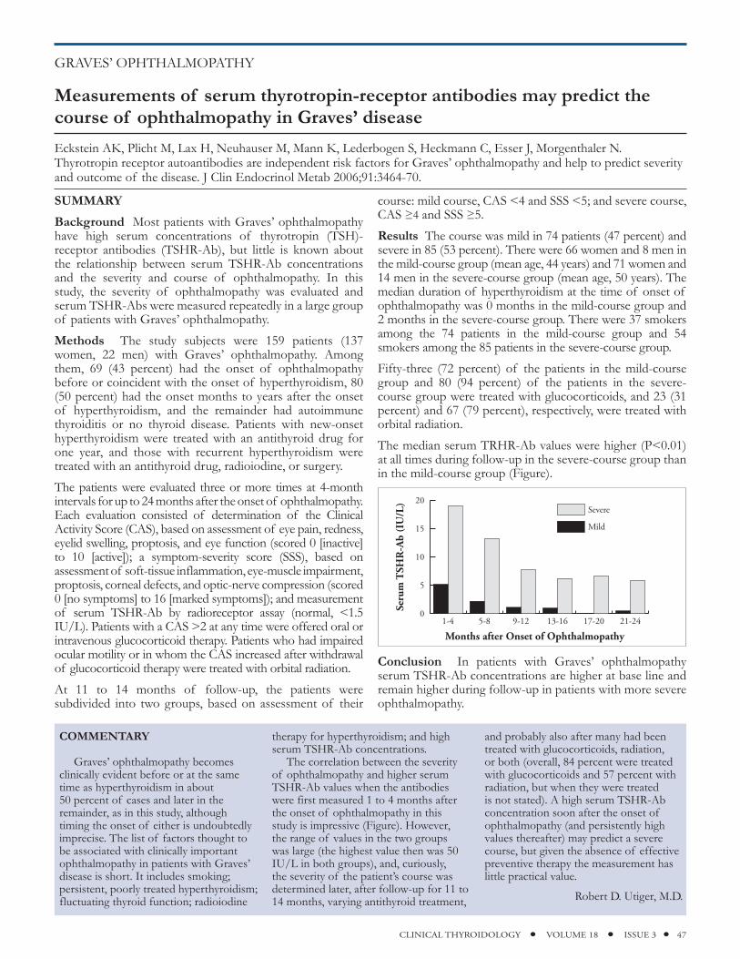
The median serum TRHR-Ab values were higher (P<0.01) at all times during follow-up in the severe-course group than in the mild-course group (Figure).
Conclusion In patients with Graves’ ophthalmopathy serum TSHR-Ab concentrations are higher at base line and remain higher during follow-up in patients with more severe ophthalmopathy.
COMMENTARY
Graves’ ophthalmopathy becomes clinically evident before or at the same time as hyperthyroidism in about 50 percent of cases and later in the remainder, as in this study, although timing the onset of either is undoubtedly imprecise. The list of factors thought to be associated with clinically important ophthalmopathy in patients with Graves’ disease is short. It includes smoking; persistent, poorly treated hyperthyroidism; fluctuating thyroid function; radioiodine
therapy for hyperthyroidism; and high serum TSHR-Ab concentrations.
The correlation between the severity of ophthalmopathy and higher serum TSHR-Ab values when the antibodies were first measured 1 to 4 months after the onset of ophthalmopathy in this study is impressive (Figure). However, the range of values in the two groups was large (the highest value then was 50 IU/L in both groups), and, curiously, the severity of the patient’s course was determined later, after follow-up for 11 to 14 months, varying antithyroid treatment,
and probably also after many had been treated with glucocorticoids, radiation, or both (overall, 84 percent were treated with glucocorticoids and 57 percent with radiation, but when they were treated is not stated). A high serum TSHR-Ab concentration soon after the onset of ophthalmopathy (and persistently high values thereafter) may predict a severe course, but given the absence of effective preventive therapy the measurement has little practical value.
Robert D. Utiger, M.D.
0
5
10
15
20Severe
Mild
21-2417-2013-169-125-81-4
Months after Onset of Ophthalmopathy
Seru
m T
SHR
-Ab
(IU
/L)

48 ● CLINICAL THYROIDOLOGY ● VOLUME 18 ● ISSUE 3
HYPOTHYROIDISM
The risk of coronary heart disease is increased in subclinical hypothyroidism
Rodondi N, Aujesky D, Vittinghoff E, Cornuz J, Bauer DC. Subclinical hypothyroidism and the risk of coronary heart disease: a meta-analysis. Am J Med 2006;119:541-51.
SUMMARY
Background Patients with subclinical hypothyroidism may have high serum concentrations of cholesterol and an increase in the thickness of the intima and media of the carotid arteries, changes associated with an increase in the risk of coronary heart disease. This meta-analysis was done to determine whether the incidence of coronary heart disease is increased in patients with subclinical hypothyroidism.
Methods The literature was reviewed to identify articles describing studies of patients with subclinical hypothyroidism, defined as a high serum thyrotropin (TSH) and normal serum thyroxine (T4) concentration, that included estimates of the risk of coronary heart disease or cardiovascular mortality in the patients and normal subjects. Information was obtained from each article about study design, patient characteristics, criteria for selection of control subjects, criteria for diagnosis of coronary heart disease, matching and adjudication procedures, and results. Several aspects of study quality were assessed, including how study patients were identified and how outcomes were adjudicated.
Results Fourteen studies of the risk of coronary heart disease or cardiovascular mortality in patients with subclinical hypothyroidism were identified (five prospective cohort studies, six cross-sectional studies, and three case–control studies). Most included women and men, and most were limited to older subjects; they lived in the United States, Europe, and Japan. The number of study subjects varied from 194 to 2592 (total, 10,540). There were 1362 cardiovascular events, including angina, myocardial infarction, cardiac revascularization, death from coronary heart disease, and cardiovascular death.
The odds ratios for risk of coronary heart disease in patients with subclinical hypothyroidism in the individual studies ranged from 0.27 (95 percent confidence interval, 0.03 to 1.48) to 6.35 (95 percent confidence interval, 2.08 to 19.63). The summary odds ratio was 1.65 (95 percent confidence interval, 1.28 to 2.12); the ratios varied little in analyses adjusted for various factors and analyses limited to various subgroups and type of study (Table).
Table. Results of Summary, Adjusted, and Subgroup Analyses of the Risk of Coronary Heart Disease in Patients with Subclinical Hypothyroidism.
Study Characteristics Odds Ratio (95% CI) No. of StudiesSummary 1.65 (1.28–2.12) 14Adjusted for cardiovascular risk factors 2.38 (1.53–3.69) 3Subjects with serum TSH values <4.5mU/L excluded 1.70 (1.08–2.68) 7
Subjects taking T4 excluded 2.06 (1.35–3.14) 7Prospective cohort studies 1.42 (0.91–2.21) 5Cross-sectional and case–control studies 1.72 (1.25–2.38) 9
CI denotes confidence interval.
The odds ratios were similar to the summary odds ratio if the two studies in which the end point was cardiovascular death or the two studies in which the end point was acute myocardial infarction were excluded, or if the analysis was limited to the six studies in which there were formal procedures to adjudicate the presence of coronary heart disease or the seven studies in which there was adjudication of coronary heart disease without knowledge of thyroid status.
Conclusion Based on cumulative data from these 14 studies, patients with subclinical hypothyroidism have an increased risk of coronary heart disease.
COMMENTARY
The possibility that the risk of cardiovascular disease is increased is an important component of the controversy regarding the detection and treatment of subclinical hypothyroidism. This meta-analysis was performed according to accepted recommendations for meta-analyses of observational studies. However, problems remain with respect to the definitions of subclinical hypothyroidism and coronary heart disease, the effects of nonthyroidal illness, and the combination of prevalence and incidence data in the various studies. The impact of the conventional cardiovascular risk factors such as age, sex, smoking, hypertension, hyperglycemia, and serum lipid abnormalities is so large compared with the potential effect of subclinical
hypothyroidism that it will always be problematic whether observational studies or meta-analyses have the power to determine such an association. Indeed, three more recently published large epidemiologic studies (1-3) continue to yield conflicting data regarding the association between subclinical hypothyroidism and cardiovascular disorders.
It should not be forgotten that similar analyses suggested that estrogen therapy protected against cardiovascular disease in postmenopausal women. The question of whether subclinical hypothyroidism is associated with cardiovascular disease is also likely to be answered only by a prospective therapeutic trial.
Mark P. J. Vanderpump. M.D.Royal Free Hospital,
London, United Kingdom
References
1. Walsh JP, Bremner AP, Bulsara MK, et al. Subclinical thyroid dysfunction as a risk factor for cardiovascular disease. Arch Intern Med 2005;165:2467-72. (Clinical Thyroidology 2006;18:1.)
2. Rodondi N, Newman AB, Vittinghoff E, et al. Subclinical hypothyroidism and the risk of heart failure, other cardiovascular events, and death. Arch Intern Med 2005; 165:2460-6. (Clinical Thyroidology 2006;18:2.)
3. Cappola AR, Fried LP, Arnold AM, et al. Thyroid status, cardiovascular risk, and mortality in older adults. JAMA 2006;295:1033-41. (Clinical Thyroidology 2006;18:22.)
CLINICAL THYROIDOLOGY ● VOLUME 18 ● ISSUE 3 ● 49
HYPOTHYROIDISM
Patients with adrenal insufficiency may have reversible hypothyroidism
Abdullatif HD, Ashraf AP. Reversible subclinical hypothyroidism in the presence of adrenal insufficiency. Endocr Pract 2006;12:572-5.
SUMMARY
Background Some patients with adrenal insufficiency also have hypothyroidism, which may improve when the former is treated. This article describes three patients with adrenal insufficiency and hypothyroidism in whom the latter improved spontaneously when they were treated with cortisol.
Case Reports
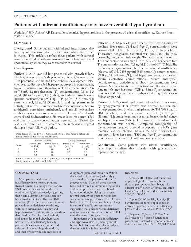
Patient 1 A 14-year-old boy presented with growth failure. His height was at the 50th percentile, his weight was at the 10th percentile, and he had little pubertal development. Bio-chemical studies revealed hypergonadotropic hypogonadism, hypothyroidism (serum thyrotropin [TSH] concentrations, 6.6 to 7.8 mU/L; free thyroxine [T4] concentrations, 0.8 to 1.3 ng/dl [10 to 17 pmol/L]) (Table), and adrenal insufficiency (plasma corticotropin [ACTH], 1445 pg/ml [318 pmol/L]; serum cortisol, 1.2 µg/dl [33 nmol/L], and high plasma renin activity, but normal serum electrolyte concentrations). Serum antithyroid peroxidase, antiadrenal, and anti-21-hydroxylase antibody concentrations were normal. He was treated with cortisol and fludrocortisone. Six weeks later, his serum TSH and free thyroxine concentrations were normal (Table). He was later treated with testosterone. He remained euthyroid during a 4-year follow-up period.
Table. Serum TSH and Free T4 Concentrations in Three Patients before and during Treatment for Adrenal Insufficiency.
Before Treatment During Treatment
Patient Serum TSH (mU/L)
Serum Free T4 (ng/dl)
Serum TSH (mU/L)
Serum Free T4 (ng/dl)
1 7.8 0.8 0.5 1.42 7.7 0.8 2.7 1.03 6.2 0.8 1.7 1.4
Normal values: TSH, 0.4-5.8 mU/L; free T4, 0.9-1.7 ng/dl. To convert free T4 values to pmol/L, multiply by 12.9.
Patient 2 A 12-year-old girl presented with type 1 diabetes mellitus. Her serum TSH and free T4 concentrations were normal (TSH, 1.8 mU/L; free T4, 1.1 ng/dl [14 pmol/L]). Thereafter, her glycemic control was good, and she grew normally, but gained little weight. Two years later, her serum TSH concentration was high (7.7 mU/L) and her serum free T4 concentration was low (0.8 ng/dl [10 pmol/L]) (Table). She had no hyperpigmentation, but she had adrenal insufficiency (plasma ACTH, 2495 pg/ml [549 pmol/L]; serum cortisol, <1.0 µg/dl [28 nmol/L], and hyperreninemia, but normal serum electrolyte concentrations). Serum antithyroid peroxidase and antiadrenal antibody concentrations were normal. She was treated with cortisol and fludrocortisone. One month later, her serum TSH and free T4 concentrations were normal. She remained euthyroid during a three-year follow-up period.
Patient 3 A 2-year-old girl presented with seizures caused by hypoglycemia. Her growth was normal, but she had hyperpigmentation. She had high plasma ACTH (2098 pg/ml [461 pmol/L]) and low serum cortisol (<1.0 µg/dl [28 nmol/L]) concentrations, but not aldosterone deficiency, and hypothyroidism (Table). Her serum antiadrenal antibody concentration was normal. Computed tomography of the abdomen revealed adrenal hypoplasia; a DAX1 gene mutation was not detected. She was treated with cortisol, and one month later her serum TSH and free T4 concentrations were normal. She was still euthyroid one year later.
Conclusion Some patients with adrenal insufficiency have hypothyroidism that subsides with glucocorticoid replacement therapy.
COMMENTARY
Most patients with adrenal insufficiency have normal pituitary-thyroid function, although their serum TSH concentrations during the day tend to be slightly increased, suggesting that normal daytime cortisol secretion has a small inhibitory effect on TSH secretion (1). A few have an autoimmune polyendocrine deficiency syndrome, with both adrenal insufficiency and hypothyroidism. Others, like the children described by Abdullatif and Ashraf, and adults described elsewhere (2,3), have adrenal insufficiency (usually primary, but sometimes central) and subclinical or overt hypothyroidism, and their hypothyroidism improves or
disappears (increased thyroid secretion, decreased TSH secretion) when they are treated with replacement doses of glucocorticoid. Most of these patients have had chronic autoimmune thyroiditis, and the improvement was attributed to its amelioration, implying that even a normal quantity of glucocorticoid has some immunosuppressive activity. Others had a fall in TSH secretion, but no change in serum T4 and T3 concentrations, suggesting that glucocorticoid deficiency may sometimes result in secretion of TSH with decreased biologic activity.
In patients with adrenal insufficiency and hypothyroidism, T4 therapy should be withheld for several weeks in order to determine if it is indeed needed.
Robert D. Utiger, M.D.
References
1. Samuels MH. Effects of variations in physiological cortisol levels on thyrotropin secretion in subjects with adrenal insufficiency: a Clinical Research Center Study. J Clin Endocrinol Metab 2000;85:1388-93.
2. Topliss DJ, White EL, Stockigt JR. Significance of thyrotropin excess in untreated primary adrenal insufficiency. J Clin Endocrinol Metab 1980;50:52-6.
3. Shigemasa C, Kouchi T, Ueta Y, et al. Evaluation of thyroid function in patients with isolated adrenocorticotropin deficiency. Am J Med Sci 1992;304:279-84.
50 ● CLINICAL THYROIDOLOGY ● VOLUME 18 ● ISSUE 3
HYPOTHYROIDISM
Small changes in thyroxine dose do not alter well-being or symptoms in patients with hypothyroidism
Walsh JP, Ward LC, Burke V, Bhagat CI, Shiels L, Henley D, Gillett MJ, Gilbert R, Tanner M, Stuckey BG. Small changes in thyroxine dosage do not produce measurable changes in hypothyroid symptoms, well-being, or quality of life: results of a double-blind, randomized clinical trial. J Clin Endocrinol Metab 2006;91:2624-30.
SUMMARY
Background Patients with hypothyroidism often continue to have symptoms despite treatment that restores their serum thyrotropin (TSH) concentrations to within the normal range. In this study, patients with hypothyroidism were treated with slightly varying doses of thyroxine (T4) to determine if there were differences in symptoms when their serum TSH concentrations varied within the normal range.
Methods The study subjects were 56 patients (52 women, 4 men; mean age, 53 years) with hypothyroidism. The inclusion criteria were treatment with at least 100 µg of T4 for at least six months (mean, 9 years), no change in the dose in the preceding two months, and a serum TSH concentration of 0.1 to 4.8 mU/L at the screening visit.
The patients received three different doses of T4, each for eight weeks, in random order. The doses were intended to result in serum TSH concentrations of 2.0 to 4.8 mU/L (low dose), 0.3 to 1.9 mU/L (middle dose), and <0.3 mU/L (high dose).
At base line and at the end of each 8-week period, the patients completed 10 four-point visual analog scales that assessed general well-being (the primary outcome measure), happiness/sadness, confusion, anxiety, irritability, tiredness, and other symptoms. At the same times, quality of life was assessed using the Short Form-36 (eight individual scores and summary physical and mental component scores); psychological function was assessed using the General Health Questionnaire-28; hypothyroid symptoms were assessed using a 12-item Thyroid Symptom Questionnaire; and cognitive function was assessed using the Symbol Digit Modalities test, the Trail Making test, and the Digit Span test. Serum TSH and free T4 also were measured.
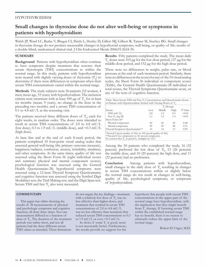
Results Fifty patients completed the study. The mean daily T4 doses were 103 µg for the low-dose period, 127 µg for the middle-dose period, and 152 µg for the high-dose period.
There were no differences in weight, pulse rate, or blood pressure at the end of each treatment period. Similarly, there were no differences in the scores for any of the 10 visual analog scales, the Short Form-36 individual or component scores (Table), the General Health Questionnaire-28 individual or total scores, the Thyroid Symptoms Questionnaire score, or any of the tests of cognitive function.
Table. Mean Serum TSH and Free T4 Concentrations and Questionnaire Scores in Patients with Hypothyroidism Treated with Varying Doses of T4.
T4 DosageSerum Low Middle High P Value TSH (mU/L) 2.8 1.0 0.3 <0.01 Free T4 (ng/dl) 1.1 1.2 1.4 <0.01Short Form-36* Physical component 42 42 42 0.48 Mental component 50 50 48 0.31Thyroid Symptom Questionnaire** 14 14 13 0.99
*Scored 0 (poor quality of life) to 100 (good quality of life).**Scored 0 (no symptoms) to 36 (severe symptoms).To convert free T4 to pmol/L, multiply by 12.9.
Among the 50 patients who completed the study, 16 (32 percent) preferred the low dose of T4, 13 (26 percent) the middle dose, and 10 (20 percent) the high dose, and 11 (22 percent) had no preference.
Conclusion Among patients with hypothyroidism, small changes in the daily dose of T4 resulting in changes in serum TSH concentrations within or slightly below the normal range do not result in changes in well-being, quality of life, psychological symptoms, or symptoms of hypothyroidism.
COMMENTARY
This paper has tables showing the results of 38 measurements of physical and psychologic symptoms and cognitive function, all done three times. Not one measurement differed as a function of dose of T4. The duration of the treatment periods was rather short, and not all patients had the three different serum TSH values as intended. These limitations
do not negate the key findings—treatment with a relatively low dose of T4 was no less effective than higher doses, and treatment that resulted in serum TSH concentrations of 2.0 to 4.8 mU/L was no less effective than treatment that reduced serum TSH concentrations to 0.3 to 1.9 mU/L or even <0.3 mU/L.
In short, if some T4 is good, more is not necessarily better. Furthermore, the results provide no support for the
hypothesis that people with serum TSH concentrations in the upper part of the normal range have hypothyroidism, with the implication that they might benefit from T4 therapy. If lowering serum TSH within the established normal range has no benefit, there is no reason to arbitrarily reduce the upper limit of the normal range.
Robert D. Utiger, M.D.
CLINICAL THYROIDOLOGY ● VOLUME 18 ● ISSUE 3 ● 51
HYPOTHYROIDISM
Combined thyroxine and triiodothyronine therapy is not more effective than thyroxine alone in patients with hypothyroidism
Grozinsky-Glasberg S, Fraser A, Nahshoni E, Weizman A, Leibovici L. Thyroxine-triiodothyronine combination therapy versus thyroxine monotherapy for clinical hypothyroidism: meta-analysis of randomized controlled trials. J Clin Endocrinol Metab 2006;91:2592-9.
SUMMARY
Background Patients with hypothyroidism may not feel well despite therapy with thyroxine (T4) in doses that restores their serum T4 and thyrotropin (TSH) concentrations to normal. This has led to studies of combined T4 and triiodothyronine (T3) therapy, based on the assumption that thyroidal production of T3 is important. The results of these studies have been somewhat conflicting, which led to this summary analysis of the individual studies.
Methods Multiple databases were searched to identify randomized and quasi-randomized trials of T4 monotherapy and T4 and T3 combination therapy in patients with hypothyroidism. The search yielded 11 studies. The characteristics of each trial, including the details of patient selection, treatment doses, end points evaluated, and methods of analysis, were then summarized.
The predefined primary outcomes were bodily pain, fatigue, depression, and quality of life, as determined by the patients’ responses to questionnaires. The questionnaires used varied among the studies, and not all outcomes were determined in all studies. The questionnaires used included the Short Form-36, Symptom Check List-90, Hospital Anxiety and Depression Scale, and Health-Related Quality-of-Life Scale. Other outcomes studied were changes in cognitive function and serum TSH concentrations. The results were summarized as standardized mean differences between T4 and T3 combination therapy and T4 monotherapy, weighted according to the numbers of patients studied.
COMMENTARY
There is a logical attraction in replacing the two missing hormones, T4 and T3, in patients with primary hypothyroidism. After all, both are produced and secreted by the thyroid gland. It is disappointing, therefore, that combinations of T4 and T3 do not have any advantage over T4 alone in patients with hypothyroidism.
What then is to be done for patients with hypothyroidism who have not achieved the anticipated sense of well-being with restoration of their serum TSH concentration to the reference range? In those whose serum TSH concentration is in the upper part of that range, a small increase in the dose of T4 may be all that is necessary. Other patients are most content when their serum TSH concentrations are slightly low, and there
is no reason not to give sufficient T4 to achieve this. In this regard, it is notable that patients with thyroid carcinoma deliberately treated with high doses of T4 rarely have symptoms that can be attributed to inadequate T4 therapy. However, many clinicians are unwilling to prescribe a dose of T4 that results in low serum TSH concentrations (except in patients with thyroid carcinoma), for fear of an increase in the risk of osteoporosis or atrial fibrillation, notwithstanding the sparse evidence that the risk of either is increased in T4-treated patients (1).
For many patients with serum TSH concentrations within the normal range, neither adding T3 nor changing the dose of T4 (2) will help. For them, an alternative explanation for their symptoms has to be sought. Perhaps the explanation lies with autoimmune thyroid
disease, the usual cause of spontaneously occurring hypothyroidism both in these studies and in general.
Anthony D. Toft, M.D.Royal Infirmary,
Edinburgh, Scotland
References
1. Toft A. Which thyroxine? Thyroid 2005; 5:124-6.
2. Walsh JP, Ward LC, Burke V, et al. Small changes in thyroxine dosage do not produce measurable changes in hypothyroid symptoms, well-being, or quality of life: results of a double-blind, randomized clinical trial. J Clin Endocrinol Metab 2006;91:2624-30. (See preceding page.)
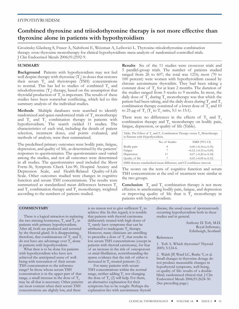
Results Six of the 11 studies were crossover trials and 5 parallel-group trials. The number of patients studied ranged from 26 to 607; the total was 1216; most (70 to 100 percent) were women with hypothyroidism caused by chronic autoimmune thyroiditis. They had been taking a constant dose of T4 for at least 2 months. The duration of the studies ranged from 5 weeks to 9 months. In most, the daily dose of T4 during T4 monotherapy was that which the patient had been taking, and the daily doses during T4 and T3 combination therapy consisted of a lower dose of T4 and 10 to 25 µg of T3 (T4 to T3 ratio, 5:1 to 15:1).
There were no differences in the effects of T4 and T3 combination therapy and T4 monotherapy on bodily pain, fatigue, depression, or quality of life (Table).
Table. The Effect of T4 and T3 Combination Therapy versus T4 Monotherapy in Patients with Hypothyroidism.
No. of Studies SMD (95% CI)
Bodily pain 4 0.00 (−0.34 to 0.35)Fatigue 7 −0.12 (−0.33 to 0.09)Depression 11 0.07 (−0.20 to 0.34)Quality of life 7 0.03 (−0.09 to 0.15)SMD denotes standardized mean difference, and CI confidence interval.
The scores on the tests of cognitive function and serum TSH concentrations at the end of treatment were similar in the two groups.
Conclusion T4 and T3 combination therapy is not more effective in ameliorating bodily pain, fatigue, and depression or improving quality of life than is T4 monotherapy in patients with hypothyroidism.
52 ● CLINICAL THYROIDOLOGY ● VOLUME 18 ● ISSUE 3
HYPOTHYROIDISM
Intra-amniotic administration of thyroxine is effective therapy in fetuses with goiter and hypothyroidism
Hashimoto H, Hashimoto K, Suehara N. Successful in utero treatment of fetal goitrous hypothyroidism: case report and review of the literature. Fetal Diagn Ther 2006;21:360-5.
SUMMARY
Background Hypothyroidism can occur during fetal life, but is rarely recognized unless the mother is receiving antithyroid drug therapy or fetal ultrasonography reveals thyroid enlargement. This article describes the detection and treatment of goitrous hypothyroidism in a fetus and summarizes previous reports of treatment of similar fetuses.
Case Report A 33-year-old pregnant woman was seen because routine fetal ultrasonography at 30 weeks’ gestation had revealed a craniofacial mass. Repeat ultrasonography revealed diffuse symmetric thyroid enlargement (2.6×1.9×2.8 cm; total volume, 14.5 cm3); the echo texture was homogeneous, and blood flow was increased. The fetal neck was hyperextended. The mother was well, and had no history of thyroid disease. Her physical examination was normal, except for a small diffuse goiter, as was her thyroid function.
Amniocentesis and cordocentesis were done at 32 weeks. The amniotic fluid TSH concentration was high (0.98 mU/L; normal, 0.15 to 0.55). The cord serum TSH concentration also was high (38.3 mU/ml; normal mean, 7.3) and the cord serum free thyroxine (T4) concentration was low (0.7 ng/dl [9 pmol/L]) (normal mean, 1.1 [14]).
Four intra-amniotic injections of T4, each of 150 µg, were given weekly from 33 and 36 weeks, resulting in a decrease in the fetal goiter to 5.3 cm3, and a decrease in amniotic fluid TSH concentration, but no change in amniotic fluid T4 concentration. The fetal neck became flexed. Fetal heart rate and growth were normal.
Labor was induced at 37 weeks because of polyhydramnios. The mother delivered a healthy boy who weighed 3062 g. Physical examination was normal except for a small goiter (2.2×1.2 cm). At birth and on day 3, the infant’s serum TSH and free T4 concentrations were normal. At age four weeks the infant was normal except that his thyroid remained enlarged, and at two years growth and development were normal.
Literature Review Nineteen fetuses, including this one, with goiter and hypothyroidism confirmed by cordocentesis have been treated with intra-amniotic administration of T4. Their gestational age at the time of diagnosis ranged from 20 to 33 weeks. Eight mothers had hyperthyroidism and were taking an antithyroid drug. The other 11 mothers had no thyroid disease; the goitrous hypothyroidism in these fetuses may have been caused by thyroid dyshormonogenesis or transplacental passage of some goitrogenic substance—for example, iodine.
The doses of T4 ranged from 150 to 600 µg, given once in seven fetuses and up to 10 times, usually weekly, in the others. There was a decrease in goiter size in all the fetuses, and no maternal or fetal complications of therapy, except for decreased growth in one fetus. At birth, psychomotor development was normal. Postnatally, seven infants had normal thyroid function, three had transient hypothyroidism, and nine had hypothyroidism (causes not stated).
Conclusion Fetuses found to have a goiter and hypothyroidism respond appropriately to intra-amniotic injection of T4 with reduction in the size of their goiter and amelioration of hypothyroidism.
COMMENTARY
Fetal goiter may be detected incidentally, as in the fetus described by Hashimoto et al. and many of the others in their review, in which case it is nearly always associated with fetal hypothyroidism. More often, it is detected because ultrasonography is done as an indirect test of thyroid function in a fetus whose mother has (or had) Graves’ hyperthyroidism. In these fetuses, the question is whether the fetus has thyroid enlargement as an indicator of hyperthyroidism, caused by transplacental passage of TSH-receptor stimulating antibodies, or hypothyroidism, caused by transplacental passage of an antithyroid drug. Information regarding fetal thyroid status may be gained by assessing fetal growth and cardiovascular function, and maternal thyroid function, TSH-receptor antibody status, and treatment. If more
precise information is needed, TSH and free T4 can be measured in amniotic fluid or serum collected by cordocentesis. Neither is without risk, and therefore neither is suitable for monitoring the response to therapy.
With respect to fetal goiter caused by hypothyroidism, which fetuses benefit from treatment with T4? The fetuses most at risk are those with a large goiter late in gestation, because of the risks of dystocia, malpresentation, and tracheal obstruction. Intra-amniotic T4 treatment may be beneficial, but how much to give and how often to give it are not clearly established. These uncertainties are complicated by the inability to assess fetal thyroid function without invasive testing. While ultrasonography can be useful, more direct methods of repeatedly assessing fetal thyroid function are needed. Maternal serum measurements of fetally derived
diiodothyronine sulfate–like compounds (so named because they cross-react in a radioimmunoassay for diiodothyronine sulfate but are chromatographically distinct from it) could be useful for this purpose (1). Fetal and maternal serum concentrations of these compounds rise progressively during normal pregnancy, and the values in fetuses with hypothyroidism are low in both fetus and mother.
Daniel H. Polk, M.D.Children’s Memorial Hospital
Chicago, IL
Reference
1. Cortelazzi D, Morpurgo PS, Zamperini P, et al. Maternal compound W serial measurements for the management of fetal hypothyroidism. Eur J Endocrinol 1999;141:570-8.

CLINICAL THYROIDOLOGY ● VOLUME 18 ● ISSUE 3 ● 53
NODULAR GOITER
No single ultrasonographic finding reliably distinguishes thyroid carcinomas from benign thyroid nodules
Cappelli C, Castellano M, Pirola I, Gandossi E, De Martino E, Cumetti D, Agosti B, Rosei EA. Thyroid nodule shape suggests malignancy. Eur J Endocrinol 2006;155:27-31.
SUMMARY
Background Thyroid nodules are common, but only a few are carcinomas. This study was done to determine whether benign thyroid nodules can be distinguished from carcinomas by the shape of the nodule or other characteristics, as determined by ultrasonography.
Methods The ultrasound and cytologic characteristics of 7455 thyroid nodules in 5198 patients seen between 1991 and 2004 were retrospectively evaluated. Among the nodules, 2865 (38 percent) were <1 cm in diameter.
The ultrasound scans were evaluated by two investigators, who recorded the following information for each nodule: anteroposterior and transverse (A/T) diameter, degree of echogenicity, presence of calcification, characteristics of nodule margin (blurred or well-defined), and vascularity. The nodules were biopsied using 25-gauge needles, and the results were reported as benign, suspicious (including follicular and Hurthle-cell tumor and suspicious for papillary carcinoma), malignant, or inadequate (<6 large clusters of follicular cells). All patients who had biopsies reported as suspicious or malignant underwent surgery. Nodules that not been biopsied that were carcinomas were not included in the analysis.
Results Among the 7455 nodules, 6135 biopsies (82 percent) were adequate and 1320 (18 percent) were inadequate; the respective numbers of patients were 4495 (3118 women, 1377 men) and 703. Nodules for which the biopsy was inadequate were excluded. Multiple nodules were successfully biopsied in 1351 patients (30 percent), of whom 1118 had 2 nodules and 233 had ≥3 nodules.
Among the 6135 nodules that were successfully biopsied, the mean longest dimension was 16 mm (range, 6 to 100);
2120 (35 percent) were <1 cm (small) and 4015 (65 percent) were ≥1 cm (large). The biopsy was reported as suspicious or malignant in 349 of the 4495 patients (8 percent), and 284 (6 percent) proved to have carcinoma (papillary carcinoma, 242; follicular carcinoma, 37; and medullary carcinoma, 5); no patient had 2 or more suspicious nodules and none had 2 carcinomas. Overall, 5 percent of the nodules that were successfully biopsied were carcinomas (3 percent of the small nodules and 6 percent of the large, P<0.01). Carcinomas were slightly more common when multiple nodules were present (5 vs. 4 percent, P=0.02).
The value of the ultrasound characteristics for predicting the presence of carcinoma is shown in the Table.
Table. Value of Ultrasonography for Identifying Malignant Thyroid Nodules.
Ultrasound Finding Sensitivity SpecificityPositive
Predictive Value
Negative Predictive
ValueA/T ratio ≥1 76% 60% 8% 98%Hypoechoic 81% 47% 7% 98%Calcifications 72% 71% 11% 98%Blurred margins 53% 81% 12% 97%Hypervascularity 62% 50% 6% 96%Size ≥1 cm 77% 35% 5% 97%
The sensitivity and specificity of an A/T ratio ≥1 and any two of the following findings—hypoechogenicity, blurred margins, and calcifications—were 99 and 57 percent, respectively, and the positive and negative predictive values were 6 and 99 percent, respectively.
Conclusion No single ultrasonographic feature reliably distinguishes thyroid carcinomas from benign thyroid nodules. The combination of an A/T ratio ≥1 and any two of three other ultrasonographic findings (hypoechogenicity, blurred margins, and calcifications) is the most sensitive indicator that a nodule is a carcinoma.
COMMENTARY
Thyroid ultrasonography is without question the best way to define the size and characteristics of thyroid nodules, but attempts to identify ultrasound findings that reliably distinguish nodules that are thyroid carcinomas from those that are benign have been unsuccessful. As described in this (curiously titled) paper, there are findings that suggest a nodule may be a carcinoma, but even combinations of these findings have a low predictive value. Another problem that limits the generalizability of ultrasound features of nodules is the considerable interobserver variation in determining
the presence of these features and also nodule dimensions and shape.
Note that shape in this study meant shape in the horizontal plane, meaning width and depth. Why a thyroid carcinoma might have a depth/width (A/T) ratio ≥1 more often than a benign nodule (76 vs. 40 percent) is not clear, and the authors do not speculate on why this might be so. One explanation may be that the carcinomas were larger, having grown more anteriorly (the path of least resistance?), and therefore become visible or palpable. Why nodule length and therefore three-dimensional shape were not evaluated also is not clear. In a study in which three-dimensional shape was evaluated, the
carcinomas were more often spherical or nearly so than were benign nodules (1).
Currently, few patients with thyroid nodules can be assured that their nodules are benign in the absence of biopsy. It is unlikely that any combination of ultrasound findings or improved imaging technology (ultrasound or other) will reduce the need for biopsy.
Robert D. Utiger, M.D.
Reference
1. Alexander EK, Marqusee E, Orcutt J, et al. Thyroid nodule shape and prediction of malignancy. Thyroid 2004;14:953-8.
54 ● CLINICAL THYROIDOLOGY ● VOLUME 18 ● ISSUE 3
THYROID CANCER
The frequency of carcinoma is similar in patients with one thyroid nodule and those with multiple nodules
Frates MC, Benson CB, Doubilet PM, Kunreuther E, Contreras M, Cibas ES, Orcutt J, Moore FD Jr, Larsen PR, Marqusee E, Alexander EK. Prevalence and distribution of carcinoma in patients with solitary and multiple thyroid nodules on sonography. J Clin Endocrinol Metab 2006;91:3411-7.
SUMMARY
Background Patients with either a solitary thyroid nodule or a multinodular goiter may have a thyroid carcinoma, but whether the frequency of carcinoma varies according to the number of thyroid nodules is not clear. In this study, the prevalence of carcinoma in patients with a solitary nodule and those with multiple nodules was determined.
Methods The study subjects were 1985 patients (1742 women, 243 men) with one or more thyroid nodules >1 cm in longest dimension, as determined by ultrasonography, who underwent successful fine-needle aspiration biopsy of up to three nodules (any additional >1-cm nodules were biopsied later). Among them, 1181 (60 percent) had a single >1-cm nodule and 804 (40 percent) had two or more >1-cm nodules. All patients in whom the biopsy was suspicious or positive for papillary carcinoma, follicular tumor, or atypical (confirmed by a second biopsy) underwent bilateral thyroidectomy; the final diagnosis in these patients was based on the histopathology of all nodules >1 cm. Carcinomas ≤1 cm found on histopathologic examination of the thyroid were excluded.
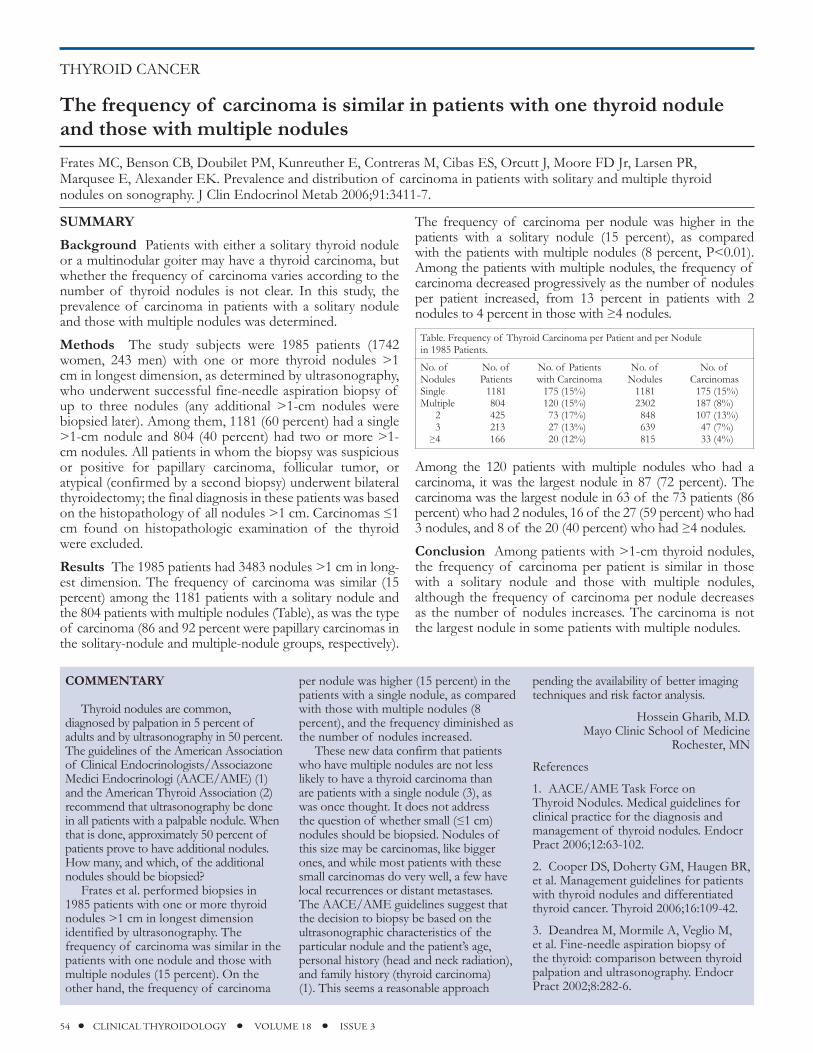
Results The 1985 patients had 3483 nodules >1 cm in long-est dimension. The frequency of carcinoma was similar (15 percent) among the 1181 patients with a solitary nodule and the 804 patients with multiple nodules (Table), as was the type of carcinoma (86 and 92 percent were papillary carcinomas in the solitary-nodule and multiple-nodule groups, respectively).
The frequency of carcinoma per nodule was higher in the patients with a solitary nodule (15 percent), as compared with the patients with multiple nodules (8 percent, P<0.01). Among the patients with multiple nodules, the frequency of carcinoma decreased progressively as the number of nodules per patient increased, from 13 percent in patients with 2 nodules to 4 percent in those with ≥4 nodules.
Table. Frequency of Thyroid Carcinoma per Patient and per Nodule in 1985 Patients.
No. of NodulesSingleMultiple 2 3 ≥4
No. of Patients
1181 804 425 213 166
No. of Patients with Carcinoma
175 (15%) 120 (15%) 73 (17%) 27 (13%) 20 (12%)
No. of Nodules
11812302 848 639 815
No. of Carcinomas
175 (15%) 187 (8%) 107 (13%) 47 (7%) 33 (4%)
Among the 120 patients with multiple nodules who had a carcinoma, it was the largest nodule in 87 (72 percent). The carcinoma was the largest nodule in 63 of the 73 patients (86 percent) who had 2 nodules, 16 of the 27 (59 percent) who had 3 nodules, and 8 of the 20 (40 percent) who had ≥4 nodules.
Conclusion Among patients with >1-cm thyroid nodules, the frequency of carcinoma per patient is similar in those with a solitary nodule and those with multiple nodules, although the frequency of carcinoma per nodule decreases as the number of nodules increases. The carcinoma is not the largest nodule in some patients with multiple nodules.
COMMENTARY
Thyroid nodules are common, diagnosed by palpation in 5 percent of adults and by ultrasonography in 50 percent. The guidelines of the American Association of Clinical Endocrinologists/Associazone Medici Endocrinologi (AACE/AME) (1) and the American Thyroid Association (2) recommend that ultrasonography be done in all patients with a palpable nodule. When that is done, approximately 50 percent of patients prove to have additional nodules. How many, and which, of the additional nodules should be biopsied?
Frates et al. performed biopsies in 1985 patients with one or more thyroid nodules >1 cm in longest dimension identified by ultrasonography. The frequency of carcinoma was similar in the patients with one nodule and those with multiple nodules (15 percent). On the other hand, the frequency of carcinoma
per nodule was higher (15 percent) in the patients with a single nodule, as compared with those with multiple nodules (8 percent), and the frequency diminished as the number of nodules increased.
These new data confirm that patients who have multiple nodules are not less likely to have a thyroid carcinoma than are patients with a single nodule (3), as was once thought. It does not address the question of whether small (≤1 cm) nodules should be biopsied. Nodules of this size may be carcinomas, like bigger ones, and while most patients with these small carcinomas do very well, a few have local recurrences or distant metastases. The AACE/AME guidelines suggest that the decision to biopsy be based on the ultrasonographic characteristics of the particular nodule and the patient’s age, personal history (head and neck radiation), and family history (thyroid carcinoma) (1). This seems a reasonable approach
pending the availability of better imaging techniques and risk factor analysis.
Hossein Gharib, M.D.Mayo Clinic School of Medicine
Rochester, MN
References
1. AACE/AME Task Force on Thyroid Nodules. Medical guidelines for clinical practice for the diagnosis and management of thyroid nodules. Endocr Pract 2006;12:63-102.
2. Cooper DS, Doherty GM, Haugen BR, et al. Management guidelines for patients with thyroid nodules and differentiated thyroid cancer. Thyroid 2006;16:109-42.
3. Deandrea M, Mormile A, Veglio M, et al. Fine-needle aspiration biopsy of the thyroid: comparison between thyroid palpation and ultrasonography. Endocr Pract 2002;8:282-6.
CLINICAL THYROIDOLOGY ● VOLUME 18 ● ISSUE 3 ● 55
THYROID CANCER
The follicular-variant subtype of papillary thyroid carcinoma may be either encapsulated or nonencapsulated
Liu J, Singh B, Tallini G, Carlson DL, Katabi N, Shaha A, Tuttle RM, Ghossein RA. Follicular variant of papillary thyroid carcinoma: a clinicopathologic study of a problematic entity. Cancer 2006;107:1255-64.
SUMMARY
Background One subgroup of differentiated thyroid carcinomas consists of tumors called follicular variant of papillary carcinoma (FVPTC). These tumors are composed of follicles lined by cells that have the nuclear features of papillary carcinoma (large, irregular nuclei, and nuclear clearing, grooves, and pseudoinclusions). They may be encapsulated, like follicular carcinomas, or nonencapsulated and infiltrative, like papillary carcinomas. This study was done to determine the relative frequency and course of these two subgroups of FVPTC.
Methods From 1980 to 1995, 552 follicular tumors of all types were identified at the Memorial Sloan-Kettering Cancer Center in New York. After review by four pathologists, 78 patients were determined to have FVPTCs, as defined by the presence of follicles lined by cells with the nuclear features of papillary carcinoma and the absence of papillary architecture, that were >1 cm in longest dimension.
The tumors were characterized as encapsulated, if completely surrounded by a capsule, with or without foci of capsular or vascular invasion, and nonencapsulated (infiltrative), if there was little or no capsule and the tumor extended into the surrounding thyroid tissue. The patients’ records were reviewed to obtain information about extent of surgery, operative findings, other treatment, and outcome.
Results The 78 patients with FVPTC included 59 women and 19 men (median age, 43 years; range, 6 to 72). The tumor was ≤4 cm in 69 patients (88 percent) and >4 cm in 9 (12 percent), and it was encapsulated in 61 patients (78 percent)
and infiltrative in 17 (22 percent). Fifty-four patients (69 percent) were treated by thyroid lobectomy, and only 15 (19 percent) were treated with radioiodine.
The clinical characteristics of the patients with encapsulated and infiltrative tumors and the pathologic characteristics of the tumors were similar, except that more of the patients with infiltrative FVPTCs had extrathyroidal extension or lymph node metastases (Table).
Table. Clinical and Pathologic Features of Encapsulated and Infiltrative FVPTCs.
Encapsulated (n=61) Infiltrative (n=17) P ValueWomen/men 49/12 10/7 0.11Tumor size ≤4 cm 52 (85%) 17 (100%) 0.19Capsular invasion 14 (23%) No capsuleVascular invasion 10 (16%) 4 (24%) 0.49Extrathyroidal extension 3 (5%) 11 (65%) <0.01Lymph node metastases 3 (5%) 11 (65%) <0.01
Sixty-nine patients were followed for >1 year, with a median follow-up of 11 years (range, 1 to 21). None of the 42 patients with a noninvasive encapsulated FVPTC had a recurrence, including 31 patients treated by thyroid lobectomy alone. Among 13 patients with invasive encapsulated tumors, 1 (8 percent) had a recurrence, and among 14 patients with infiltrative tumors none had a recurrence.
Conclusion The follicular variant of papillary thyroid carcinoma appears to take two forms. Most are encapsulated tumors that may invade the tumor capsule or vessels, but rarely involve lymph nodes, and which therefore resemble follicular adenomas or follicular carcinomas. The others are infiltrative tumors that may involve lymph nodes, resembling classical papillary carcinomas.
COMMENTARY
The key findings that have defined FVPTCs in most studies have been follicular architecture, the presence of the nuclear features of papillary carcinoma in many if not all cells in the tumor, and the scarcity of papillae. But precisely how to determine if an individual tumor is a FVPTC and whether FVPTC is being overdiagnosed are debated (1,2). With respect to molecular characteristics. FVPTC seem to be more similar to folli-cular adenomas and carcinomas than to papillary carcinomas. On the other hand, the prognosis of patients with FVPTC seems to be similar to that of patients with classical papillary carcinoma (3).
This subdivision of FVPTC into encapsulated and infiltrative subgroups and the conclusions that the encapsulated
tumors are follicular adenomas (if there is no capsular or blood-vessel invasion) or follicular carcinomas (if capsular or vascular invasion is seen) and the infiltrative tumors are papillary carcinomas seem reasonable. Of particular importance is the identification of a group of patients who have encapsulated tumors with no capsular or vascular invasion that are considered carcinomas only because the tumor-cell nuclei look like those of papillary carcinoma. These patients may well have follicular adenomas, notwithstanding the appearance of the nuclei of the tumor cells, and therefore it is no surprise that their prognosis is excellent when they are treated by thyroid lobectomy alone. The clinical problem is how to identify them preoperatively. At present, no one is likely to accept the suggestion that thyroid cells with abnormal nuclei
may not be cancer cells, and fine-needle aspiration biopsy cannot reveal the absence of capsular or vascular invasion.
Robert D. Utiger, M.D.
References
1. Chan JK. Strict criteria should be applied in the diagnosis of encapsulated follicular variant of papillary thyroid carcinoma. Am J Clin Pathol 2002;117:16-8.
2. Renshaw AA, Gould EW. Why there is the tendency to “overdiagnose” the follicular variant of papillary thyroid carcinoma. Am J Clin Pathol 2002;117:19-21.
3. Passler C, Prager G, Scheuba C, et al. Follicular variant of papillary thyroid carcinoma: a long-term follow-up. Arch Surg 2003;138:1362-6.

56 ● CLINICAL THYROIDOLOGY ● VOLUME 18 ● ISSUE 3
THYROID CANCER
Radioiodine therapy can cause testicular dysfunction in men with thyroid carcinoma
Rosario PW, Barroso AL, Rezende LL, Padrao EL, Borges MA, Guimaraes VC, Purisch S. Testicular function after radioiodine therapy in patients with thyroid cancer. Thyroid 2006;16:667-70.
SUMMARY
Background Men with thyroid carcinoma who are treated with radioiodine (I-131) may have abnormal pituitary–gonadal function, but the extent of the abnormalities and their relationship to the dose of I-131 are not well defined. In this study, pituitary–gonadal function was assessed in men with thyroid carcinoma who received single or multiple doses of I-131.
Methods Serum follicle-stimulating hormone (FSH), luteinizing hormone (LH), and free testosterone were measured before and 6, 12, and 18 months after a single dose of 100 or 150 mCi (3.7 or 5.6 GBq) of I-131 in 52 men with thyroid carcinoma. Their mean age was 45 years, and the mean dose of I-131 was 115 mCi (4.25 GBq).
The same measurements were done before and 18 months after the last dose of I-131 in 22 men with thyroid carcinoma who had pulmonary metastases and who received several doses of I-131. In addition, semen analyses were done in the men who had high serum FSH concentrations at the latter time. Their mean age was 51 years, and the mean total dose of I-131 was 550 mCi (20.3 GBq) (range, 350 to 750 [13.0 to 27.8 GBq]). The mean interval between the hormonal measurements was 60 months in these men.
All the men were taking thyroxine and had serum thyrotropin concentrations <0.5 mU/L when studied.
Results Among the 52 men given a single dose of I-131, the mean serum FSH concentration was normal (≤14 mIU/ml) before treatment, high 6 months after treatment, and normal 12 and 18 months after treatment (Table). Their mean serum LH and free testosterone concentrations were within the normal
range at all times. Five men (10 percent) had a high serum LH concentration (>10 mIU/ml) at 6 months, but no man had a low serum free testosterone concentration at any time. The results were similar in the men who received 100 mCi (3.7 MBq) of I-131 and those who received 150 mCi (5.6 GBq).
Table. Mean Serum FSH and LH Concentrations in 52 Men with Thyroid Carcinoma before and after I-131 Therapy.
I-131 TherapyBefore 6 Months 12 Months 18 months
Serum FSH (mIU/ml) 6.8 24.2* 9.5 5.9 High 0% 100% 29% 0%Serum LH (mIU/ml) 3.2 6.7* 2.9 3.4 High 0% 10% 0% 0%
*P<0.01, as compared with before therapy.
Among the 22 men given several doses of I-131 and studied 18 months after their last dose, 12 (54 percent) had a high serum FSH concentration and 5 (23 percent) had a high serum LH concentration; none had a low serum free testosterone concentration. Among the 12 men with high serum FSH concentrations at this time, 8 (67 percent) had oligospermia (sperm concentration <20×106/ml, mean of three samples). The total dose of I-131 was higher in the 8 men who had oligospermia (770 mCi [28.5 GBq]) than in the 4 men who did not (500 mCi [18.5 GBq]).
Conclusion Men with thyroid carcinoma who are treated with single doses of 100 to 150 mCi (3.7 to 5.6 GBq) of I-131 may have transient testicular damage, manifested by high serum FSH or LH concentrations, and those treated with higher doses may have oligospermia.
COMMENTARY
It is clear from this and other studies that I-131 therapy decreases spermatogenesis and, to a lesser extent, testosterone secretion. The changes are usually transient, and long-term fertility is not impaired unless the dose is very high. The most common abnormality is an increase in serum FSH concentrations. Here, the concentrations were highest when first measured six months after treatment. In a study of men treated with 80 mCi (3.0 GBq), serum FSH concentrations were highest one month after treatment, and all the men had normal concentrations by nine months (1). The increase in serum FSH is presumably a result of a decrease in
spermatogenesis (and a concomitant decrease in serum inhibin). However, semen analyses have rarely been done, and it is possible that spermatogenesis is not always decreased when serum FSH concentrations are high. While Rosario et al. did find oligospermia in many of the men who had high serum FSH concentrations 18 months after the last of several doses of I-131, they did not perform semen analyses either in the other men who received several doses or those who received single doses and had repeated serum measurements.
When should sperm banking be advised in men with thyroid carcinoma who are to be treated with I-131? Testicular radiation doses of 1 Gy (100 rads) may cause transient oligospermia,
and doses of 4 to 6 Gy (400 to 600 rads) may cause permanent azoospermia. These doses may be achieved in men treated with 100 mCi (3.7 GBq) and 400 to 600 mCi (14.8 to 22.2 GBq), respectively. It seems prudent, therefore, to advise men who are to be treated with the former dose or more to consider sperm preservation should they wish to have children in the future.
Robert D. Utiger, M.D.
Reference
1. Hyer S, Vini L, O’Connell M, et al. Testicular dose and fertility in men following I131 therapy for thyroid cancer. Clin Endocrinol (Oxf) 2002;56:755-8. [Clinical Thyroidology 2002;14:54.]

CLINICAL THYROIDOLOGY ● VOLUME 18 ● ISSUE 3 ● 57
AUTOIMMUNE THYROID DISEASE
Five to 10 percent of pregnant women have postpartum thyroiditis
Nicholson WK, Robinson KA, Smallridge RC, Ladenson PW, Powe NR. Prevalence of postpartum thyroid dysfunction: a quantitative review. Thyroid 2006;16:573-82.
SUMMARY
Background Postpartum thyroiditis is a form of autoimmune thyroiditis that by definition occurs within one year after parturition. It is characterized by transient subclinical or overt hyperthyroidism, transient (and occasionally permanent) subclinical or overt hypothyroidism, or the former followed by the latter. Most of the women have high serum antithyroid antibody concentrations. Its frequency has varied, due at least in part to variations in definition and ascertainment. In this study, studies of postpartum thyroiditis were systematically reviewed and summary estimates of its prevalence were calculated.
Methods Studies were included in the review if they contained the results of measurements of serum thyrotropin (TSH) in women <1 year postpartum, original prevalence data, and five or more cases of postpartum thyroiditis. Eight criteria of study quality were assessed: adequacy of description of study subjects; inclusion of results for subgroups of subjects; no bias in selection of subjects for evaluation; separate evaluation of screening and diagnostic test results; adequate statistical analysis; reporting of indeterminate results; reproducibility of the screening test; and rate of follow-up ≥85 percent.
Crude prevalence estimates were calculated as the number of women with postpartum thyroiditis (high or low serum TSH concentrations at any time) divided by the total number of women studied. Pooled prevalence rates weighted by sample size were then calculated.
Results Twenty-one studies met the criteria for inclusion in the analysis. The studies included 8081 women from 12 countries. Seventeen were prospective cohort studies, two were cross-sectional studies, one was a case–control study, and one study was of unclear design. Fifteen studies focused on postpartum women in general, whereas others focused
on women with a history of thyroid disease, a family history of thyroid disease, or type 1 diabetes mellitus. Serum TSH was measured from 1 to 12 times. No study fulfilled all eight criteria of study quality (mean, 2.3; range, 0 to 5).
Among the 15 studies of postpartum women in the general population, the mean prevalence of postpartum thyroiditis was 8.1 percent (95 percent confidence interval, 6.6 to 10.0) (Table). The prevalence was slightly greater when serum TSH was measured for up to 12 months postpartum than when it was measured for 6 months, and it was considerably higher in women with a history of thyroid disease, a family history of thyroid disease, or type 1 diabetes. The prevalence rates were highest in Spain (9.3 percent) and Sweden (7.3 percent), and were 5.7 percent in the United States and 4.4 percent in Asia.
Table. Prevalence of Postpartum Thyroiditis.
GroupNo. of Women Studied
No. with Postpartum Thyroiditis
Prevalence*
General population 7846 470 8.1 Serum TSH measured up to 6 months 3568 205 7.9 Serum TSH measured up to 12 months 3186 214 9.2Women with a history of thyroid disease 119 51 43.2Women with a family history of thyroid disease 64 9 22.3
Women with type 1 diabetes 125 22 19.6
*Weighted by sample size.
The prevalence of a high serum antithyroid peroxidase antibody concentration was 16.2 percent, and a high concentration increased the risk of postpartum thyroiditis compared with women with a normal concentration (risk ratio, 5.7; 95 percent confidence interval, 5.3 to 6.1).
Conclusion Among postpartum women, 5 to 10 percent have thyroiditis, as manifested by an abnormal serum TSH concentration on one or more occasions.
COMMENTARY
The diagnosis of postpartum thyroiditis in the studies summarized in this review was based on abnormal serum TSH concentrations, sometimes measured only once or twice, and no distinction was made between hyperthyroidism and hypothyroidism. In another summary of 13 studies (most included in the summary by Nicholson et al.), which included 371 women with postpartum thyroiditis, 119 (32 percent) had hyperthyroidism alone, 159 (43 percent) had hypothyroidism alone, and 93 (25 percent) had hyperthyroidism followed by hypothyroidism (1). In general, hyperthyroidism occurs 2 to 6 months postpartum and lasts 2 to 8 weeks, and
hypothyroidism occurs 4 to 8 months postpartum and also lasts 2 to 8 weeks, but is permanent in a few women (and recurs later in others).
How often is hyperthyroidism or hypothyroidism clinically important in these women? Most women with postpartum thyroiditis have subclinical hyperthyroidism or subclinical hypo-thyroidism, and would be unlikely to have many symptoms. In the few studies in which symptoms were assessed, some symptoms were slightly more common, as compared with women with no postpartum thyroid dysfunction (summarized in ref. 1). Whether treatment has any benefit, as opposed to waiting for the thyroid dysfunction to
subside, is not known, but presumably women destined to have permanent hypothyroidism would benefit.
Given the available information about the frequency of postpartum thyroiditis, and the fact that thyroid dysfunction is usually transient, both screening for it and doing anything other than watchful waiting in most women found to have it seem inappropriate.
Robert D. Utiger, M.D.
Reference
1. Stagnaro-Green A. Postpartum thyroiditis. J Clin Endocrinol Metab 2002;87:4042-7.

58 ● CLINICAL THYROIDOLOGY ● VOLUME 18 ● ISSUE 3
THYROID FUNCTION IN PREGNANCY
Thyroxine treatment reduces the rates of miscarriage and preterm delivery in pregnant women with autoimmune thyroid disease
Negro R, Formoso G, Mangieri T, Pezzarossa A, Dazzi D, Hassan H. Levothyroxine treatment in euthyroid pregnant women with autoimmune thyroid disease: effects on obstetrical complications. J Clin Endocrinol Metab 2006;91:2587-9.
SUMMARY
Background The frequency of miscarriage is increased in women with normal thyroid function who have autoimmune thyroid disease, as manifested by high serum antithyroid antibody concentrations. In this study, the effect of thyroxine (T4) treatment on the outcome of pregnancy was evaluated in pregnant women with high serum concentrations of these antibodies.
Methods Serum antithyroid peroxidase (anti-TPO) antibodies, thyrotropin (TSH), and free thyroxine (T4) were measured at the first prenatal visit (mean, 10 weeks of gestation), at 20 and 30 weeks, and 3 days postpartum in 984 women. Among the 984 women, 869 (88 percent) had a normal serum anti-TPO antibody concentration (≤100 U/L) and 115 (12 percent) a high concentration. The latter women were randomly divided into two groups; 57 were treated with T4 for the remainder of their pregnancy and 58 were not treated. The dose of T4 was varied from 0.5 to 1.0 µg/kg daily, according to the base-line serum TSH value (mean dose, 50 µg daily). The T4 treatment was started by the 8th week of gestation in 23 women (40 percent) and by the 12th week in 45 (79 percent).
The study outcomes were the rates of miscarriage and preterm (<37 weeks) birth; infant weight, length, and Apgar score; gestational hypertension (systolic blood pressure ≥140 mm Hg or diastolic blood pressure ≥90 mm Hg); and severe preeclampsia (persistent headache or scotomata, blood pressure ≥160/110 mm Hg, serum creatinine >1.0 mg/dl [88.4 µmol/L], platelet count <100,000/µl, high serum aminotransferase concentration, or proteinuria).
Results At base line, the mean serum TSH concentrations were slightly higher in the women with high serum anti-TPO antibody concentrations. Thereafter, the serum TSH concentrations increased slightly in the no-treatment group, but not in the other two groups. Serum anti-TPO antibody concentrations decreased by 62 percent in the 115 women with high values at base line.
Among the women with high serum anti-TPO antibody concentrations, 2 (4 percent) of those in the T4-treatment group had a miscarriage, as compared with 8 (14 percent) of those in the no-treatment group (P<0.01) and 21 (2 percent) of the normal women (Table). All but 2 of the 31 miscarriages occurred during the first trimester. Preterm delivery was also less common in the T4-treatment group than in the no-treatment group (7 vs. 22 percent, P<0.05). There were no differences in any of the other outcomes.Table. Outcomes of Pregnancy in Women with High or Normal Serum Anti-TPO Antibody Concentrations.
Serum Anti-TPO High, T4
Treatment
Serum Anti-TPO High, No
TreatmentNormal
Miscarriage 2 (4%) 8 (14%) 21 (2%)Preterm delivery 4 (7%) 13 (22%) 71 (8%)Gestational hypertension 5 (9%) 7 (12%) 63 (7%)Preeclampsia 2 (4%) 3 (5%) 63 (7%)
Conclusion Among pregnant women with autoimmune thyroid disease, the rates of miscarriage and preterm delivery are increased, as compared with normal women, and the rates are decreased if they are treated with T4.
COMMENTARY
In this randomized trial of T4 treatment in women with normal serum TSH and high serum anti-TPO antibody concentrations in the first trimester of pregnancy, the rates of miscarriage and preterm delivery were lower in the women who were treated than in those not treated. The frequency of high serum anti-TPO antibody concentrations in women in the first trimester of their pregnancies in this study (12 percent) was similar to that in other studies, as were the rates of miscarriage and preterm delivery in the women with high serum anti-TPO antibody concentrations who were not treated (1). These similarities suggest that the benefits of T4 treatment are likely to extend to other groups of pregnant women with high serum anti-TPO antibody concentrations. One caveat is that T4 treatment needs to be initiated early in pregnancy, because all the miscarriages
in the women with high serum anti-TPO antibody concentrations occurred during the first trimester.
The reduction in rates of miscarriage and preterm delivery in the T4-treated women is most likely related to correction of mild hypothyroidism, since T4 treat-ment would not be expected to ameliorate autoimmune thyroid disease. One problem with the generalizibility of these results is that the study was performed in Italy, an area of mild dietary iodine deficiency. Pregnant women who have mild iodine deficiency are likely to have mild hypothyroidism (2), especially if they have autoimmune thyroid disease. T4 treatment might not be as effective in pregnant women whose iodine intake was higher (although it is important to remember that the criterion for T4 treatment in this study was a high serum anti-TPO antibody concentration, not a high serum TSH concentration).
Whether pregnant women should
be screened for hypothyroidism (or autoimmune thyroid disease) is controversial. Replication of these results would end this controversy, and measurements of serum TSH and anti-TPO antibodies in women who are contemplating pregnancy or are newly pregnant will become the standard of care.
Elizabeth N. Pearce, M.D., M.Sc.Boston University Medical Center,
Boston, MA
References
1. Stagnaro-Green A, Glinoer D. Thyroid autoimmunity and the risk of miscarriage. Best Pract Res Clin Endocrinol Metab 2004; 18:167-81.
2. Glinoer D. The regulation of thyroid function during normal pregnancy: importance of iodine nutrition status. Best Pract Res Clin Endocrinol Metab 2004; 18:133-52.

CLINICAL THYROIDOLOGY ● VOLUME 18 ● ISSUE 3 ● 59
COMMENTARY
This study serves as a reminder that serum concentrations of thyroglobulin are rather easily perturbed, and therefore as a warning that it should not be measured soon after biopsy, just as it should not be measured soon after thyroid surgery or radioiodine therapy. It is unfortunate that it was not measured at other times between 60 minutes and 15 days in this study, so that its serum half-life could be determined. In other studies, the serum half-life of thyroglobulin varied from 30 hours, calculated from the clearance of exogenously administered thyroglobulin (1), to 65 hours, calculated from the clearance after thyroidectomy (2).
The important question is not when, but whether, serum thyroglobulin should
be measured in patients with thyroid nodules. The values may be high in patients with either benign nodules or carcinoma, and among patients with carcinoma the pretreatment values are no higher in those with lymph node or distant metastases than in those with tumor confined to the thyroid (3). Among patients with carcinoma who have been operated on, are hypothyroid, and about to be treated with a first dose of radioiodine, higher values are associated with a larger thyroid remnant but also are predictive of later recurrence. This, therefore, is the time when serum thyroglobulin should first be measured in patients with thyroid carcinoma, and it need not be measured at all in patients with benign nodules.
Robert D. Utiger, M.D.
References
1. Izumi M, Kubo I, Taura M, et al. Kinetic study of immunoreactive human thyroglobulin. J Clin Endocrinol Metab 1986;62:410-2.
2. Hocevar M, Auersperg M, Stanovnik L. The dynamics of serum thyroglobulin elimination from the body after thyroid surgery. Eur J Surg Oncol 1997;23:208-10.
3. Guarino E, Tarantini B, Pilli T, et al. Presurgical serum thyroglobulin has no prognostic value in papillary thyroid cancer. Thyroid 2005;15:1041-5.
THYROID DIAGNOSIS
Serum thyroglobulin concentrations increase markedly soon after biopsy of a thyroid nodule
Luboshitzky R, Lavi I, Ishay A. Serum thyroglobulin levels after fine-needle aspiration of thyroid nodules. Endocr Pract 2006;12:264-9.
SUMMARY
Background Serum thyroglobulin concentrations are high in patients with many thyroid disorders, including hyperthyroidism (unless caused by exogenous thyroid hormone), thyroid inflammation, and thyroid nodules (benign or malignant), and after thyroid surgery. In this study, serum thyroglobulin was measured after fine-needle aspiration biopsy and after palpation of the thyroid in patients who had thyroid nodules.
Methods The study subjects were 50 women with thyroid nodules and 15 normal women. All were clinically euthyroid and had normal serum thyrotropin, free thyroxine, and antithyroglobulin and antithyroid peroxidase antibody concentrations. Serum thyroglobulin was measured before and 60 minutes and 15 days after ultrasound-guided fine-needle aspiration of a solitary or a dominant thyroid nodule in 25 of the women with thyroid nodules (biopsy group), before and 60 minutes after palpation of the thyroid in the other 25 women with thyroid nodules (palpation group), and twice, at a 60-minute interval, in the 15 normal women (normal group). The mean age of the women in the three groups was 50, 53, and 55 years, respectively.
Results The mean serum thyroglobulin concentrations in the biopsy group were 113 ng/ml at base line, 341 ng/ml 60 minutes after the biopsy, and 109 ng/ml 15 days later (Figure). The 60-minute values were >20 ng/ml higher than at base line in 21 of these 25 women, and the magnitude of the increase tended to be greater in women with lower base-line values.
The mean base-line and 60-minute values in the palpation group were 69 and 70 ng/ml, respectively, and the 60-minute values were >10 ng/ml higher than at base line in three of these women. The mean base-line and 60-minute values in the normal women were both 19 ng/ml.
The serum free thyroxine concentrations were similar in the biopsy group and the palpation group, but the thyroid volume, nodule size, and number of nodules were smaller in the women in the palpation group (data not given). The results of the biopsies also were not given.
Conclusion In women with thyroid nodules serum thyroglobulin concentrations increase markedly soon after fine-needle aspiration biopsy of a nodule, whereas the concentrations change little after palpation of the thyroid.
0
100
200
300
400
Base Line 60 Minutes 15 Days
Biopsy GroupPalpation GroupNormal Group
Time
Seru
m T
hyro
glob
ulin
(ng
/ml)

60 ● CLINICAL THYROIDOLOGY ● VOLUME 18 ● ISSUE 3
SUMMARY
Background Thyroid hormones, primarily triiodothyronine (T3), act by binding to specific T3-nuclear receptors, and the T3-receptor complexes activate or inhibit gene expression. However, some rapid actions of the hormone—particularly vascular actions—cannot be explained by this mechanism. This study tested the hypothesis that T3-receptor complexes activate the phosphatidylinositol 3-kinase/protein kinase Akt signaling pathway, which in turn activates endothelial nitric oxide synthase (Figure), resulting in an acute decrease in blood pressure and an increase in cerebral blood flow.
Methods and Results The messenger RNA for the α1
subtype of the T3-receptor and T3-receptor-α1 protein were detected in bovine aortic endothelial cells and endothelial cells from human umbilical veins. In contrast, little of the β1 subtype of the T3-receptor was detected in these tissues. T3 (1 nmol/L) stimulated phosphorylation of Akt and an increase in Akt kinase activity in these cells within 20 minutes, and the increases were inhibited by compounds that inhibit phosphatidylinositol 3-kinase, but not by actinomycin D, which inhibits gene expression. T3 did not stimulate Akt kinase activity in cells lacking T3 receptors.
THYROID HORMONE ACTION
Triiodothyronine has vasodilatory and neuroprotective actions mediated by a nongenomic increase in endothelial-cell nitric acid synthase activity
Hiroi Y, Kim HH, Ying H, Furuya F, Huang Z, Simoncini T, Noma K, Ueki K, Nguyen NH, Scanlan TS, Moskowitz MA, Cheng SY, Liao JK. Rapid nongenomic actions of thyroid hormone. Proc Natl Acad Sci USA 2006;103:14104-9.
Incubation of endothelial cells with T3 (0.1 nmol/L and higher) resulted in dose-dependent increases in phosphorylation of endothelial-cell nitric oxide synthase and nitric oxide synthesis in 10 to 20 minutes. These increases also were blocked by co-incubation with inhibitors of phosphatidylinositol 3-kinase.
Intravenous administration of T3 (500 ng) to normal mice resulted in a small decrease in mean blood pressure in 5 minutes, and by 30 minutes it had fallen from 84 to 80 mm Hg. The fall was greater, from 88 to 79 mm Hg, in 30 minutes in hypothyroid mice. In contrast, there was little or no fall in mean blood pressure in normal or hypothyroid mice lacking endothelial nitric oxide synthase (eNOS-/- mice).
In normal mice, cerebral blood flow increased from 134 to 190 ml/100 g body weight/min after administration of T3, whereas there was no increase in eNOS-/- mice. In mice in which one middle cerebral artery was occluded for 2 hr and then reperfused for 22 hr, administration of T3 (500 ng) 30 minutes before occlusion decreased infarct size from 114 to 85 mm3. The observed neurologic deficit also was less in the mice given T3 before occlusion. T3 had little effect on infarct size and neurologic deficit in eNOS-/- mice, mice given an inhibitor of phosphatidylinositol 3-kinase, or mice lacking T3 receptors.
Conclusion T3 rapidly stimulates phosphatidylinositol 3-kinase/Akt signaling and increases nitric acid synthase activity in endothelial cells by a receptor-mediated nongenomic action. This stimulation results in vasodilatation, increased cerebral blood flow, and decreased brain injury after cerebral artery occlusion in mice.
COMMENTARY
Most of the nongenomic actions of T3 that have been described have involved changes in vascular reactivity. They have included increases in ion fluxes in vascular tissue, decreases in peripheral vascular resistance and mean blood pressure, and increases in cerebral and forearm blood flow in animals and humans (1,2). For example, forearm blood flow is increased in patients with hyperthyroidism, and it falls disproportionately more than in normal subjects during infusion of
L-NG-monomethyl-Larginine (L-NMMA), which decreases endothelial nitric oxide formation (3). Many of these vascular effects of T3 are rapid, occurring within minutes. They tend to require rather high doses of T3, which of course raises questions as to their physiologic importance. And, at least according to Hiroi et al., they are mediated by T3-nuclear receptor complexes, which indicates that these complexes can bind to the components of signaling pathways as well as to DNA.
Robert D. Utiger, M.D.
References
1. Kahaly GJ, Dillmann WH. Thyroid hormone actions in the heart. Endocr Rev 2005;26:704-28.
2. Schmidt BM, Martin N, Georgens AC, et al. Nongenomic cardiovascular effects of triiodothyronine in euthyroid male volunteers. J Clin Endocrinol Metab 2002;87:1681-6.
3. Napoli R, Biondi B, Guardasole V, et al. Impact of hyperthyroidism and its correction on vascular reactivity in humans. Circulation 2001;104:3076-80.
T3-receptor complex ↓Phosphatidylinositol 3-kinase →↑Akt ↓ ↑Endothelial nitric oxide synthase ↓ Vasodilatation

Review Articles
Bianco AC, Kim BW. Deiodinases: implications of the local control of thyroid hormone action. J Clin Invest 2006;116:2571-9.
Bunevicius R, Prange AJ Jr. Psychiatric manifestations of Graves’ hyperthyroidism. Pathophysiology and treatment options. CNS Drugs 2006;20:897-909.
Casey BM. Subclinical hypothyroidism and pregnancy. Obstet Gynecol Surv 2006;61:415-20.
Ferracci F, Carnevale A. The neurological disorder associated with thyroid autoimmunity. J Neurol 2006;253:975-84.
Filetti S, Durante C, Torlontano M. Nonsurgical approaches to the management of thyroid nodules. Nat Clin Pract Endocrinol Metab 2006;2:384-94.
Garrity JA, Bahn RS. Pathogenesis of Graves’ ophthalmopathy: implications for prediction, prevention, and treatment. Am J Ophthalmol 2006;142:147-53.
Kung AW. Thyrotoxic periodic paralysis: a diagnostic challenge. J Clin Endocrinol Metab 2006:91:2490-5.
Laurberg P, Jorgensen T, Perrild H, Ovesen L, Knudsen N, Pedersen IB, Rasmussen LB, Carle A, Vejbjerg P. The Danish investigation on iodine intake and thyroid disease, DanThyr: status and perspectives. Eur J Endocrinol 2006;155:219-28.
Pearce EN. Diagnosis and management of thyrotoxicosis. BMJ 2006;332:1369-73.
Trovisco V, Soares P, Sobrinho-Simoes M. B-RAF mutations in the etiopathogenesis, diagnosis, and prognosis of thyroid carcinomas. Human Pathol 2006;37:781-6.
Vanderpas J. Nutritional epidemiology and thyroid hormone metabolism. Annu Rev Nutr 2006;26:293-322.
Zimmermann MB. The influence of iron status on iodine utilization and thyroid function. Annu Rev Nutr 2006;26:367-89.

ThThyroidyroid and and AginggingThThyroidyroid
and and Agingging
May 18, 2007May 18, 2007Washington, DCWashington, DC
ATA spring symposium endorsed by the American Geriatrics Society
American Thyroid AssociationAmerican Thyroid Association
Save te the Dhe Date!te!
American Thyroid Association6066 Leesburg Pike, Suite 550Falls Church, VA 22041
PRESORTEDFIRST-CLASS
U.S. POSTAGEPAID
BOWIE, MDPERMIT NO. 4434
CL
INIC
AL
TH
YR
OID
OL
OG
Y
CLINICAL THYROIDOLOGY is supported by an unrestricted educational grant from King Pharmaceuticals, Inc., Bristol, Tennessee.