Embed Size (px)
Citation preview

CTOT-C Confidential Page 1 of 92
B Cell Induction in Pediatric Lung Transplantation Version 4.0-July 16, 2018
CLINICAL TRIALS IN ORGAN TRANSPLANTATION IN CHILDREN
CTOTC-08
B Cell Targeted Induction to Improve Outcomes in Pediatric Lung Transplantation Short Title: B Cell Induction in Pediatric Lung Transplantation
4.0 / July 16, 2018
NIAID Funding Mechanism: The National Institute of Allergy and Infectious Diseases (NIAID)
NIAID Funding Mechanism: U01 AI077810-06
IND Sponsor/Number: NIAID-DAIT/IND Number 121403
Study Drug Manufacturer/Provider: Genentech, Incorporated
PRINCIPAL INVESTIGATOR Stuart Sweet, MD, PhD
Washington University
School of Medicine
Department of Pediatrics
Campus Box 8116
St. Louis, MO 63110
Phone: 314-454-4131
E-mail: [email protected]
PROTOCOL CHAIR Lara Danziger-Isakov, MD, MPH
Cincinnati Children’s Hospital Medical Center
Division of Infectious Diseases
ML 7017
Cincinnati, OH 45229
Phone: 513-636-9109
E-mail: [email protected]
MEDICAL MONITOR Jonah Odim, MD, PhD
Division of Allergy, Immunology, and
Transplantation
National Institute of Allergy and
Infectious Diseases
5601 Fishers Lane, Room 6B28
MSC 9828
Rockville, MD 20852
Phone: 240-627-3540
E-mail: [email protected]
BIOSTATISTICIAN Karen Kesler, PhD
Rho Federal Systems
6330 Quadrangle Drive
Chapel Hill, NC 27517
Phone: 919-595-6244
E-mail:
PROJECT MANAGER Nikki Williams
Division of Allergy, Immunology, and
Transplantation
National Institute of Allergy and Infectious
Diseases
5601 Fishers Lane, Room 6B39
MSC 9828
Rockville, MD 20852
Phone: 240-627-3537
E-mail: [email protected]
REGULATORY OFFICER Julia Goldstein, MD
Office of Regulatory Affairs
Division of Allergy, Immunology, and
Transplantation
National Institute of Allergy and
Infectious Diseases
5601 Fishers Lane, Room 7B29
MSC 9828
Rockville, MD 20852
Phone: 240-627-3509
E-mail: [email protected] Confidentiality Statement
The information contained within this document is not to be disclosed in any way without the prior permission of the Protocol Chair, or the Division of
Allergy, Immunology and Transplantation, National Institute of Allergy and Infectious Diseases of the National Institutes of Health.

CTOT-C Confidential Page 2 of 92
B Cell Induction in Pediatric Lung Transplantation Version 4.0-July 16, 2018
INVESTIGATOR SIGNATURE PAGE Protocol: CTOTC-08
Version/Date: 4.0/July 16, 2018
Title:
B cell Targeted Induction to Improve Outcomes in Pediatric Lung Transplantation
Study Sponsor: The National Institute of Allergy and Infectious Diseases (NIAID) INSTRUCTIONS: The site Principal Investigator should print, sign, and date at the indicated location below. A copy should be kept for your records and the original signature page sent. After signature, please return the original of this form by surface mail to:
DAIT REGULATORY MANAGEMENT CENTER
Pharmaceutical Product Development
3900 Paramount Parkway
Morrisville, NC 27560
I confirm that I have read the above protocol in the latest version. I understand it, and I will work
according to the principles of Good Clinical Practice (GCP) as described in the United States Code of
Federal Regulations (CFR) – 45 CFR part 46 and 21 CFR parts 50, 56, and 312, and in the International
Conference on Harmonization (ICH) document Guidance for Industry: E6 Good Clinical Practice: Consolidated Guidance dated April 1996. Further, I will conduct the study in keeping with local legal
and regulatory requirements.
As the site Principal Investigator, I agree to carry out the study by the criteria written in the protocol
and understand that no changes can be made to this protocol without the written permission of the
IRB and NIAID.
____________________________________ Site Principal Investigator (Print)
____________________________________ _________________
Site Principal Investigator (Signature) Date

CTOT-C Confidential Page 3 of 92
B Cell Induction in Pediatric Lung Transplantation Version 4.0-July 16, 2018
Protocol Synopsis
Title B cell Targeted Induction to Improve Outcomes in Pediatric Lung Transplantation
Clinical Phase Phase 2
Number of Sites 7 US Sites
IND Sponsor/Number NIAID/IND Number 121403
Primary Study Objective To determine whether rituximab induction along with standard of care immunosuppression will
improve outcomes following pediatric lung transplantation.
Secondary Study Objectives
1. To determine the effects of rituximab induction on post-transplant immunity in pediatric lung
transplant recipients.
2. To assess the safety and tolerability of rituximab.
3. To assess the feasibility of a phone based intervention to decrease tacrolimus trough level
variability.
Study Design/Treatment Description
Phase 2, prospective, multi-center, double-blind, randomized, placebo-controlled clinical trial in which
50 primary pediatric lung transplant recipients will be randomized (1:1) to receive either induction
therapy with anti-CD20 mAb (375 mg/m2) IV or placebo (IV day 0 and day 12 post-transplant) plus
standard of care immunosuppression (thymoglobulin induction, tacrolimus or generic equivalent,
MMF or generic equivalent, and steroids).
Primary Endpoint(s) The primary endpoint will be the earliest time to any of the following events during the follow-up
period:
Chronic Allograft Dysfunction
Listed for Retransplant
Death
Secondary Clinical Endpoints
Post-transplant outcomes including:
1. Incidence of chronic allograft dysfunction, listing for retransplantation and death during the
follow-up period, which will be a minimum of 12 months post-transplant;
2. Incidence of Primary Graft Dysfunction;
3. Incidence of Grade A Acute Rejection during the follow-up period, which will be a minimum
of 12 months post-transplant;
4. Incidence of Antibody Mediated Rejection during the follow-up period, which will be a
minimum of 12 months post-transplant;
5. Incidence of tacrolimus variability threshold during the follow-up period, which will be a
minimum of 12 months post-transplant;
6. Percentage of participants meeting tacrolimus variability threshold who complete tacrolimus
variability intervention;
7. Magnitude of change in standard deviation of tacrolimus levels following intervention.
Post-transplant safety outcomes including:
1. Incidence and severity of infection episodes;
CTOT-C Confidential Page 4 of 92
B Cell Induction in Pediatric Lung Transplantation Version 4.0-July 16, 2018
2. Serious adverse events related to rituximab.
Secondary Mechanistic Endpoints
1. Incidence and kinetics of DSA and autoantibodies, specifically Collagen V (ColV) and k-alpha-1
tubulin (kα1T);
2. Frequency, kinetics, phenotype and function of peripheral B cells;
3. Frequency, kinetics and cytokine profiles of allo- and auto reactive T cells;
4. Incidence and quantity of B cells and B cell proximity to other cells in the graft tissue.
Accrual Objective 50 randomized pediatric lung transplant recipients
Study Duration 4.5 years (3.5 year accrual + 1-4.5 year follow-up period)
All participants will be followed for a minimum of 1 year post-transplant. Participants will continue
follow up visits (up to 4.5 years post-transplant) until the last participant completes 1 year of follow-
up.
Enrollment Inclusion Criteria
1. Subject and/or parent/guardian must be able to understand and provide
informed consent;
2. Less than or equal to 21 years of age;
3. Candidate for primary lung transplant (listed for lung transplant);
4. Female and male subjects with reproductive potential must agree to use FDA approved methods of birth
control for 12-months after completion of treatment;
5. Adequate bone marrow function based on the following criteria:
a. ANC > 1000mm3
b. Platelets > 100,000/mm3
c. Hemoglobin > 7 gm/dL
d. AST or ALT <2x Upper Limit of Normal unless related to primary disease
Enrollment Exclusion Criteria
1. Inability or unwillingness of a participant to give written informed consent or
comply with study protocol;
2. Multi-organ transplant;
3. Previous treatment with rituximab (Rituxan®);
4. History of severe allergic anaphylactic reactions to humanized or murine
monoclonal antibodies;
5. History of severe reaction to previous therapy with IVIG;
6. History of Burkholderia cenocepacia;
7. History of anti-CD20 therapy;
8. Persistent hypogammaglobulinemia (IgG < lower level of normal for age based on local laboratory
ranges or 400 gm/dL for >2 months) and/or
IVIG replacement therapy;
9. Positive blood culture, sepsis or other disease process with hemodynamic instability at time of
enrollment;
10. Any history of serologic positivity to HIV, HBsAg, HBcAb and HCV Ab;
11. History of malignancy less than 2 years in remission of malignancy (any history

CTOT-C Confidential Page 5 of 92
B Cell Induction in Pediatric Lung Transplantation Version 4.0-July 16, 2018
of adequately treated in-situ cervical carcinoma, or adequately treated basal or squamous cell
carcinoma of the skin will be permitted);
12. Any condition, including psychiatric disorders, that in the opinion of the
investigator would interfere with the subject’s ability to comply with study requirements;
13. Participation in another investigational trial within 4 weeks of enrollment;
14. Currently lactating or plans to become pregnant during the timeframe of the study follow-up
period;
15. Past or current medical problems or findings from physical examination or laboratory testing that
are not listed above, which, in the opinion of the investigator, may pose additional risks from
participation in the study, may interfere with the participant’s ability to comply with study
requirements or that may impact the quality or interpretation of the data obtained from the
study.
Randomization Inclusion Criteria
1. Serum IgG immunoglobulin level greater than lower level of normal for age based on local
laboratory ranges or 400 mg/dL within 90 days prior to randomization;
2. Female subjects of childbearing potential must have a negative pregnancy test
within 48 hours of transplant;
3. Negative for Hepatitis B infection (if at time of transplant, subject does not exhibit effective
immunization, the subject should be re-tested).
Randomization Exclusion Criteria
1. Use of an induction agent other than Thymoglobulin®;
2. Renal insufficiency requiring hemodialysis or ultrafiltration;
3. Inability to obtain intravenous access;
4. Positive blood culture, sepsis or other disease process with hemodynamic
instability at time of transplant;
5. Use of investigational agent(s) within 5 half-lives of the investigational drug or 4 weeks,
whichever is longer;
6. Receipt of a MMR vaccine within 30 days prior to randomization;
7. Any condition that, in the opinion of the investigator, would interfere with the subject’s ability to
comply with study requirements.
Study Stopping Rules Satisfaction of any of the following stopping rules in study subjects at any time of follow-up in the
treatment arms will trigger an ad hoc DSMB Safety Review:
• Any occurrence of confirmed PML.
• Incidence of death of 30% or more subjects.
• Incidence of at least mild acute rejection of 35% or more.
• Incidence of humoral rejection of 25% or more.
• Incidence of primary graft dysfunction of 50% or more.
• Incidence of PTLD of 5% or more.
• Incidence of infections of any type requiring hospitalization of 40% or more.
Individual Subject Stopping Rules
Individuals who meet any of the criteria listed below will not receive the second dose of rituximab.
1. Serious adverse event casually related to the rituximab infusion;

CTOT-C Confidential Page 6 of 92
B Cell Induction in Pediatric Lung Transplantation Version 4.0-July 16, 2018
2. Acute pulmonary infectious process with evidence of graft dysfunction;
3. Positive blood culture, sepsis or other disease process with hemodynamic instability;
4. Renal insufficiency requiring hemodialysis or ultrafiltration;
5. Inability to obtain intravenous access;
6. Use of an investigational drug after the first dose of placebo or rituximab;
7. Any other event which in the opinion of the principal investigator may pose additional risk to the
participant.

CTOT-C Confidential Page 7 of 92
B Cell Induction in Pediatric Lung Transplantation Version 4.0-July 16, 2018
Study Contacts: Participating Centers
SITE PRINCIPAL INVESTIGATOR
Carol Conrad, MD
Lucile Packard Children’s Hospital
Stanford University
700 Welch Road, Suite 301
Phone: 650-497-8791
Email: [email protected]
SITE PRINCIPAL INVESTIGATOR
Joshua Blatter, MD
St. Louis Children’s Hospital
Washington University
660 South Euclid, Box 8116
St. Louis, MO 63110
Phone: 314-454-2694
Email: [email protected]
SITE PRINCIPAL INVESTIGATOR
Samuel Goldfarb, MD
Children’s Hospital of Philadelphia
3501 Civic Center Boulevard
Philadelphia, PA 19104
Phone: 215-590-0388
Email: [email protected]
SITE PRINCIPAL INVESTIGATOR
Don Hayes, MD
Nationwide Children’s Hospital
700 Children’s Drive
Columbus, OH 43205
Phone: 614-722-4818
Email: [email protected]
SITE PRINCIPAL INVESTIGATOR
Ernestina Melicoff-Portillo, MD
Texas Children’s Hospital
6621 Fannin Street
CCC 1040.0
Houston, TX 77030
Phone: 832-822-3300
Email: [email protected]
SITE PRINCIPAL INVESTIGATOR
Gary Visner, MD
Boston Children’s Hospital
Harvard University
300 Longwood Avenue
Boston, MA 02115
Phone: 617-730-0843
Email:
[email protected] SITE PRINCIPAL INVESTIGATOR
Marc Schecter, MD
Cincinnati Children’s Hospital Medical Center
3333 Burnet Avenue
MC 2021
Cincinnati, OH 45229
Phone: 513-636-0776
Email: [email protected]

CTOT-C Confidential Page 8 of 92
B Cell Induction in Pediatric Lung Transplantation Version 4.0-July 16, 2018
Study Contacts: Core Laboratories
LABORATORY INVESTIGATOR
Michael Donovan, MD, PhD
The Mt. Sinai School of Medicine
Ricanati Miller Transplant Institute
Annenberg Building
1 Gustave L. Levey Place 2366B, Box 1198
New York, NY 10029
Phone: 212-241-6710
Email: [email protected]
CORE PATHOLOGIST
Carol Farver, MD
Cleveland Clinic Foundation
9500 Euclid Avenue
Cleveland, OH 44195
Phone: 216-445-7695
Email: [email protected]
LABORATORY INVESTIGATOR
Peter Heeger, MD
The Mt. Sinai School of Medicine
Ricanati Miller Transplant Institute
Annenberg Building
1 Gustave L. Levey Place 236B, Box 1243
New York, NY 10029
Phone: 212-241-6324
Email: [email protected]
LABORATORY INVESTIGATOR
Thalachallour Mohanakumar, PhD
Norton Thoracic Institute
St. Joseph’s Hospital and Medical Center
124 W. Thomas Road, Suite 105
Phoenix, AZ 85013
Phone: 602-406-8347
Email: [email protected]
LABORATORY INVESTIGATOR
Ignacio Sanz, MD
Emory University
615 Michael Street, Room 241
Atlanta, GA 30322
Phone: 404-712-2945
Email: [email protected]
LABORATORY INVESTIGATOR
Gregory Storch, MD
Washington University
660 South Euclid Avenue, Box 8116
St. Louis, MO 63110
Phone: 314-454-6079
Email: [email protected]
CORE PATHOLOGIST
Frances White, MD
Washington University
660 South Euclid Avenue, Box 8118
St. Louis, MO 63110
Phone: 314-362-0147
Email: [email protected]

CTOT-C Confidential Page 9 of 92
B Cell Induction in Pediatric Lung Transplantation Version 4.0-July 16, 2018
Table of Contents Glossary of Abbreviations ......................................................................................................................................... 12
Study Definitions Page ............................................................................................................................................. 16
1. Study Hypothesis/Objectives ............................................................................................................................ 18
1.1. Hypothesis ................................................................................................................................................ 18
1.2. Primary Objective ..................................................................................................................................... 18
1.3. Secondary Objectives ................................................................................................................................ 18
2. Background and Rationale ................................................................................................................................ 19
2.1. Background and Scientific Rationale.......................................................................................................... 19
2.2. Rationale for Selection of Investigational Product ..................................................................................... 21
2.3. Preclinical Experience ............................................................................................................................... 23
2.4. Clinical Studies .......................................................................................................................................... 24
3. Study Design..................................................................................................................................................... 28
3.1. Description of Study Design ...................................................................................................................... 28
3.2. Primary Endpoints..................................................................................................................................... 29
3.3. Secondary Endpoints ................................................................................................................................ 29
3.4. Mechanistic Endpoints .............................................................................................................................. 29
3.5. Stratification, Randomization, and Blinding/Masking ................................................................................ 29
4. Selection of Participants and Clinical Sites ........................................................................................................ 31
4.1. Rationale for Study Population .................................................................................................................. 31
4.2. Enrollment Inclusion Criteria ..................................................................................................................... 31
4.3. Enrollment Exclusion Criteria .................................................................................................................... 31
4.4. Randomization Inclusion Criteria ............................................................................................................... 32
4.5. Randomization Exclusion Criteria .............................................................................................................. 32
4.6. Selection of Clinical Sites ........................................................................................................................... 32
5. Known and Potential Risks and Benefits to Participants .................................................................................... 33
5.1. Risks of rituximab (Rituxan®) ..................................................................................................................... 33
5.2. Risks of Immunosuppression Medications ................................................................................................. 35
5.3. Risks of Study Procedures ......................................................................................................................... 36
5.4. Potential Benefits ..................................................................................................................................... 37
6. Investigational Agent ........................................................................................................................................ 38
6.1. Rituximab (Rituxan®)................................................................................................................................. 38

CTOT-C Confidential Page 10 of 92
B Cell Induction in Pediatric Lung Transplantation Version 4.0-July 16, 2018
6.2. Infusion Supervision (Rituximab and Rituximab Placebo)........................................................................... 40
6.3. Drug Accountability .................................................................................................................................. 41
6.4. Toxicity Prevention and Management ....................................................................................................... 41
6.5. Modification or Premature Discontinuation of Investigational Agent ........................................................ 41
7. Other Medications............................................................................................................................................ 43
7.1. Immunosuppression Medications ............................................................................................................. 43
7.2. Viral Prophylaxis and Therapy ................................................................................................................... 44
7.3. Prohibited Medications ............................................................................................................................. 44
7.4. Treatment for Hypogammaglobulinemia................................................................................................... 45
7.5. Diagnosis and Treatment of Rejection ....................................................................................................... 45
8. Study Procedures ............................................................................................................................................. 49
8.1. Enrollment ................................................................................................................................................ 49
8.2. Screening/Baseline Visit ............................................................................................................................ 49
8.3. Randomization .......................................................................................................................................... 49
8.4. Study Assessments .................................................................................................................................... 49
8.5. Unscheduled Visits .................................................................................................................................... 51
8.6. Visit Windows ........................................................................................................................................... 51
8.7. Study Treatment Assignment Procedures.................................................................................................. 52
9. Mechanistic Assays ........................................................................................................................................... 53
9.1. Humoral Laboratory Core.......................................................................................................................... 53
9.2. Markers of Graft Injury Core Laboratory ................................................................................................... 54
9.3. Molecular Immunology Studies ................................................................................................................. 54
9.4. Cellular Immunology Studies ..................................................................................................................... 55
9.5. Immunohistochemistry ............................................................................................................................. 58
9.6. Microbiology and Viral Detection Core ...................................................................................................... 58
10. Biospecimen Storage and Future Use ............................................................................................................ 60
11. Criteria for Participant and Study Completion and Premature Study Termination ......................................... 61
11.1. Participant Completion ......................................................................................................................... 61
11.2. Participant Stopping Rules and Withdrawal Criteria .............................................................................. 61
11.3. Participant Replacement ....................................................................................................................... 61
11.4. Follow-up after Early Study Withdrawal ................................................................................................ 61
12. Safety Monitoring and Reporting .................................................................................................................. 62

CTOT-C Confidential Page 11 of 92
B Cell Induction in Pediatric Lung Transplantation Version 4.0-July 16, 2018
12.1 Overview .................................................................................................................................................. 62
12.2 Definitions ................................................................................................................................................ 62
12.3 Grading and Attribution of Adverse Events ............................................................................................... 63
12.4 Collection and Recording of Adverse Events ............................................................................................. 65
12.5 Reporting of Serious Adverse Events and Adverse Events ......................................................................... 65
12.6 Pregnancy Reporting ............................................................................................................................... 67
12.7 Reporting of Other Safety Information ..................................................................................................... 67
12.8 Review of Safety Information ................................................................................................................... 67
13. Statistical Considerations and Analytical Plan................................................................................................ 72
13.1 Overview .................................................................................................................................................. 72
13.2 Endpoints and Safety Outcomes................................................................................................................ 72
13.3 Measures to Minimize Bias ....................................................................................................................... 72
13.4 Analysis Plan ............................................................................................................................................. 72
13.5 Interim Analyses ....................................................................................................................................... 76
13.6 Sample Size Considerations ....................................................................................................................... 76
14. Identification and Access to Source Data ...................................................................................................... 78
14.1. Source Data .......................................................................................................................................... 78
14.2. Access to Source Data ........................................................................................................................... 78
15. Protocol Deviations ...................................................................................................................................... 79
15.1. Protocol Deviation Definitions ............................................................................................................... 79
15.2. Reporting and Managing Protocol Deviations ........................................................................................ 79
16. Ethical Considerations and Compliance with Good Clinical Practice .............................................................. 80
16.1. Statement of Compliance ...................................................................................................................... 80
16.2. Informed Consent Process .................................................................................................................... 80
16.3. Privacy and Confidentiality .................................................................................................................... 80
17. Publication Policy ......................................................................................................................................... 81
18. References.................................................................................................................................................... 82
APPENDICES
Appendix 1: Schedule of Events
Appendix 2: Schedule of Events (Donor)

CTOT-C Confidential Page 12 of 92
B Cell Induction in Pediatric Lung Transplantation Version 4.0-July 16, 2018
Glossary of Abbreviations
Abs Antibodies
Ags Antigens
ACR Acute Cellular Rejection
AE Adverse Event
AEC Airway Epithelial Cells
AIB Autoimmune B cells
ALT Alanine Aminotransferase
AMR Antibody Mediated Rejection
ANC Absolute Neutrophil Count
APC Antigen Presenting Cell
AST Aspartate Aminotransferase
BAL Bronchoalveolar Lavage
BOS Bronchiolitis Obliterans Syndrome
cc Cubic Centimeters
CBC Complete Blood Count
CDC Center for Disease Control
cDNA Complementary DNA
CF Cystic Fibrosis
CFR Code of Federal Regulations
CMV Cytomegalovirus
CNI Calcineurin Inhibitor
CoIV Type V Collagen
CRF Case Report Form
CT Computed Tomography
CTCAE Common Terminology Criteria for Adverse Events
CTOT Clinical Trials in Organ Transplantation
CTOT-02 B-Cell Depletion by Anti-CD20 (Rituximab) in Renal Allograft Recipients Who
Develop Early de Novo Anti-HLA Alloantibodies Will Result in Inhibition of
Alloantiody Production and Attenuation of Chronic Humoral Rejection
(NCT00307125)
CTOT-11 Prevention of Cardiac Allograft Vasculopathy Using Rituximab (Rituxan®)
Therapy in Cardiac Tranpslantation (NCT01278745)

CTOT-C Confidential Page 13 of 92
B Cell Induction in Pediatric Lung Transplantation Version 4.0-July 16, 2018
CTOT-C Clinical Trials in Organ Transplantation in Children
CTOTC-03 Viral Triggers of Alloimmunity and Autoimmunity in Pediatric Lung
Transplantation (NCT00891865)
CXR Chest X-Ray
DAIT Division of Allergy, Immunology, and Transplantation
dL Deciliters
DLBCL Diffuse Large B cell Lymphoma
DSA Donor Specific Antibody
DSMB Data Safety Monitoring Board
eCRF Electronic Case Report Form
EBV Epstein Barr Virus
eCRF Electronic Case Report Form
ELISA Enzyme-Linked Immunosorbent Assay
FDA Food and Drug Administration
GCP Good Clinical Practice
gm Gram
H&E Hematoxylin and Eosin Stain
HACA Human Anti-Chimeric Antibody
HBcAb Hepatitis B Antibody
HBsAg Hepatitis B Surface Antigen
HBV Hepatitis B Virus
hCOV Human Coronavirus
HCV Hepatitis C Virus
HIV Human Immunodeficiency Virus
HLA Human Leukocyte Antigen
hMPV Human Metapneumovirus
ICH International Conference on Harmonization
ICU Intensive Care Unit
IFNg Interferon Gamma
IL-1B Interleukin-1 beta
IL-10 Interleukin-10
IL-17 Interleukin-17
IND Investigational New Drug

CTOT-C Confidential Page 14 of 92
B Cell Induction in Pediatric Lung Transplantation Version 4.0-July 16, 2018
IPLTC International Pediatric Lung Transplant Collaborative
IRB Institutional Review Board
ISHLT The International Society for Heart & Lung Transplantation
ISMMS Icahn School of Medicine at Mount Sinai
ITN Immune Tolerance Network
ITT Intent to Treat
IVIG Intraveneous Immunoglobulin
JC John Cunningham (Virus)
Kα1T K-A1-Tubulin
kD Kilo Dalton
Kg Kilogram
LTx Lung Transplanation
LTxR Lung Transplant Recipients
mg Milligram
mH Minor Antigenic
µL Microliter
mm Millimeter
MMF Mycophenolate Mofetil
MMR Measles Mumps and Rubella
MOP Manual of Procedures
ng Nanogram
NG Nasogastric
NHL Non-Hodgkin Lymphoma
NIAID National Institute of Allergy and Infectious Diseases
nM Nanomolar
NP Nasopharyngeal
OAD Obliterative Airway Disease
OB Obliterative Bronchilitis
PCR Polymerase Chain Reaction
PDT Protocol Development Team
PFT Pulmonary Function Test
PGD Primary Graft Dysfunction
pg picogram

CTOT-C Confidential Page 15 of 92
B Cell Induction in Pediatric Lung Transplantation Version 4.0-July 16, 2018
PI [Site] Principal Investigator
PML Progressive Multifocal Leukoencephalopathy
PO Per Oral
PP Per Patient
PRA Panel Reactive Antibody
PTLD Post-Transplant Lymphoproliferative Disorder
RA Rheumatoid Arthritis
rATG rabbit Anti-thymocyte Globulin
RESTARRT Research Study of ATG and Rituximab in Renal Transplantation (NCT01318915)
RSV Respiratory Syncytial Virus
RVI Respiratory Viral Infection
SAE Serious Adverse Event
SACCC Statistical and Clinical Coordinating Center
SAP Statistical Analysis Plan
SAR Suspected Adverse Reaction
SD Standard Deviation
SLE Systemic Lupus Erythematosis
SOP Standard Operating Procedure
SUSAR Serious Unexpected Suspected Adverse Reaction
TBBx Transbronchial Biopsy
TLR Toll-like Receptors
Treg Regulatory T-Cells
TVI Tacrolimus Variability Intervention
TVT Tacrolimus Variability Threshold
UNOS United Network for Organ Sharing

CTOT-C Confidential Page 16 of 92
B Cell Induction in Pediatric Lung Transplantation Version 4.0-July 16, 2018
Study Definitions Page
Abnormal Histology Abnormal histology will be defined as one or more of the following:
• Neutrophilic capillaritis or septal margination;
• high grade (³A3) or persistent /recurrent acute rejection;
• acute lung injury/diffuse alveolar damage;
• high grade (B2R) or persistent low grade (B1R) lymphocytic bronchitis
obliterative bronchiolitis;
• arteritis in the absence of acute rejection or other finding not explained
by clinical circumstances (i.e. infectious causes thoroughly excluded).
Acute Cellular Rejection Acute rejection, Grade A, will be defined based on pathology specimens
obtained from transbronchial biopsy according to the working formulation for
the revision of the classification of pulmonary allograft rejection in 2007.103
Antibody Mediated
Rejection
Antibody Mediated Rejection (AMR) will be defined based on the Revision of the
1996 Working Formulation for the Standardization of Nomenclature in the
Diagnosis of Lung Rejection and the Pathology of pulmonary antibody mediated
rejection and the 2012 update from the Pathology Council of the ISHLT.103,104
AMR will be diagnosed based on the presence of one or more of the following:
• Donor Specific Antibody (DSA) or autoantibody;
• Abnormal Histology;
• Graft Dysfunction.
Anonymized A sample that was previously identifiable, has had all identifiers removed and
can no longer be linked back to the subject or the subject’s medical record by
any means.
Chronic Allograft
Dysfunction
Chronic allograft dysfunction will be diagnosed locally based on ISHLT criteria for
BOS or histologic evidence of oblitereans bronchilitis.103, 105
Local sites will rule
out acute rejection, acute infection and airway stenosis or narrowing prior to
diagnosing chronic allograft dysfunction.
Donor Specific
Antibodies or
Autoantibodies
Circulating antibody to HLA or other antigens expressed on donor endothelial
cells.
Inadequate Weight Gain Drop between 5-10 percentile points
Infectious Events Infectious events will be defined using published criteria from the ISHLT.106
The
2010 guidelines on defining infections in cardiothoracic transplantation provide
classification of bacterial, fungal and viral pulmonary infections in lung transplant
recipients.
Lost to Follow-Up The CTOTC-08 subject may be considered “lost to follow up” after the subject
misses a minimum of 3 consecutive study visits, and the site personnel has made
a number of unsuccessful phone contacts. The decision to early terminate the
subject will be the decision of the site PI, all attempts to establish contact with
the subject will be documented in the study files.

CTOT-C Confidential Page 17 of 92
B Cell Induction in Pediatric Lung Transplantation Version 4.0-July 16, 2018
Tacrolimus Variability
Intervention
A series of calls with the participant, parent(s)/guardian(s) and specifically
trained call center personnel to address barriers to adherence, using a
manualized intervention.
Tacrolimus Variabililty
Threshold
SD of 2.0 or more (outpatient tacrolimus trough levels) or an undetectable
tacrolimus trough level.
Primary Graft
Dysfunction
PGD is defined according to the recent summary statement from the ISHLT and
will be graded from 0 to 3 based on radiographic changes and the ratio of
PaO2/FiO2.107
Retransplantation Listed for a second lung transplant
Protocol Mandated
Procedures
Any procedure performed solely for the purpose of this research study (not site-
specific standard of care)
Randomized A subject who met all eligibility criteria (inclusion and exclusion); met with the
study investigator or designee to discuss the study purpose, requirements (i.e.,
time requirements, schedule of events, etc.), discussed all risks and benefits, and
signed the informed consent document.
Study Termination The subject will no longer be seen for any study related procedure; including
clinical assessments, local laboratory assessments, study therapy, core
mechanistic studies. No data will be submitted on any subject as of the date of
termination.
Study Therapy The investigational agent and all protocol required therapies include the
following; Rituximab (Rituxan®) or Rituximab Placebo, Standard of Care
Immunosuppression (Thymoglobulin® (induction), Tacrolimus, Mycophenolate
Mofetil and corticosteroids).

CTOT-C Confidential Page 18 of 92
B Cell Induction in Pediatric Lung Transplantation Version 4.0-July 16, 2018
1. Study Hypothesis/Objectives
1.1. Hypothesis Rituximab induction along with standard of care immunosuppression will improve outcomes following pediatric lung
transplantation by reducing the development of antibodies reactive to donor mismatched HLA (DSA) and to lung
expressed self-antigens and by limiting T-cell autoimmunity and alloimmunity without compromising patient safety.
1.2. Primary Objective To determine whether rituximab induction along with standard of care immunosuppression will improve outcomes
following pediatric lung transplantation.
1.3. Secondary Objectives 1. To determine the effects of rituximab induction on post-transplant immunity in pediatric lung transplant
recipients.
2. To assess the safety and tolerability of rituximab.
3. To assess the feasibility of a phone based intervention to decrease tacrolimus trough level variability.

CTOT-C Confidential Page 19 of 92
B Cell Induction in Pediatric Lung Transplantation Version 4.0-July 16, 2018
2. Background and Rationale
2.1. Background and Scientific Rationale
Outcomes after pediatric lung Pediatric lung transplantation is an accepted treatment for end-stage lung disease. Despite advancements in
recipient selection, surgical techniques, prophylaxis against infection and development of new immunosuppressive
regimens, annual reports from the International Society for Heart and Lung Transplantation (ISHLT) indicate no
statistical improvement in long term outcome or survival for pediatric lung transplant recipients1. Further, data from
the United Network for Organ Sharing (UNOS) supports that while survival has improved marginally, improvements
in short-term survival account for this difference2. In addition, outcomes in both pediatric and adult lung
transplantation are significantly worse than outcomes following transplantation of other solid organs. Despite
increasing use of T-cell depleting induction therapies, 5-year survival has not been significantly impacted1. Five year
survival has only increased incrementally from 51% to 53% in the most recent report.
Bronchiolitis obliterans syndrome and morbidity/mortality BOS is the primary and most significant cause of long term morbidity and mortality after human lung transplantation
(LTx) and reports from two large centers have shown the prevalence of BOS is 60% to 80% in 5 years post LTx in
adults 3-6
. The most recent statistics show 14% of pediatric patients develop bronchiolitis obliterans syndrome (BOS)
within the first year after transplantation, which is unchanged from 5 years ago1,7
. The incidence of BOS within 4
years of transplant ranges from 31% in infants < 1 year at transplant to 54% in children > 12 at transplant and up to
80% between 5 and 10 years in adults 1,7-9
. Between 25 and 40% of lung transplant recipients will die directly or
indirectly from BOS 1,3
. While retransplantation for late lung allograft failure is feasible, limited organ availability can
preclude rapid retransplantation and, more importantly, the survival after a second pediatric lung transplant is
significantly worse than the primary transplantation particularly if performed within one year of the primary
transplant 10-13
.
Immunologic basis for bronchiolitis obliterans syndrome BOS is a fibroproliferative process leading to obliteration of tubular structures in the organ
9,14. Putative etiologies of
BOS include acute rejection, infection, reperfusion injury, drug toxicity, and lung denervation. However, the bulk of
evidence suggests BOS is caused by an immunological injury to pulmonary epithelial and endothelial cells 15
. Current
understanding, derived from animal models of skin, heart, islet, tracheal and more recently, heterotopic lung
transplant rejection, are that allograft injury, including many forms of chronic allograft injury, are T cell dependent16
.
Together this body of literature indicates both naïve and memory T cells, either directly recognizing donor allogeneic
MHC or indirectly recognizing donor derived allogeneic (and or minor antigenic, mH) peptides expressed in the
context of recipient MHC are central mediators of allograft injury16-18
. Increasing experimental evidence further
indicates that transplantation-associated inflammation can bypass self-tolerance mechanisms resulting in expansion
of autoreactive T (and B cells) that contribute to allograft injury19-22
. T cells reactive to heart-expressed cardiac
myosin (CM) participate in murine heart transplant rejection and T cells reactive to lung-expressed type V collagen
(ColV) mediate lung transplant injury. In addition to the antigen specificity, the frequency of responding CD4 and
CD8 T cells and their induced effector functions (including cytokine profiles and cytotoxic potential) contribute to the
pathogenicity of the transplant-induced immune response23,24
.

CTOT-C Confidential Page 20 of 92
B Cell Induction in Pediatric Lung Transplantation Version 4.0-July 16, 2018
While a pathogenic role for alloreactive and autoreactive T cells mediating human lung allograft injury including BOS
is implied from animal studies, there is less direct evidence to support this contention from human lung transplant
recipients. As one example, Wilkes, Burlingham and colleagues showed that ColV specific, IL-17 producing T cells are
specifically detectable prior to the clinical recognition of BOS in adult lung transplant recipients.25
Further,
Mohanakumar showed expansion of both ColV-specific and donor-specific HLA class II IFNg- producing T cells and IL-
17- producing T cells in adult lung transplant recipients with BOS.26-28
Data from the CTOTC-03 study include the
most comprehensive analysis of T cell immunity in children ever reported. Preliminary findings reveal donor
reactive T cells, as well as kα1T-reactive T cells, are detectable in a significant minority of children prior to and
following lung transplantation.
It is essential to note that T cells function optimally in the context of interactions with other immune cells, including
B cells. T-B interactions are bidirectional. T cell expressed CD154 interactions with B cell expressed CD40, along
with T cell derived cytokines, provide helper signals for B cells to undergo an isotype switch (e.g. from IgM to IgG).
The resultant antibodies to mismatched donor-MHC and or autoantigen are key contributors to acute and chronic
allograft injury16
. Work from Mohanakumar and colleagues showed that antibodies to kα1T and ColV are strongly
associated with lung transplant injury in adults29,30
.
Antibodies to MHC as well as autoantibodies can cause obliterative airway disease in mice.31
As one example,
Mohanakumar et al demonstrated that administration of anti-HLA in the absence of T cells could cause obliterative
airway disease (OAD) in a transgenic murine model 32
. Recently his group has also shown that antibodies to kα1T
when given intraperitoneally following syngeneic lung transplantations in a murine model of orthotopic left lung
transplantation can result in OAD (abstract presented at ISHLT, Prague, 2012). Potential mechanisms of antibody
mediated injury at the effector site, largely derived from studies in animals, include complement mediated injury,
macrophage/NK cell mediated injury via FcR binding, and direct stimulation of the target cell by the antibody.
Vascular rejection and development of anti-HLA antibodies were associated with chronic allograft rejection in
humans 33,34
. Mohanakumar and colleagues showed anti-HLA is associated with development of BOS after LTx in
adults 5 . Based on the findings of shed donor HLA in the lungs following LTx
35, it is likely that donor HLA are
processed and presented to T helper cells engaged in indirect recognition, production of cytokines and secretion of
allo-Ab 36-38
. Mohanakumar’s studies demonstrated T-cells from LTx recipients (LTxR) with BOS can recognize donor
HLA-I & II peptides 36,39,40
. Moreover, this work showed anti-HLA can activate human airway epithelial cells (AEC)
resulting in growth factor production which play an important role in BOS 41
.
Independent of antibody production, B cells are also found within the lung at sites of inflammation/rejection and
were shown to be associated with the late development of BOS. Intriguingly, while mice administered anti-MHC
developed OAD associated with an increased number of lung infiltrating B cells (2.3±0.6x10⁶
vs. 0.9±0.4x10⁶cells,
p<0.05), mice lacking B cells did not develop OAD following administration of anti-MHC antibodies.31,42
Testing
whether B cell depletion prevents B cell infiltrates in the lung and thereby limits BOS in children is one goal of the
proposed work.
B cells also process and present donor antigen with appropriate costimulatory signals to T cells, raising the intriguing
hypothesis that B cell depletion could prevent T cell activation by removing a key antigen presenting cell (APC) in
cellular immune responses. Experiments performed in mice43,44
as well as in primates45,46
support this hypothesis, as
the absence or depletion of B cells prevented chronic heart allograft injury and the development of OAD in a murine

CTOT-C Confidential Page 21 of 92
B Cell Induction in Pediatric Lung Transplantation Version 4.0-July 16, 2018
model42
associated with diminished T cell activation. These experimental studies provide important rationale for our
proposed work in children with lung transplants, in which we will test the clinical and mechanistic impact of B cell
depletion on outcome and T cell alloimmunity respectively.
While the above discussion focuses on the pathogenic role of T-cells and B cells, both T-cells and B cells can function
as regulatory/suppressor cells to control pathogenic immunity. Regulatory T-cells (Treg) have many phenotypes
with the principal one being CD4+CD25+Foxp3+. These regulatory cells exert control over other T cells through
multiple mechanisms including CTLA4 induced inhibition of APCs, secretion of immunomodulatory cytokines (TGFb,
IL-10) and adenosine metabolism via CD39. Regulatory, IL-10 producing B cells have also recently been described in
animals47-50
, and potentially in humans. 51-55
Regulatory B cells may directly inhibit pathogenic T cells but additionally
have been reported to function in part by inducing/propagating Treg. Intriguing evidence from animal models and
from spontaneously tolerant human kidney transplant recipients suggest that B cells are required for tolerance
induction and that B cell depletion can occasionally precipitate rejection, potentially by removing Breg (and as a
consequence Treg).50,52,56
B cell depletion with rituximab in pediatrics and transplanation Rituximab is a chimeric mouse/human monoclonal antibody approved as early as 1997 for use in Non-Hodgkin’s
Lymphoma, Chronic Lymphocytic Leukemia, Rheumatoid Arthritis (RA) in combination with methotrexate in adult
patients with moderately-to severely-active RA who have inadequate response to one or more TNF antagonist
therapies, Wegener’s Granulomatosis and Microscopic Polyangiitis in combination with glucocorticoids. Rituximab
targets CD20, a cell surface expressed protein found immature, transitional, mature and memory B cells but is
absent on antibody producing plasma cells. While its exact mechanism of action is not fully understood, multiple
studies in adults with autoimmune diseases such as Systemic Lupus Erythematosis (SLE) or RA showed that rituximab
markedly depletes CD19+HLA-DR+ 57
and memory CD19+CD27+ B cells.58
Switched memory B cells (CD27+IgD-) are
relatively spared compared to CD27-IgD+ naïve B cells and CD27+IgD+ unswitched B cells.58
Further in RA patients,
responders to rituximab therapy showed a significant decrease in CD19+CD27+ memory B cells compared to non-
responders indicating that response may be impacted by the subset of B cells affected.57
One important
consequence of rituximab-induced B cell depletion is that the host B cell repertoire repopulates over the ensuing 3-6
months. This reconstitution process appears to recapitulate B cell ontogeny, where immature B cells undergo
negative selection on stromal cells in the bone marrow and emerge with a transitional phenotype into the
periphery.59
Transitional B cells, characterized by expression of CD38hiCD24hiIgD+CD27-CD10+/-,60
require survival
signals provided by the cytokines BAFF and APRIL and are key intermediaries in the development of the mature B
cell repertoire. Evidence suggests that the transitional B cell population contains Breg and that transitional B cells
are subject to mechanisms of self-tolerance in the periphery.
2.2. Rationale for Selection of Investigational Product Rituximab is a chimeric mouse/human monoclonal antibody approved as early as 1997 for use in Non-Hodgkin’s
Lymphoma, Chronic Lymphocytic Leukemia, Rheumatoid Arthritis (RA) in combination with methotrexate in adult
patients with moderately-to severely-active RA who have inadequate response to one or more TNF antagonist
therapies, Wegener’s Granulomatosis and Microscopic Polyangiitis in combination with glucocorticoids. Rituximab
targets CD20, a cell surface expressed protein found immature, transitional, mature and memory B cells but is
absent on antibody producing plasma cells. While its exact mechanism of action is not fully understood, multiple
studies in adults with autoimmune diseases such as Systemic Lupus Erythematosis (SLE) or RA showed that rituximab
markedly depletes CD19+HLA-DR+ 57 and memory CD19+CD27+ B cells.58 Switched memory B cells (CD27+IgD-) are

CTOT-C Confidential Page 22 of 92
B Cell Induction in Pediatric Lung Transplantation Version 4.0-July 16, 2018
relatively spared compared to CD27-IgD+ naïve B cells and CD27+IgD+ unswitched B cells.58
Further in RA patients,
responders to rituximab therapy showed a significant decrease in CD19+CD27+ memory B cells compared to non-
responders indicating that response may be impacted by the subset of B cells affected.57
One important
consequence of rituximab-induced B cell depletion is that the host B cell repertoire repopulates over the ensuing 3-6
months. This reconstitution process appears to recapitulate B cell ontogeny, where immature B cells undergo
negative selection on stromal cells in the bone marrow and emerge with a transitional phenotype into the
periphery.59
Transitional B cells, characterized by expression of CD38hiCD24hiIgD+CD27-CD10+/-,60
require survival
signals provided by the cytokines BAFF and APRIL and are key intermediaries in the development of the mature B
cell repertoire. Evidence suggests that the transitional B cell population contains B reg and that transitional B cells
are subject to mechanisms of self-tolerance in the periphery.
Rituximab has been safely used in pediatric patients
In pediatrics, rituximab is safe when used for multiple indications including B-cell leukemia/lymphoma, relapsing
minimal change disease and focal segmental glomerulosclerosis, calcineurin-dependent nephrotic syndrome and
progressive IgA nephropathy.61
Treatment of post-transplant lymphoproliferative disease in solid organ transplant
recipients with rituximab appears both promising and safe.62
Further, rituximab has been reported in limited
numbers of pediatric lung transplant recipients for treatment of PTLD, AMR and capillaritis.63,64
Several protocols for
hematopoietic stem cell transplant report safely using rituximab as adjunctive therapy to T-cell depleting induction;
this is similar to the proposed intervention arm but in a novel population, pediatric lung transplant recipients. The
safety of rituximab in pediatric transplantation is underscored by a recent review of 42 pediatric children’s hospitals
indicating that 30% of pediatric rituximab use is in pediatric transplant recipients.65
Rituximab has been used with some efficacy in transplant recipients to remove pre-existing antibodies.
In adult transplantation, trials evaluating rituximab as induction in sensitized kidney recipients, unsensitized kidney
(RESTARRT) and heart recipients (CTOT-11) are ongoing. Induction with rituximab was successfully reported in a
double-blind, placebo controlled study of adult kidney transplant recipients in Sweden without an increased
incidence of post-transplant infection.63
In pediatric renal transplantation, Chaudhuri and colleagues reported the
resolution of post-transplant nephrotic syndrome recurrence (mediated by autoantibody to nephrin proteins) with
rituximab.64
Additionally, 4 weekly doses of rituximab was successfully used in trials as a treatment in combination
with thymoglobulin and steroids for acute kidney rejection with B-cell infiltrates in pediatric recipients.65
No
increased risk of infection was observed. Furthermore, pediatric and adult kidney recipients who develop early post-
transplant anti-HLA antibodies were randomized to receive 2 (adults) or 4 (children) doses of rituximab or placebo to
assess for differences in chronic allograft function (CTOT-02). Data from kidney transplantation suggests that at
least 2 doses are needed to achieve effective B-cell clearance. 69,70
Use in adult pre-transplant desensitization and adult lung transplantation antibody therapy
While isolated B-cell depleting induction (without T-cell directed induction) was found to be inadequate with
increased risk of acute T-cell mediated cellular rejection in one study,71
B-cell depletion with rituximab in multiple
adult studies has been shown to improve outcomes when used as part of ABOi desensitization protocols prior to
kidney transplantation.72,73
Rituximab has been employed successfully in adult lung transplantation for the same
indication in case reports.74,75
Furthermore, in adult lung transplant recipients found to have DSA and self-antigens
(Ka1T and colV) rituximab was used as part of an antibody depleting regimen. Patients who cleared either DSA or
self-directed antibodies were less likely to progress to BOS than patients with persistent antibody.76,77
However,

CTOT-C Confidential Page 23 of 92
B Cell Induction in Pediatric Lung Transplantation Version 4.0-July 16, 2018
only a subset of subjects cleared either DSA or auto-antibodies with the intervention suggesting that prevention of
antibody development from the time of transplantation may be a more effective approach to BOS prevention.
Hypothesis and Dosing rationale Taken together these studies suggest that rituximab can effectively deplete subsets of circulating B-cells and reduce
antibody formation without significantly increasing infectious risk. Moreover its successful use in autoimmune
disease suggests that, following reconstitution of the B-cell compartment, autoimmunity may not return. We
hypothesize that, due to the injury associated with donor death and the transplant procedure, the early post-
transplant period is critical for the development of adaptive immunity (both cellular and humoral). Therefore
depletion of the B-cell population during this period will reduce or eliminate the development of adaptive humoral
responses to donor antigens and exposed cryptic lung self-proteins and add to the reduction in cellular immunity by
reducing the pool of B-cells available to serve as APCs.
We considered other potential interventions to affect antibody mediated pathology including intravenous
immunoglobulin or bortezomib (which depletes plasma cells); however, rituximab was chosen due to the
established safety profile in pediatric transplant patients coupled with the greatest potential biological impact (i.e.
prevention of antibody development via depletion of specific B-cell subsets as opposed to strategies directed at
antibody removal).
Thus our primary hypothesis is that induction with rituximab (in combination with Thymoglobulin) will improve
outcomes following pediatric lung transplantation through a decrease in antibody development post-transplant.
The regimen of 375mg/m2 was chosen based on manufacturer labeling for current indications, safety profile and
historical pediatric experience with rituximab. In light of the information from prior studies using a single dose of
rituximab a second dose is scheduled at day 12 post-transplant consistent with the study design of the adult heart
transplant study (CTOT-11). We will assess the level of B cell reduction in the first 8 participants using flow
cytometry to ensure B cell reduction in this patient population is equivalent to B cell reduction obtained in other
studies.
2.3. Preclinical Experience In the murine model for OAD previously
established in the Mohanakumar lab, non-
complement fixing MHC class I Abs were
administered intra-tracheally to wild type mice
and complement factor C3 knock out (C3KO)
mice. Both groups developed OAD as evidenced
by increase in cellular infiltration and fibrosis
around the vessels and bronchioles (Figure 1).
This was associated with development of T-
helper (Th)-17 type immune responses to ColV
and Kα1T and up-regulation of profibrotic
cytokines and growth factors. This indicates
complement activation is not necessary in the
pathogenesis of chronic rejection following lung
transplantation.
Figure 1: OAD lesions upon ligation with non-complement fixing Abs in murine alloimmune OAD model

CTOT-C Confidential Page 24 of 92
B Cell Induction in Pediatric Lung Transplantation Version 4.0-July 16, 2018
2.4. Clinical Studies The funded and ongoing CTOTC-03 trial is a prospective,
observational study to evaluate the impact of respiratory viral
infections on the development of allo-and autoreactivity in
pediatric lung transplant recipients. The team has laboratory
results on 36 patients thus far.
While positive class I (3%) or class II (0%) DSA pre-transplant were
rare, significant numbers developed class I and class II DSA within
3 months post-transplant (Figure 2).
Autoantibodies to ColV and kα1T were detectable prior to transplant in 33% and 44% of subjects, respectively. Post-
transplant, de novo DSA and autoantibodies to both self-antigens occurred frequently, with kα1T most common.
Correlations between antibodies and outcomes have not been assessed to date.
These results indicate both allo- and autoantibodies develop frequently and early following pediatric lung
transplantation supporting our contention that induction therapy targeting B cells will be required to prevent and or
eliminate these antibodies.
We analyzed recipient T cells for reactivity to donor antigens (when donor
tissue was available) and to ColV and kα1T as putative autoantigens. We
observed 9/26 (35%), 2/36 (6%) and 21/36 (58%) of the recipients exhibited
significant frequencies of IFNg producing cells to donor and/or autoantigens
(ColV and Kα1T) posttransplant, respectively.
To expand upon these techniques to test for IL-17 production (IL-17 has been
associated with BOS in recent adult transplant recipients25
) with limited
available cells from pediatric patients we developed a 2 color IFNg/IL-17 ELISPOT assay (Figure 3).
CTOTC-03 tested for respiratory viral infections as a potential trigger for immune activation. We evaluated
prospective serial nasopharyngeal (NP) and bronchoalveolar lavage (BAL) specimens and interrogated by respiratory
multiplex PCR (Luminex xTAG) that identifies 17 viruses.
Preliminary epidemiologic data revealed 23 of 31 (74%) of subjects had at least one positive viral specimen. The
median time to virus-positive specimens was 65 days post-transplant (range 1-599). Rhino/enterovirus was
recovered most frequently (40 episodes) compared to coronavirus (3), hMPV (1), influenza (2), RSV (2), adenovirus
(1), parainfluenza (1). Rhino/enterovirus occurred throughout the year but the other viruses were detected in the
winter/spring months. Along with previous infectious diseases epidemiology, we have the capacity to monitor for
and respond to changes in the rates of infectious episodes in our patient population.
Figure 2
0
20
40
60
80
100
Pre-Transplant de novo Post-transplant
Perc
ent
Percent +antibodies from those at risk
DSA I
DSA II
ka1T
ColV
Figure 3: 2 color IFNg/IL-17 ELISPOT assay
Figure 2: Percent + antibodies from those at risk
CTOT-C Confidential Page 25 of 92
B Cell Induction in Pediatric Lung Transplantation Version 4.0-July 16, 2018
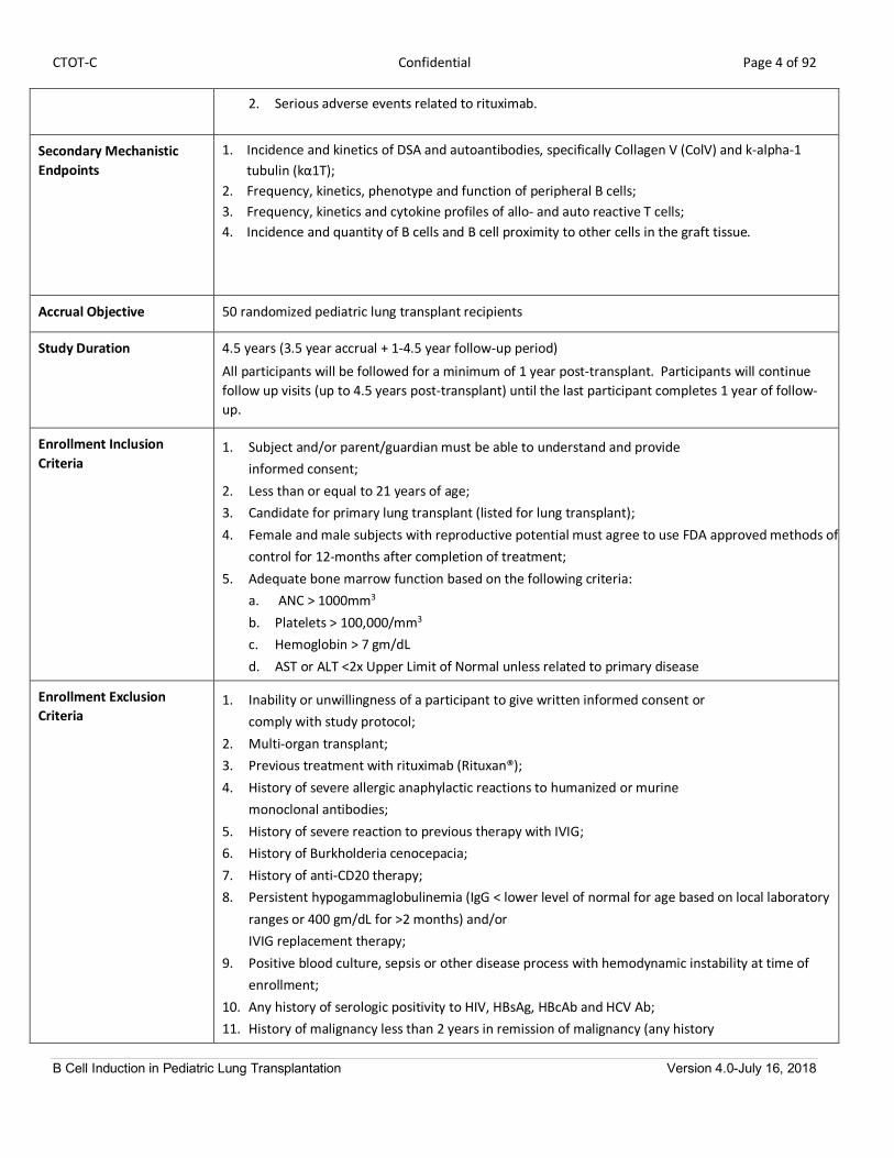
Development of Abs to HLA following Lung Transplant and its impact on the development of BOS: Since 2006, the Washington University adult lung program and the
Mohanakumar lab instituted serial analysis for development of Abs to
donor mismatched HLA during the post-transplant period. All adult LTxR
had negative donor cross-matches. Of 116 patients serially analyzed, 65
developed DSA (56%), with 52 in the first 90 days, again supporting our
contention that early intervention will be required to prevent induction.
To test the hypothesis that antibodies contribute to late graft injury, the
Mohanakumar group used IVIG (500 mg/kg x 6 monthly doses) and
rituximab (375 mg/m2
x 1 dose) for desensitization. Of the 44 patients
treated, 27 (61%) cleared the DSA, and 17 (39%) had persistent DSA. An
additional 17 patients were treated with IVIG alone and 11 (65%) cleared
the DSA. More importantly, patient and graft survival was significantly better in successfully desensitized patients
(Figure 4) providing strong correlative evidence that DSA participate in the development of allograft injury.78
A critical role for Abs to self-Ags in the pathogenesis of BOS: Among adult LTxR who developed
DSA (n=57) and subsequently
cleared DSA following Ab directed
therapy (n=34), 9 patients
nonetheless developed BOS. We
detected persistence of Abs to self-
Ags, ColV and kα1T in most of these
individuals, strongly implicating
pathogenic roles for Abs to self-Ags. As shown in Figure 5, the LTxR who
cleared both Abs to HLA and self-Ags
demonstrated improved freedom from BOS in comparison to patients that cleared only DSA but not Abs to self Ags.
Taken together, these preliminary results strongly support our hypothesis that Abs to self–Ags play a critical role in
the pathogenesis of BOS and our proposed study will assess both the impact of intervention and further determine
significance and mechanisms of antibodies to self-antigens.
Successful post-transplant desensitization of Abs to self-Ags in adult LTxR:
To determine whether a post-transplant desensitization regimen for DSA with rituximab and IVIG has an impact on
the Abs to self-Ags, we analyzed 123 LTxR who developed DSA. Forty-five LTxR had Abs to kα1T and 31 had Abs to
ColV prior to desensitization. Following treatment 46.7% of the kα1T+ and 48.4% of the ColV+ became negative and
another 25% demonstrated a significant decrease. This study demonstrates that rituximab-based desensitization of
Abs to self-Ags is feasible.
Figure 4: Improved survival following clearance of DSA.
Figure 4: Improved survival following clearance of DSA.
Figure 5: Increased incidence of BOS in DSA cleared LTx patients following Ab directed treatment when Abs to self-Ags persist

CTOT-C Confidential Page 26 of 92
B Cell Induction in Pediatric Lung Transplantation Version 4.0-July 16, 2018
Correlation between antibodies to self-Ags and pro-inflammatory milieu
persists with continued Abs to self-Ags even after DSA clears: To determine
the mechanisms by which Abs against HLA and self-Ags may contribute to the
pathogenesis of BOS, were performed serum cytokine analysis using 25-plex
Luminex assay. We analyzed sera from three groups of patients: group I DSA
and Abs to self-Ags present (prior to therapy), group II cleared DSA but Abs to
self-Ags persisted and group III cleared both DSA and Abs to self-Ags.
Results presented in Figure 6 demonstrate that group I patients who had
both DSA and Abs to self-Ag (n= 23) as well as group II that cleared DSA but
persisted Abs to self-Ag (n=9) had high levels of pro-inflammatory cytokines.
In contrast, group III cleared DSA and Abs to self-Ags (n=25) and did not
develop BOS had decreased levels of pro-inflammatory cytokines (namely IL-
1β (3.2 fold decrease), IL-17 (3.0), and IFN-γ (2.3)) and increased IL-10 (3.7
fold, p<0.01 for all) within 6 months. These results support persistent
immune responses to self-Ags can lead to a pro-inflammatory milieu that can
facilitate the development of BOS.
Increased frequency of B cell with autoimmune phenotype in BOS:
Data from mouse models and humans with autoimmune diseases suggest an
important role for a specific subset of B cells in the pathogenesis of
autoimmune diseases79-81
. These B cells express CD11c, produce
autoantibodies and can be induced to proliferate in response to TLR stimuli.
To determine whether this subpopulation of B cells is increased in adult LTxR
with BOS we determined the frequency of the autoimmune B cells (AIB) with
phenotype of CD19+, CD11c+ B220+ in peripheral blood of LTxR with BOS and
Abs to self-Ags. Results presented in Figure 7 demonstrate a five-fold increase
in the AIB (9.6±2.8% of B cells) over normal (1.6±0.7% of B cells). To determine
whether those B cells are present in the lung allografts, BAL cells were
characterized. Results demonstrate that the BOS(+) LTxR who had Abs to self-
Ag demonstrate a 3.5 fold increase over BOS(-) LTxR (9.3±2.1% vs. 2.8±1.2% of B cells, n=6). It is of interest that
BOS(+) anti self-Ag(-) LTxR showed similarly low frequency of AIB as in BOS(-) LTxR (data not shown). These results
strongly favor our contention that AIB may play a role in immune responses to self-Ags and development of BOS,
which will be tested in the mechanistic study.
Circulating B cells in BOS(+) LTxR augment the frequency of self-Ag specific Th17 cells:
To determine the role of B cells to activate self-Ag specific T cells , T cells isolated from
BOS(+) self-Ab(+) or BOS(-) self Ab(-) LTxR were cultured with sub-optimal concentrations
of kα1T or ColV (100ng/ml) with or without B cells. At the end of 2 weeks, the frequency of
Th17 cells was analyzed by ELISPOT.
Figure 6: Increased Pro-inflammatory cytokines in LTx patients with persistent DSA and Abs to self-Ags correlated with BOS.
Figure 7: Increased frequency of AIB in
PBMC and BAL.
PBMC BAL
Figure 8: Frequency of Ag specific
Th17 cells increases in the
presence of B cells.
0
20
40
60
80
100
120
KAT Collagen V
SPM
B+B-
CTOT-C Confidential Page 27 of 92
B Cell Induction in Pediatric Lung Transplantation Version 4.0-July 16, 2018
Results in Figure 8 demonstrate that in the presence of B cells there was a significant increase in the frequency of
Th17 cells specific for kα1T and ColV when compared without B cells. BOS(-) self-Ab(-) LTxR or cells from normal
volunteers failed to respond.
BOS(+) LTxR with significant increase in pro-inflammatory environment even without Abs to HLA, ColV and kα1T:
Sera from LTxR with BOS but neither DSA nor Abs to self-Ags (ColV, Kα1T) were analyzed for serum cytokines using
Luminex. LTxR who developed BOS (24%, 12/51) but did not develop Abs to HLA or ColV and kα1T had elevated
levels of IL-17 (8.2), IFN-γ (3.7) and decreased levels of IL-10 (2.9) suggesting that induction of Th17 immune
responses may play a role even in the absence of humoral responses to HLA, ColV and kα1T.

CTOT-C Confidential Page 28 of 92
B Cell Induction in Pediatric Lung Transplantation Version 4.0-July 16, 2018
3. Study Design
3.1. Description of Study Design This is a phase 2, prospective, multi-center, double-blind, randomized, placebo-controlled clinical trial in which 50
primary pediatric lung transplant recipients will be randomized (1:1) to receive either induction therapy with anti-
CD20 mAb (375 mg/m2) IV or placebo (IV day 0 and day 12 post-transplant) plus standard of care
immunosuppression (thymoglobulin induction, tacrolimus or equivalent, MMF or equivalent, and steroids). The
study will randomize 50 primary pediatric lung transplant recipients from seven participating centers. Subjects will
be screened, consented, and enrolled while on the UNOS waitlist. When the recipient has received the transplant
and is deemed hemodynamically stable, randomization will occur.
Figure 9: Study Design
While on UNOS Waitlist: Enrollment criteria met and subject is enrolled.
N=50 Randomized
1:1
/:/ Placebo Induction Therapy Plus Standard of Care Immunosuppression
N=25
Rituximab Induction Therapy Plus Standard of Care Immunosuppression
N=25
Primary Endpoint at 12 months post-transplant
Randomization criteria met within 12h of
returning to the ICU following transplant?
Yes
No Terminated
Transplant occurs and subject is considered stable enough to safely tolerate rituximab infusion.
Baseline mechanistic samples collected

CTOT-C Confidential Page 29 of 92
B Cell Induction in Pediatric Lung Transplantation Version 4.0-July 16, 2018
3.2. Primary Endpoints The primary endpoint is the earliest time to any of the following events during the follow-up period:
• Chronic Allograft Dysfunction
• Listed for Retransplant
• Death
3.3. Secondary Endpoints The following secondary clinical endpoints will be assessed:
1. Post-transplant outcomes including:
a) Incidence of chronic allograft dysfunction, relisting and death during the follow-up period, which will be a
minimum of 12 months post-transplant;
b) Incidence of Primary Graft Dysfunction;
c) Incidence of Grade A Acute Rejection during the follow up period, which will be a minimum of 12 months
post-transplant;
d) Incidence of Antibody Mediated Rejection during the follow up period, which will be a minimum of 12
months post-transplant;
e) Incidence of tacrolimus variability threshold during the follow up period, which will be a minimum of 12
months post-transplant;
f) Percentage of participants meeting tacrolimus variability threshold who complete tacrolimus variability
intervention;
g) Magnitude of change in standard deviation of tacrolimus levels following intervention.
2. Post-transplant safety outcomes including:
a) Incidence and severity of infection episodes; See previous comment in the synopsis above
b) Serious adverse events related to rituximab.
3.4. Mechanistic Endpoints The following mechanistic endpoints will be assessed:
1. Incidence and kinetics of DSA and autoantibodies, specifically Collagen V (ColV) and k-alpha-1 tubulin (kα1T);
2. Frequency, kinetics, phenotype and function of peripheral B cells;
3. Frequency, kinetics and cytokine profiles of allo- and auto reactive T cells;
4. Incidence and quantity of B cells and B cell proximity to other cells in the graft tissue.
3.5. Stratification, Randomization, and Blinding/Masking This is a double-blinded study; therefore, medication assignments will be blinded to the study participants as well as
to the site clinical personnel. Only the site research pharmacist will have access to the unblinded randomization schedule for that site. In the event the subject undergoes a life-threatening reaction, then study subject and
treating physician will be unblinded to the treatment assignment. In the event the subject undergoes repeat severe
infusion or hypersensitivity reactions, despite appropriate treatment, then study subject and treating physician will
be unblinded to the treatment assignment. IND safety reports will be reported to the FDA, DSMB, and IRBs in an
unblinded fashion.

CTOT-C Confidential Page 30 of 92
B Cell Induction in Pediatric Lung Transplantation Version 4.0-July 16, 2018
Randomization treatment assignments will be developed by the SACCC statisticians and stored on the SACCC server.
The SACCC will maintain a central web-based randomization system. Once the subject is determined to be eligible
based on the criteria, the site personnel will be provided the appropriate treatment assignment.
3.5.1 Procedure for Unblinding/Unmasking Unblinding must be approved by the study Medical Monitor unless an immediate life threatening condition
has developed and the Medical Monitor is not accessible. The site investigator will notify the protocol
chair(s) and the study Statistical and Clinical Coordinating Center team of the unblinding event on the next
business day. The emergency unblinding will also be reported to the Data and Safety Monitoring Board
(DSMB).
A full account of the event will be recorded on the Study Treatment Unblinding eCRF, including the date and
time of the unblinding, the reason for the decision to unblind, and the name of the individual who made the
decision and the names of the Medical Monitor and others who were notified. The reasons for unblinding of
a participant’s treatment will be included in the final study report.
Unblinding the study due to an approved interim analysis, final analysis, or study termination will require
written approval from NIAID.

CTOT-C Confidential Page 31 of 92
B Cell Induction in Pediatric Lung Transplantation Version 4.0-July 16, 2018
4. Selection of Participants and Clinical Sites
4.1. Rationale for Study Population Although early outcomes in pediatric transplantation have improved in the past 2 decades, conditional survival
beyond the early post-transplant period has not improved significantly, due primarily to the development of chronic
allograft dysfunction. This is particularly evident for pediatric lung transplant recipients. In recent years,
accumulating evidence has implicated antibodies as an important contributor to chronic allograft dysfunction.
These include antibodies to donor histocompatibility antigens or cryptic self-antigens exposed during organ
transplantation or subsequent graft injury. The mechanistic connection between antibody development and chronic
allograft dysfunction in lung transplantation has yet to be determined. Moreover, effective therapies for prevention
and management of pathologic antibodies for chronic allograft dysfunction have yet to be developed.
This study and the associated mechanistic studies will dissect the role B cells play in this process, including direct
antibody mediated effects and indirect effects on cellular alloimmune and autoimmune response.
4.2. Enrollment Inclusion Criteria Individuals who meet all of the following criteria are eligible for enrollment as study participants:
1. Subject and/or parent guardian must be able to understand and provide informed consent;
2. Less than or equal to 21 years of age;
3. Candidate for a primary lung transplant (listed for lung transplant);
4. Adequate bone marrow function based on the following criteria:
a. ANC: >1000mm3
b. Platelets: >100,000/mm3
c. Hemoglobin: >7 gm/dL
d. AST or ALT< 2x Upper Limit of Normal unless related to primary disease
5. Female and male subjects with reproductive potential must agree to use FDA approved methods of birth control
for 12-months after completion of treatment.
4.3. Enrollment Exclusion Criteria Individuals who meet any of these criteria are not eligible for enrollment as study participants:
1. Inability or unwillingness of a participant to give written informed consent or comply with study protocol;
2. Multi-organ transplant;
3. Previous treatment with rituximab (Rituxan®);
4. History of severe allergic anaphylactic reactions to humanized or murine monoclonal antibodies;
5. History of severe reaction to previous therapy with IVIG;
6. History of Burkholderia cenocepacia;
7. History of anti-CD20 therapy;
8. Persistent hypogammaglobulinemia (IgG < lower level of normal for age based on local laboratory ranges or 400
gm/dL for >2 months) and/or IVIG replacement therapy;
9. Positive blood culture, sepsis or other disease process with hemodynamic instability at time of enrollment;

CTOT-C Confidential Page 32 of 92
B Cell Induction in Pediatric Lung Transplantation Version 4.0-July 16, 2018
10. Any history of serologic positivity to HIV, HBsAg, HBcAb and HCV Ab;
11. History of malignancy less than 2 years in remission of malignancy (any history of adequately treated in-situ cervical
carcinoma, or adequately treated basal or squamous cell carcinoma of the skin will be permitted);
12. Any condition, including psychiatric disorders, that in the opinion of the investigator would interfere with the subject’s
ability to comply with study requirements;
13. Participation in another investigational trial within 4 weeks of enrollment;
14. Currently lactating or plans to become pregnant during the timeframe of the study follow-up period;
15. Past or current medical problems or findings from physical examination or laboratory testing that are not listed
above, which, in the opinion of the investigator, may pose additional risks from participation in the study, may
interfere with the participant’s ability to comply with study requirements or that may impact the quality or
interpretation of the data obtained from the study.
4.4. Randomization Inclusion Criteria Individuals who meet all of the following criteria are eligible for randomization:
1. Serum IgG immunoglobulin level greater than lower level of normal for age based on local laboratory ranges or 400
gm/dL within 90 days prior to randomization;
2. Female subjects of childbearing potential must have a negative pregnancy test within 48 hours of transplant;
3. Negative for Hepatitis B infection (if at time of transplant, participant does not exhibit effective immunization, the
participant should be re-tested).
4.5. Randomization Exclusion Criteria Individuals who meet any of these criteria are not eligible for randomization:
1. Use of an induction agent other than Thymoglobulin®;
2. Renal insufficiency requiring hemodialysis or ultrafiltration;
3. Inability to obtain intravenous access;
4. Positive blood culture, sepsis or other disease process with hemodynamic instability at time of transplant;
5. Use of investigational agent(s) within 5 half-lives of the investigational drug or 4 weeks, whichever is longer;
6. Receipt of a MMR vaccine within 30 days prior to randomization;
7. Any condition that, in the opinion of the investigator, would interfere with the subject’s ability to comply with study
requirements.
4.6. Selection of Clinical Sites The target population for this study consists of pediatric lung transplant recipients. Subjects will be enrolled at sites
throughout the United States. The sites participating in the study have performed the majority of pediatric lung
transplants annually in the United States. The target population therefore is a fair representation of the pediatric
lung population.

CTOT-C Confidential Page 33 of 92
B Cell Induction in Pediatric Lung Transplantation Version 4.0-July 16, 2018
5. Known and Potential Risks and Benefits to Participants
5.1. Risks of rituximab (Rituxan®) No dose-limiting effects were observed in Phase I/II studies. Reported adverse events including fever, chills,
headache, nausea, vomiting, rhinitis, asthenia, and hypotension, occurred primarily during rituximab infusions and
typically responded to an interruption of the infusion and resumption at a slower rate.
Fatal Infusion Reactions: Severe and fatal cardiopulmonary events, including angioedema, hypoxia, pulmonary
infiltrates, acute respiratory distress syndrome, myocardial infarction, and cardiogenic shock, have been reported.
These severe reactions typically occurred during the first infusion with time to onset of 30-120 minutes.
Cardiac Events: Patients with preexisting cardiac conditions, including arrhythmia and angina, have had recurrences
of these cardiac events during rituximab infusions.
Tumor Lysis Syndrome: Tumor lysis syndrome, some with fatal outcome, has been reported and is characterized in
patients with a high number of circulating malignant cells (≥25,000 ul) by rapid reduction in tumor volume, renal
insufficiency, hyperkalemia, hypocalcemia, hyperuricemia, and hyperphosphatemia.
Renal Events: Rituximab has been associated with severe renal toxicity including acute renal failure requiring
dialysis, and in some cases has led to death. Renal toxicity has occurred in patients with high numbers of circulating
malignant cells (≥25,000/mm2) or high tumor burden who experience tumor lysis syndrome and in patients
administered concomitant cisplatin.
Mucocutaneous Reactions: Severe bullous skin reactions, including fatal cases of toxic epidermal necrolysis and
paraneoplastic pemphigus, have been reported in patients treated with rituximab. The onset of reaction has varied
from 1 to 13 weeks following rituximab exposure.
Nervous System: a. Posterior reversible encephalopathy syndrome (PRES)/Reversible Posterior Leukoencephalopathy Syndrome
(RPLS): Cases of posterior reversible encephalopathy syndrome (PRES)/reversible posterior leukoencephalopathy
syndrome (RPLS) have been reported. Signs and symptoms include visual disturbance, headache, seizures and
altered mental status, with or without associated hypertension. A diagnosis of PRES/RPLS requires confirmation by
brain imaging. The reported cases had recognized risk factors for PRES/RPLS, including hypertension,
immunosuppressive therapy and/or other concomitant therapies.
b. Progressive multifocal leukoencephalopathy (PML): PML is a rare disease caused by the reactivation of latent JC
virus in the brain. Immunosuppression allows reactivation of the JC virus which causes demyelination and
destruction of oligodendrocytes resulting in death or severe disability. Rare cases of PML, some resulting in death,
have been reported in patients with hematologic malignancies who have received rituximab. The majority of
these patients had received rituximab in combination with chemotherapy or as part of a hematopoietic stem cell
transplant. Cases of PML resulting in death have also been reported following the use of rituximab for the
treatment of autoimmune diseases. The reported cases had multiple risk factors for PML, including the underlying
disease and long-term immunosuppressive therapy or chemotherapy. Most cases of PML were diagnosed within
12 months of their last infusion of rituximab.

CTOT-C Confidential Page 34 of 92
B Cell Induction in Pediatric Lung Transplantation Version 4.0-July 16, 2018
Physicians should consider PML in any patient presenting with new onset neurologic manifestations. Consultation
with a neurologist, brain MRI, and lumbar puncture should be considered as clinically indicated. In patients who
develop PML, rituximab should be discontinued and reductions or discontinuation of any concomitant
chemotherapy or immunosuppressive therapy should be considered.
Hematologic Events: In clinical trials, Grade 3 and 4 cytopenias were reported in 48% of patients treated with
rituximab; these include: lymphopenia (40%), neutropenia (6%), leukopenia (4%), anemia (3%), and
thrombocytopenia (2%). The median duration of lymphopenia was 14 days (range, 1 to 588 days) and of
neutropenia was 13 days (range, 2 to 116 days). A single occurrence of transient aplastic anemia (pure red cell
aplasia) and two occurrences of hemolytic anemia following Rituximab therapy were reported.
In addition, there have been a limited number of postmarketing reports of prolonged pancytopenia, marrow
hypoplasia, and late onset neutropenia.
Infectious Events: Rituxan® induced B cell depletion in 70% to 80% of patients with NHL and was associated with
decreased serum immunoglobulins in a minority of patients; the lymphopenia lasted a median of 14 days (range, 1-
588 days). Infectious events occurred in 31% of patients: 19% of patients had bacterial infections, 10% had viral
infections, 1% had fungal infections, and 6% were unknown infections. Serious infectious events (Grade 3 or 4),
including sepsis, occurred in 2% of patients.
Hepatitis B Reactivation: Hepatitis B virus (HBV) reactivation with fulminant hepatitis, hepatic failure, and death
has been reported in some patients with hematologic malignancies treated with rituximab. The majority of patients
received rituximab in combination with chemotherapy. The median time to the diagnosis of hepatitis was
approximately four months after the initiation of rituximab and approximately one month after the last dose.
Participants with pre-existing Hepatitis B infection will not be eligible to participate in this study.
Other Serious Viral Infections: The following additional serious viral infections, either new, reactivated or
exacerbated, have been identified in clinical studies or postmarketing reports. The majority of patients received
Rituxan® in combination with chemotherapy or as part of a hematopoietic stem cell transplant. These viral infections
included JC virus (progressive multifocal leukoencephalopathy [PML]), cytomegalovirus, herpes simplex virus,
parvovirus B19, varicella zoster virus, West Nile virus, and hepatitis C. In some cases, the viral infections occurred up
to one year following discontinuation of Rituxan® and have resulted in death.
Bowel Obstruction and Perforation: Abdominal pain, bowel obstruction and perforation, in some cases leading to
death, were observed in patients receiving Rituxan® in combination with chemotherapy for DLBCL. In post-
marketing reports, which include both patients with low-grade or follicular NHL and DLBCL, the mean time to onset
of symptoms was 6 days (range 1−77) in patients with documented gastro-intestinal perforation. Complaints of
abdominal pain, especially early in the course of treatment, should prompt a thorough diagnostic evaluation and
appropriate treatment.
Immunogenicity: Patients may develop a human anti-chimeric antibody (HACA) response with rituximab treatment.
The clinical significance of this is unclear.
Pregnancy: B cell lymphocytopenia generally lasting less than 6 months can occur in infants exposed to rituximab in
utero.

CTOT-C Confidential Page 35 of 92
B Cell Induction in Pediatric Lung Transplantation Version 4.0-July 16, 2018
Immunization: Response rates may be reduced with non-live vaccines.
Additional Safety Signals: The following serious adverse events have been reported to occur in patients following
completion of rituximab infusions: arthritis, disorders of blood vessels (vasculitis, serum sickness and lupus-like
syndrome), eye disorders (uveitis and optic neuritis), lung disorders including pleuritis and scarring of the lung
(bronchiolitis obliterans), that may result in fatal outcomes, and fatal cardiac failure.
See the rituximab Investigator Brochure for additional details regarding safety experience with rituximab.
5.2. Risks of Immunosuppression Medications The safety and effectiveness of immunosuppression medications in pediatric patients is not established in controlled
trials. However, the dose, efficacy and adverse event profile are not thought to be different from adults based on
studies in the literature.
5.2.1 Thymoglobulin® [Anti-thymocyte globulin (rabbit)] Risks associated with Thymoglobulin® include immune-mediated reactions, infections, reactivation of infection,
malaise, dizziness, sepsis, thrombocytopenia, leukopenia, and increased incidence of malignancies, including
lymphoma or post-transplant lymphoproliferative disease (PTLD). Occasional reactions are observed at the infusion
site including pain, swelling and erythema.
The majority of pediatric lung transplant centers reporting to the International Society of Heart and Lung
Transplantation (ISHLT) registry use antibody based induction immunosuppressive therapy.
5.2.2 CellCept® (Mycophenolate mofetil-MMF)
CellCept® (Mycophenolate mofetil - MMF) is approved (in combination with cyclosporine and corticosteroids) as an
immunosuppressive agent for renal, cardiac, and hepatic solid organ transplantation. Adverse events reported in >
30% of renal, cardiac or liver transplant patients receiving MMF were pain, fever, headache, asthenia, anemia,
leukopenia, thrombocytopenia, leukocytosis, urinary tract infection, hypertension, hypotension, peripheral edema,
hypercholesteremia, hypokalemia, hyperglycemia, increased creatinine and BUN, cough, hypomagnesaemia,
diarrhea, constipation, nausea, vomiting, respiratory infection, dyspnea, lung disorder, pleural effusion, tremor and
insomnia.
There is an increased risk of developing lymphomas and other malignancies, particularly of the skin.
Lymphoproliferative disease or lymphoma developed in 0.4% to 1% of patients receiving MMF 1 - 1.5 mg BID. Severe
neutropenia developed in up to 2% of renal transplant recipients receiving MMF 1.5 mg BID. MMF can cause fetal
harm when administered to a pregnant woman. Cases of progressive multifocal leukoencephalopathy (PML),
sometimes fatal, and pure red cell aplasia have been reported in patients treated with MMF. Gastrointestinal
bleeding (requiring hospitalization) has been observed in approximately 3% of renal, in 1.7% of cardiac, and in 5.4%
of hepatic transplant patients treated with MMF 1.5 g BID.
Use of CellCept® during pregnancy is associated with an increased risk of first trimester pregnancy loss and an
increased risk of congenital malformations, especially external ear and other facial abnormalities including cleft lip
and palate, and anomalies of the distal limbs, heart, esophagus, and kidney.
Females of reproductive potential will receive contraceptive counseling and use acceptable birth control during the
study, and for 6 weeks after stopping CellCept®, unless the participant chooses abstinence (she chooses to avoid
heterosexual intercourse completely).

CTOT-C Confidential Page 36 of 92
B Cell Induction in Pediatric Lung Transplantation Version 4.0-July 16, 2018
For those females using CellCept® at any time during pregnancy and those becoming pregnant within 6 weeks of
discontinuing therapy, the investigator or healthcare practitioner should report the pregnancy to the
Mycophenolate Pregnancy Registry. The investigator or healthcare practitioner should strongly encourage the
patient to enroll in the pregnancy registry.
Risks and benefits of CellCept® should be discussed with the patient. When appropriate, consider alternative
immunosuppressants with less potential for embryofetal toxicity. In certain situations, the patient and her
healthcare practitioner may decide that the maternal benefits outweigh the risks to the fetus. If this drug is used
during pregnancy, or if the patient becomes pregnant while taking this drug, the patient should be apprised of the
potential hazard to the fetus.
Additional information about MMF can be found in the package insert.
5.2.2 Prograf® (Tacrolimus)
Side effects of Prograf® (Tacrolimus) include hypertension, glucose intolerance, peripheral neuropathy, renal
insufficiency, abnormal liver function studies, seizures, nausea, vomiting, confusion, hypomagnesaemia,
tremulousness, neurotoxicity, posterior reversible encephalopathy syndrome (PRES), progressive multifocal
leukoencephalopathy (PML), interstitial lung disease, BK nephropathy, and increased risk of secondary malignancies.
Additional information about Tacrolimus can be found in the package insert.
5.3. Risks of Study Procedures
5.3.1 Risk of Blood Draw (Venipuncture) The amount of blood that may be drawn from pediatric subjects for research purposes will not exceed 3cc/kg per
day or 7cc/kg per six week period. All blood samples for the mechanistic study will be obtained at the time of
scheduled blood draws, so there will be minimal additional risk associated with obtaining the study samples.
The subject may experience some discomfort at the site of the needle entry, bruising, swelling, redness, fainting, or
local infection. The additional amount of blood could contribute to the development of anemia. The subject’s
clinical condition will be taken into consideration to determine whether research blood tests can be performed.
5.3.2. Risk of Bronchoalveolar Lavage and Bronchoscopy Bronchoalveolar lavage (BAL) will be obtained during standard of care bronchoscopies according to IPLTC guidelines.
The risks associated with BAL collected for this study are no different than routine management.
5.3.3. Risk of Tacrolimus Standard Deviation Threshold Non-adherence may be identified during the intervention. If non-adherence is identified, this may negatively affect
the participant’s candidacy for a second transplant.
The initial call for the intervention will be facilitated by the site coordinator at the transplant center. During this call
the team at ICMMS will ask the participant or parent/guardian to provide contact information to the team at ICMMS
for future calls. Any PHI shared with ISMMS may be disclosed.

CTOT-C Confidential Page 37 of 92
B Cell Induction in Pediatric Lung Transplantation Version 4.0-July 16, 2018
5.4. Potential Benefits Receipt of the study treatment (rituximab) may reduce the participant’s risk of antibody-related complications from
lung transplant. Participants may benefit by having their health status closely followed. In the future, other people
may benefit from this study because the information learned from this study may influence future care of transplant
patients.

CTOT-C Confidential Page 38 of 92
B Cell Induction in Pediatric Lung Transplantation Version 4.0-July 16, 2018
6. Investigational Agent
6.1. Rituximab (Rituxan®) Rituximab is a is a genetically engineered, chimeric, murine/human monoclonal antibody directed against the CD20
antigen found on the surface of normal and malignant pre-B and mature B cells. The antibody is an IgG1 κ
immunoglobulin containing murine light-and heavy-chain variable region sequences and human constant region
sequences. Rituximab is composed of two heavy chains of 451 amino acids and two light chains of 213 amino acids
(based on cDNA analaysis) and has an approximate molecular mass of 145 kD. Rituximab has a binding affinity for
the CD20 antigen of ~8.0 nM.
6.1.1. Rituximab (Rituxan®)
6.1.1.1. Formulation, Packaging, and Labeling Rituximab® is a sterile, clear, colorless, preservative-free liquid concentrate for intravenous (IV)
administration. Rituximab is supplied at a concentration of 10 mg/mL in either 100 mg (10 mL) or
500 mg (50 mL) single-use vials. The products are formulated for intravenous administration in 9.0
mg/mL sodium chloride, 7.35 mg/mL sodium citrate dihydrate, 0.7 mg/mL polysorbate 80, and
Sterile Water for Injection. The pH is adjusted to 6.5.
The vials provided to the pharmacy will have occluded, study-specific investigational agent labels.
6.1.1.2. Dosage and Preparation Rituximab will be given as an intravenous infusion.
2 Doses: 375 mg/m2 on Day 0 (within 12 hours of return to ICU following transplant) and Day 12 (+/-
2 days).
Rituximab should be prepared using appropriate aseptic techniques. Withdraw the necessary
amount of Rituximab or Rituximab Placebo and dilute to a final concentration of 1 to 4 mg/mL into
an infusion bag containing either 0.9% Sodium Chloride, USP, or 5% Dextrose in Water, USP. Gently
invert the bag to mix the solution. Discard any unused portion left in the vial. Parenteral drug
products should be inspected visually for particulate matter and discoloration prior to
administration.
Rituximab or Rituximab Placebo should be given as a slow intravenous infusion. It should not be administered as an intravenous push or bolus. Do not infuse rituximab concomitantly with another
IV solution or other IV medications. Rituximab infusions should be made through a dedicated line.
Participants will be premedicated using:
• Tylenol® (Acetaminophen), 15 mg/kg (to a maximum of 1 gram) should be administered (by
mouth, enteral or rectal administration) 30 to 60 minutes prior to the start of an infusion;
and
• Benadryl® (Diphenhydramine) by mouth 30-60 minutes prior to the start of an infusion as
described in Table 1 or by IV 1 mg/kg (to a maximum of 50 mg) 30 minutes prior to the start
of an infusion.

CTOT-C Confidential Page 39 of 92
B Cell Induction in Pediatric Lung Transplantation Version 4.0-July 16, 2018
Age (years) Diphenhydramine Oral Dose (mg)
2-6 6.25
6-<12 12.5
12+ 25
Table 1: Diphenhydramine Pre-medication Dose
Premedication may attenuate infusion-related events. Since transient hypotension may occur during
rituximab infusion, consideration should be given to withholding anti-hypertensive medications 12
hours prior to rituximab infusion.
6.1.1.3. Administration Guidelines First infusion (Day 0): Rituximab should be administered within 12 hours of returning from the ICU
following transplant, based on hemodynamic stability. Rituximab should be administered
intravenously through a dedicated line at an initial rate of 0.5 mg/kg/hr (to a maximum rate of 50
mg/hr) for the first hour.
If hypersensitivity or infusion-related events do not occur, the infusion rate will be escalated by 0.5
mg/kg/hr every 30 minutes, to a maximum rate of 400 mg/hr.
If a hypersensitivity or infusion-related reaction event develops, the infusion rate should be
temporarily slowed or interrupted. The infusion can continue at one-half the previous rate upon
improvement of patient symptoms.
Subsequent Infusion (Day 12 +/- 2 days):
A second dose of rituximab will be administered to subjects considered stable enough to receive the
infusion.
Individual Subject Stopping Rules:
Individuals who meet any of the criteria listed below will not receive the second dose of rituximab.
1. Serious adverse event causally related to the rituximab infusion;
2. Acute pulmonary infectious process with evidence of graft dysfunction;
3. Positive blood culture, sepsis or other disease process with hemodynamic instability;
4. Renal insufficiency requiring hemodialysis or ultrafiltration;
5. Inability to obtain intraveneous access;
6. Use of an investigational drug after the first dose of placebo or rituximab;
7. Any other event which in the opinion of the principal investigator may pose additional risk to the
participant.
If the first infusion was well tolerated, the subsequent infusion may be administered intraveneously
through a dedicated line at an initial rate of 1.0 mg/kg/hr (to a maximum rate of 100 mg/h) for the

CTOT-C Confidential Page 40 of 92
B Cell Induction in Pediatric Lung Transplantation Version 4.0-July 16, 2018
first hour. If hypersensitivity or infusion-related events do not occur, the infusion rate will be
escalated by 1.0 mg/kg/hr (maximum rate of 100 mg increase per hour) every 30 minutes, to a
maximum rate of 400 mg/hr. If hypersensitivity or infusion-related event develops, the infusion
should be temporarily slowed or interrupted. The infusion can continue at one half the previous
rate upon improvement of patient symptoms.
If hypersensitivity or infusion-related events occurred during the first infusion, the subsequent
infusion should be administered in the same manner as the first infusion.
6.1.2. Rituximab Placebo
6.1.2.1. Formulation, Packaging, and Labeling Rituximab placebo is a sterile, clear, colorless, preservative-free liquid concentrate for intravenous
(IV) administration. Rituximab is supplied at a concentration of 10 mg/mL in either 100 mg (10 mL)
or 500 mg (50 mL) single-use vials. The product is formulated for intravenous administration in 9.0
mg/mL sodium chloride, 7.35 mg/mL sodium citrate dihydrate, 0.7 mg/mL polysorbate 80, and
Sterile Water for Injection. The pH is adjusted to 6.5. The vials provided to the pharmacy will have
occluded, study-specific investigational agent labels.
6.1.2.2. Dosage, Preparation, and Administration Rituxmab placebo will be administered according to the guidelines for rituximab outlined in section
6.1.1.2.
6.2. Infusion Supervision (Rituximab and Rituximab Placebo) Rituximab and rituximab placebo should be administered in a hospital environment where full resuscitation facilities
are immediately available. Rituximab and rituximab placebo may be administered on an outpatient basis however
patients may be hospitalized for observation at the discretion of the investigator.
The infusion will be supervised by the clinical staff (i.e., clinical nurse, physician) at the participating institutions. A
history of each infusion and any adverse side effects will be recorded and reported to the SACCC using the
appropriate case report forms.
Vital signs (temperature, blood pressure, pulse, and respiratory rate) and standard laboratory testing (CBC, basic
metabolic panel) will be obtained prior to the start of each rituximab or rituximab placebo infusion. The local
investigator will determine whether to hold the infusion due to lab abnormalities. Vital signs will be monitored
every 15 minutes for the first hour of the infusion, every hour until the end of the infusion, and every hour
thereafter for a total of 3 hours after the infusion. The IV line should remain in place for at least 1 hour following
the infusion to enable the administration of drugs, if necessary. Additional vital signs may be obtained as clinically
indicated. Fluid resuscitation will be provided as needed for changes in vital signs suggesting hydration would be
beneficial.

CTOT-C Confidential Page 41 of 92
B Cell Induction in Pediatric Lung Transplantation Version 4.0-July 16, 2018
6.3. Drug Accountability Under Title 21 of the Code of Federal Regulations (21CFR §312.62) the investigator will maintain adequate records of
the disposition of the investigational agent, including the date and quantity of the drug received, to whom the drug
was dispensed (participant-by-participant accounting), and a detailed accounting of any drug accidentally or
deliberately destroyed.
Records for receipt, storage, use, and disposition will be maintained by the study site. A drug-dispensing log will be
kept current for each participant. This log will contain the identification of each participant and the date and
quantity of drug dispensed.
All records regarding the disposition of the investigational product will be available for inspection.
Any unused study drug (unopened or within expiration date) may be returned to the manufacturer at the end of the
study.
6.4. Toxicity Prevention and Management Patients enrolled in this study will be evaluated clinically and with standard laboratory tests before and during their
participation in this study. Safety evaluations will consist of medical interviews, recording of adverse events,
physical examinations, blood pressure, and laboratory measurements. Subjects will be evaluated for adverse
events, serious adverse events, and adverse events requiring study drug interruption or discontinuation at each
study visit for the duration of their participation in the study.
6.5. Modification or Premature Discontinuation of Investigational Agent Rituximab (Rituxan®) and Rituximab Placebo may be modified or prematurely discontinued for any participant for any of
the following reasons:
v General Infusion-Related Reactions. If an infusion-related reaction develops, the infusion rate should be temporarily
reduced or interrupted. The infusion may be continued at one-half the previous rate upon improvement of the
patient’s symptoms.
v Life-Threatening Reaction. In the event of a life-threatening reaction, the infusion should be discontinued and the
patient should receive medical treatment. Patients who experience a life-threatening reaction will not receive
additional Rituximab/Placebo, and will remain in the study, and be included in the ITT analysis. The study treatment
assignment will be unblinded to the site personnel and the study participant. (Protocol section 3.5.1)
v Severe Infusion Reaction. The infusion should be interrupted for severe reactions, and supportive care measures
should be instituted as medically indicated (e.g., IV fluids, vasopressors, oxygen, bronchodilators, diphenhydramine,
and acetaminophen). The infusion should not be restarted until all the symptoms have disappeared. On the
restarting of the infusion, the rate should be half of that which precipitated the reaction. Patients who experience
repeat severe infusion reactions, despite appropriate treatment, will not receive additional Rituximab/Placebo, and
will remain in the study, and be included in the ITT analysis. The study treatment assignment will be unblinded to the
site personnel and the study participant. (Protocol section 3.5.1)
v Hypersensitivity Reactions. Hypersensitivity reactions (non-IgE-mediated reactions) may respond to adjustments in
the infusion rate and in medical management. The infusion should be interrupted for severe hypersensitivity
reactions and can be resumed at a 50% reduction in rate (e.g., from 100 mg/hr to 50 mg/hr) when symptoms have
completely resolved. Treatment of these symptoms with diphenhydramine and acetaminophen (or their equivalents)
is recommended; additional treatment with bronchodilators or IV saline may be indicated. In most cases, patients
who have experienced non-life-threatening hypersensitivity reactions have been able to complete the full course of

CTOT-C Confidential Page 42 of 92
B Cell Induction in Pediatric Lung Transplantation Version 4.0-July 16, 2018
therapy. Medications for the treatment of hypersensitivity reactions (e.g., epinephrine, antihistamines, and
corticosteroids) should be available for immediate use in the event of a reaction during administration. Patients who
experience repeat hypersensitivity reactions, despite appropriate treatment, will not receive additional
Rituximab/Placebo, and will remain in the study, and be included in the ITT analysis. The study treatment assignment
will be unblinded to the site personnel and the study participant. (Protocol section 3.5.1)
v Cardiovascular. Infusions should be discontinued in the event of serious or life-threatening cardiac arrhythmias.
Patients who develop clinically significant arrhythmias should undergo cardiac monitoring during and after
subsequent infusions. Patients should be monitored throughout the infusion and the immediate post-infusion
period.

CTOT-C Confidential Page 43 of 92
B Cell Induction in Pediatric Lung Transplantation Version 4.0-July 16, 2018
7. Other Medications
7.1. Immunosuppression Medications 7.1.1. Protocol-mandated
7.1.1.1 Thymoglobulin® [Anti-thymocyte Globulin (Rabbit)] All subjects will receive Thymoglobulin® induction therapy. Thymoglobulin® will be
administered for 5 days as an intravenous infusion at a dose of 1.5 mg/kg/day beginning on
the day of transplant.
7.1.1.2 Tacrolimus (Prograf®) Tacrolimus (Prograf®), or generic equivalent, will be administered to attain target trough levels outlined
in Table 3. Suggested dosing is outlined in Table 2.
Timepoint Tacrolimus Dose
Post-operatively (by 72 hours following
transplant operation) *
0.01-0.04 mg/kg/day IV continuous infusion OR
0.06-0.1 mg/kg/day (maximum 5 mg per dose) via NG
or G-tube divided into 2 equal doses OR
0.08 mg/kg/day sublingual divided into 2 equal doses
Once taking PO 0.1-0.3 mg/kg/day PO divided into 2 equal doses
*Dosing may be adjusted to every 8 hours in infants and young children if therapeutic levels are hard to
achieve (due to rapid metabolism of the drug in this age group).
Table 2: Tacrolimus Dose
Dosing may be adjusted for underlying medical conditions and/or interactions with other medications.
Target whole blood tacrolimus concentrations are as follows:
Timepoint Target Trough Level After Transplant
First 2 Weeks Post-Transplant 10-20 ng/mL
Week 2-1-year Post-transplant 10-15 ng/mL
After 1 Year 6-10 ng/mL
The above given recommendations for therapeutic drug monitoring of tacrolimus may not be
suitable for infants secondary to the different pharmacokinetics in infants.
Table 3: Tacrolimus Trough Levels

CTOT-C Confidential Page 44 of 92
B Cell Induction in Pediatric Lung Transplantation Version 4.0-July 16, 2018
7.1.1.3. Mycophenolate Mofetil Mycophenolate Mofetil (CellCept®), or generic equivalent, will be administered at an initial pediatric
dose of 600-700 mg/m2 twice daily. The dose may be modified to an initial dose of 800-900 mg/m
2
twice daily for cystic fibrosis subjects at puberty and above. Initial adult dosing is 1000-1500 mg twice
daily.
Mycophenolate Mofetil (CellCept®), or generic equivalent, dosing will be adjusted based on clinical
complications. Dosing outside of the target dose range will not be considered a protocol deviation.
7.1.1.4. Steroids Methylprednisolone/Prednisone will be administered according to each center’s standard practice. After
the first negative biopsy, prednisone will be tapered, according to each center’s protocol, to reach a
target dose of 0.1- 0.2 mg/kg/day by one year post-transplant.
7.2. Viral Prophylaxis and Therapy Participating sites will follow IPLTC guidelines for viral prophylaxis and treatment.
7.2.1. Respiratory Syncytial Virus Subjects less than 2 years of age will receive palivizumab per the local standard of care. Respiratory
Syncytial Virus will be treated with ribavirin per the local center standard of care.
7.2.2. Cytomegalovirus Sero Positive Donor and/or Recipient (D+/R+, D+/R-, D-/R+) will receive ganciclovir/valganciclovir
prophylaxis, per the local standard of care, for a minimum of 6 months following transplantation.
7.2.3. Fungal All subjects will receive fungal prophylaxis post-transplant according to local center standard of care. Fungal
therapies will be determined by the local standard of care.
7.2.4. Vaccination Efficacy and/or safety of immunization during periods of B cell depletion have not been adequately studied.
It is recommended that a patient’s vaccination record and possible requirements be reviewed. Per the
investigator’s discretion, the patient may have any required vaccination/booster administered at least
4 weeks prior to the initiation of study treatment. Review of the patient’s immunization status for the
following vaccinations is recommended: tetanus; diptheria; influenza; pneumoccocal polysaccharide;
Varicella; measles, mumps and rubella (MMR); and hepatitis B. Patients who are considered to be at high
risk for hepatitis B virus (HBV) infection and for whom the investigator has determined that immunization is
indicated should complete the entire HBV vaccine series at least 4 weeks prior to participation in the study.
All participants eligible for randomization will be encouraged to receive the influenza vaccine per Center for
Disease Control guidelines.
7.3. Prohibited Medications The following is a list of prohibited medications:
• Induction agent, other than Thymoglobulin®;
• Other investigational agents;
• Rituximab (Rituxan®), except for treatment of PTLD or treatment of AMR as described in section 7.4.2;

CTOT-C Confidential Page 45 of 92
B Cell Induction in Pediatric Lung Transplantation Version 4.0-July 16, 2018
• IVIG, except for treatment of post-transplant hypogammaglobulinemia or humoral rejection as described in
sections 7.4 and 7.5.2.
7.4. Treatment for Hypogammaglobulinemia Intraveneous immune globulin will be administered at 400-500 mg/kg every four weeks to patients who develop
hypogammaglobulinemia (IgG less than lower level of normal for age based on local laboratory ranges or 400 mg/dL)
following transplant. For subjects with severe hypogammaglobulinemia (IgG less than 200 mg/dL), an IV loading
dose of 1 gram/kg will be administered. The maintenance IV dose of 400-500 mg/dL at four week intervals will be
titrated to response. IgG trough levels will be measured prior to an infusion.
7.5. Diagnosis and Treatment of Rejection Rejection will be defined according to the current ISHLT working formulation of pulmonary allograft rejection. Each
transbronchial biopsy (TBBx) will be evaluated at the local center and all therapeutic decisions will be based on the
local pathologist’s findings.
7.5.1. Acute Cellular Rejection Acute Cellular Rejection will be graded based on ISHLT guidelines
103 described in Table 4.
ACR Grade ACR Classification A0 None A1 Minimal A2 Mild A3 Moderate A4 Severe
Table 4: Grading for Acute Cellular Rejection
Acute Cellular Rejection (Grade A2 or greater) will be treated based on IPLTC guidelines described in Table 5.
Table 5: Treatment for Acute Cellular Rejection
Initial Treatment Methylprednisolone 10-20 mg/kg/day (max = 1 g/day) for 3 to 4 days AND
return to maintenance steroid dosing
Repeat biopsy in 2-4 weeks depending on histology and/or clinical status (treatment success = ≤ A2)
Persistent/Recurrent acute rejection*
Antibody-mediated rejection should be excluded If same or lower ACR grade (i.e. A3 to A2): Repeat high dose (IV) steroid protocol outlined in initial treatment.
If worsening lung function and/or ACR grade, severe ACR (Grade A4) or failure of two courses of steroids:
Rabbit (rATG): 1.5 mg/kg/day,(max 150 mg/day) for 10-14 days
Alternatives for refractory ACR
One or more of the following may be used:
1. Campath 1H (Alemtuzumab)
2. Total lymphoid irradiation
3. Photopheresis
4. Methotrexate
5. Rapamycin

CTOT-C Confidential Page 46 of 92
B Cell Induction in Pediatric Lung Transplantation Version 4.0-July 16, 2018
7.5.2. Antibody Mediated Rejection Antibody Medicated Rejection (AMR) will be graded locally as shown in Table 5.
DSA1 or
Autoantibody
Abnormal
Histology2
Graft Dysfunction3
I: Latent humoral response X
II: Subclinical humoral rejection X X
III: Humoral rejection X X X
1 Circulating antibody to HLA or other antigens expressed in lung tissue.
2 Including neutrophilic capillaritis or septal margination,high grade (³A3) or persistent / recurrent acute rejection, acute lung injury/diffuse
alveolar damage, high grade (B2R) or persistent low grade (B1R) lymphocytic bronchitis, obliterative bronchiolitis, arteritis in the absence of
acute rejection or other finding not explained by clinical circumstances (i.e. infectious causes thoroughly excluded)
3 Unexplained (i.e. infectious causes thoroughly excluded) persistent (> 1 week) change in respiratory status manifested by two MAJOR OR
one MAJOR plus one or more MINOR criteria listed below:
MAJOR CRITERIA:
I. Increase in resting respiratory rate ³ 10%
II. Decrease in resting SaO2 ³ 4%
III. Persistent drop in FEV1 of ³ 10%
MINOR CRITERIA:
I. Daily repetitive cough
II. Auscultatory findings
III. Inadequate weight gain defined as a drop between 5-10 percentile points
IV. CXR or CT changes
Table 5: Antibody Mediated Rejection Diagnostic Grading
Study participants will be screened for donor specific antibodies (DSA) monthly for the first 3 months
following transplantation and then every 3 months if negative. C4d immunohistochemistry will be
performed on all TBBx. AMR will be treated as described below.
I: Latent humoral response
No treatment, consider monitoring DSA more frequently
II: Subclinical humoral rejection
1. Administer methylprednisolone 10-20 mg/kg/day (max = 1 g/day) for 3 to 4 days AND
return to maintenance steroid dosing.
2. Follow-up TBBx performed 2-4 weeks following completion of therapy. The local
investigator may consider open lung biopsy if graft dysfunction is present.
3. If subclinical humoral rejection persists on the follow up biopsy, plasmapheresis will be
performed.
III: Humoral rejection 1. Pulse steroids (10 mg/kg) day 1-3.
2. Plasmapheresis x 5 days without IgG replacement.
3. Bortezomib (1.3 mg/M2) on days 1, 4, 8, & 11 (given after plasmapheresis) with IgG
replacement after last dose of Bortezomib.
4. Rituximab (375 mg/m2) weekly weeks 1-4 for patients >1 month post-transplant.
Table 6: Treatment for Antibody Mediated Rejection

CTOT-C Confidential Page 47 of 92
B Cell Induction in Pediatric Lung Transplantation Version 4.0-July 16, 2018
Subjects will be monitored for clinical improvement following treatment of AMR. Sites will consider repeat
PRA for DSA at 1, 4 and 8 weeks post intervention and/or repeat biopsy for persistent symptoms.
7.5.3. Chronic Allograft Dysfunction Chronic allograft dysfunction is defined as BOS, Grade 0p and above, or histologic evidence of oblitereans
bronchilitis. 103, 105
For participants old enough to perform spirometry, BOS will be diagnosed locally based on
the 2001 ISHLT diagnostic criteria shown in Table 7. Local sites will rule out acute rejection, acute infection
and airway stenosis or narrowing prior to diagnosing BOS. Spirometry will be supervised by a trained
technologist at each site.
BOS Grade Classification
0 FEV1 > 90% of baseline and FEF25-75 > 75% of baseline
0p FEV1 81 to 90% of baseline and/or FEF25-75 < 75% of baseline
1 FEV1 > 66-80% of baseline
2 FEV1 > 51-65% of baseline
3 FEV1 > 50% or less of baseline
Table 7: Classification of BOS
For participants unable to perform a pulmonary function test, BOS will not be determined. Obliterative
bronchiolitis (OB) will be used as the outcome in this population. OB will be identified by histology
according to the ISHLT report.
Biopsy to evaluate for OB will also be obtained from participants with unexplained air trapping on high
resolution expiratory CT images or ventilation perfusion scanning and participants with unexplained
obstructive changes on infant PFTs.
Chronic allograft dysfunction will be evaluated and treated as described in Table 8.
BOS Grade 0p 1. Reassess for contribution of reflux and/or infection and treat as
appropriate.
2. Perform bronchoscopy and transbronchial biopsy.
3. If no OB on transbronchial biopsy consider open lung biopsy if
rapidly progressive and monitor study participant closely.
BOS progressing to Grade1 despite above interactions
1. Reassess for ongoing contribution of reflux and/or infection and
treat as appropriate.
2. Re-biopsy (transbronchial or open lung biopsy).
3. Pulse with steroids (Methylprednisolone 10 mg/kg/day x 3 days)
and use cytolytic therapy (Thymoglobulin®) in patients with
lymphocytic inflammation in active OB lesions.
Evidence of OB on biopsy
Pulse with steroids and use cytolytic therapy (Thymoglobulin®; dosing
as above) in patients with lymphocytic inflammation in active OB
lesions.
Table 8: Evaluation and Treatment for Chronic Allograft Dysfunction

CTOT-C Confidential Page 48 of 92
B Cell Induction in Pediatric Lung Transplantation Version 4.0-July 16, 2018
Subjects will be re-evaluated every 3 months following the diagnosis of chronic allograft dysfunction. If BOS
grade progresses the local PI may consider:
• Photopheresis
• Changing sirolimus to methotrexate (5-15 mg/M2 PO/IM weekly) for study participants who are not
candidates for photopheresis.
• Re-transplant evaluation if consistent with local center guidelines.
7.5.4. Primary Graft Dysfunction Primary Graft Dysfunction (PGD) will be evaluated at 4 time points as defined by the ISHLT Working Group
on Primary Lung Graft Dysfunction.107
The ISHLT grading scheme for primary graft dysfunction (PGD)
described in Table 9 is based on the P/F ratio and chest X-ray.
Grade PaO2/FiO2 Radiographic infiltrates consistent with pulmonary edema
0 >300 Absent 1 >300 Present 2 200-300 Present 3 <200 Present
Table 9: ISHLT Recommendations for Grading of Primary Graft Dysfunction (PGD) Severity
If multiple blood gas values are available, the worst P/F ratio will be used for the purposes of this grading
scheme.
The following caveats to the grading scheme will be used.
• Absence of infiltrates on chest radiograph is Grade 0, even if PaO2/FiO2 ratio < 300.
• If the subject is on nasal cannula for oxygen of FiO2 < 0.3, the subject is graded as 0 or 1, based on
chest radiograph.
• Any subject on extracorporeal oxygenation is Grade 3.
• Any subject mechanically ventilated with FiO2 > 0.5 on nitric oxide beyond 48 hours from the time of
transplant is Grade 3.

CTOT-C Confidential Page 49 of 92
B Cell Induction in Pediatric Lung Transplantation Version 4.0-July 16, 2018
8. Study Procedures
8.1. Enrollment This research study will be explained in lay terms to each potential research participant. As the part of the informed
consent process outlined in CFR Title 21 Part 50, the investigator or physician listed on the Investigator of Record (FDA
1572) will conduct a face-to-face meeting with the study candidate to review all required elements of informed consent.
The potential study participant and/or parent/guardian will sign an informed consent form before undergoing any
screening study procedures. Age of assent will be determined locally. Once the informed consent process is complete, the
participant is considered enrolled in the study. All enrolled participants will be assigned a unique participant number and
their disposition must be accounted for at the end of the study.
8.2. Screening/Baseline Visit At the time of listing, potential participants will be entered on a site specific screening log. Age at listing, diagnosis and
reason the patient was not enrolled into the study, if applicable, will be captured on the screening log. If the patient
and/or parent(s)/guardian(s) are in agreement to participate in the study then the informed consent document should be
obtained while the participant is on the wait list.
During the screening period study personnel will review the subject’s medical record for previous and current medical
history, perform a physical examination, and record the subject’s demographic information (age, gender, and race). Blood
specimens will be obtained for screening and mechanistic studies as specified in Appendix 1. Pre-transplant blood may be
collected up to 60 days prior to transplant surgery. Blood must be recollected every 60 days until the transplant occurs.
The baseline visit will occur at the time of transplant. During the baseline visit, donor demographics (age, gender,
and race), cause of death, HLA, Blood Type, serum creatinine, cytomegalovirus (CMV), Epstein - Barr virus (EBV),
Hepatitis B, Hepatitis C, and HIV information will be collected.
8.3. Randomization Eligibility for randomization will be confirmed. Hematology (CBC with differential and platelet count) and serum
chemistries will be obtained within 2 weeks of randomization.
Randomization will occur within 12 hours after returning to the ICU following transplant.
Randomization will only occur if there are still spaces available at the time of transplantation. If the targeted accrual has
been reached at the time of transplantation the participant will be excluded from further procedures and randomization.
8.4. Study Assessments Subjects enrolled in this study will be followed for a minimum of 12 months post-transplant. Follow up visits will
continue as detailed in Appendix 1 until the last participant completes 12 months of follow up (up to 54 months).
Subjects may have blood and BAL samples collected at the following timepoints post-transplant:
• Transplant/Randomization-Day 0
• Day: 12
• Weeks: 4-6
• Months: 2, 3, 6, 9, 12, 18 and 24

CTOT-C Confidential Page 50 of 92
B Cell Induction in Pediatric Lung Transplantation Version 4.0-July 16, 2018
Clinical safety will be monitored through routine physical examinations and appropriate laboratory assessments. During
this period participants will have repeated clinical/laboratory evaluations, as specified in the Schedule of Events (Appendix
1, Schedule of Events- Recipient).
Assessments for the development of adverse events, serious adverse events, infections, rejections, death, PTLD, relisted
for transplant or re-transplantation and hospitalizations will be completed at each study visit. All events will be reported
using a designated electronic case report form (eCRF).
Chronic allograft dysfunction, relisting for transplant and death data will be collected up to 54 months post-transplant.
Protocol Biopsies
Transbronchial biopsies will be performed routinely as part of standard of care. The histologic evaluations from
routine biopsies at day 12, week 4-6, month 3, month 6, month 12, month 18, and at the time of a symptomatic
episode will be evaluated by the local pathologist and interpretations recorded on electronic case report forms
(eCRFs).
Tacrolimus Standard Deviation Marker
Variability of tacrolimus will be monitored continuously starting 3 months post-transplant, using the calculation of the
degree of fluctuation of tacrolimus blood levels over time, described by the standard deviation (SD) of tacrolimus levels.82
All outpatient levels obtained at least 3 months post-transplant will be used. In order to ensure the validity of tacrolimus
trough levels, clinical sites will agree on quality control measures for obtaining trough levels. A rolling SD will be calculated
for every patient using 1-year of tacrolimus levels (at least 3 trough levels). A SD of 2.0 or more (Shemesh et al. manuscript
in preparation), an undetectable level or the absence of a tacrolimus trough level for 3 months will trigger a phone call
among the site coordinator, the participant, parent(s)/guardian(s) and trained personnel at ISMMS to address barriers to
adherence.
Individuals who meet the criteria listed below will not participate in the phone calls for the tacrolimus
standard deviation marker:
1. Inpatient status
2. Any condition that, in the opinion of the investigator, may impact the quality or interpretation of the data
obtained from the intervention.
The frequency of the calls is described in the table below.
Timepoint
(following SD of 2.0 or more or undetectable level)
Call Frequency
Month 1 Weekly
Month 2 Monthly
Month 3-Month 24 Every other month
Table 10: Tacrolimus Standard Deviation Marker Call Schedule
Per the center clinician’s discretion, calls may be escalated to 2 times per week for up to 1 month at any time during
the intervention.

CTOT-C Confidential Page 51 of 92
B Cell Induction in Pediatric Lung Transplantation Version 4.0-July 16, 2018
After one month, calls may revert to a weekly schedule for the following reasons:
o Participant discloses continuing non-adherence during the call;
o The most recent trough level is “undetectable”;
o The parent/guardian and/or team are concerned about ongoing non-adherence;
o A deterioration in medical outcomes as determined by the Center that is not explained by
physiological factors;
o Per the clinical discretion of the call center staff member (i.e., the call center and parents/guardians
are working on a remedy to an identified barrier).
The team at the call center will determine when the participant will return to the schema outlined in Table 10.
With the consent of the participant, information obtained during the intervention will be shared with the participant’s
parent(s)/guardian(s) and clinical team.
Neurological Assessment All participants who receive at least 1 dose of Rituximab (Rituxan®) or placebo will undergo a screening neurological
exam at the time points specified in Appendix 1. Subjects who have objective findings will be evaluated by their local
Neurology consult service.
8.5. Unscheduled Visits
“Unscheduled” visits will occur if a participant develops symptoms of a respiratory viral infection (RVI) or a bronchoscopy
is performed for suspicion of rejection or infection. Nasopharyngeal swab and blood specimens will be obtained at the
time of an “unscheduled” visit. BAL will also be collected if specimens are obtained during a bronchoscopy for diagnostic
purposes.
8.6. Visit Windows Study visits should take place within the time limits specified below: the designated visit windows (i.e. +/- n days) for
each scheduled visit are also indicated on the Table of Events.
Visit No. Visit Visit Window
00 Baseline Pre-transplant blood may be collected up to 60 days prior to transplant
surgery. Blood must be recollected every 60 days.
01 Transplant Infusion within 12 hours of returning to the ICU following transplant
Labs within 72 hours of transplant
02 Day 12 +/-2 days
03 Week 4-Week 6 +6/- 7 days
04-05 Month 2-Month 3 +/- 7 days
06-07 Month 6-Month 9 +/- 14 days
08-10 Month 12-Month 24 +/- 30 days
Table 11: Visit Windows

CTOT-C Confidential Page 52 of 92
B Cell Induction in Pediatric Lung Transplantation Version 4.0-July 16, 2018
8.7. Study Treatment Assignment Procedures
8.7.1. Randomization Procedures
Randomization treatment assignments will be developed by the SACCC statisticians and stored on the SACCC
server. The SACCC will maintain a central web-based randomization system. Once the subject is determined
to be eligible, the site personnel will enter the information in the web-based system, which will then
generate an automatic email and/or faxed communication to the site staff indicating successful
randomization. Only the research pharmacist will receive a communication identifying the treatment
assignment.
8.7.2. Blinding This is a double-blinded study; therefore, medication assignments will be blinded to the study participants as
well as to the site clinical personnel. Only the site research pharmacist will have access to the unblinded randomization schedule for that site. In the event that the subject undergoes a life-threatening infusion
reaction, then the study subject and the treating physician will be unblinded to treatment assignment.
Safety events that do not occur during an infusion of the study drug, but that in the opinion of the
investigator cannot be adequately treated without knowledge of the group assignment can also be cause for
unblinding.
IND safety reports will be reported to the FDA, DSMB, and IRBs in an unblinded fashion.
8.7.3. Unblinding Authorization Emergency unblinding of a participant for safety purposes is to be handled through the site Investigators, the
medical monitor, and the site pharmacist. Whenever possible the medical monitor should be notified prior
to and involved in the decision to unblind. In all cases, including when the medical monitor is not notified
prior to the decision to unblind, prompt notification and reporting is required. Except in the case of a life
threatening infusion reaction, a request to unblind a study subject should be based on the belief that
knowledge of the treatment assignment is necessary in order to treat the patient appropriately. In the case
of a life-threatening infusion reaction every effort should be made to contact the medical monitor prior to
the unblinding, however ultimately the site Investigator must act in the study subject’s best interest.
8.7.4. Reporting Unblinding Events
Any unblinding event will require a full report on the Study Treatment Unblinding eCRF. The information
captured on the eCRF will include 1) a brief description of the medical events which led to the decision to
unblind, 2) whether the NIAID medical monitor was notified 3) the date and time of unblinding, and 4) the
reason for unblinding the subject. If the unblinding occurred in a situation other than the infusion of the
study drug, the eCRF should include how knowledge of the treatment assignment was expected to influence
therapeutic decisions. All unblinding information will be reported to the DSMB and will be included in the
final study report to the FDA.
During site visits, the site monitor must verify that the medical monitor of the trial was notified and that a
written account (described above) was completed.

CTOT-C Confidential Page 53 of 92
B Cell Induction in Pediatric Lung Transplantation Version 4.0-July 16, 2018
9. Mechanistic Assays Biological specimens obtained for this study will be used for development of future assays or validation of current
assays to evaluate the biological response after lung transplantation. The assays described in this section and their
clinical correlation will constitute the focus of this protocol and are described in detail below.
9.1. Humoral Laboratory Core The Humoral Core Laboratory is located at the Washington University in St. Louis, MO (PI: Thalachallour Mohanakumar, MD)
9.1.1. Antibodies to Mismatched Donor HLA We will measure serum anti-HLA antibodies using standardized reagents and protocols developed within the
CTOTC/CTOT consortium.85
Dr. Mohanakumar’s laboratory will function as the core lab for this work and has
been an integral participant in the cross validation studies performed by the consortium. Serum will be
screened by Luminex panel of reactive antibodies (PRA) and if positive, tested for reactivity to single antigen
beads. We will also perform flow crossmatch testing using donor cells as targets. This latter approach is
important because it will permit us to assess whether there are donor reactive antibodies that are
undetected by the flow bead panels. Such antibodies may react to HLA molecules which may be under
represented in the beads (e.g. some of the HLA C alleles, or DP, DQ alleles) or may react with non-HLA
antigens. As part of the mechanistic analysis, we will determine whether donor specific antibodies (DSA) are
complement fixing using complement dependent lymphocytotoxicity assay against the donor cells86
We will
perform analyses/correlations with outcome using any alloantibody, donor specific alloantibody and either
class I reactivity or class II reactivity as test variables. All assays are standard in the Mohanakumar lab and
have been adapted to conform with CTOT/CTOTC mechanistic studies procedures. For initial evaluation, a
test will be considered positive with >5% reactivity present. This variable can also be evaluated as a
continuous variable as described in the statistical consideration below. 87-89
9.1.2. Autoantibodies For detection of antibodies to lung associated self-antigens the team will test for reactivity to well-defined
autoantigens using ELISA methods which have been standardized in Mohanakumar’s lab. Detected
antibodies will be analyzed for their titer and immunoglobulin isotype. 90
Pre and post-transplant sera
collected longitudinally will also be tested and titers determined. Development of antibodies and the
amount of antibodies to K α1 tubulin, Collagen V and Collagen I will be determined. Collagens II, III and IV
and cardiac myosin will be included in the panel as controls. Antigens will be obtained commercially or
produced in house (kα1T). The Mohanakumar lab has cloned the human K-alpha 1 tubulin gene with a N-
terminal histidine tag under the control of lac Z promoter. The protein is overexpressed in E coli by induction
with IPTG and purified using metal affinity chromatography. The purified recombinant protein is passed
through a polymyxin column to remove the contaminating endotoxins and the preparations used for the
assays are free of detectable endotoxin by Limulus Amoebocyte Lysate assay.91
This antigen has been used
to detect autoantibodies and autoreactive T-cells in the CTOTC-03 trial. Polyclonal and monoclonal
antibodies against the human kα1T have already been raised and are available for this study as controls. The
titers of anti-kα1T and anti-ColV specific antibodies present in the normal subjects will be used to determine
the cutoff values to determine positivity in the lung transplant recipients defined as weak positive (one
standard deviation above the mean from normal subjects) and strong positive (>2 standards above the
mean) responses. Sera obtained before transplantation will be used to determine the baseline titers of these

CTOT-C Confidential Page 54 of 92
B Cell Induction in Pediatric Lung Transplantation Version 4.0-July 16, 2018
antibodies in the lung transplant recipients. The isotype specificity and isotype switching in these recipients
will also be characterized in an ELISA using specific secondary antibodies that recognize IgM, IgG1, IgG2,
IgG3 and IgG4. Therefore, all of the antibodies when detected will be defined for their complement fixing
ability by C3d binding assay as well as by determining the Ig isotypes (IgG1,2,3 ,4 and IgM) using ELISA
method.
9.2. Markers of Graft Injury Core Laboratory The Core Laboratory is located at the Washington University in St. Louis, MO (PI: Thalachallour Mohanakumar, MD)
A portion of the serum aliquoted for humoral studies and supernatant from the bronchoalveolar lavage specimen
will be used to perform cytokine, chemokine, and growth factors analysis studies.
9.2.1. Cytokines, Chemokines and Growth Factors In an effort to determine mechanisms through which autoantibodies could injure the allograft, in-vitro
assays will be used to test whether sera or isolated IgG that contains reactivity to either ColV or kα1T
activates airway epithelial cells in culture. These assays have been developed in the Mohanakumar lab and
have been published.92
Primary airway epithelial cells isolated from the donor tracheas which are currently
available in Mohanakumar’s lab or commercially available human bronchial epithelial cells (ATCC and
Cambrex Inc.) and human small airway epithelial cells (Cambrex Inc.) will be used to study the signaling
mediated by the antibodies to Kα1T and ColV. Epithelial cells (1X10⁶) will be exposed to various dilutions of
the patient sera (or IgG) with and without anti-epithelial and collagen specific antibodies for 1 hr, 4 hrs and
24 hrs at 37 C. Up regulation of genes including TCF-5, HSP-27, HSP-90, c-Myc, NF-kB, VEGF, HB-EGF and
TGF-b have been demonstrated upon incubation of airway epithelial cells (AECs) with serum from kα1T
Ab(+)BOS(+) adult patients.30
Production of cytokines, chemokines, and growth factors in the epithelial cells
exposed to patient sera will be quantitated by Luminex. Expression and activation levels of the signaling
intermediates c-Myc, PI3 kinase, paxillin, src, and FAK will also be quantitated in these cells by western blot
analysis. Expression of adhesion molecules (ICAM, VCAM, selectins) on the cell surface will be measured by
flow cytometry. If we find that the autoantibody containing serum specifically induces epithelial cell
activation and one or more assays, we will isolate the autoreactive fraction of the IgG using (for example)
kα1T conjugated beads and repeat the assays.
9.3. Molecular Immunology Studies The Molecular Immunology Core Laboratory is located at the Mount Sinai School of Medicine in New York, NY (PI: Peter Heeger, MD).
RNA obtained from peripheral blood collection in PAXgene RNA tubes (PreAnalytiX, a Qiagen/BD Company) will be
used for both mRNA Profiling/Gene Expression (PI: Peter Heeger, MD) and IgG VDJ Region studies (PI: Ignacio Sanz,
MD). The Molecular Core Laboratory will isolate the RNA and aliquot a portion for both studies.
9.3.1. Gene Expression Profiles, mRNA Profiling in Blood and the Bronchoalveolar Lavage RNA will be stored at the Heeger laboratory and used for gene expression studies (RNA, miRNA). We will
assess the transcriptional profile associated with rituximab based immunosuppression using array
technology.
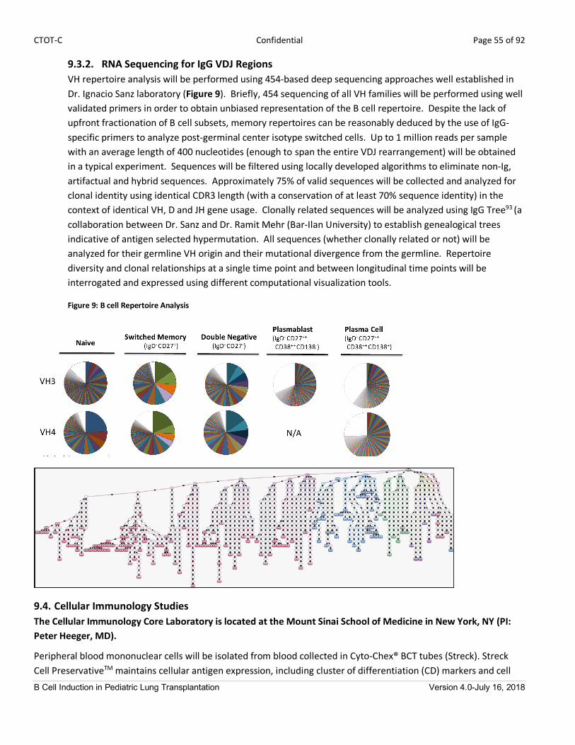
CTOT-C Confidential Page 55 of 92
B Cell Induction in Pediatric Lung Transplantation Version 4.0-July 16, 2018
9.3.2. RNA Sequencing for IgG VDJ Regions VH repertoire analysis will be performed using 454-based deep sequencing approaches well established in
Dr. Ignacio Sanz laboratory (Figure 9). Briefly, 454 sequencing of all VH families will be performed using well
validated primers in order to obtain unbiased representation of the B cell repertoire. Despite the lack of
upfront fractionation of B cell subsets, memory repertoires can be reasonably deduced by the use of IgG-
specific primers to analyze post-germinal center isotype switched cells. Up to 1 million reads per sample
with an average length of 400 nucleotides (enough to span the entire VDJ rearrangement) will be obtained
in a typical experiment. Sequences will be filtered using locally developed algorithms to eliminate non-Ig,
artifactual and hybrid sequences. Approximately 75% of valid sequences will be collected and analyzed for
clonal identity using identical CDR3 length (with a conservation of at least 70% sequence identity) in the
context of identical VH, D and JH gene usage. Clonally related sequences will be analyzed using IgG Tree93
(a
collaboration between Dr. Sanz and Dr. Ramit Mehr (Bar-Ilan University) to establish genealogical trees
indicative of antigen selected hypermutation. All sequences (whether clonally related or not) will be
analyzed for their germline VH origin and their mutational divergence from the germline. Repertoire
diversity and clonal relationships at a single time point and between longitudinal time points will be
interrogated and expressed using different computational visualization tools.
Figure 9: B cell Repertoire Analysis
9.4. Cellular Immunology Studies The Cellular Immunology Core Laboratory is located at the Mount Sinai School of Medicine in New York, NY (PI: Peter Heeger, MD).
Peripheral blood mononuclear cells will be isolated from blood collected in Cyto-Chex® BCT tubes (Streck). Streck
Cell PreservativeTM
maintains cellular antigen expression, including cluster of differentiation (CD) markers and cell

CTOT-C Confidential Page 56 of 92
B Cell Induction in Pediatric Lung Transplantation Version 4.0-July 16, 2018
morphology of biological samples for analysis by flow cytometry. In addition, the study will collect bronchoalveolar
lavage specimen in a Streck Cell Preservative 10ml screw cap vial for B Cell Phenotyping analysis. Cell integrity is
maintained at room temperature for up to 7 days in the cell preservative.
9.4.1. Peripheral Blood B cell Phenotyping Peripheral blood phenotyping will be performed using standardized panels of antibodies (8 color). The efficacy of
Rituxan® on peripheral blood CD20 positive B-cells will be assessed using flow cytometry in samples obtained at
visit 03 (4-6 weeks post-transplant). The B cell phenotypes to be analyzed and a representative flow cytometry
plot illustrating the proposed approach is included as Table 12 and Figure 10.
Table 12: B-cell phenotypes
B cell Subset Surface Phenotype
Transitional T1-T2 IgD+CD27- MTG+CD24hi CD38hi
T3 IgD+CD27- MTG+CD24int CD38int
Naïve IgD+CD27- MTG-CD24int CD38int
Memory Unswitched IgD+ CD27+
Switched Resting IgD-CD27+CD21+CD95-CD24+
Activated IgD-CD27+CD21-CD95+CD24-
Switched CD27- memory (DN) IgD-CD27-CD21+CD95-
Antibody secreting cells Plasmablasts IgD-CD27++CD38++CD19+
Autoimmune phenotype CD19+, CD11c+ CD220+
Figure 10: B cell phenotypes flow cytometry
B cell phenotyping will be performed along with analyses of T cells and APCs (see below). The team will quantify
total B cells, naïve and memory B cells, plasma cells, transitional cells, B cells with an active “autoimmune”
phenotype and Breg using the panels depicted in Table 7.52 ,94, 95
Kinetic analysis will be performed for each patient
and differences between the treatment group and the control group will be determined.
Antigen specificity analyses will focus on autoantigenic B cells reactive to ColV and Ka1T. Fluorescent labeled
pentamers of kα1T using the recombinant kα1T produced in our lab will be used to identify the number of B cells
specific for kα1T and their correlation with circulating Abs to kα1T and other self-Ags (ColV) will be analyzed.

CTOT-C Confidential Page 57 of 92
B Cell Induction in Pediatric Lung Transplantation Version 4.0-July 16, 2018
9.4.2. B-cell Phenotyping in Bronchoalveolar Lavage
Cell surface markers of BAL samples will be analyzed for B cell subsets as outlined in Table 12 above,
including quantifying the numbers of transitional B cells and those with an autoimmune phenotype (CD19,
CD11c and CD220). The effect of B cell depletion on induction of immune response to self-Ags will be
determined by monitoring the number and frequency of B cells and Abs to HLA (Luminex) and self-antigens
(ELISA). We expect that the depletion of B cells by rituximab will result in a decrease in the frequency of
CD19+CD11c+CD220+ B cells. Further, this decline will correlate to a decrease in Abs to HLA as well as Abs to
self-Ags. Additionally, based on preliminary data, we expect that BOS+ pediatric lung recipients will have an
increase in these CD19+CD11c+CD220+ B cells.
9.4.3. Peripheral Blood T-Cell Phenotyping T-cell phenotyping will be performed by flow cytometry simultaneously with the B cell phenotyping outlined
above and using validated panels for multicolor flow (Table 13), to assess CD4 and CD8 T-cell subsets and
thymic emigrants.
Table 13: Example of flow phenotyping for CD4 T-cells
T-Cell Subset Surface Phenotype
Naïve CD4+ CD45RO+ CD27+
Central Memory CD4+ CD45RA+ CD27+
Effector Memory CD4+ CD45RO+ CD27-
Tregs CD3+ CD20- CD4+ CD25+ CD127L
Naïve recent thymic emigrants CD3+ CD20- CD4+ CD8- CCR7+ CD45RA+ CD31+
Recent thymic emigrant Treg CD3+ CD20- CD4+ CD8- CD45RA+ CD31+ FoxP3+
9.4.4. T-Cell function by ELISPOT
Peripheral blood mononuclear cells (PBMC) and plasma will be isolated from whole blood collected in a Sodium
Heparin Vacutainer Tube. The PBMCs will be used to perform T-Cell Function assays (PI: Peter Heeger, MD) and the
plasma will be shipped to the Washington University in St. Louis (PI: Thalachallour Mohanakumar, MD) and used for
humoral immunology assays.
For T-cell autoreactivity, PBMCs collected longitudinally immediately prior to transplant, and then post-
transplantation and will be tested for their reactivity against self-antigens collagen V, Kα1Tand col IV as control. (All
of these proteins are available as reported above and are endotoxin free). Because cellular immunity to
autoantigens is anticipated to be relatively low frequency, we will use the highly sensitive cytokine ELISPOT as an
initial approach for detection and enumeration of frequency of cells secreting specific cytokines following
stimulation with various self-antigens described above. We have the ability to perform 2 color IFNg/IL17 ELISPOT
assays for autoantigen reactivity, which will permit us to maximize our ability to obtain information from small
amounts of blood (Figure 3 above). We will assess cellular alloreactivity by cytokine ELISPOT and if sufficient cells
are available, by CSFE dilution and intracellular cytokine staining.
Donor B cell lines (primary expansion using CD40L transfected fibroblasts and IL-4 as described (and unmatched 3P
control lines) will be made under GLP conditions in the Heeger lab.17
IFNγ ELISPOT assays will be the primary
readout. Assays for 2 different cytokines will be performed as part of the mechanistic study. Again, we will study
IFNg and IL-17 production as prototypic proinflammatory cytokines and IL-4, IL-5 and IL-10 as prototypic type Th2

CTOT-C Confidential Page 58 of 92
B Cell Induction in Pediatric Lung Transplantation Version 4.0-July 16, 2018
cytokines. Aliquots of samples will be stored in sufficient cells for potential future testing against additional
autoantigens.
9.5. Immunohistochemistry The Immunohistochemistry Core Laboratory is located at the Mount Sinai School of Medicine (PI: Michael J. Donovan, MD).
M Donovan, MD, PhD, ISMMS is an innovator in systems pathology in which he has developed computer assisted
approaches for morphometric and multicolor image analysis of IF stained tissues. Using these published methods
multicolor IF staining for B cells (CD20), T-cells (CD4, CD8), Treg (CD4, foxp3), and macrophages (CD68) will be
performed on representative histology slides. Computer assisted image analysis will be used to determine the
numbers of each cell type within the area being studied and to assess correlations among cell subsets. Examples
demonstrating feasibility are shown in Figure 11.
Results will be correlated with the graded score of the biopsy, the presence or absence of AMR features (capillaritis,
C4d capillary loop staining) and outcome, including effects of treatment for acute rejection and the long term
development of BOS.
9.6. Microbiology and Viral Detection Core The Microbiology and Viral Detection Core Laboratory is located at the Washington University in St. Louis, MO (PI: Gregory Storch, MD).
9.6.1. Viral Detection by PCR in Bronchoalveolar Lavage (BAL) and Nasopharyngeal Swabs (NP) Bronchoalveolar Lavage (BAL) samples will be obtained during scheduled surveillance or unscheduled
symptomatic bronchoscopies. Nasopharyngeal swab (NP) samples will be collected during scheduled
surveillance and at symptomatic episodes that do not clinically require bronchoscopy. With all
bronchoscopies, samples for bacterial, fungal, and viral testing will be taken even if infection was not initially
suspected. Mycobacterial testing will be performed if indicated. After processing for immediate clinical
testing by conventional methods at local clinical sites with immediate reporting of infectious episodes, the
remainder of the samples will be stored at -70oC and batch shipped to the Microbiology and Viral Detection
Core Laboratory. If we find that the batched analysis by this highly sensitive method detects potentially
Figure 11: Detection of B cell infiltrates in paraffin embedded transbronchial biopsy samples from the CTOTC-03 trial.
Representative 2 color IF staining for CD20 (red) and CD8 (green) showing A) rare T or B cells in an ISHLT grade A0B0
biopsy, B) diffuse T and B cell infiltration near an airway in an ISHLT grade A0B1C biopsy with bronchiolitis obliterans, and
C) prominent B cell infiltrate adjacent to a vessel in an ISHLT grade A2B1 biopsy from a patient who later developed
bronchiolitis obliterans
A B C

CTOT-C Confidential Page 59 of 92
B Cell Induction in Pediatric Lung Transplantation Version 4.0-July 16, 2018
treatable infection that are not detected by the individual clinical sites (which did not occur to date in
CTOTC-03), we will adjust the design so that it is performed in real time so as to provide the clinically
relevant information to the sites for therapeutic intervention.
9.6.2. Viral Detection in Whole Blood by Quantitative PCR CMV has been epidemiologically linked to acute and chronic rejection including initial data from our
research team showing an increased risk of early mortality in children with CMV viremia.97
EBV, associated
with post-transplant lymphoproliferative disease (PTLD), has also been identified as a potential co-factor for
outcome. Due to known inter-laboratory variability in quantitative measurements of CMV and EBV viral
load,98,99
whole blood will be collected and sent to the Microbiology and Viral Detection Core.
Whole blood will be stored for potential evaluation of additional interacting viruses to include HHV-6.

CTOT-C Confidential Page 60 of 92
B Cell Induction in Pediatric Lung Transplantation Version 4.0-July 16, 2018
10. Biospecimen Storage and Future Use Biological specimens obtained under this protocol may be used in future assays to reevaluate biological responses as
additional research tests are developed over time. These specimens will be collected at time points already
scheduled for the core mechanistic studies outlined in the Schedule of Events (Appendices 1 and 2), in order to allow
specimens to be stored for use in new assays that have yet to be optimized or conceived, or assays performed by
other CTOT-C members for cross-validation studies. Appropriate informed consent will be obtained for both the
collection and storing of samples. The specimens from these evaluations may be stored beyond the funding period.
During the funding period, samples will be identifiable, which means samples will be coded with a subject ID number
that could be directly linked to the subject and the subject’s medical record. When the funding period is over,
samples will be anonymized, which means a sample that was previously identifiable, has had all identifiers removed
and can no longer be linked back to the subject or the subject’s medical record by any means.
Study participants will be informed that they may be approached about additional clinical evaluations or studies that
have received the full approval of the NIAID as new evaluations are identified. If additional evaluations are
determined to be desirable, this protocol (and other appropriate study documents, e.g., the informed consent and
the statistical analysis plan) will be amended and submitted to the appropriate regulatory authorities, ethics
committees, and IRBs for approval. Each participant’s signature will be obtained on the revised informed consent
form before additional evaluations are performed. The specimens from these evaluations may be stored up to the
end of the grant— approximately 5 years, or longer if the grant is extended.

CTOT-C Confidential Page 61 of 92
B Cell Induction in Pediatric Lung Transplantation Version 4.0-July 16, 2018
11.Criteria for Participant and Study Completion and Premature Study Termination
11.1. Participant Completion All participants will be followed for a minimum of12 months and up to 54 months after transplantation.
11.2. Participant Stopping Rules and Withdrawal Criteria Participants may be prematurely terminated from the study for the following reasons:
1. The participant elects to withdraw consent from all future study activities, including follow-up.
2. The participant is “lost to follow-up” (i.e., no further follow-up is possible because attempts to reestablish
contact with the participant have failed).
3. The participant dies.
4. The Investigator no longer believes participation is in the best interest of the participant.
5. The study is stopped by the site, the sponsor (NIAID), or the Food and Drug Administration.
11.3. Participant Replacement Participants who withdraw or are withdrawn will not be replaced if they have received at least one dose of
rituximab.
11.4. Follow-up after Early Study Withdrawal Participants who receive at least one dose of rituximab will follow the schedule of events-Appendix 1.

CTOT-C Confidential Page 62 of 92
B Cell Induction in Pediatric Lung Transplantation Version 4.0-July 16, 2018
12.Safety Monitoring and Reporting
12.1 Overview This section defines the types of safety data that will be collected under this protocol and outlines the procedures
for appropriately collecting, grading, recording, and reporting those data. Adverse events that are classified as
serious according to the definition of health authorities must be reported promptly (per Section 12.5, Reporting of Serious Adverse Events and Adverse Events) to DAIT/NIAID. Appropriate notifications will also be made to site
principal investigators, Institutional Review Boards (IRBs) and health authorities.
Information in this section complies with ICH Guideline E2A: Clinical Safety Data Management: Definitions and Standards for Expedited Reporting, ICH Guideline E-6: Guideline for Good Clinical Practice, 21CFR Parts 312 and 320,
and applies the standards set forth in the National Cancer Institute (NCI), Common Terminology Criteria for Adverse
Events (CTCAE), Version 4.0: http://ctep.cancer.gov/reporting/ctc.html.
12.2 Definitions
12.2.1 Adverse Event (AE) Any untoward or unfavorable medical occurrence associated with the subject’s participation in the research,
whether or not considered related to the subject’s participation in the research (modified from the
definition of adverse events in the 1996 International Conference on Harmonization E-6 Guidelines for Good
Clinical Practice) (from OHRP "Guidance on Reviewing and Reporting Unanticipated Problems Involving Risks
to Subjects or Others and Adverse Events (1/15/07)"
http://www.hhs.gov/ohrp/policy/advevntguid.html#Q2)
For this study, an adverse event will include any untoward or unfavorable medical occurrence associated
with:
• Study therapy regimen: Rituximab or Rituximab Placebo
• Study mandated procedures:
o Bronchoscopy: Any AE occurring within 24 hours after a study related bronchoscopy.
o Blood Draw: Any AE occurring within 24 hours after a study related blood draw.
12.2.1.1 Suspected Adverse Reaction (SAR)
Any adverse event for which there is a reasonable possibility that the investigational drug [or
investigational study therapy regimen] caused the adverse event. For the purposes of safety
reporting, ‘reasonable possibility’ means there is evidence to suggest a causal relationship between
the drug and the adverse event. A suspected adverse reaction implies a lesser degree of certainty
about causality than adverse reaction, which means any adverse event caused by a drug (21 CFR
312.32(a)).
12.2.2 Unexpected Adverse Event
An adverse event or suspected adverse reaction is considered “unexpected” if it is not listed in the
Investigator Brochure or package insert or is not listed at the specificity, severity or rate of occurrence that
has been observed; or is not consistent with the risk information described in the general investigational
plan or elsewhere in the IND.
CTOT-C Confidential Page 63 of 92
B Cell Induction in Pediatric Lung Transplantation Version 4.0-July 16, 2018
“Unexpected” also refers to adverse events or suspected adverse reactions that are mentioned in the
Investigator Brochure or package insert as occurring with a class of drugs or as anticipated from the
pharmacological properties of the drug, but are not specifically mentioned as occurring with the particular
drug under investigation (21 CFR 312.32(a)]
12.2.3 Serious Adverse Event (SAE) An adverse event or suspected adverse reaction is considered “serious” if, in the view of either the
investigator or DAIT/NIAID, it results in any of the following outcomes (21 CFR 312.32(a)):
1. Death.
2. A life-threatening event: An AE or SAR is considered “life-threatening” if, in the view of either the
investigator or DAIT/NIAID, its occurrence places the subject at immediate risk of death. It does not
include an AE or SAR that, had it occurred in a more severe form, might have caused death.
3. Inpatient hospitalization or prolongation of existing hospitalization.
4. Persistent or significant incapacity or substantial disruption of the ability to conduct normal life
functions.
5. Congenital anomaly or birth defect.
6. Important medical events that may not result in death, be life threatening, or require hospitalization
may be considered serious when, based upon appropriate medical judgment, they may jeopardize the
subject and may require medical or surgical intervention to prevent one of the outcomes listed above.
The NIAID medical monitor will review each SAE and assess the relationship of the event to the study
procedure. Events meeting serious criteria that occur outside of these parameters should also be reported if
the investigator deems a possible association with a protocol mandated procedure. Elective hospitalizations
or hospital admissions for the purpose of conduct of protocol mandated procedures are not to be reported
as an SAE unless hospitalization is prolonged due to complications.
12.3 Grading and Attribution of Adverse Events
12.3.1 Grading Criteria The study site will grade the severity of adverse events experienced by the study subjects according to the
criteria set forth in the National Cancer Institute’s Common Terminology Criteria for Adverse Events (CTCAE) version 4. This document (referred to herein as the NCI-CTCAE manual) provides a common language to
describe levels of severity, to analyze and interpret data, and to articulate the clinical significance of all
adverse events. The NCI-CTCAE has been reviewed by the Protocol Chair and Principal Investigator and has
been deemed appropriate for the subject population to be studied in this protocol.
CTOT-C Confidential Page 64 of 92
B Cell Induction in Pediatric Lung Transplantation Version 4.0-July 16, 2018
Adverse events will be graded on a scale from 1 to 5 according to the following standards in the NCI-CTCAE
manual:
Grade 1 = mild adverse event.
Grade 2 = moderate adverse event.
Grade 3 = severe and undesirable adverse event.
Grade 4 = life-threatening or disabling adverse event.
Grade 5 = death.
Events grade 1 and 2 will not be collected. Events grade 3 or higher will be recorded on the appropriate AE
electronic case report form except for hematologic cytopenias. Only hematologic cytopenias meeting
serious criteria outlined in 12.2.3 and occurring from the time of first rituximab infusion to 6 weeks following
the last rituximab infusion will be collected and reported to the sponsor. Outside of this period,
hematologic cytopenias meeting grade 3 and higher will be collected and reported to the sponsor.
For grading an abnormal value or result of a clinical or laboratory evaluation (including, but not limited to, a
radiograph, an ultrasound, an electrocardiogram etc.), a treatment-emergent adverse event is defined as an
increase in grade from baseline or from the last post-baseline value that doesn’t meet grading criteria.
Changes in grade from screening to baseline will also be recorded as adverse events, but are not treatment-
emergent. If a specific event or result from a given clinical or laboratory evaluation is not included in the
NCI-CTCAE manual, then an abnormal result would be considered an adverse event if changes in therapy or
monitoring are implemented as a result of the event/result.
12.3.2 Attribution Definitions The relationship, or attribution, of an adverse event to the study therapy regimen or study procedure(s) will
initially be determined by the site investigator and recorded on the appropriate AE/SAE eCRF. Final
determination of attribution for safety reporting will be determined by DAIT/NIAID. The relationship of an
adverse event to study therapy regimen or procedures will be determined using the descriptors and
definitions provided in Table 14.
For additional information and a printable version of the NCI-CTCAE manual, consult the NCI-CTCAE web
site: http://ctep.cancer.gov/reporting/ctc.html.
Table 14: Attribution of Adverse Events
Code Descriptor Relationship (to primary investigational product and/or other concurrent mandated study therapy or
study procedure)
UNRELATED CATEGORY
1 Unrelated The adverse event is clearly not related: there is
insufficient evidence to suggest a causal relationship.
RELATED CATEGORIES
2 Possible The adverse event has a reasonable possibility to be
related; there is evidence to suggest a causal
relationship.
3 Definite The adverse event is clearly related.

CTOT-C Confidential Page 65 of 92
B Cell Induction in Pediatric Lung Transplantation Version 4.0-July 16, 2018
12.4 Collection and Recording of Adverse Events
12.4.1 Collection Period Adverse events will be collected from the time of enrollment until a subject completes study participation or
until 30 days after he/she prematurely withdraws (without withdrawing consent) or is withdrawn from the
study.
12.4.2 Collecting Adverse Events Adverse events (including SAEs) may be discovered through any of these methods:
• Observing the subject.
• Interviewing the subject [e.g., using a checklist, structured questioning, diary, etc.].
• Receiving an unsolicited complaint from the subject.
• In addition, an abnormal value or result from a clinical or laboratory evaluation can also indicate an
adverse event, as defined in Section 12.3, Grading and Attribution of Adverse Events.
12.4.3 Recording Adverse Events Throughout the study, the investigator will record adverse events and serious adverse events as described
previously (Section 12.2, Definitions) on the appropriate AE/SAE eCRF regardless of the relationship to study
therapy regimen or study procedure.
Once recorded, an AE/SAE will be followed until it resolves with or without sequelae, or until the end of
study participation, or until 30 days after the subject prematurely withdraws (without withdrawing
consent)/or is withdrawn from the study, whichever occurs first.
12.5 Reporting of Serious Adverse Events and Adverse Events
12.5.1 Reporting of Serious Adverse Events to Sponsor This section describes the responsibilities of the site investigator to report serious adverse events to the
sponsor via the SACCC eCRF. Timely reporting of adverse events is required by 21 CFR and ICH E6 guidelines.
Site investigators will report all serious adverse events (see Section 12.2.3, Serious Adverse Event),
regardless of relationship or expectedness within 24 hours of discovering the event.
For serious adverse events, all requested information on the AE/SAE eCRF will be provided. However,
unavailable details of the event will not delay submission of the known information. As additional details
become available, the AE/SAE eCRF will be updated and submitted.
12.5.2 Reporting to Health Authority After an adverse event requiring 24 hour reporting (per Section 12.5.1, Reporting of Serious Adverse Events
to Sponsor) is submitted by the site investigator and assessed by DAIT/NIAID, there are two options for
DAIT/NIAID to report the adverse event to the appropriate health authorities:

CTOT-C Confidential Page 66 of 92
B Cell Induction in Pediatric Lung Transplantation Version 4.0-July 16, 2018
12.5.2.1 Annual Reporting DAIT/NIAID will include in the annual study report to health authorities all adverse events classified
as:
o Serious, expected, suspected adverse reactions (see Section 12.2.1.1, Suspected Adverse Reaction, and Section 12.2.2, Unexpected Adverse Event).
o Serious and not a suspected adverse reaction (see Section 12.2.2, Suspected Adverse Reaction).
o Pregnancies.
Note that all adverse events (not just those requiring 24-hour reporting) will be reported in the
Annual IND Report.
12.5.2.2 Expedited Safety Reporting
This option, with 2 possible categories, applies if the adverse event is classified as one of the
following:
Category 1: Serious and unexpected suspected adverse reaction [SUSAR] (see Section 12.2.1.1,
Suspected Adverse Reaction and Section 12.2, Unexpected Adverse Event and 21 CFR 312.32(c)(1)i).
The sponsor shall report any suspected adverse reaction that is both serious and
unexpected. The sponsor shall report an adverse event as a suspected adverse reaction
only if there is evidence to suggest a causal relationship between the study drug and the
adverse event, such as:
1. A single occurrence of an event that is uncommon and known to be strongly
associated with drug exposure (e.g., angioedema, hepatic injury, or Stevens-Johnson
Syndrome);
2. One or more occurrences of an event that is not commonly associated with drug
exposure, but is otherwise uncommon in the population exposed to the drug (e.g.,
tendon rupture);
3. An aggregate analysis of specific events observed in a clinical trial (such as known
consequences of the underlying disease or condition under investigation or other
events that commonly occur in the study population independent of drug therapy)
that indicates those events occur more frequently in the drug treatment group than
in a concurrent or historical control group.
Category 2: Any findings from studies that suggests a significant human risk
The sponsor shall report any findings from other epidemiological studies, analyses of
adverse events within the current study or pooled analysis across clinical studies or animal
or in vitro testing (e.g. mutagenicity, teratogenicity, carcinogenicity) that suggest a
significant risk in humans exposed to the drug that would result in a safety-related change in
the protocol, informed consent, investigator brochure or package insert or other aspects of
the overall conduct of the study.
DAIT/NIAID shall notify the FDA and all participating investigators of expedited Safety Reports within 15
calendar days; unexpected fatal or immediately life-threatening suspected adverse reaction(s) shall be
reported as soon as possible or within 7 calendar days.

CTOT-C Confidential Page 67 of 92
B Cell Induction in Pediatric Lung Transplantation Version 4.0-July 16, 2018
12.5.3 Reporting of Adverse Events to IRBs/IECs All investigators shall report adverse events, including expedited reports, in a timely fashion to their
respective IRBs/IECs in accordance with applicable regulations and guidelines. All Safety Reports to the
FDA shall be distributed by DAIT/NIAID or designee to all participating institutions for site IRB/IEC
submission.
12.6 Pregnancy Reporting The investigator shall be informed immediately of any pregnancy in a study subject or a partner of a study subject. A
pregnant subject shall be instructed to stop taking study medication. The investigator shall counsel the subject and
discuss the risks of continuing with the pregnancy and the possible effects on the fetus. Monitoring of the pregnant
subject shall continue until the conclusion of the pregnancy.
The investigator shall report to the SACCC all pregnancies within 1 business day of becoming aware of the event
using the Pregnancy eCRF. All pregnancies identified during the study shall be followed to conclusion and the
outcome of each must be reported. The Pregnancy eCRF shall be updated and submitted to SACCC when details
about the outcome are available. When possible, similar information shall be obtained for a pregnancy occurring in
a partner of a study subject.
Information requested about the delivery shall include:
o Gestational age at delivery
o Birth weight, length, and head circumference
o Gender
o Appearance, pulse, grimace, activity, and respiration (APGAR) score at 1 minute, 5 minutes, and 24
hours after birth, if available
o Any abnormalities.
All pregnancy complications that result in a congenital abnormality, birth defect, miscarriage, and medically
indicated abortion - an SAE shall be submitted to the SACCC using the SAE reporting procedures described
above.
12.7 Reporting of Other Safety Information An investigator shall promptly notify the site IRB as well as the SACCC when an “unanticipated problem
involving risks to subjects or others” is identified, which is not otherwise reportable as an adverse event.
12.8 Review of Safety Information
12.8.1 Medical Monitor Review The DAIT/NIAID Medical Monitor shall receive monthly reports from the SACCC compiling new and
accumulating information on AEs, SAEs, and pregnancies recorded by the study site(s) on appropriate eCRFs.
In addition, the Medical Monitor shall review and make decisions on the disposition of the SAE and
pregnancy reports received by the SACCC (See Sections 12.5.1, Reporting of Serious Adverse Events to Sponsor, and 12.6, Pregnancy Reporting).

CTOT-C Confidential Page 68 of 92
B Cell Induction in Pediatric Lung Transplantation Version 4.0-July 16, 2018
12.8.2 DSMB Review 12.8.2.1 Planned DSMB Reviews
The Data and Safety Monitoring Board (DSMB) shall review safety data at least yearly during
planned DSMB Data Review Meetings. Data for the planned safety reviews will include, at a
minimum, a listing of all reported AEs and SAEs.
The DSMB will be informed of an Expedited Safety Report in a timely manner.
12.8.2.2 Ad hoc DSMB Reviews
In addition to the pre-scheduled data reviews and planned safety monitoring, the DSMB may be
called upon for ad hoc reviews. The DSMB will review any event that potentially impacts safety at
the request of the study principal investigator or DAIT/NIAID. In addition, satisfaction of any of the
following stopping rules in study subjects at any time of follow-up in the treatment arms will trigger
an ad hoc DSMB Safety Review:
o Any occurrence of confirmed PML.
o Incidence of death of 30% or more subjects.
o Incidence of at least mild acute rejection of 35% or more.
o Incidence of humoral rejection of 25% or more.
o Incidence of primary graft dysfunction of 50% or more.
o Incidence of PTLD of 5% or more.
o Incidence of infections of any type requiring hospitalization of 40% or more.
The thresholds for stopping rules involving subject-level incidence rates have been formulated
based on an analysis of adverse events in the CTOTC-03 (NCT00891865) observational study of
subjects who are similar to those in the control arm of this study. They will be evaluated routinely by
the SACCC with the occurrence of any of the triggering events throughout follow-up of subjects
within either randomized treatment arm. They will be implemented by comparing the exact lower
95% confidence limit on the currently observed subject-level incidence rate to the threshold rate
described for that event in the stopping rule. If the lower confidence limit is greater than the
threshold, the stopping rule will be considered to have been met and the Medical Monitor, DSMB
and study PI will be notified.
Following are tables illustrating, for each stopping rule, the minimum numbers of subjects with the
respective event that would meet the stopping rule for selected numbers of subjects within either
randomized treatment arm:

CTOT-C Confidential Page 69 of 92
B Cell Induction in Pediatric Lung Transplantation Version 4.0-July 16, 2018
Table 15: Minimum Numbers of Subjects with the Event in Either Randomized Treatment Arm that meet the Stopping Rule for Death with a Threshold of Concern of 30%
Table 16: Minimum Numbers of Subjects with the Event in Either Randomized Treatment Arm that meet the Stopping Rule for Mild Acute Rejection with a Threshold of Concern of 35%
Table 17: Minimum Numbers of Subjects with the Event in Either Randomized Treatment Arm that meet the Stopping Rule for Humoral Rejection with a Threshold of Concern of 25%
Number of Subjects with Event
Number of Randomized Subjects
Cumulative Incidence Rate
Lower 95% Confidence Limit
4 4 100.00 47.29
5 6 83.33 41.82
6 8 75.00 40.03
6 10 60.00 30.35
7 12 58.33 31.52
8 14 57.14 32.50
9 16 56.25 33.34
10 18 55.56 34.06
10 20 50.00 30.20
11 22 50.00 31.13
Number of Subjects with Event
Number of Randomized Subjects
Cumulative Incidence Rate
Lower 95% Confidence Limit
4 4 100.00 47.29
5 6 83.33 41.82
6 8 75.00 40.03
7 10 70.00 39.34
8 12 66.67 39.09
9 14 64.29 39.04
10 16 62.50 39.10
11 18 61.11 39.22
11 20 55.00 34.69
12 22 54.55 35.25
Number of Subjects with Event
Number of Randomized Subjects
Cumulative Incidence Rate
Lower 95% Confidence Limit
4 4 100.00 47.29
4 6 66.67 27.13
5 8 62.50 28.92
6 10 60.00 30.35
7 12 58.33 31.52
7 14 50.00 26.36
8 16 50.00 27.86
9 18 50.00 29.12
9 20 45.00 25.87
10 22 45.45 27.13

CTOT-C Confidential Page 70 of 92
B Cell Induction in Pediatric Lung Transplantation Version 4.0-July 16, 2018
Table 18: Minimum Numbers of Subjects with the Event in Either Randomized Treatment Arm that meet the Stopping Rule for Primary Graft Dysfunction with a Threshold of Concern of 50%
Table 19: Minimum Numbers of Subjects with the Event in Either Randomized Treatment Arm that meet the Stopping Rule for PTLD with a Threshold of Concern of 5%
Number of Subjects with Event
Number of Randomized Subjects
Cumulative Incidence Rate
Lower 95% Confidence Limit
5 4 100.00 54.93
6 6 100.00 60.70
7 8 87.50 52.93
9 10 90.00 60.58
10 12 83.33 56.19
11 14 78.57 53.43
12 16 75.00 51.56
13 18 72.22 50.22
15 20 75.00 54.44
16 22 72.73 53.15
Number of Subjects with Event
Number of Randomized Subjects
Cumulative Incidence Rate
Lower 95% Confidence Limit
2 4 50.00 9.76
2 6 33.33 6.28
3 8 37.50 11.11
3 10 30.00 8.73
3 12 25.00 7.19
3 14 21.43 6.11
3 16 18.75 5.31
4 18 22.22 7.97
4 20 20.00 7.14
4 22 18.18 6.46

CTOT-C Confidential Page 71 of 92
B Cell Induction in Pediatric Lung Transplantation Version 4.0-July 16, 2018
Table 20: Minimum Numbers of Subjects with the Event in Either Randomized Treatment Arm that meet the Stopping Rule for Infection Requiring Hospitalization with a Threshold of Concern of 40%
o
After review of the data, the DSMB will make recommendations regarding study conduct and/or
continuation.
12.8.2.2.1 Temporary Suspension of enrollment and drug dosing for ad hoc DSMB Safety Review
A temporary halt in enrollment, randomization and administration of rituximab/rituximab
placebo will be implemented if an ad hoc DSMB safety review is required. In the event a
study participant has already received the first dose of rituximab/rituximab placebo, every
attempt will be made to expedite review such that a prompt decision can be made
regarding the appropriate management for these subjects.
Number of Subjects with Event
Number of Randomized Subjects
Cumulative Incidence Rate
Lower 95% Confidence Limit
4 4 100.00 47.29
5 6 83.33 41.82
6 8 75.00 40.03
8 10 80.00 49.31
9 12 75.00 47.27
10 14 71.43 46.00
11 16 68.75 45.17
12 18 66.67 44.60
13 20 65.00 44.20
14 22 63.64 43.91

CTOT-C Confidential Page 72 of 92
B Cell Induction in Pediatric Lung Transplantation Version 4.0-July 16, 2018
13.Statistical Considerations and Analytical Plan
13.1 Overview In this randomized controlled clinical trial, we will compare primary pediatric lung transplant recipients given
rituximab induction therapy plus standard immmunosuppression to subjects given a placebo induction therapy plus
standard immmunosuppression and assess whether rituximab induction therapy improves outcomes in this
population.
13.2 Endpoints and Safety Outcomes The primary endpoint will be the earliest time to any of the following events during the follow up period: Chronic
Allograft Dysfunction, listing for retransplantation, and death.
The following secondary clinical endpoints will be assessed during the post-transplant follow-up period, which will
be a minimum of 12 months:
• incidence of chronic allograft dysfunction, listing for retransplant or death,
• incidence of Primary Graft Dysfunction,
• incidence of Grade A Acute Rejection,
• incidence of Antibody Mediated Rejection,
• incidence of TVT,
• percentage of participants identified to participate completing the TVI,
• and magnitude of change in standard deviation of tacrolimus levels following intervention.
The following secondary clinical post-transplant safety outcomes will be assessed: incidence and severity of infection
episodes, and serious adverse events related to rituximab.
The following mechanistic endpoints will be assessed: incidence and kinetics of DSA and auto-antibodies,
specifically Collagen V (ColV) and k-alpha-1 tubulin (kα1T); frequency, kinetics, phenotype and function of peripheral
B cells; frequency, kinetics and cytokine profiles of allo- and auto reactive T cells; the incidence and quantity of B
cells and B cell proximity to other cells in the graft tissue.
13.3 Measures to Minimize Bias Study treatments will be randomly assigned to eligible subjects and the treatment assignment of each subject will be
double-blinded. In addition, the central laboratories that perform the mechanistic assays will be blinded to both the
treatment assignment and clinical status of all study subjects.
13.4 Analysis Plan The overall focus of our statistical analysis of the clinical portion of the study will be on the estimation of hazard
ratios (HR) and risk ratios (RR) and their two-sided 90% confidence intervals comparing the rituximab and placebo
groups on each endpoint of the study. Focusing on estimates and confidence intervals will allow us to assess the
range potential efficacies as best we can within the limitations imposed by studying such a rare pediatric population.
Further detail describing the analysis plan will be contained in a Statistical Analysis Plan (SAP) that will be maintained
by the SACCC and updated as necessary.

CTOT-C Confidential Page 73 of 92
B Cell Induction in Pediatric Lung Transplantation Version 4.0-July 16, 2018
13.4.1 Analysis Populations
Intent-to-Treat (ITT) Sample. All randomized subjects who receive at least a portion of the initial
rituximab/placebo infusion will constitute the ITT sample. This sample will be used for evaluation of the
primary and secondary clinical endpoints. Subjects will be analyzed in the group to which they were
randomized, regardless of compliance with the protocol or withdrawal from the study.
Per-Protocol (PP) Sample. Subjects from the ITT sample who complete the full induction protocol, with
complete administration of the prescribed dose at both the initial and subsequent infusions, will make up
the PP sample. This sample will capture subjects who both tolerate rituximab well and recover as expected
from their transplant.
Safety Sample. All subjects from the ITT sample will constitute the safety sample.
13.4.2 Primary Analysis of Primary Endpoints /Outcomes We will use competing-risks Cox proportional hazards models to compare treatment groups on the time to
endpoint, right-censored at the end of the two-year follow-up period or at time of loss to follow-up. This
model, as opposed to a time-to-first-event model, will allow us to estimate a separate hazard function for
each type of outcome as well as the overall outcome, and thus assess whether the effect of rituximab is the
same for all outcome types100
. Our focus will be on the estimates of these hazard ratios (HR) and their two-
sided 90% confidence intervals, with HR < 1 indicating rituximab efficacy. We will assess whether HR is
constant throughout follow-up, or if the effect of rituximab is confined to early or late portions of the study
periods through the use of time-dependent covariates. Model diagnostics will include assessment of the
proportional hazards assumption and of each observation for influence on model parameters. The model
will be run on both the ITT and PP populations, as events allow.
13.4.3 Supportive Analyses of the Primary Endpoints We will also model the time-to-first-event model within the Kaplan-Meier framework for the primary
outcome, as needed, given the likelihood of little to no events for one or more of the 3 outcomes.
Secondary analyses will model each component of the outcome separately with the other components as
risk factors, as allowed by the number of events and the sample size.

CTOT-C Confidential Page 74 of 92
B Cell Induction in Pediatric Lung Transplantation Version 4.0-July 16, 2018
13.4.4 Analyses of Secondary Endpoints and Safety Outcomes All secondary endpoint analyses will use the ITT sample and will be assessed during the post-transplant
follow-up period, which will be a minimum of 12 months.
Separately for each of the secondary incidence endpoints, we will compare the rituximab and control groups
on the proportion of patients in each group with at least one episode of the endpoint by estimating the
relative risk of the endpoint with its two-sided 90% confidence interval. The absolute risk difference with
90% confidence interval will also be estimated. These methods assume no, or very low rates of, death and
re-transplant in the study period. However, if meaningful numbers of subjects have these outcomes, the risk
ratio for each incidence endpoint can be estimated through a Cox proportional hazards model with the
incidence endpoint as the outcome and right-censoring at death, re-transplant, or other loss to follow-up,
following the modeling process described above for the primary endpoint.
Among subjects who are identified as qualifying for and agreeing to participate in the TVI, we will compare
the rituximab and control groups on change in standard deviation of tacrolimus levels following the TVI
(described in sections 5.3.3 and 8.4) using an analysis of covariance (ANCOVA) model, in which the standard
deviation of tacrolimus levels following intervention is the outcome, and study group and pre-intervention
standard deviation of tacrolimus levels are the predictors. The group effect with its 90% confidence interval
will be estimated.
For safety outcomes, we will analyze each type of infection separately (bacterial, fungal, or viral, including
RVI, CMV, and EBV), as well as for all types of infections combined. We will first determine the incidence of
bacterial, fungal and viral infections in the rituximab and control groups. ‘Incidence rate’ will be the number
of infections per patient-month, and we will focus on the estimates and associated 90% confidence intervals
for the ratio of such rates (RR), where RR > 1.0 indicates greater infection rates for rituximab. Of course,
infection events are biologically related within patient, thus, relative to perfect independence of events,
greater proportions of patients will experience more episodes of the same type of infection and greater
proportions of patients will experience no episodes. Accordingly, a Poisson regression models offset for
months of follow-up and adjusted for overdispersion will be used to compare groups on the rate of each
type of infection and any infection.
Serious adverse events related to rituximab will be reported by their incidence rate per patient and per
infusion with 90% confidence intervals. Adverse events will be summarized by treatment group, with 90%
confidence intervals.
13.4.5 Analyses of Exploratory Endpoints We will correlate pre and/or post-transplant serum reactivity to each autoantigen and to donor HLA with
our primary endpoint to prospectively and independently test the hypothesis that autoantibodies can be
used as predictors of incipient injury. We will also correlate the presence of serum antibodies with positive
C4d staining of the graft tissue (to be collected as part of the clinical component of the study).
To test the hypothesis that rituximab prevents induction of de novo autoantibodies and or DSA, we will
compare titers of autoantibodies (or DSA) between the rituximab and control groups. Development of
antibodies to either DSA or self-antigens or both and the kinetics of antibody development (time course
following transplantation) will be correlated with clinical evidence of AMR and BOS as determined in the

CTOT-C Confidential Page 75 of 92
B Cell Induction in Pediatric Lung Transplantation Version 4.0-July 16, 2018
clinical part of study described above and the ISHLT criterion. While the absolute numbers of patients in
each group with preexisting autoantibodies or DSA is anticipated to be small (thus limiting power) we will
compare and report pre vs post-transplant titers in each case to determine if rituximab impacts preexisting
antibody.
The results will be analyzed for development of PGD, development of DSA, diagnosis of AMR and BOS. In
addition we will also determine any correlation which may exist between the strength of antibodies
detected for DSA, HLA and each antibody to self-antigens either alone or cumulatively and clinical outcomes
such as PGD, AMR and BOS.
We will analyze DSA and each type of antibody separately. The DSA and autoantibody data will consist of
binary (positive/negative) or ordinal (negative/weak positive/strong positive) responses measured
repeatedly at scheduled and unequally-spaced intervals. With these features of the data and at this sample
size, it may not be feasible to obtain estimates of treatment effect at each time-point using a single
statistical model with rituximab and time main effects and their interaction as covariates. Therefore we will
describe the treatment effect over the course of the follow-up period.
The hazard ratio for the association between development of antibodies and clinical outcomes (the
endpoint and its components, and the secondary incidence endpoints) will be estimated with its 90%
confidence interval using a Cox proportional hazard model with presence or absence of antibodies as time-
dependent repeated measures covariates, stratified by treatment group. We will model the association of
each antibody on each outcome separately and in combination.
We will also estimate odds ratios with 90% confidence intervals for the associations between each of the
various serum antibodies and C4d staining of the graft tissue. We will estimate these odds ratios at each
time point between each pair of antibodies and each antibody and C4d result, both cross-sectional, and
cumulative, in which case we would treat any subject with a current or prior positive result as positive for
that particular antibody.

CTOT-C Confidential Page 76 of 92
B Cell Induction in Pediatric Lung Transplantation Version 4.0-July 16, 2018
13.4.6 Descriptive Analyses Selected subject baseline clinical and demographic characteristics, as well as use of concomitant
medications and study subject disposition will be summarized using standard descriptive statistics for
continuous and categorical variables.
13.5 Interim Analyses No interim analyses are planned. However, as described in section 12.8.2., the DAIT Transplant DSMB will
periodically review safety data in open and/or closed sessions.
13.6 Sample Size Considerations Planning this study indicates a sample-size analysis that is congruent with our focus on estimates and confidence
intervals. Accordingly, simulated study data using a Weibull distribution with parameters chosen to match our
expectation that the outcome rate in the control group will be 74% at one year and 53% at two years, with hazard
ratios of 0.40 and 0.33. For the purposes of the study only large group differences are clinically relevant, and
therefore this effect size is reasonable.
Sample-sizes between N = 25 + 25 and N = 30+ 30 are feasible, as argued elsewhere. We also looked at N = 35 + 35,
but acknowledge this probably not obtainable.
5000 individual trials were run for each combination of total sample size (N = 50, 60, 70) and true hazard ratio (0.40,
0.33). Each trial’s dataset was analyzed using a standard Cox proportional hazards model and the estimated log (HR)
and its standard error were obtained. Using the criteria given in Table 21, each trial’s estimate and upper confidence
limit was rated as being “excellent,” “good,” “fair,” or “lacking” in supporting the rituximab efficacy. The obtained
proportions are given in Table 22.
Table 21: Criteria for Estimating Upper Confidence Limit Evidence for
Efficacy Estimated Hazard
Ratio Upper 90%
Confidence Limit “Excellent” <0.55 <0.85
“Good” <0.60 0.85 – 1.10
“Fair” <0.70 1.10 – 1.20
“Lacking” Any >1.20
Table 22: Upper 90% Confidence Limits by HR and Sample Size
Sample Sizes 25+25 30+30 35+35 True Hazard Ratio 0.04 0.33 0.04 0.33 0.04 0.33 Evidence for Efficacy
“Excellent” 0.39 0.50 0.46 0.59 0.51 0.64
“Good” 0.22 0.21 0.23 0.21 0.24 0.20
“Fair” 0.07 0.06 0.06 0.05 0.06 0.04
“Lacking” 0.33 0.24 0.25 0.16 0.19 0.12

CTOT-C Confidential Page 77 of 92
B Cell Induction in Pediatric Lung Transplantation Version 4.0-July 16, 2018
In addition, although the primary analysis is based on a Cox regression model, a simple power and sample size
assessment using the less efficient log-rank test indicates that 25 subjects in each group will provide greater than
80% power to detect a HR of 0.45 for the treatment effect using a one-sided test at the 0.05 level of significance.

CTOT-C Confidential Page 78 of 92
B Cell Induction in Pediatric Lung Transplantation Version 4.0-July 16, 2018
14.Identification and Access to Source Data
14.1. Source Data Source documents and source data are considered to be the original documentation where subject information,
visits consultations, examinations and other information are recorded. Documentation of source data is necessary
for the reconstruction, evaluation and validation of clinical findings, observations and other activities during a
clinical trial.
14.2. Access to Source Data The site investigators and site staff will make all source data available to the DAIT/NIAID, as well as to relevant
health authorities. Authorized representatives as noted above are bound to maintain the strict confidentiality of
medical and research information that may be linked to identified individuals.
CTOT-C Confidential Page 79 of 92
B Cell Induction in Pediatric Lung Transplantation Version 4.0-July 16, 2018
15.Protocol Deviations
15.1. Protocol Deviation Definitions Protocol Deviation – The investigators and site staff will conduct the study in accordance to the protocol; no
deviations from the protocol are permitted. Any change, divergence, or departure from the study design or
procedures constitutes a protocol deviation. As a result of any deviation, corrective actions will be developed by the
site and implemented promptly.
Major Protocol Deviation (Protocol Violation) - A Protocol Violation is a deviation from the IRB approved protocol
that may affect the subject's rights, safety, or well-being and/or the completeness, accuracy and reliability of the
study data. In addition, protocol violations include willful or knowing breaches of human subject protection
regulations, or policies, any action that is inconsistent with the NIH Human Research Protection Program’s research,
medical, and ethical principles, and a serious or continuing noncompliance with federal, state, local or institutional
human subject protection regulations, policies, or procedures.
Non-Major Protocol Deviation - A non-major protocol deviation is any change, divergence, or departure from the
study design or procedures of a research protocol that does not have a major impact on the subject's rights, safety
or well-being, or the completeness, accuracy and reliability of the study data.
15.2. Reporting and Managing Protocol Deviations The study site principal investigator has the responsibility to identify, document and report protocol deviations.
However, protocol deviations may also be identified during site monitoring visits or during other forms of study
conduct review.
Deviations that impact the ability to assess study outcomes will be collected for this study. Upon determination that
a protocol deviation has occurred, the study staff will notify the SACCC by completing a Protocol Deviation eCRF.
NIAID, the study PI and DSMB will review deviations on a regular basis. The site will be responsible for reporting
deviations to the local IRB according to site-specific guidelines.

CTOT-C Confidential Page 80 of 92
B Cell Induction in Pediatric Lung Transplantation Version 4.0-July 16, 2018
16.Ethical Considerations and Compliance with Good Clinical Practice
16.1. Statement of Compliance This clinical study will be conducted using good clinical practice (GCP), as delineated in Guidance for Industry: E6 Good Clinical Practice Consolidated Guidance, and according to the criteria specified in this study protocol. Before
study initiation, the protocol and the informed consent documents will be reviewed and approved by the IRB. Any
amendments to the protocol or to the consent materials will also be approved by the IRB before they are
implemented.
16.2. Informed Consent Process The consent process will provide information about the study to a prospective participant and will allow adequate
time for review and discussion prior to his/her decision. The principal investigator or designee listed on the FDA
1572 will review the consent and answer questions. The prospective participant will be told that being in the trial is
voluntary and that he or she may withdraw from the study at any time, for any reason. All participants (or their
legally acceptable representative) will read, sign, and date a consent form before undergoing any study procedures.
Consent materials will be presented in participants’ primary language. A copy of the signed consent form will be
given to the participant.
The consent process will be ongoing. The consent form will be revised when important new safety information is
available, the protocol is amended, and/or new information becomes available that may affect participation in the
study.
16.3. Privacy and Confidentiality A participant’s privacy and confidentiality will be respected throughout the study. Each participant will be assigned
a unique identification number and these numbers rather than names will be used to collect, store, and report
participant information. Site personnel will not transmit documents containing personal health identifiers (PHI) to
the study sponsor or their representatives.

CTOT-C Confidential Page 81 of 92
B Cell Induction in Pediatric Lung Transplantation Version 4.0-July 16, 2018
17.Publication Policy
The CTOT-C policy on the publication of study results will apply to this trial.

CTOT-C Confidential Page 82 of 92
B Cell Induction in Pediatric Lung Transplantation Version 4.0-July 16, 2018
18. References 1. Benden C, Aurora P, Edwards LB, et al. The Registry of the International Society for Heart and Lung
Transplantation: Fourteenth Pediatric Lung and Heart-Lung Transplantation Report--2011. J Heart Lung
Transplant;30:1123-32.
2. Zafar F, Heinle JS, Schecter MG, et al. Two decades of pediatric lung transplant in the United States: have we
improved? J Thorac Cardiovasc Surg;141:828-32, 32 e1.
3. Sundaresan S, Trulock EP, Mohanakumar T, Cooper JD, Patterson GA. Prevalence and outcome of bronchiolitis
obliterans syndrome after lung transplantation. Washington University Lung Transplant Group. Ann Thorac Surg
1995;60:1341-6; discussion 6-7.
4. Valentine VG, Robbins RC, Berry GJ, et al. Actuarial survival of heart-lung and bilateral sequential lung transplant
recipients with obliterative bronchiolitis. J Heart Lung Transplant 1996;15:371-83.
5. Smith MA, Sundaresan S, Mohanakumar T, et al. Effect of development of antibodies to HLA and cytomegalovirus
mismatch on lung transplantation survival and development of bronchiolitis obliterans syndrome. J Thorac
Cardiovasc Surg 1998;116:812-20.
6. Smith KG, Isbel NM, Catton MG, Leydon JA, Becker GJ, Walker RG. Suppression of the humoral immune response
by mycophenolate mofetil. Nephrol Dial Transplant 1998;13:160-4.
7. Waltz DA, Boucek MM, Edwards LB, et al. Registry of the International Society for Heart and Lung Transplantation:
ninth official pediatric lung and heart-lung transplantation report--2006. J Heart Lung Transplant 2006;25:904-11.
8. Webber SA, McCurry K, Zeevi A. Heart and lung transplantation in children. Lancet 2006;368:53-69.
9. Boehler A, Kesten S, Weder W, Speich R. Bronchiolitis obliterans after lung transplantation: a review. Chest
1998;114:1411-26.
10. Smits JM, Vanhaecke J, Haverich A, et al. Three-year survival rates for all consecutive heart-only and lung-only
transplants performed in Eurotransplant, 1997-1999. Clin Transpl 2003:89-100.
11. Magee JC, Barr ML, Basadonna GP, et al. Repeat organ transplantation in the United States, 1996-2005. Am J
Transplant 2007;7 Suppl 1:1424-33.
12. Starnes VA, Bowdish ME, Woo MS, et al. A decade of living lobar lung transplantation: recipient outcomes. J
Thorac Cardiovasc Surg 2004;127:114-22.
13. Scully BB, Zafar F, Schecter MG, et al. Lung retransplantation in children: appropriate when selectively applied.
Ann Thorac Surg;91:574-9.
14. Boehler A, Estenne M. Post-transplant bronchiolitis obliterans. [Review] [144 refs]. EuropeanRespiratoryJournal
2003;22:1007-18.
15. Griffith BP, Paradis IL, Zeevi A, et al. Immunologically mediated disease of the airways after pulmonary
transplantation. Ann Surg 1988;208:371-8.

CTOT-C Confidential Page 83 of 92
B Cell Induction in Pediatric Lung Transplantation Version 4.0-July 16, 2018
16. Wood KJ, Goto R. Mechanisms of rejection: current perspectives. Transplantation;93:1-10.
17. Poggio ED, Augustine JJ, Clemente M, et al. Pretransplant cellular alloimmunity as assessed by a panel of reactive
T cells assay correlates with acute renal graft rejection. Transplantation 2007;83:847-52.
18. Gokmen MR, Lombardi G, Lechler RI. The importance of the indirect pathway of allorecognition in clinical
transplantation. Curr Opin Immunol 2008;20:568-74.
19. Benichou G, Alessandrini A, Charrad RS, Wilkes DS. Induction of autoimmunity after allotransplantation. Front
Biosci 2007;12:4362-9.
20. Veillette GR, Sahara H, Meltzer AJ, et al. Autoimmune sensitization to cardiac myosin leads to acute rejection of
cardiac allografts in miniature swine. Transplantation;91:1187-91.
21. Fedoseyeva EV, Kishimoto K, Rolls HK, et al. Modulation of tissue-specific immune response to cardiac myosin
can prolong survival of allogeneic heart transplants. J Immunol 2002;169:1168-74.
22. Wilkes DS. Autoantibody formation in human and rat studies of chronic rejection and primary graft dysfunction.
Semin Immunol;24:131-5.
23. Jones ND, Turvey SE, Van Maurik A, et al. Differential susceptibility of heart, skin, and islet allografts to T cell-
mediated rejection. J Immunol 2001;166:2824-30.
24. He C, Schenk S, Zhang Q, et al. Effects of T cell frequency and graft size on transplant outcome in mice. J
Immunol 2004;172:240-7.
25. Burlingham WJ, Love RB, Jankowska-Gan E, et al. IL-17-dependent cellular immunity to collagen type V
predisposes to obliterative bronchiolitis in human lung transplants. J Clin Invest 2007;117:3498-506.
26. Bharat A, Fields RC, Steward N, Trulock EP, Patterson GA, Mohanakumar T. CD4+25+ regulatory T cells limit Th1-
autoimmunity by inducing IL-10 producing T cells following human lung transplantation. Am J Transplant
2006;6:1799-808.
27. Bharat A, Narayanan K, Street T, et al. Early posttransplant inflammation promotes the development of
alloimmunity and chronic human lung allograft rejection. Transplantation 2007;83:150-8.
28. Saini D, Weber J, Ramachandran S, et al. Alloimmunity-induced autoimmunity as a potential mechanism in the
pathogenesis of chronic rejection of human lung allografts. J Heart Lung Transplant;30:624-31.
29. Bharat A, Saini D, Steward N, et al. Antibodies to self-antigens predispose to primary lung allograft dysfunction
and chronic rejection. Ann Thorac Surg;90:1094-101.
30. Goers TA, Ramachandran S, Aloush A, Trulock E, Patterson GA, Mohanakumar T. De novo production of K-alpha1
tubulin-specific antibodies: role in chronic lung allograft rejection. J Immunol 2008;180:4487-94.
31. Fukami N, Ramachandran S, Saini D, et al. Antibodies to MHC class I induce autoimmunity: role in the
pathogenesis of chronic rejection. J Immunol 2009;182:309-18.
CTOT-C Confidential Page 84 of 92
B Cell Induction in Pediatric Lung Transplantation Version 4.0-July 16, 2018
32. Maruyama T, Jaramillo A, Narayanan K, Higuchi T, Mohanakumar T. Induction of obliterative airway disease by
anti-HLA class I antibodies. Am J Transplant 2005;5:2126-34.
33. Rose EA, Smith CR, Petrossian GA, Barr ML, Reemtsma K. Humoral immune responses after cardiac
transplantation: correlation with fatal rejection and graft atherosclerosis. Surgery 1989;106:203-7; discussion 7-8.
34. Liu Z, Harris PE, Colovai AI, Reed EF, Maffei A, Suciu Foca N. Indirect recognition of donor MHC class II antigens in
human transplantation. Clin Immunol Immunopath 1996;78:228-35.
35. Rizzo M, Sundaresan S, Lynch J, et al. Increased concentration of soluble human leukocyte antigen class I levels in
the bronchoalveolar lavage of human pulmonary allografts. J Heart Lung Transplant 1997;16:1135-40.
36. SivaSai KS, Smith MA, Poindexter NJ, et al. Indirect recognition of donor HLA class I peptides in lung transplant
recipients with bronchiolitis obliterans syndrome. Transplantation 1999;67:1094-8.
37. Reznik SI, Jaramillo A, Zhang L, Patterson GA, Cooper JD, Mohanakumar T. Anti-HLA antibody binding to hla class
I molecules induces proliferation of airway epithelial cells: a potential mechanism for bronchiolitis obliterans
syndrome. J Thorac Cardiovasc Surg 2000;119:39-45.
38. Reznik SI, Jaramillo A, Sayegh M, Patterson GA, Mohanakumar T. Sensitization against donor mismatched HLA
class II peptides in lung transplant recipients with bronchiolitis oblitierans syndrome. Transplantation 1999;67:S42.
39. Lu KC, Jaramillo A, Mendeloff EN, et al. Concomitant allorecognition of mismatched donor HLA class I- and class
II-derived peptides in pediatric lung transplant recipients with bronchiolitis obliterans syndrome. J Heart Lung
Transplant 2003;22:35-43.
40. Reznik SI, Jaramillo A, SivaSai KS, et al. Indirect allorecognition of mismatched donor HLA class II peptides in lung
transplant recipients with bronchiolitis obliterans syndrome. Am J Transplant 2001;1:228-35.
41. Jaramillo A, Smith, C.R., Maruyama, T., Zhang, L., Patterson, G.A., Mohanakumar, T. Anti-HLA class I antibody
binding to airway epithelial cells induces production of fibrogenic growth factors and apoptotic cell death: a possible
mechanism for bronchiolitis obliterans syndrome. Hum Immunol 2003;64:521-9.
42. Fukami N, Ramachandran S, Takenaka M, Weber J, Subramanian V, Mohanakumar T. An Obligatory Role for Lung
Infiltrating B Cells in the Immunopathogenesis of Obliterative Airway Disease Induced by Antibodies to MHC Class I
Molecules. Am J Transplant 2012;12:867-76.
43. Parsons RF, Yu M, Vivek K, et al. Murine Islet Allograft Tolerance Upon Blockade of the B-Lymphocyte Stimulator,
BLyS/BAFF. Transplantation 2012;93:676-85.
44. Redfield RR, 3rd, Rodriguez E, Parsons R, et al. Essential role for B cells in transplantation tolerance. Curr Opin
Immunol;23:685-91.
45. Mohiuddin MM, Corcoran PC, Singh AK, et al. B cell Depletion Extends the Survival of GTKO.hCD46Tg Pig Heart
Xenografts in Baboons for up to 8 Months. Am J Transplant;12:763-71.
46. Liu C, Noorchashm H, Sutter JA, et al. B lymphocyte-directed immunotherapy promotes long-term islet allograft
survival in nonhuman primates. Nat Med 2007;13:1295-8.

CTOT-C Confidential Page 85 of 92
B Cell Induction in Pediatric Lung Transplantation Version 4.0-July 16, 2018
47. Horikawa M, Minard-Colin V, Matsushita T, Tedder TF. Regulatory B cell production of IL-10 inhibits lymphoma
depletion during CD20 immunotherapy in mice. J Clin Invest;121:4268-80.
48. Watanabe R, Ishiura N, Nakashima H, et al. Regulatory B cells (B10 cells) have a suppressive role in murine lupus:
CD19 and B10 cell deficiency exacerbates systemic autoimmunity. J Immunol;184:4801-9.
49. Yanaba K, Bouaziz JD, Haas KM, Poe JC, Fujimoto M, Tedder TF. A regulatory B cell subset with a unique
CD1dhiCD5+ phenotype controls T cell-dependent inflammatory responses. Immunity 2008;28:639-50.
50. Ding Q, Yeung M, Camirand G, et al. Regulatory B cells are identified by expression of TIM-1 and can be induced
through TIM-1 ligation to promote tolerance in mice. J Clin Invest;121:3645-56.
51. Newell KA, Phippard D, Turka LA. Regulatory cells and cell signatures in clinical transplantation tolerance. Curr
Opin Immunol;23:655-9.
52. Newell KA, Asare A, Kirk AD, et al. Identification of a B cell signature associated with renal transplant tolerance in
humans. J Clin Invest;120:1836-47.
53. Hernandez-Fuentes MP, Lechler RI. A 'biomarker signature' for tolerance in transplantation. Nat Rev
Nephrol;6:606-13.
54. Sagoo P, Perucha E, Sawitzki B, et al. Development of a cross-platform biomarker signature to detect renal
transplant tolerance in humans. J Clin Invest;120:1848-61.
55. Brouard S, Mansfield E, Braud C, et al. Identification of a peripheral blood transcriptional biomarker panel
associated with operational renal allograft tolerance. Proc Natl Acad Sci U S A 2007;104:15448-53.
56. DiLillo DJ, Horikawa M, Tedder TF. B-lymphocyte effector functions in health and disease. Immunol Res;49:281-
92.
57. Nakou M, Katsikas G, Sidiropoulos P, et al. Rituximab therapy reduces activated B cells in both the peripheral
blood and bone marrow of patients with rheumatoid arthritis: depletion of memory B cells correlates with clinical
response. Arthritis Res Ther 2009;11:R131.
58. Rehnberg M, Amu S, Tarkowski A, Bokarewa MI, Brisslert M. Short- and long-term effects of anti-CD20 treatment
on B cell ontogeny in bone marrow of patients with rheumatoid arthritis. Arthritis Res Ther 2009;11:R123.
59. Abulayha AM, Tabal SA, Shawesh EI, et al. Depletion of peripheral blood B cells with Rituximab and phenotype
characterization of the recovering population in a patient with follicular lymphoma. Leuk Res;34:307-11.
60. Palanichamy A, Barnard J, Zheng B, et al. Novel human transitional B cell populations revealed by B cell depletion
therapy. J Immunol 2009;182:5982-93.
61. Accessed February 27, 2012, at clinicaltrials.gov.
62. Gupta S, Fricker FJ, Gonzalez-Peralta RP, Slayton WB, Schuler PM, Dharnidharka VR. Post-transplant
lymphoproliferative disorder in children: recent outcomes and response to dual rituximab/low-dose chemotherapy
combination. Pediatr Transplant;14:896-902.

CTOT-C Confidential Page 86 of 92
B Cell Induction in Pediatric Lung Transplantation Version 4.0-July 16, 2018
63. Gross TG, Orjuela, MA, Perkins, SL, et al. Low -Dose Chemotherapy and Rituximab for Posttransplant
Lymphoproliferative Disease (PTLD): A Children's Oncology Group Report. Am J Tranplant 2012;12:3069-75.
64. Astor, TL, Galantowicz, M, Phillips, A, Palafox, J, Baker, P. Pulmonary Capillaritis as a Manifestiation of Acute
Humoral Allograft Rejection Following Infant Lung Transplanation. Am J Transplant 2009;9: 409-412.
65. Kavcic, M, Fisher, BT, Seif, AE, Li, Y, Huang, YS, Walker, D, Aplenc R. Leveraging Administrative Data to Monitor
Rituximab Use in 2875 Patients at 42 Freestanding Children's Hospitals Across the United States. J Pediatr
2013;162:1252-58.
66. Tyden G, Genberg H, Tollemar J, et al. A randomized, doubleblind, placebo-controlled, study of single-dose
rituximab as induction in renal transplantation. Transplantation 2009;87:1325-9.
67. Chaudhuri A, Kambham N, Sutherland S, et al. Rituximab treatment for recurrence of nephrotic syndrome in a
pediatric patient after renal transplantation for congenital nephrotic syndrome of Finnish type. Pediatr Transplant
2011:doi: 10.1111/j.399-3046.
68. Zarkhin V, Li L, Kambham N, Sigdel T, Salvatierra O, Sarwal MM. A randomized, prospective trial of rituximab for
acute rejection in pediatric renal transplantation. Am J Transplant 2008;8:2607-17.
69. Genberg H, Hansson A, Wernerson A, Wennberg L, Tyden G. Pharacodynamics of Rituximab in Kidney
Allotransplanation. Am J Transplant 2006;6:2418-28.
70. Kamburova EG, Koenen HJPM, Borgman KJE, ten Berge IJ, Joosten I, Hilbrands LB. A Single Dose of Rituximab
Does Not Deplete B Cells in Secondary Lymphoid Organs but Alters Phenotype and Function. Am J Transplant 2013;
13:1503-11.
71. Clatworthy M, Watson CJE, Plotne, G, Bardsley V, et al. B-Cell-Depleting Induction Therapy and Acute Cellular
Rejection. N Engl J Med 2009;360:2683-2685.
72. Vo AA, Petrozzino J, Yeung K, Sinha A, Kahwaji J, Peng A, Villicana R, Mackowiak J, Jordan SC. Efficacy,
outcomes, and cost-effectiveness of desensitization using IVIG and rituximab. Transplantation 2013;95:852-8.
73. Kong JM, Ahn J, Park JB, Chung BH, Yang J, Kim JK, Huh KH, Kim JM. ABO incompatible living donor kidney
transplantation in Korea: highly uniform protocols and good medium-term outcome. Clin Transplant 2013;27:875-
81.
74. Banner NR, Rose ML, Cummins D, et al. Management of an ABO-Incompatible Lung Transplant. Am J Transplant
2004;4:1192-1196.
75. Struber M, Warnecke G, Hafer C, Goudeva L, et al. Intentional ABO-Incompatible Lung Transplantation. Am J
Transplant 2008;8:2476-78.
76. Hachem RR, Tiriveedhi V, Patterson GA, Aloush A, Turlock EP, Mohanakumar T. Anitbodies to K-α 1 Tubulin and
Collagen V Are Associated With Chronic Rejection After Lung Transplantation. Am J Transplant 2012;12:2164-71.

CTOT-C Confidential Page 87 of 92
B Cell Induction in Pediatric Lung Transplantation Version 4.0-July 16, 2018
77. Hachem RR, Yusen RD, Meyers BF, Aloush AA, Mohanakumar T, Patterson GA, Trulock EP. Anti-human
Leukocyte Antigen Antibodies and Preemptive Antibody-Directed Therapy After Lung Transplanation. J Heart Lung
Transplant 2010;29:973-80.
78. Hachem RR, Yusen RD, Meyers BF, et al. Anti-human leukocyte antigen antibodies and preemptive antibody-
directed therapy after lung transplantation. J Heart Lung Transplant;29:973-80.
79. Shlomchik MJ. Sites and stages of autoreactive B cell activation and regulation. Immunity 2008;28:18-28.
80. Rubtsov AV, Rubtsova K, Fischer A, et al. Toll-like receptor 7 (TLR7)-driven accumulation of a novel CD11c B cell
population is important for the development of autoimmunity. Blood;118:1305-15.
81. Hao Y, O'Neill P, Naradikian MS, Scholz JL, Cancro MP. A B cell subset uniquely responsive to innate stimuli
accumulates in aged mice. Blood;118:1294-304.
82. Shemesh E, Shneider BL, Savitzky JK, et al. Medication adherence in pediatric and adolescent liver transplant
recipients. Pediatrics 2004;113:825-32.
83. Venkat VL, Nick TG, Wang Y, Bucuvalas JC. An objective measure to identify pediatric liver transplant recipients
at risk for late allograft rejection related to non-adherence. Pediatr Transplant 2008;12:67-72.
84. Shemesh E, Fine RN. Is calculating the standard deviation of tacrolimus blood levels the new gold standard for
evaluating non-adherence to medications in transplant recipients? Pediatr Transplant;14:940-3.
85. Reed EF, Rao P, Zhang Z, et al. Comprehensive Assessment and Standardization of Solid Phase Multiplex-Bead
Arrays for the Detection of Antibodies to HLA. Am J of Transplant 2013;13:1859-1870.
86. Thammanichanond D, Athimang W, Paisooksantivatana K, et al. Cytotoxic flow cytometric crossmatch in renal
transplantation: a single assay to simultaneously detect antibody binding and cytotoxicity. Transplant Proc;44:62-5.
87. Costa AN, Scolari MP, Iannelli S, et al. The presence of posttransplant HLA-specific IgG antibodies detected by
enzyme-linked immunosorbent assay correlates with specific rejection pathologies. Transplantation 1997;63:167-9.
88. Fuller TC, Phelan D, Gebel HM, Rodey GE. Antigenic specificity of antibody reactive in the antiglobulin-
augmented lymphocytotoxicity test. Transplantation 1982;34:24-9.
89. Sundaresan S, Mohanakumar T, Smith MA, et al. HLA-A locus mismatches and development of antibodies to HLA
after lung transplantation correlate with the development of bronchiolitis obliterans syndrome. Transplantation
1998;65:648-53.
90. Wahrmann M, Exner M, Schillinger M, et al. Pivotal role of complement-fixing HLA alloantibodies in presensitized
kidney allograft recipients. Am J Transplant 2006;6:1033-41.
91. Tiriveedhi V, Angaswamy N, Weber J, Mohanakumar T. Lipid raft facilitated ligation of K-alpha1-tubulin by
specific antibodies on epithelial cells: Role in pathogenesis of chronic rejection following human lung
transplantation. Biochem Biophys Res Commun;399:251-5.

CTOT-C Confidential Page 88 of 92
B Cell Induction in Pediatric Lung Transplantation Version 4.0-July 16, 2018
92. Tiriveedhi V, Gelman AE, Mohanakumar T. HIF-1alpha signaling by airway epithelial cell K-alpha1-tubulin: role in
fibrosis and chronic rejection of human lung allografts. Cell Immunol;273:59-66.
93. Zuckerman NS, McCann KJ, Ottensmeier CH, et al. Ig gene diversification and selection in follicular lymphoma,
diffuse large B cell lymphoma and primary central nervous system lymphoma revealed by lineage tree and mutation
analyses. International Immunology 2010;22:875-87.
94. Wei C, Jung J, Sanz I. OMIP-003: phenotypic analysis of human memory B cells. Cytometry A;79:894-6.
95. Qian Y, Wei C, Eun-Hyung Lee F, et al. Elucidation of seventeen human peripheral blood B cell subsets and
quantification of the tetanus response using a density-based method for the automated identification of cell
populations in multidimensional flow cytometry data. Cytometry B Clin Cytom;78 Suppl 1:S69-82.
96. Lund FE. Cytokine-producing B lymphocytes-key regulators of immunity. Curr Opin Immunol 2008;20:332-8.
97. Danziger-Isakov LA, Delamorena M, Hayashi RJ, et al. Cytomegalovirus viremia associated with death or
retransplantation in pediatric lung transplant recipients. Transplantation 2003;75:1538-43.
98. Preiksaitis JK, Pang XL, Fox JD, Fenton J, Miller G, Caliendo A. Intger-Laboratory Compison of Cytmegalovirus
(CMV) Viral Load (VL) Assays. Am J Transplant 2007;7:197.
99. Preiksaitis JK, Pang XL, Fox JD, Fenton J, Miller G, Caliendo A. Inter-Laboratory Comparison of Epstein-Barr Virus
(EBV) Viral Load (VL) Assays. Am J Transplant 2007;7:500.
100. Therneau TM, Grambsch PM. Modeling Survival Data: Extending the Cox Model. New York: Springer-Verlag;
2000.
101. White IR, Royston P. Imputing missing covariate values for the Cox model. Stat Med 2009;28:1982-98.
102. Little R. Modeling the drop-out mechanisms in repeated-measures studies. JASA 1995;90:1112-21.
103. Stewart S, Fishbein MC, Snell GI, et al. Revision of the 1996 working formulation for the standardization of
nomenclature in the diagnosis of lung rejection. J Heart Lung Transplant 2007;26:1229-42.
104. Berry G, Burke M, Andersen C, et al. Pathology of Pulmonary Antibody-Mediated Rejection: 2012 Update from
the Pathology Council of the ISHLT. Heart and Lung Transplantation 2013;32:14-21.
105. Estenne M, Maurer JR, Boehler A, et al. Bronchiolitis obliterans syndrome 2001: an update of the diagnostic
criteria. J Heart Lung Transplant 2002;21:297-310.
106. Husain S, Mooney ML, Danziger-Isakov L, et al. A 2010 working formulation for the standardization of
definitions of infections in cardiothoracic transplant recipients. J Heart Lung Transplant;30:361-74.
107. Christie JD, Carby M, Bag R, Corris P, Hertz M, Weill D. Report of the ISHLT Working Group on Primary Lung
Graft Dysfunction part II: definition. A consensus statement of the International Society for Heart and Lung
Transplantation. J Heart Lung Transplant 2005;24:1454-9.

CTOT-C Confidential Page 89 of 92
B Cell Induction in Pediatric Lung Transplantation Version 4.0-July 16, 2018
Appendix 1. Schedule of Events (Recipient)
Transplant Study Day -1 0 12 Unscheduled Visit
within 3 months of transplant5
Unscheduled Visit
greater than 3 months
from transplant5
Transplant Study Week
4-6
Transplant Study Month
2 3 6 9 12 186 246 25-546
Visit Number 001 012 023 03 04 05 06 074 08 09 10 Informed Consent, Screening
Original retained, copy provided to study participant X
Assessment of Eligibility Criteria
Enrollment and Randomization Inclusion/Exclusion Criteria X X
Demographics Date of birth, Race, Ethnicity and Gender X Medical and Respiratory History
Body Systems X
Physical Exam Height, weight, temperature, blood pressure, pulse, heart rate, screening neurological assessment
X X X X X X X X X X X X
Pulmonary Function Test
Age Restrictions (collect if performed as standard of care) X X X X X X X X X X X X
Imaging Chest X-Ray, Chest CT, Perfusion Scan (collect if performed as standard of care) X X X X X X X X X X X Medications Concomitant, Immunosuppressive and Prophylactic Medications → → → → → → → → → → → → → Assessments of Events AE/SAE, PGD, Infection, Rejection, Hospitalization, PTLD, or Re-transplant → → → → → → → → → → → → → Assessment of Endpoint Events
Chronic Allograft Dysfunction, Death, Re-Listed → → → → → → → → → → → → → →
Blood Type/HLA Typing A, B, O, Rh factor, Class I (A, B, C), Class II (DR, DP, DQ) if available X Panel Reactive Antibody Current X PRA Highest PRA or cPRA (within 12 weeks of transplant) X Crossmatch T cell and B cell X Pregnancy Test Serum or Urine (within 48 hours of transplant) X X X X X X X Hematology Panel CBC (including differential and platelets) X X X X X X X X X X X X Trough Levels Tacrolimus (all tacrolimus trough levels collected as SOC will be collected after 3
months post-transplant) X X X X X X X X X X X X
Chemistry Panel Glucose, BUN, creatinine, uric acid, total bilirubin, alkaline phosphatase, LDH, total protein, SGOT (AST), SGPT (ALT), calcium
X X X X X X X X X X X X X
CMV/EBV Only collect if performed as standard of care, serology collected at baseline and viral X X X X X X X X X X X X X

CTOT-C Confidential Page 90 of 92
B Cell Induction in Pediatric Lung Transplantation Version 4.0-July 16, 2018
Transplant Study Day -1 0 12 Unscheduled Visit
within 3 months of transplant5
Unscheduled Visit
greater than 3 months
from transplant5
Transplant Study Week
4-6
Transplant Study Month
2 3 6 9 12 186 246 25-546
Visit Number 001 012 023 03 04 05 06 074 08 09 10 load at post-transplant visits
Serology HIV, HBsAg, HBcAb, HCV Ab (within 12 months of transplant) X Immunoglobulin Levels IgG X X X X X X X X X X X X X Pathology Local Biopsy Histology Scoring (ISHLT), including c1q fixation and C4d X X X X X X X X X Rituximab or Placebo (375 mg/kg) X X
HUMORAL ANTIBODY CORE – WUSTL (PI: THALACHALLOUR MOHANAKUMAR) Serum- Anti-HLA Ab, Non-HLA Ab, Crossmatch Testing X X X X X X X X X X X X X Serum- Autoantibodies X X X X X X X X X X X X X
MARKERS OF GRAFT INJURY CORE – WUSTL (PI: THALACHALLOUR MOHANAKUMAR) Serum- Cytokine/Chemokine/GF X X X X X X X X X Bronchoalveolar Lavage (BAL) – Supernatant for Cytokine Analysis X X X X X X X X X X
MOLECULAR IMMUNOLOGY CORE – ISMMS (PI: PETER HEEGER) Blood-mRNA Profiling, Gene Exp. X X X X X X X Bronchoalveolar Lavage – Pellet for Gene Expression Profiles X X X X X X X X X X RNA Sequencing for IgG VDJ Regions (I. Sanz, Emory) X X X X
CELLULAR IMMUNOLOGY CORE – ISMMS (PI: PETER HEEGER) Blood- T and B Cell Phenotyping (Flow Cytometry) X X X X X X X Bronchoalveolar Lavage –in Cell Preservative: Phenotyping B Cells X X X X X X X X X X T Cell Function (ELISPOT) and Plasma to Archive X X X X X
IMMUNOHISTOCHEMISTRY CORE –ISMMS (PI: MICHAEL DONOVAN) Immunohistochemistry: CD4, FoxP3, CD20 and CD8, CD20 and CD68: (5 unstained slides: Minimum of 3 slides prepared with 2 sections each representative of more than one tissue section)
X X X X X X X X X
VIRAL DETECTION CORE –WUSTL (PI: GREGORY STORCH) Bronchoalveolar Lavage – Whole specimen for Viral Detection X X X X X X X X X X Nasal Swab-Viral PCR X X X X X X X X X X Whole Blood Quantitative Viral Studies (CMV/EBV) X X X X X X X X X X X X X

CTOT-C Confidential Page 91 of 92
B Cell Induction in Pediatric Lung Transplantation Version 4.0-July 16, 2018
Transplant Study Day -1 0 12 Unscheduled Visit
within 3 months of transplant5
Unscheduled Visit
greater than 3 months
from transplant5
Transplant Study Week
4-6
Transplant Study Month
2 3 6 9 12 186 246 25-546
Visit Number 001 012 023 03 04 05 06 074 08 09 10 BIOSPECIMEN REPOSITORY – ISMMS (PI: PETER HEEGER)
Blood to Archive X 1. Visit 00: Samples may be collected up to 60 days prior to transplant surgery.
2. Visit 01: Samples may be collected up to 72 hours following transplant. 3. Visit 02: Samples may be collected 1-2 weeks after transplant. 4. Visit 07: The month 9 visit will occur if the subject is seen as part of the local standard of care at this timepoint. 5. Unscheduled Visits: BAL obtained if performed by the clinical site for diagnostic purposes. For visits occurring greater than 3 months blood will be collected if visit is conducted at the transplant center. 6. All subjects will complete 12 months of follow-up. A sub-set of subjects will also complete Month 18 – Month 54 visits.

CTOT-C Confidential Page 92 of 92
B Cell Induction in Pediatric Lung Transplantation Version 4.0-July 16, 2018
Appendix 2. Schedule of Events (Donor)
Transplant Study Day -1 0 12 Unscheduled Visit
within 3 months of transplant
Unscheduled Visit
greater than 3 months from
transplant
Transplant Study Week 4-6 8 12 Transplant Study Month 2 3 6 9 12 18 24 Visit Number 00 01 02 03 04 05 06 07 08 09 10
GENERAL ASSESSMENTS Demographics Age, Race, Ethnicity, Gender X Donor Information Cause of Death X
LOCAL LABORATORY ASSESSMENTS Blood Type A, B, O, Rh X HLA Typing I (A, B, C), II (DR, DP, DQ) X Viral Panel CMV IgG, EBV IgG, HBsAg, HBcAb, HCV Ab, HIV X
MECHANISTIC ASSAYS – ISMMS (PI: PETER HEEGER) T Cell Function (ELISPOT) Blood or Spleen X Blood to Archive Biospecimen Repository-ISMMS X