Embed Size (px)
Citation preview
Hindawi Publishing CorporationClinical and Developmental ImmunologyVolume 2012, Article ID 520970, 5 pagesdoi:10.1155/2012/520970
Clinical Study
Clinical Usefulness of the Serological Gastric Biopsy forthe Diagnosis of Chronic Autoimmune Gastritis
Antonio Antico,1 Marilina Tampoia,2 Danilo Villalta,3 Elio Tonutti,4
Renato Tozzoli,5 and Nicola Bizzaro6
1 Laboratorio Analisi Chimico-Cliniche, Ospedale Civile, 35013 Cittadella, Italy2 Laboratorio di Patologia Clinica, Azienda Ospedaliero-Universitaria, Policlinico di Bari, 70124 Bari, Italy3 Allergologia e Immunologia Clinica, Azienda Ospedaliera S. Maria degli Angeli, 33170 Pordenone, Italy4 Allergologia e Immunopatologia, Azienda Ospedaliero-Universitaria S. Maria della Misericordia, 33100 Udine, Italy5 Laboratorio di Patologia Clinica, Azienda Ospedaliera S. Maria degli Angeli, 33170 Pordenone, Italy6 Laboratorio di Patologia Clinica, Ospedale San Antonio, 33028 Tolmezzo, Italy
Correspondence should be addressed to Nicola Bizzaro, [email protected]
Received 14 September 2012; Accepted 18 October 2012
Academic Editor: Dimitrios P. Bogdanos
Copyright © 2012 Antonio Antico et al. This is an open access article distributed under the Creative Commons AttributionLicense, which permits unrestricted use, distribution, and reproduction in any medium, provided the original work is properlycited.
Aim. To assess the predictive value for chronic autoimmune gastritis (AIG) of the combined assay of anti-parietal-cell antibodies(PCA), anti-intrinsic-factor antibodies (IFA), anti-Helicobacter pylori (Hp) antibodies, and measurement of blood gastrin.Methods. We studied 181 consecutive patients with anemia, due to iron deficiency resistant to oral replacement therapy or tovitamin B12 deficiency. Results. 83 patients (45.8%) tested positive for PCA and underwent gastroscopy with multiple gastricbiopsies. On the basis of the histological diagnosis, PCA-positive patients were divided into 4 groups: (1) 30 patients with chronicatrophic gastritis; they had high concentrations of PCA and gastrin and no detectable IFA; (2) 14 subjects with metaplastic gastricatrophy; they had high PCA, IFA, and gastrin; (3) 18 patients with nonspecific lymphocytic inflammation with increased PCA,normal gastrin levels, and absence of IFA; (4) 21 patients with multifocal atrophic gastritis with “borderline” PCA, normal gastrin,absence of IFA and presence of anti-Hp in 100% of the cases. Conclusions. The assay of four serological markers proved particularlyeffective in the diagnostic classification of gastritis and highly correlated with the histological profile. As such, this laboratorydiagnostic profile may be considered an authentic “serological biopsy.”
1. Introduction
Chronic autoimmune gastritis (AIG), also known as type Achronic atrophic gastritis, is an organ-specific disease thatcauses malabsorption of essential elements and perniciousor microcytic anemia, and it is a predisposing factor tocarcinoid tumour and gastric adenocarcinoma. It is generallyasymptomatic up to an advanced stage of atrophy and/ordysplasia of the mucosa [1, 2]. Histologically, it is charac-terised by a chronic inflammatory disorder of the gastricbody and the fundus, sustained by a cell-mediated aggres-sion by CD4+CD25− Th1 lymphocyte effectors. The maintarget of immunological injury is the H+/K+-adenosine-triphosphate enzyme (ATPase), a protein of the membranethat coats the secretory canaliculi of the parietal cells and
is responsible for the secretion of the hydrogen ions inexchange for the potassium ions (proton pump) [3, 4].Induced by a triggering factor not yet entirely identified,the CD4+CD25− T-cells, together with macrophages and Blymphocytes, infiltrate the submucosa, the lamina propria,and the gastric glands causing the loss of parietal, principal,and ghrelin-producing cells or P/D1 cells [5, 6]. The damageof the body and fundus mucosa results in [3, 7]
(a) hypo/achlorhydria, as a result of destruction of theparietal cells;
(b) hypergastrinemia, as a result of alteration of the neg-ative feedback mechanism modulated by gastricacidity and governed by somatostatin via paracrinemechanism;
2 Clinical and Developmental Immunology
(c) malabsorption, which causes iron-deficiency anemiaresistant to oral treatment or vitamin B12-deficiencyanemia;
(d) presence of circulating autoimmune antibodiesdirected against the proton pump and the intrinsicfactor, a consequence of the effector cytokine actionof the T lymphocytes on humoral immunity;
(e) reduction of serum levels of pepsinogen and ghrelin,the principal and P/D1 cells being destroyed asbystanders of the parietal cells.
The presence of immunological markers and/or thechange in the biochemical markers may be indicative of auto-immune gastritis, even in a clinically asymptomatic subject.Moreover, since these markers appear long before the clinicalsymptoms in the natural history of the disease, their assayought to make it possible to identify AIG in an earlier phase[8, 9].
The purpose of our study was to assess the predictivevalue (VP) for AIG of the combined testing for anti-parietal-cell antibodies (PCA), anti-intrinsic-factor antibodies (IFA),anti-Helicobacter pylori (Hp) antibodies and gastrin, inpatients suffering from iron-deficiency or vitamin B12deficiency anemia, and to analyse the diagnostic efficacyof this laboratory profile in the selection of the subjectscandidate to endoscopy.
2. Patients and Methods
Over the span of 14 months, 181 consecutive patients wererecruited (19 males and 162 females; age: 25–81 years),referred by their GPs with diagnosis of microcytic iron-deficiency anemia resistant to oral treatment (39.8%) orvitamin B12 deficiency macrocytic anemia (60.2%), clinicalconditions that are correlated to AIG [3]. None of thepatients had bleeding of the gastroenteric tract nor, in thecase of the female sex, menometrorrhagia, or liver disease,chronic inflammatory gut disease, or other diseases causingmalabsorption. In all patients, blood cell count and themicroscopic analysis of the peripheral blood smear showedmoderate anisopoikilocytosis of the erythrocytes and, in 67%of the cases of macrocytic anemia, hypersegmentation of theneutrophils.
The assay of serum iron and vitamin B12 was carried outusing enzymatic and immunological methods, respectively,on automated analysers with detection by modular structureenhanced chemiluminescence (Modular P and Modular E170, Roche Diagnostics, Basle, Switzerland).
In the 181 selected patients the following markers wereassayed:
(a) anti-parietal-cell antibodies (PCA), using quantita-tive immunoenzymatic method (ELISA) (Aesku.Di-agnostics, Wendelsheim, Germany (cutoff value:30 U/mL));
(b) anti-intrinsic-factor antibodies (IFA), using quan-titative ELISA method (Aesku.Diagnostics—cutoffvalue: 20 U/mL);
(c) gastrin, using automated quantitative immun-chem-iluminescent method (Immulite 2000, SiemensHealthCare Diagnostics, Flanders, NY, USA—refer-ence interval: 35–115 pg/mL);
(d) anti-Helicobacter pylori antibodies (anti-Hp) usingquantitative ELISA method (Orgentec Diagnostika,Mainz, Germany—cutoff value: 6 U/mL).
The cut-off values adopted were those suggested by themanufacturers.
The study protocol scheduled that the recruited sub-jects that presented positivity for PCA and/or IFA should,after informed consent, be subjected to esophagogastroduo-denoscopy (EGDS), irrespective of the levels of gastrinemia.The site and number of the biopsy samples were decided inaccordance with recent recommendations [1, 10, 11]: onebiopsy from the greater curvature of the fundus, two biopsiesfrom the greater curvature of the body, and two biopsiesfrom the lesser curvature of the body, that is, from the sitesinvolved in the chronic inflammation process typical of AIG.The statistical analysis was carried out using Student’s t-test for nonpaired data and the statistics software SPSS 13.0(Chicago, IL, USA).
3. Results
Eighty-three out of 181 patients (45.8%) tested positive forPCA, 14 of them (16.8%) had also positive IFA. Among the69 patients who were positive for PCA only, 36 (52.2%) hadmicrocytic, and 33 (47.8%) had macrocytic anemia. In the14 patients with positivity for both PCA and IFA, microcyticanemia was present in 4 cases (28.6%) and macrocyticanemia in 10 cases (71.4%). Forty-four patients out of 83(53%) had raised gastrin, whereas gastrin was normal in allthe seronegative subjects. Anti-Hp antibodies were presentin 33 (39.7%) patients. All 83 PCA-positive subjects and, ascontrol, 11 PCA-negative subjects with gastrin within thenormal range, referred by the GP to the gastroenterologistfor recurrent dyspeptic disturbances, underwent EGDS withmultiple gastric biopsies. On the basis of the histologicaldiagnosis it was possible to divide the patients into fourgroups (Table 1): Group 1: 30 (36%) patients (mean age: 53±20) with a histological profile of chronic atrophic gastritisof body and fundus; Group 2: 14 (17%) subjects (meanage: 70 ± 16) with metaplasic gastric atrophy of body andfundus, histological lesion more severe and extensive thanthat observed in Group 1; Group 3: 18 (22%) patients (meanage: 46 ± 12) had nonspecific lymphocytic gastritis; Group4: 21 patients (25%) (mean age: 71 ± 12) had multifocalatrophic gastritis.
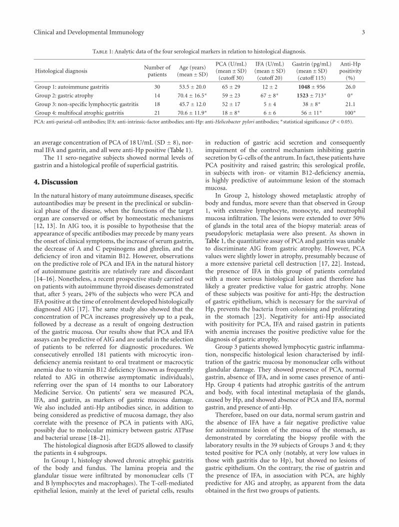
Serological analysis showed that Group 1 patients hadhigh concentrations of PCA (mean 65 U/mL), high gastrin(mean 1048 pg/mL), absence of IFA, and presence of anti-Hp antibodies in 26% of the cases; Group 2 patients hadan average concentration of PCA of 59 U/mL, gastrin of1523 pg/mL, positivity for IFA, and absence of anti-Hp;Group 3 patients had an average concentration of PCAof 52 U/mL, normal gastrin, absence of IFA, and presenceof anti-Hp in 21.1% of the cases; Group 4 patients had
Clinical and Developmental Immunology 3
Table 1: Analytic data of the four serological markers in relation to histological diagnosis.
Histological diagnosisNumber of
patientsAge (years)
(mean± SD)
PCA (U/mL)(mean± SD)
(cutoff 30)
IFA (U/mL)(mean± SD)
(cutoff 20)
Gastrin (pg/mL)(mean± SD)(cutoff 115)
Anti-Hppositivity
(%)
Group 1: autoimmune gastritis 30 53.5± 20.0 65± 29 12± 2 1048± 956 26.0
Group 2: gastric atrophy 14 70.4± 16.5∗ 59± 23 67± 8∗ 1523± 713∗ 0∗
Group 3: non-specific lymphocytic gastritis 18 45.7± 12.0 52± 17 5± 4 38± 8∗ 21.1
Group 4: multifocal atrophic gastritis 21 70.6± 11.9∗ 18± 8∗ 6± 6 56± 11∗ 100∗
PCA: anti-parietal-cell antibodies; IFA: anti-intrinsic-factor antibodies; anti-Hp: anti-Helicobacter pylori antibodies; ∗statistical significance (P < 0.05).
an average concentration of PCA of 18 U/mL (SD ± 8), nor-mal IFA and gastrin, and all were anti-Hp positive (Table 1).
The 11 sero-negative subjects showed normal levels ofgastrin and a histological profile of superficial gastritis.
4. Discussion
In the natural history of many autoimmune diseases, specificautoantibodies may be present in the preclinical or subclin-ical phase of the disease, when the functions of the targetorgan are conserved or offset by homeostatic mechanisms[12, 13]. In AIG too, it is possible to hypothesise that theappearance of specific antibodies may precede by many yearsthe onset of clinical symptoms, the increase of serum gastrin,the decrease of A and C pepsinogens and ghrelin, and thedeficiency of iron and vitamin B12. However, observationson the predictive role of PCA and IFA in the natural historyof autoimmune gastritis are relatively rare and discordant[14–16]. Nonetheless, a recent prospective study carried outon patients with autoimmune thyroid diseases demonstratedthat, after 5 years, 24% of the subjects who were PCA andIFA positive at the time of enrolment developed histologicallydiagnosed AIG [17]. The same study also showed that theconcentration of PCA increases progressively up to a peak,followed by a decrease as a result of ongoing destructionof the gastric mucosa. Our results show that PCA and IFAassays can be predictive of AIG and are useful in the selectionof patients to be referred for diagnostic procedures. Weconsecutively enrolled 181 patients with microcytic iron-deficiency anemia resistant to oral treatment or macrocyticanemia due to vitamin B12 deficiency (known as frequentlyrelated to AIG in otherwise asymptomatic individuals),referring over the span of 14 months to our LaboratoryMedicine Service. On patients’ sera we measured PCA,IFA, and gastrin, as markers of gastric mucosa damage.We also included anti-Hp antibodies since, in addition tobeing considered as predictive of mucosa damage, they alsocorrelate with the presence of PCA in patients with AIG,possibly due to molecular mimicry between gastric ATPaseand bacterial urease [18–21].
The histological diagnosis after EGDS allowed to classifythe patients in 4 subgroups.
In Group 1, histology showed chronic atrophic gastritisof the body and fundus. The lamina propria and theglandular tissue were infiltrated by mononuclear cells (Tand B lymphocytes and macrophages). The T-cell-mediatedepithelial lesion, mainly at the level of parietal cells, results
in reduction of gastric acid secretion and consequentlyimpairment of the control mechanism inhibiting gastrinsecretion by G-cells of the antrum. In fact, these patients havePCA positivity and raised gastrin; this serological profile,in subjects with iron- or vitamin B12-deficiency anemia,is highly predictive of autoimmune lesion of the stomachmucosa.
In Group 2, histology showed metaplastic atrophy ofbody and fundus, more severe than that observed in Group1, with extensive lymphocyte, monocyte, and neutrophilmucosa infiltration. The lesions were extended to over 50%of glands in the total area of the biopsy material: areas ofpseudopyloric metaplasia were also present. As shown inTable 1, the quantitative assay of PCA and gastrin was unableto discriminate AIG from gastric atrophy. However, PCAvalues were slightly lower in atrophy, presumably because ofa more extensive parietal cell destruction [17, 22]. Instead,the presence of IFA in this group of patients correlatedwith a more serious histological lesion and therefore haslikely a greater predictive value for gastric atrophy. Noneof these subjects was positive for anti-Hp; the destructionof gastric epithelium, which is necessary for the survival ofHp, prevents the bacteria from colonising and proliferatingin the stomach [23]. Negativity for anti-Hp associatedwith positivity for PCA, IFA and raised gastrin in patientswith anemia increases the positive predictive value for thediagnosis of gastric atrophy.
Group 3 patients showed lymphocytic gastric inflamma-tion, nonspecific histological lesion characterised by infil-tration of the gastric mucosa by mononuclear cells withoutglandular damage. They showed presence of PCA, normalgastrin, absence of IFA, and in some cases presence of anti-Hp. Group 4 patients had atrophic gastritis of the antrumand body, with focal intestinal metaplasia of the glands,caused by Hp, and showed absence of PCA and IFA, normalgastrin, and presence of anti-Hp.
Therefore, based on our data, normal serum gastrin andthe absence of IFA have a fair negative predictive valuefor autoimmune lesion of the mucosa of the stomach, asdemonstrated by correlating the biopsy profile with thelaboratory results in the 39 subjects of Groups 3 and 4; theytested positive for PCA only (notably, at very low values inthose with gastritis due to Hp), but showed no lesions ofgastric epithelium. On the contrary, the rise of gastrin andthe presence of IFA, in association with PCA, are highlypredictive for AIG and atrophy, as apparent from the dataobtained in the first two groups of patients.
4 Clinical and Developmental Immunology
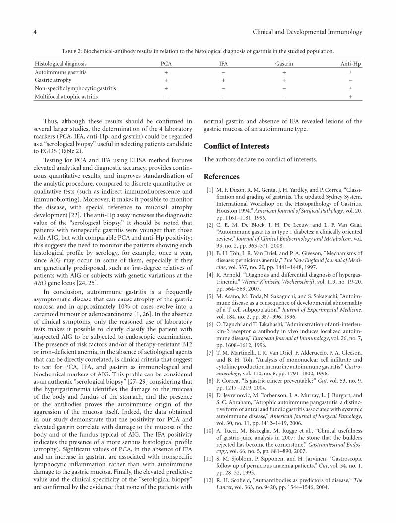
Table 2: Biochemical-antibody results in relation to the histological diagnosis of gastritis in the studied population.
Histological diagnosis PCA IFA Gastrin Anti-Hp
Autoimmune gastritis + − + ±Gastric atrophy + + + −Non-specific lymphocytic gastritis + − − ±Multifocal atrophic astritis − − − +
Thus, although these results should be confirmed inseveral larger studies, the determination of the 4 laboratorymarkers (PCA, IFA, anti-Hp, and gastrin) could be regardedas a “serological biopsy” useful in selecting patients candidateto EGDS (Table 2).
Testing for PCA and IFA using ELISA method featureselevated analytical and diagnostic accuracy, provides contin-uous quantitative results, and improves standardisation ofthe analytic procedure, compared to discrete quantitative orqualitative tests (such as indirect immunofluorescence andimmunoblotting). Moreover, it makes it possible to monitorthe disease, with special reference to mucosal atrophydevelopment [22]. The anti-Hp assay increases the diagnosticvalue of the “serological biopsy.” It should be noted thatpatients with nonspecific gastritis were younger than thosewith AIG, but with comparable PCA and anti-Hp positivity;this suggests the need to monitor the patients showing suchhistological profile by serology, for example, once a year,since AIG may occur in some of them, especially if theyare genetically predisposed, such as first-degree relatives ofpatients with AIG or subjects with genetic variations at theABO gene locus [24, 25].
In conclusion, autoimmune gastritis is a frequentlyasymptomatic disease that can cause atrophy of the gastricmucosa and in approximately 10% of cases evolve into acarcinoid tumour or adenocarcinoma [1, 26]. In the absenceof clinical symptoms, only the reasoned use of laboratorytests makes it possible to clearly classify the patient withsuspected AIG to be subjected to endoscopic examination.The presence of risk factors and/or of therapy-resistant B12or iron-deficient anemia, in the absence of aetiological agentsthat can be directly correlated, is clinical criteria that suggestto test for PCA, IFA, and gastrin as immunological andbiochemical markers of AIG. This profile can be consideredas an authentic “serological biopsy” [27–29] considering thatthe hypergastrinemia identifies the damage to the mucosaof the body and fundus of the stomach, and the presenceof the antibodies proves the autoimmune origin of theaggression of the mucosa itself. Indeed, the data obtainedin our study demonstrate that the positivity for PCA andelevated gastrin correlate with damage to the mucosa of thebody and of the fundus typical of AIG. The IFA positivityindicates the presence of a more serious histological profile(atrophy). Significant values of PCA, in the absence of IFAand an increase in gastrin, are associated with nonspecificlymphocytic inflammation rather than with autoimmunedamage to the gastric mucosa. Finally, the elevated predictivevalue and the clinical specificity of the “serological biopsy”are confirmed by the evidence that none of the patients with
normal gastrin and absence of IFA revealed lesions of thegastric mucosa of an autoimmune type.
Conflict of Interests
The authors declare no conflict of interests.
References
[1] M. F. Dixon, R. M. Genta, J. H. Yardley, and P. Correa, “Classi-fication and grading of gastritis. The updated Sydney System.International Workshop on the Histopathology of Gastritis,Houston 1994,” American Journal of Surgical Pathology, vol. 20,pp. 1161–1181, 1996.
[2] C. E. M. De Block, I. H. De Leeuw, and L. F. Van Gaal,“Autoimmune gastritis in type 1 diabetes: a clinically orientedreview,” Journal of Clinical Endocrinology and Metabolism, vol.93, no. 2, pp. 363–371, 2008.
[3] B. H. Toh, I. R. Van Driel, and P. A. Gleeson, “Mechanisms ofdisease: pernicious anemia,” The New England Journal of Medi-cine, vol. 337, no. 20, pp. 1441–1448, 1997.
[4] R. Arnold, “Diagnosis and differential diagnosis of hypergas-trinemia,” Wiener Klinische Wochenschrift, vol. 119, no. 19-20,pp. 564–569, 2007.
[5] M. Asano, M. Toda, N. Sakaguchi, and S. Sakaguchi, “Autoim-mune disease as a consequence of developmental abnormalityof a T cell subpopulation,” Journal of Experimental Medicine,vol. 184, no. 2, pp. 387–396, 1996.
[6] O. Taguchi and T. Takahashi, “Administration of anti-interleu-kin-2 receptor α antibody in vivo induces localized autoim-mune disease,” European Journal of Immunology, vol. 26, no. 7,pp. 1608–1612, 1996.
[7] T. M. Martinelli, I. R. Van Driel, F. Alderuccio, P. A. Gleeson,and B. H. Toh, “Analysis of mononuclear cell infiltrate andcytokine production in murine autoimmune gastritis,” Gastro-enterology, vol. 110, no. 6, pp. 1791–1802, 1996.
[8] P. Correa, “Is gastric cancer preventable?” Gut, vol. 53, no. 9,pp. 1217–1219, 2004.
[9] D. Jevremovic, M. Torbenson, J. A. Murray, L. J. Burgart, andS. C. Abraham, “Atrophic autoimmune pangastritis: a distinc-tive form of antral and fundic gastritis associated with systemicautoimmune disease,” American Journal of Surgical Pathology,vol. 30, no. 11, pp. 1412–1419, 2006.
[10] A. Tucci, M. Bisceglia, M. Rugge et al., “Clinical usefulnessof gastric-juice analysis in 2007: the stone that the buildersrejected has become the cornerstone,” Gastrointestinal Endos-copy, vol. 66, no. 5, pp. 881–890, 2007.
[11] S. M. Sjoblom, P. Sipponen, and H. Jarvinen, “Gastroscopicfollow up of pernicious anaemia patients,” Gut, vol. 34, no. 1,pp. 28–32, 1993.
[12] R. H. Scofield, “Autoantibodies as predictors of disease,” TheLancet, vol. 363, no. 9420, pp. 1544–1546, 2004.
Clinical and Developmental Immunology 5
[13] N. Bizzaro, R. Tozzoli, and Y. Shoenfeld, “Are we at a stage topredict autoimmune rheumatic diseases?” Arthritis and Rheu-matism, vol. 56, no. 6, pp. 1736–1744, 2007.
[14] D. Leslie, P. Lipsky, and A. L. Notkins, “Autoantibodies as pre-dictors of disease,” Journal of Clinical Investigation, vol. 108,no. 10, pp. 1417–1422, 2001.
[15] R. Tozzoli, “The diagnostic role of autoantibodies in the pre-diction of organ-specific autoimmune diseases,” ClinicalChemistry and Laboratory Medicine, vol. 46, no. 5, pp. 577–587, 2008.
[16] D. Villalta, R. Tozzoli, E. Tonutti, and N. Bizzaro, “The labo-ratory approach to the diagnosis of autoimmune diseases: isit time to change?” Autoimmunity Reviews, vol. 6, no. 6, pp.359–365, 2007.
[17] R. Tozzoli, G. Kodermaz, A. R. Perosa et al., “Autoantibodiesto parietal cells as predictors of atrophic body gastritis: a five-year prospective study in patients with autoimmune thyroiddiseases,” Autoimmunity Reviews, vol. 10, no. 2, pp. 80–83,2010.
[18] C. Hershko, A. Ronson, M. Souroujon, I. Maschler, J. Heyd,and J. Patz, “Variable hematologic presentation of autoim-mune gastritis: age-related progression from iron deficiencyto cobalamin depletion,” Blood, vol. 107, no. 4, pp. 1673–1679,2006.
[19] A. Amedei, M. P. Bergman, B. J. Appelmelk et al., “Molecularmimicry between helicobacter pylori antigens and H+ ,K+-adenosine triphosphatase in human gastric autoimmunity,”Journal of Experimental Medicine, vol. 198, no. 8, pp. 1147–1156, 2003.
[20] M. M. D’Elios, B. J. Appelmelk, A. Amedei, M. P. Bergman,and G. Del Prete, “Gastric autoimmunity: the role of Heli-cobacter pylori and molecular mimicry,” Trends in MolecularMedicine, vol. 10, no. 7, pp. 316–323, 2004.
[21] F. Presotto, B. Sabini, A. Cecchetto et al., “Helicobacter pyloriinfection and gastric autoimmune diseases: is there a link?”Helicobacter, vol. 8, no. 6, pp. 578–584, 2003.
[22] I. R. van Driel, A. G. Baxter, K. L. Laurie et al., “Immunopath-ogenesis, loss of T cell tolerance and genetics of autoimmunegastritis,” Autoimmunity Reviews, vol. 1, no. 5, pp. 290–297,2002.
[23] A. Oksanen, P. Sipponen, R. Karttunen et al., “Atrophic gastri-tis and Helicobacter pylori infection in outpatients referred forgastroscopy,” Gut, vol. 46, no. 4, pp. 460–463, 2000.
[24] K. Varis, T. Ihamaki, and M. Harkonen, “Gastric morphol-ogy, function, and immunology in first-degree relatives ofprobands with pernicious anemia and controls,” ScandinavianJournal of Gastroenterology, vol. 14, no. 2, pp. 129–139, 1979.
[25] M. Nakao, K. Matsuo, H. Ito et al., “ABO genotype and the riskof gastric cancer, atrophic gastritis, and Helicobacter pyloriinfection,” Cancer Epidemiology Biomarkers and Prevention,vol. 20, no. 8, pp. 1665–1672, 2011.
[26] A. Antico, “La gastrite autoimmune,” RIMeL/IJLaM, vol. 4, pp.125–133, 2008 (Italian).
[27] A. Korstanje, G. den Hartog, I. Biemond, and C. B. H. W.Lamers, “The serological gastric biopsy: a non-endoscopicaldiagnostic approach in management of the dyspeptic patient:significance for primary care based on a survey of the liter-ature,” Scandinavian Journal of Gastroenterology, Supplement,vol. 37, no. 236, pp. 22–26, 2002.
[28] T. Storskrubb, P. Aro, J. Ronkainen et al., “Serum biomarkersprovide an accurate method for diagnosis of atrophic gastritisin a general population: the Kalixanda study,” ScandinavianJournal of Gastroenterology, vol. 43, no. 12, pp. 1448–1455,2008.
[29] M. D. Burkitt, A. Varro, and D. M. Pritchard, “Importance ofgastrin in the pathogenesis and treament of gastric tumors,”World Journal of Gastroenterology, vol. 15, no. 1, pp. 1–16,2009.

Submit your manuscripts athttp://www.hindawi.com
Stem CellsInternational
Hindawi Publishing Corporationhttp://www.hindawi.com Volume 2014
Hindawi Publishing Corporationhttp://www.hindawi.com Volume 2014
MEDIATORSINFLAMMATION
of
Hindawi Publishing Corporationhttp://www.hindawi.com Volume 2014
Behavioural Neurology
EndocrinologyInternational Journal of
Hindawi Publishing Corporationhttp://www.hindawi.com Volume 2014
Hindawi Publishing Corporationhttp://www.hindawi.com Volume 2014
Disease Markers
Hindawi Publishing Corporationhttp://www.hindawi.com Volume 2014
BioMed Research International
OncologyJournal of
Hindawi Publishing Corporationhttp://www.hindawi.com Volume 2014
Hindawi Publishing Corporationhttp://www.hindawi.com Volume 2014
Oxidative Medicine and Cellular Longevity
Hindawi Publishing Corporationhttp://www.hindawi.com Volume 2014
PPAR Research
The Scientific World JournalHindawi Publishing Corporation http://www.hindawi.com Volume 2014
Immunology ResearchHindawi Publishing Corporationhttp://www.hindawi.com Volume 2014
Journal of
ObesityJournal of
Hindawi Publishing Corporationhttp://www.hindawi.com Volume 2014
Hindawi Publishing Corporationhttp://www.hindawi.com Volume 2014
Computational and Mathematical Methods in Medicine
OphthalmologyJournal of
Hindawi Publishing Corporationhttp://www.hindawi.com Volume 2014
Diabetes ResearchJournal of
Hindawi Publishing Corporationhttp://www.hindawi.com Volume 2014
Hindawi Publishing Corporationhttp://www.hindawi.com Volume 2014
Research and TreatmentAIDS
Hindawi Publishing Corporationhttp://www.hindawi.com Volume 2014
Gastroenterology Research and Practice
Hindawi Publishing Corporationhttp://www.hindawi.com Volume 2014
Parkinson’s Disease
Evidence-Based Complementary and Alternative Medicine
Volume 2014Hindawi Publishing Corporationhttp://www.hindawi.com


![Capitolo 3 IMMUNOLOGIA E IMMUNOPATOLOGIAfupress.com/Archivio/pdf/2470.pdf · Capitolo 3 IMMUNOLOGIA E IMMUNOPATOLOGIA 'H¿QL]LRQH L’immunità è stata definita come la capacità](https://img.pdfslide.net/doc/110x75/5f71d07d27cdc829de282033/capitolo-3-immunologia-e-im-capitolo-3-immunologia-e-immunopatologia-hqllrqh.jpg)