Embed Size (px)
Citation preview
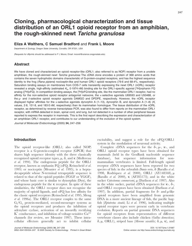
Cloning, pharmacological characterization and tissuedistribution of an ORL1 opioid receptor from an amphibian,the rough-skinned newt Taricha granulosa
Eliza A Walthers, C Samuel Bradford and Frank L MooreDepartment of Zoology, Oregon State University, Corvallis, OR 97331, USA
(Requests for offprints should be addressed to F L Moore; Email: [email protected])
Abstract
We have cloned and characterized an opioid receptor-like (ORL1; also referred to as NOP) receptor from a urodeleamphibian, the rough-skinned newt Taricha granulosa The cDNA clone encodes a protein of 368 amino acids thatcontains the seven hydrophobic domains characteristic of G-protein-coupled receptors, and has the highest sequenceidentity to the frog (Rana pipiens) nociceptin-like and human ORL1 opioid receptors (79·6 and 68·4%, respectively).Saturation binding assays on membranes from COS-7 cells transiently expressing the newt ORL1 (nORL) receptorrevealed a single, high-affinity (estimated Kd, 0·1974 nM) binding site for the ORL1-specific agonist [3H]orphanin FQanalog ([3H]oFQ). In competition binding assays, the [3H]oFQ-binding site, like the mammalian ORL1 receptor, had noaffinity for the non-selective opioid receptor antagonist naloxone, the �-selective agonists U69593 and U50488, orthe µ- and �-selective opioid receptor agonists DAMGO and DPDPE, respectively. However, the nORL receptordisplayed higher affinities for the �-selective agonists dynorphin A (1–13), dynorphin B, and dynorphin A (1–8) (Ki
values, 2·8, 151·8, and 183·0 nM, respectively) than its mammalian homologue. The tissue distribution of the nORLreceptor, as determined by reverse transcriptase PCR, was also found to differ from reports on the mammalian ORL1receptor, with mRNA detected in brain, spinal cord, and lung, but not detected in a number of other peripheral tissuesreported to express the receptor in mammals. This is the first report describing the expression and characterization ofan amphibian ORL1 receptor, and contributes to our understanding of the evolution of the opioid system.
Journal of Molecular Endocrinology (2005) 34, 247–256
Introduction
The opioid receptor-like (ORL1; also called NOP)receptor is a G-protein-coupled receptor (GPCR) thatshares high sequence identity with the three classicallyrecognized opioid receptor types, µ, �, and � (Mollereauet al. 1994). The endogenous peptide for the ORL1receptor, known as orphanin FQ (oFQ; Reinscheid et al.1995) or nociceptin (Meunier et al. 1995), is a hepta-decapeptide whose N-terminal tetrapeptide sequence isrelated to that of the opioid peptides (FGGF or YGGF),and whose basic core is similar to dynorphin (Dyn), theendogenous peptide for the � receptor. In spite of thesesimilarities, the ORL1 receptor does not recognize themajority of opioid ligands, and oFQ has low affinity forthe classic opioid receptors (Reinscheid et al. 1995, Menget al. 1996a). The ORL1 receptor couples to the sameGi/Go protein-mediated, second-messenger systems asthe opioid receptors and produces the inhibition ofadenylate cyclase, activation of an inwardly rectifyingK+ conductance, and inhibition of voltage-sensitive Ca2+
channels (for review, see Meunier 1997). These intra-cellular effectors generally act to inhibit cellular
excitability, and suggest a role for the oFQ/ORL1system in the modulation of neuronal activity.
Complete cDNA sequences for the �-, µ-, �-, andORL1 opioid receptor types have been obtained formammals (held in the GenBank nucleotide sequencedatabase), but sequence information for non-mammalian vertebrates is limited. Full-length opioidreceptor cDNA sequences have been reported for twospecies of fish: in the zebrafish Danio rerio, � (Barallo et al.1998, Rodriquez et al. 2000), ORL1 (AY148348), µ(Barallo et al. 2000), � (AF285173); and in the whitesucker Catostomus commersoni, µ (Darlison et al. 1997). Alsoin the white sucker, partial cDNA sequences for �-, �-,and ORL1 receptors have been obtained (Darlison et al.1997). In addition, partial fragments for �- and µ-likeopioid receptors have been amplified from genomicDNA in a more ancient lineage of fish, the pacific hag-fish (Eptatretus stoutii; Li et al. 1996), indicating multipleopioid receptor types were present early in vertebrateevolution. Reports on partial genomic DNA sequencesfor opioid receptors from representatives of differentvertebrate classes also include chicken (Gallus domesticus;�, µ, ORL1), striped bass (Morone saxatilis; �, µ, �), and
247
Journal of Molecular Endocrinology (2005) 34, 247–2560952–5041/05/034–247 © 2005 Society for Endocrinology Printed in Great Britain
DOI: 10.1677/jme.1.01687Online version via http://www.endocrinology-journals.org
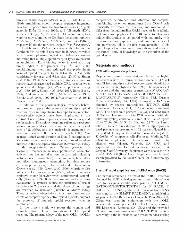
thresher shark (Alopias vulpinus; �, µ, ORL1; Li et al.1996). Amphibian opioid receptor sequence fragmentshave been reported from bullfrog (Rana catesbiana; �, µ, �)genomic DNA (Li et al. 1996), and full-length cDNAsequences for µ-, �-, �-, and ORL1 opioid receptorswere recently submitted to GenBank (accession numbersAF530571, AF530572, AF530573, and AY434690,respectively) for the northern leopard frog (Rana pipiens).
The definitive cDNA sequences recently submitted toGenBank for the opioid receptors in R. pipiens correlatewith previous pharmacological and behavioral studiesindicating that multiple opioid receptor types are presentin amphibians. Early binding assays in toad and frogbrains indicated the presence of µ-, �-, and �-likeopioid-binding sites, and estimated the predominantform of opioid receptor to be �-like (60–70%), withconsiderably fewer µ- and �-like sites (20–30%; Simonet al. 1982, 1984). Since these early studies, numerousreports have characterized multiple ORL receptor types(µ, �, �) and subtypes (�1, �2) in amphibians (Ruegget al. 1980, 1981, Simon et al. 1985, 1987, Borsodi et al.1986, Makimura et al. 1988, Mollereau et al. 1988,Benyhe et al. 1990, 1992 1994, Wollemann et al. 1994,Newman et al. 2002).
In addition to the pharmacological evidence, behav-ioral studies support the presence of multiple opioidreceptor types in amphibians. In amphibians, receptor-type-selective opioids have been implicated in thecontrol of nociceptive responses, locomotor activity, andsex behaviors. The opiate morphine sulfate (a µ-receptoragonist) is an analgesic when injected into the spinalcord of R. pipiens, and the analgesia is attenuated bynaloxone (Pezalla 1983, Stevens & Pezalla 1983). Alsoin frogs, spinal administration of Dyn, �-endorphin, orMet-enkephalin produces a potent, dose-dependentincrease in the nociceptive threshold (Stevens et al. 1987).In the rough-skinned newt, Taricha granulosa, the�-agonist bremazocine reduces spontaneous locomotoractivity, but has no affect on corticotropin-releasingfactor-induced locomotion; whereas, morphine doesnot affect spontaneous locomotion, but does reducecorticotropin-releasing factor-induced locomotion(Deviche et al. 1989, Lowry et al. 1990). Morphine alsoinfluences locomotion in R. pipiens, where it inducesexplosive motor behavior when administered systemi-cally (Pezalla 1983). Both bremazocine and ethylketo-cyclazocine (� agonists) reduce the incidence of sexualbehaviors in T. granulosa, and the effects of both drugsare reversed by naloxone (Deviche & Moore 1987).These behavioral observations, which are based on theeffects of receptor-type-selective opioid agonists, supportthe presence of multiple opioid receptor types inamphibians.
In the present study we report the cloning andcharacterization of an amphibian ORL1 opioidreceptor. The pharmacology of the newt ORL1 (nORL)
receptor was determined using saturation and competi-tion binding assays on membranes from COS-7 cellstransiently expressing the receptor, and was found todiffer from the mammalian ORL1 receptor in its affinityfor Dyn-derived peptides. The nORL receptor also has aunique distribution as compared with mammals, withexpression in brain, spinal cord, and lung. To the best ofour knowledge, this is the first characterization of thistype of opioid receptor in an amphibian, and adds tothe current body of knowledge on the evolution of theopioid system.
Materials and methods
PCR with degenerate primers
Degenerate primers were designed based on highlyconserved regions in transmembrane domains (TMs) Iand III from multiple opioid receptor types identified indiverse vertebrate phyla (Li et al. 1996). The sequences ofthe sense and the antisense primers were 5�-ACCAAYATYTACATHTTYAA-3� and 5�-GGTRAACATRTTRTAGTARTC-3�, respectively (Gibco BRL CustomPrimers, Carlsbad, CA, USA). Template cDNA wasobtained by reverse transcriptase (RT)-PCR (MBIFermentas, Hanover, MD, USA) on newt brain totalRNA. The degenerate primers (0·5 µM) and newt braincDNA template were used in PCR reactions with thefollowing cycling conditions: 2 min at 94 �C, 35 cyclesof 94 �C for 30s, 50 �C for 30s, and 72 �C for 1 min,followed by a 7 min extension at 72 �C. Appropriatelysized products (approximately 152 bp) were ligated intothe pGEM T-Easy vector and transformed into JM109Escherichia coli competent cells (Promega, Madison, WI,USA) for amplification. Plasmids were purified byalkaline lysis (Qiagen, Valencia, CA, USA) andsequenced by the Central Services Laboratory atOregon State University. Sequences were analyzed witha BLAST-N 2·0 (Basic Local Alignment Search Tool)search provided by National Center for BiotechnologyInformation.
3* and 5* rapid amplification of cDNA ends (RACE)
The partial sequence (152 bp) of the nORL1 receptorobtained by PCR with degenerate primers (above) wasused to design a specific sense primer (5�-CTGGCTGATGCGCTGGTGCTCGT-3�) for 3� RACE. 3�RACE-ready cDNA, synthesized from newt brain RNAaccording to the SMART RACE cDNA amplificationkit protocol (BD Biosciences Clontech, Palo Alto, CA,USA), was used in conjunction with the nORLgene-specific sense primer (One Trick Pony; RansomHill Bioscience, Ramona, CA, USA) and the providedClontech antisense primer in a 3� RACE PCR reactionaccording to the kit protocol and recommended cycling
E A WALTHERS and others · Characterization of an amphibian ORL1 receptor248
www.endocrinology-journals.orgJournal of Molecular Endocrinology (2005) 34, 247–256
conditions. 5� RACE-ready cDNA was synthesized fromnewt brain RNA according to the Ambion RACE RLMkit protocol (Ambion, Austin, TX, USA). 5� RACE PCRreactions were carried out using the kit reagents andprotocol, the provided Outer RACE sense primer, and a5� RACE gene-specific antisense primer (5�-TTGGCTTTGTGTGGTGTACGGATGT-3�), with the followingcycling conditions: 94 �C for 3 min, four cycles of 94 �Cfor 45 s, 62 �C for 45 s, and 72 �C for 1 min, 34 cycles of94 �C for 30 s, 62 �C for 30 s, and 72 �C for 1 min,followed by 72 �C for 6 min. The RACE PCR productswere subcloned and sequenced as described above.Overlapping the 5� and 3� RACE product sequencesproduced a contig that contained the entire nORL1receptor sequence. The receptor was subsequentlyPCR-amplified for sequence confirmation three separ-ate times with primers designed at the ends of thecoding sequence and a proof-reading polymerase (Pfx;Invitrogen, Carlsbad, CA, USA).
Tissue distribution of the nORL1 receptor byRT-PCR
Six rough-skinned newts were cryoanaesthetized andrapidly decapitated. The newts were then dissected, andthe following organs were collected and frozenimmediately in liquid nitrogen: brain, spinal cord,spleen, small intestine, heart, lung, liver, sperm duct,bladder, and kidney. The tissues were stored at –80 �C.All experiments were performed in accordance with theNational Institutes of Health Guide for the Care andUse of Laboratory Animals and had been approved bythe Oregon State University Institutional Animal Careand Use Commitee. The reagents and protocol providedwith RNA STAT-60 (Tel-Test, Friendswood, TX, USA)were used to collect total RNA from the tissues. Thepurified RNA was reverse-transcribed into cDNA usingan oligo-dT primer and the Gibco Superscript Systemfor RT-PCR First-Strand Synthesis kit and protocol(Invitrogen). Up to 5 µg total RNA from each tissue wasused per reverse-transcription reaction. The resultantcDNA was stored at –20 �C.
Gene-specific primers were designed for PCR basedon the nORL receptor sequence. The sense primer(5�-GTGATGGGGTCTGCTGAAAT-3�) correspondsto amino acid residues 184–190 at the start of theputative second extracellular loop of the nORL receptor,and the antisense primer (5�-CACTGCACCAGCACAAAGAT-3�) corresponds to amino acid residues278–284 at the end of putative TMVI. PCR reactions(20 µl) were set up with the specific primers (0·5 µM) andthe cDNA (10–150 ng) from the collected tissues. Thequality and quantity of the cDNA was controlled for inPCR reactions conducted simultaneously using �-actinprimers. The following cycling conditions were used:94 �C for 2 min, 35 cycles of 94 �C for 10 s, 60 �C for
30 s, and 72 �C for 1 min, followed by 72 �C for 5 min.The PCR products were examined by 1·5% agarose gelelectrophoresis and ethidium bromide staining for thenORL-specific amplicon (303 bp) or the actin-specificamplicon (150 bp).
Transfection
The full-length nORL receptor insert was subclonedinto the mammalian expression vector pcDNA3·1/V5-His-TOPO (Invitrogen); individual clones were analyzedfor correct orientation with respect to the cytomegalo-virus promoter. COS-7 cells (ATCC, Manassas, VA,USA) were cultured in 10 cm tissue-culture plates(Falcon catalog no. 353003) at 37 �C under 5% CO2 inDulbecco’s modified Eagle’s medium (Mediatech Cell-gro; Fisher, Pittsburgh, PA, USA) supplemented with2 mM -glutamine (Invitrogen) and 10% fetal bovineserum (Hyclone, Logan, UT, USA). Transient expres-sion was achieved by transfecting plates (90–95%confluency) using Lipofectamine 2000 (Invitrogen)diluted with Opti-MEM medium (Invitrogen). 6 h aftertransfection, the lipofection mixture was aspirated andreplaced with fresh growth medium.
Cell harvesting and membrane preparation
At 48 h after transfection, cells were washed twice withroom temperature PBS and harvested under homogeni-zation buffer (2 ml/plate) using a cell scraper. Suspendedcells were homogenized in chilled buffer (25 mM Hepes,100 µM PMSF, 100 µg/ml trypsin inhibitor, 0·7 µg/mlleupeptin, and 100 µg/ml bacitracin) using a chilledDounce homogenizer. Homogenates were centrifugedfor 10 min at 45 000 g (4 �C). Pellets were resuspendedin homogenization buffer (1 ml/plate) and centrifugeda second time. Cell membrane pellets were resuspendedin binding buffer (250 µl/plate) containing MgCl2 (seebelow), snap-frozen in liquid N2, and stored at �80 �Cuntil use. In the course of this study, multiple trans-fections were performed; however, tissues from differenttransfections were not pooled, and in a given assay alltissues used were obtained from a single transfection.The concentration of total protein in membranepreparations was determined using a colorimetricprotein assay (Pierce, Rockford, IL, USA).
Binding assays
Saturation and competition binding assays wereperformed in binding buffer consisting of 25 mM Hepes(pH 7·45) containing 10 mM MgCl2, 100 µM PMSF,trypsin inhibitor (100 µg/ml), leupeptin (0·7 µg/ml), andbacitracin (100 µg/ml). Frozen membrane pellets werethawed on ice and diluted with binding buffer to achievea final protein concentration of 50 µg per binding
Characterization of an amphibian ORL1 receptor · E A WALTHERS and others 249
www.endocrinology-journals.org Journal of Molecular Endocrinology (2005) 34, 247–256
reaction. For saturation experiments, variable concen-trations of [3H]oFQ analog (102 Ci/mmol; MultiplePeptide Systems, San Diego, CA, USA) were used inthe presence of 10 µM nor-BNI (Tocris, Ballwin,MO, USA). Competition binding experiments wereperformed using 1 nM [3H]oFQ analog and variousdoses (10�12-10�5 M) of non-radioactive competitors.Porcine Dyn A (1–8), porcine Dyn A (1–13) amide, andporcine Dyn B were purchased from Phoenix Pharma-ceuticals (Belmont, CA, USA); U50488, U69593,naloxone and [D-Ala2,NMe-Phe4,Gly-ol5]-enkephalinDAGO (DAMGO) were purchased from Tocris;[D-Pen2,D-Pen5]-enkephalin (DPDPE) is a product ofMultiple Peptide Systems. Non-specific binding wasdefined as radioactivity remaining bound in the presenceof 10 µM non-radioactive nociceptin (TOCRIS). Tripli-cate samples were run at each dose for both total andnon-specific binding. Assays were conducted at 4 �C for4 h with gentle mixing (80 r.p.m. on an orbital shaker).At the end of the incubation period, bound and freeligands were separated by rapid filtration over GF/Cfilters (pre-soaked with 0·25% polyethyleneimine for30 min) under vacuum using a Brandel cell harvester(Brandel, Gaithersburg, MA, USA). Filters were washedtwice with 4 ml of chilled (4 �C) Hepes/MgCl2 (pH 7·45)buffer. Radioactivity in the filters was determined byliquid-scintillation counting on a Beckman LS 6500scintillation counter (GMI, Albertville, MN, USA).Binding data were analyzed using GraphPad Prismsoftware (version 3·0; GraphPad, San Diego, CA, USA).
Results
Sequence analysis
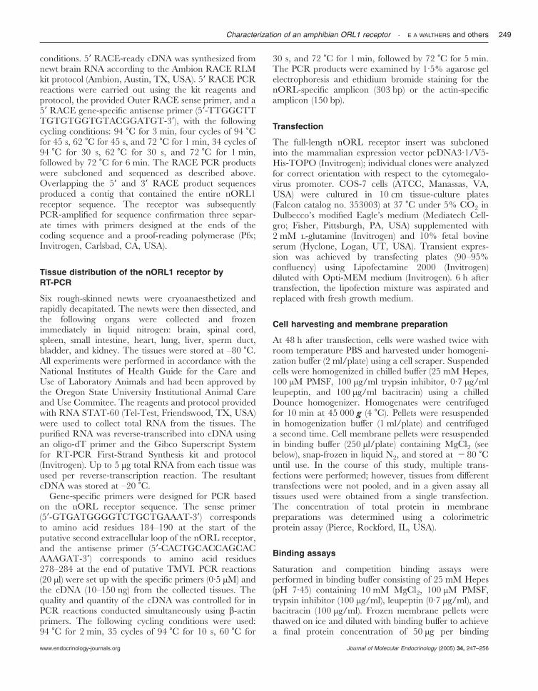
Figure 1 shows the nucleotide coding region and itsdeduced amino acid sequence for the clone isolatedby RT-PCR and 5� and 3� RACE from newt brain(GenBank accession no. AY728087). The cDNAencodes a protein that is 368 amino acids long andcontains seven hydrophobic regions that share identitywith the TMs of GPCRs. The deduced amino acidsequence also contains a number of putative consensussites for post-translational modifications common tocharacterized GPCRs, including three asparagineresidues within the N-terminal domain (glycosylationsites), two cysteine residues in the intracellularC-terminal domain (palmitoylation sites), and a cysteineresidue in extracellular loops 1 and 2 (disulfide bridge).
Of the characterized GPCRs, the newt clone sharesthe highest homology with members of the opioidreceptor family, and shares the greatest amino acidsequence identity with the ORL1 type of opioid receptor(64·2–79·6%), compared with � (54·3–56%), � (51·7–53·6%), and µ (51·2–53·6%). When compared with thehuman opioid receptor types (ORL1, �, �, µ), there is
high sequence identity in the TM regions (66–77%) andthe intracellular loops (70–81%). However, the identitybetween the newt clone and the human ORL1 receptorin the extracellular loops (60%) is definitively higherthan its identity to µ, �, and � in these regions (28–37%).Furthermore, the second extracellular loop (EL2) of thenewt receptor contains a high number of acidic residues,a characteristic shared only by the ORL1 and�-receptor types.
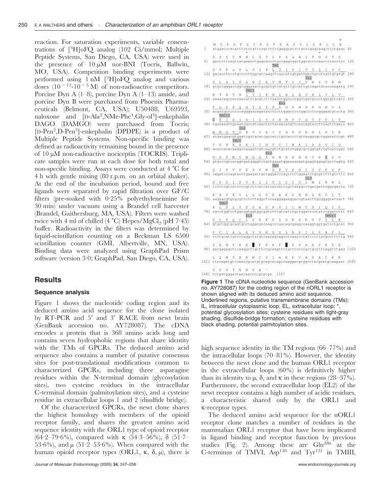
The deduced amino acid sequence for the nORL1receptor clone matches a number of residues in themammalian ORL1 receptor that have been implicatedin ligand binding and receptor function by previousstudies (Fig. 2). Among these are Gln286 at theC-terminus of TMVI, Asp130 and Tyr131 in TMIII,
Figure 1 The cDNA nucleotide sequence (GenBank accessionno. AY728087) for the coding region of the nORL1 receptor isshown aligned with its deduced amino acid sequence.Underlined regions, putative transmembrane domains (TMs);IL, intracellular cytoplasmic loop; EL, extracellular loop; *,potential glycosylation sites; cysteine residues with light-grayshading, disulfide-bridge formation; cysteine residues withblack shading, potential palmitoylation sites.
E A WALTHERS and others · Characterization of an amphibian ORL1 receptor250
www.endocrinology-journals.orgJournal of Molecular Endocrinology (2005) 34, 247–256
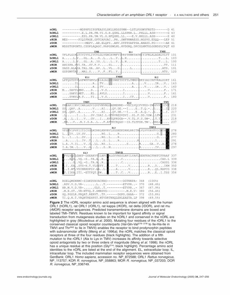
Figure 2 The nORL receptor amino acid sequence is shown aligned with the humanORL1 (hORL1), rat ORL1 (rORL1), rat kappa (rKOR), rat delta (rDOR), and rat mu(rMOR) receptor sequences. Predicted transmembrane domains are boxed andlabeled TMI–TMVII. Residues known to be important for ligand affinity or signaltransduction from mutagenesis studies on the hORL1 and conserved in the nORL arehighlighted in gray (Mouledous et al. 2000). Mutating four residues of the rORL1 to theconserved classical opioid receptor counterparts (Val-Gln-Val276–278 to Ile-His-Ile inTMVI and Thr302 to Ile in TMVII) enables the receptor to bind prodynorphin peptideswith subnanomolar affinity (Meng et al. 1996a); the nORL matches the classical opioidreceptors at three of the four residues (black highlights). The addition of a fifthmutation to the rORL1 (Ala to Lys in TMV) increases its affinity towards selectiveopioid antagonists by two or three orders of magnitude (Meng et al. 1998); the nORLhas a unique residue at this position (Gly214; black highlight). Percentage amino acididentities to the nORL are listed at the end of the alignment. EL, extracellular loop; IL,intracellular loop. The included mammalian receptor sequences were obtained fromGenBank: ORL1 Homo sapiens, accession no. NP_872588; ORL1 Rattus norvegicus,NP_113757; KOR R. norvegicus, NP_058863; MOR R. norvegicus, NP_037203; DORR. norvegicus, NP_036749.
Characterization of an amphibian ORL1 receptor · E A WALTHERS and others 251
www.endocrinology-journals.org Journal of Molecular Endocrinology (2005) 34, 247–256
Phe220 and Phe224 in TMV, and Trp276 in TMVI(human ORL1 numbering; Mouledous et al. 2000). Also,the newt receptor shares identity with the classic opioidreceptors at three residues (Ile277, Ile279, and Ile303; newtnumbering) shown to be important in the recognition of� ligands that are not conserved in the mammalianORL1 receptor (Meng et al. 1996a, 1998).
nORL receptor distribution
The expression of the nORL receptor in various tissueswas determined by RT-PCR on newt brain, spinal cord,spleen, small intestine, heart, lung, liver, sperm duct,bladder, and kidney. The cDNA generated from thetissues was tested in PCR reactions using nORLreceptor-specific primers. Appropriately sized amplicons(303 bp) were produced in reactions containing cDNAfrom brain, spinal cord, and lung (Fig. 3). Negativecontrols were reactions in which water was substitutedfor template cDNA. The cDNA from each tissue wasalso tested in PCR reactions with �-actin primers toensure that the template was of sufficient quality andquantity to amplify. The �-actin primers successfullyamplified the cDNA from each tissue (results not shown).
Characterization of the cloned receptor transientlyexpressed in COS-7 cells
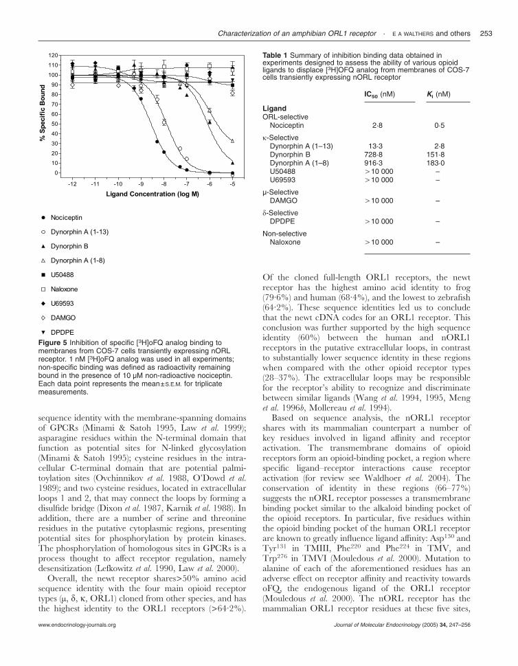
Saturation binding of [3H]oFQ analog, an ORL1-specific agonist, to membranes of transfected COS-7cells revealed the presence of a single, high-affinitybinding site with an estimated Kd value of 0·1974 nM(Fig. 4). At a dose equivalent to the estimated Kd for[3H]oFQ, specific binding was 94·5% of total binding.No specific binding was detected in membranesprepared from untransfected (control) cells. In competi-tion experiments designed to assess the ability of variousopioid ligands to displace [3H]oFQ, the � opioidreceptor-selective agonist Dyn A (1–13) had high affinity
(Ki, 2·76 nM), and the � agonists Dyn B and Dyn A (1–8)displayed sub-micromolar affinities (Ki, 151·81 and183·03 nM, respectively). In contrast, the �-selectiveagonists U50488 and U69593, the non-selective opioidreceptor antagonist naloxone, the µ-selective agonistDAMGO, and the �-selective agonist DPDPE, didnot compete for the [3H]oFQ-binding site (Fig. 5 andTable 1).
Discussion
The present study reports the molecular cloning andpharmacological characterization of an amphibianGPCR. The results indicate that the GPCR cloned fromnewt brain is the homologue of the mammalian ORL1opioid receptor. This conclusion was reached based onsequence analysis and binding studies with an ORL1-selective agonist, [3H]oFQ. To the best of ourknowledge, this is the first reported characterization ofan ORL1 opioid receptor from an amphibian.
Initial BLAST searches on the nucleotide anddeduced amino acid sequence of the newt cloneindicated it belonged to the GPCR superfamily. Thefollowing characteristics of the deduced amino acidsequence supported the GPCR classification: thepresence of seven hydrophobic regions that share
Figure 3 Distribution of the nORL receptor as determined byRT-PCR. The nORL receptor-specific amplicon (303 bp) wasdetected in the brain, spinal cord, and lung. +, Positive control;−, no-template control.
Figure 4 Saturation analysis of [3H]oFQ analog binding tomembranes isolated from COS-7 cells transiently expressingnORL receptor. Non-specific binding for each ligand dose wasdetermined in the presence of 10 µM nor-BNI. At a doseequivalent to the estimated Kd for oFQ analog, specific bindingwas 94·5% of total binding. Each data point represents themean±S.E.M. from triplicate measurements. The inset showsScatchard analysis.
E A WALTHERS and others · Characterization of an amphibian ORL1 receptor252
www.endocrinology-journals.orgJournal of Molecular Endocrinology (2005) 34, 247–256
sequence identity with the membrane-spanning domainsof GPCRs (Minami & Satoh 1995, Law et al. 1999);asparagine residues within the N-terminal domain thatfunction as potential sites for N-linked glycosylation(Minami & Satoh 1995); cysteine residues in the intra-cellular C-terminal domain that are potential palmi-toylation sites (Ovchinnikov et al. 1988, O’Dowd et al.1989); and two cysteine residues, located in extracellularloops 1 and 2, that may connect the loops by forming adisulfide bridge (Dixon et al. 1987, Karnik et al. 1988). Inaddition, there are a number of serine and threonineresidues in the putative cytoplasmic regions, presentingpotential sites for phosphorylation by protein kinases.The phosphorylation of homologous sites in GPCRs is aprocess thought to affect receptor regulation, namelydesensitization (Lefkowitz et al. 1990, Law et al. 2000).
Overall, the newt receptor shares>50% amino acidsequence identity with the four main opioid receptortypes (µ, �, �, ORL1) cloned from other species, and hasthe highest identity to the ORL1 receptors (>64·2%).
Of the cloned full-length ORL1 receptors, the newtreceptor has the highest amino acid identity to frog(79·6%) and human (68·4%), and the lowest to zebrafish(64·2%). These sequence identities led us to concludethat the newt cDNA codes for an ORL1 receptor. Thisconclusion was further supported by the high sequenceidentity (60%) between the human and nORL1receptors in the putative extracellular loops, in contrastto substantially lower sequence identity in these regionswhen compared with the other opioid receptor types(28–37%). The extracellular loops may be responsiblefor the receptor’s ability to recognize and discriminatebetween similar ligands (Wang et al. 1994, 1995, Menget al. 1996b, Mollereau et al. 1994).
Based on sequence analysis, the nORL1 receptorshares with its mammalian counterpart a number ofkey residues involved in ligand affinity and receptoractivation. The transmembrane domains of opioidreceptors form an opioid-binding pocket, a region wherespecific ligand–receptor interactions cause receptoractivation (for review see Waldhoer et al. 2004). Theconservation of identity in these regions (66–77%)suggests the nORL receptor possesses a transmembranebinding pocket similar to the alkaloid binding pocket ofthe opioid receptors. In particular, five residues withinthe opioid binding pocket of the human ORL1 receptorare known to greatly influence ligand affinity: Asp130 andTyr131 in TMIII, Phe220 and Phe224 in TMV, andTrp276 in TMVI (Mouledous et al. 2000). Mutation toalanine of each of the aforementioned residues has anadverse effect on receptor affinity and reactivity towardsoFQ, the endogenous ligand of the ORL1 receptor(Mouledous et al. 2000). The nORL receptor has themammalian ORL1 receptor residues at these five sites,
Figure 5 Inhibition of specific [3H]oFQ analog binding tomembranes from COS-7 cells transiently expressing nORLreceptor. 1 nM [3H]oFQ analog was used in all experiments;non-specific binding was defined as radioactivity remainingbound in the presence of 10 µM non-radioactive nociceptin.Each data point represents the mean±S.E.M. for triplicatemeasurements.
Table 1 Summary of inhibition binding data obtained inexperiments designed to assess the ability of various opioidligands to displace [3H]OFQ analog from membranes of COS-7cells transiently expressing nORL receptor
IC50 (nM) Ki (nM)
LigandORL-selective
Nociceptin 2·8 0·5
�-SelectiveDynorphin A (1–13) 13·3 2·8Dynorphin B 728·8 151·8Dynorphin A (1–8) 916·3 183·0U50488 .10 000 –U69593 .10 000 –
µ-SelectiveDAMGO .10 000 –
�-SelectiveDPDPE .10 000 –
Non-selectiveNaloxone .10 000 –
Characterization of an amphibian ORL1 receptor · E A WALTHERS and others 253
www.endocrinology-journals.org Journal of Molecular Endocrinology (2005) 34, 247–256
indicating their functional importance may be highlyconserved. In addition, a residue located at the end ofTMVI (Gln286), which plays a pivotal role in thetransduction of signal in the human ORL1 (Mouledouset al. 2000), is also conserved in the nORL receptor.
Although the nORL receptor has the highest overallsequence identity to the ORL1 receptors, it also shares anumber of residues with the classical opioid receptorsthat are implicated in � ligand recognition. The ratORL1 receptor was rendered capable of bindingDyn-derived peptides with high affinity after mutating asfew as four residues to the conserved classical opioidreceptor counterparts (Val-Gln-Val276–278 to Ile-His-Ilein TMVI, and Thr302 to Ile in TMVII; Meng et al.1996a). These amino acid substitutions created a mutantreceptor capable of binding both oFQ and prodynor-phin products with sub-nanomolar affinity. The additionof a fifth mutation to the rat ORL1 receptor construct(Ala213 -Lys in TMV) caused its affinity towards selectiveopioid antagonists to increase two or three orders ofmagnitude (Meng et al. 1998). Interestingly, the nORLreceptor matches the classical opioid receptors at threeof the aforementioned residues targeted for mutation(Ile303 in TMVII, Ile277 and Ile279 in TMVI; nORLamino acid numbering). Therefore, we predicted thatthe nORL receptor pharmacology may differ from itsmammalian counterpart in that it may be capable ofrecognizing both oFQ and Dyn peptides with highaffinity.
The results from saturation and competition bindingassays performed on membranes from COS-7 cellsexpressing the nORL receptor supported our predictionthat this receptor would have a unique pharmacology.In saturation binding assays, the ORL1-selectiveagonist [3H]oFQ displayed specific, high-affinity (Kd,0·1974 nM) binding to the transfected membranes; oFQdoes not bind to the classical opioid receptors, so thisresult further confirms the identity of the expressed newtclone as an ORL1 receptor. Untransfected cellsdisplayed no specific binding. Results from competitionexperiments proved interesting. The non-selectiveantagonist naloxone did not compete with [3H]oFQ forthe binding site, which is consistent with previous reportson mammalian ORL1 receptors. However, the � opioidreceptor-selective agonist, Dyn A (1–13), competed withnanomolar affinity (Ki, 2·76 nM) for the [3H]oFQ-binding site. This is in contrast to previous reportsdemonstrating that Dyn A can bind to the mammalianORL1 receptor with only moderate affinity (Butour et al.1997). The high-affinity binding of Dyn A (1–13) to thenORL1 receptor is similar to that seen in the rat ORL1mutation study (Meng et al. 1996a) described above, andindicates that the same amino acids in the newt receptormay be responsible for ligand specificity. Another Dyn Aanalog (Dyn A 1–8) displayed moderately high affinityfor the binding site (Ki, 183·03 nM), as did the �-selective
agonist Dyn B (Ki, 151·81 nM). However, two additional�-selective agonists, U50488 and U69593, did notcompete for the binding site. Therefore, the pharmaco-logical profile of the nORL1 receptor is unique amongthe mammalian profiles thus far reported.
It has been hypothesized that the four opioidprecursor genes (proenkephalin, proopiomelanocortin,prodynorphin, and proorphanin) arose by sequentialduplication of a common ancestral gene over the past500 million years, and that the emergence of the cognatereceptor proteins (�, µ, �, ORL1) paralleled thedivergence of the peptide genes (Douglass et al. 1984,Dores et al. 1993, Danielson & Dores 1999). Recently,the first complete non-mammalian vertebrate pro-orphanin cDNA gene sequence was obtained from thesturgeon, Acipencer transmontanus (Danielson et al. 2001).The sturgeon oFQ peptide retains the classical opioidpeptide core sequence (YGGF), whereas all mammalianoFQ peptides have a modified opioid-like core (FGGF).The sturgeon oFQ, with the N-terminal tyrosine, showsaffinity for the mammalian ORL1 as well as the µ, �,and � opioid receptors. The authors suggest that thesturgeon oFQ represents an intermediate stage in theevolution of the proorphanin gene, and a ‘‘transitionalstate in the eventual functional isolation of the orphaninheptadecapeptide from the classical opioids’’ (Danielsonet al. 2001). If this is true, then receptor coevolution mayhave produced cognate receptor intermediates in somespecies capable of recognizing oFQ and the classicalopioid peptides. Based on the pharmacological profilefor the nORL receptor reported here, we couldspeculate that it is potentially an evolutionary intermedi-ate between the �- and ORL1 receptor types.
The results of the RT-PCR distribution study indicatethat nORL receptor mRNA is expressed in brain, spinalcord, and lung. The detection of nORL receptor mRNAin brain and spinal cord was expected, since previousstudies have documented high ORL1 receptor densitiesin the central nervous system (see Mollereau &Mouledous 2000). However, the peripheral distributionof the nORL receptor is unique when compared withthe distribution of the mammalian opioid receptor types.nORL1 receptor mRNA expression was not detected ina number of peripheral tissues shown to express theORL1 receptor in mammals, including intestine, liver,vas deferens (sperm ducts), and spleen in rat (Wanget al. 1994), and kidney in pig (Osinski et al. 1999).High-affinity [3H]nociceptin-binding sites on rat heart(Dumont & Lemaire 1998), and the ability of oFQ toinhibit contractions of rat bladder (Giuliani et al. 1998),also suggest ORL1 receptor expression in thesemammalian organs, although none was detected byRT-PCR in the newt. In the present study, the lung wasthe one peripheral tissue found to contain nORLreceptor mRNA. The ORL1 receptor may be expressedin the lungs of mammals as well, since the oFQ peptide
E A WALTHERS and others · Characterization of an amphibian ORL1 receptor254
www.endocrinology-journals.orgJournal of Molecular Endocrinology (2005) 34, 247–256
can inhibit contractions of guinea-pig bronchus whenapplied to the isolated organ (Rizzi et al. 1999). Based onthe limited peripheral distribution of the nORLreceptor, it appears its primary role in this species is incentral nervous system processes. Future studies on thespecific distribution of the nORL1 receptor in brain andspinal cord will provide additional useful information oninterspecific differences.
The results of the present study indicate that anopioid-like receptor cDNA with high sequence identityto the mammalian ORL1 receptor is present in anamphibian. The expression of the nORL receptordemonstrated that it is capable of binding both oFQ andprodynorphin peptides with high affinity; a uniquepharmacology compared with previous reports on themammalian ORL1 receptors. In addition to beingexpressed in brain and spinal cord, nORL receptormRNA is also localized in the lung; a peripheraldistribution that is novel as compared with mammals.The results of the binding assays, sequence information,and distribution studies indicate that the nORL1receptor may represent an intermediate stage in theevolution of the opioid receptors, and contributes to thecurrent body of knowledge regarding this importantneuropeptide system.
Acknowledgements
We would like to thank Brian Searcy for his technicalassistance. E A W thanks Essau M. Perro. We would liketo thank NIDA for the gifts of DPDPE and [3H]oFQanalog. This work was supported by NIMH grant no.R01 MH62027. The authors declare that there is noconflicting interest that would prejudice the impartialityof this scientific work.
References
Barrallo A, Gonzalez-Sarmiento R, Porteros A, Garcia-Isidoro M &Rodriguez RE 1998 Cloning, molecular characterization, anddistribution of a gene homologous to delta opioid receptor fromzebrafish (Danio rerio). Biochemical and Biophysical ResearchCommunications 275 544–548.
Barrallo A, Gonzalez-Sarmiento R, Alvar F & Rodriguez RE 2000ZFOR2, a new opioid receptor-like gene from the teleostzebrafish (Danio rerio). Brain Research Molecular Brain Research 84 1–6.
Benyhe S, Varga E, Hepp J, Magyar A, Borsodi A & Wollemann M1990 Characterization of kappa 1 and kappa 2 opioid bindingsites in frog (Rana esculenta) brain membrane preparation.Neurochemical Research 15 899–904.
Benyhe S, Szucs M, Borsodi A & Wollemann M 1992 Speciesdifferences in the stereoselectivity of kappa opioid binding sites for[3H]U-69593 and [3H]ethylketocyclazocine. Life Sciences 511647–1655.
Benyhe S, Simon J, Borsodi A, Wollemann M & Barnard EA 1994[3H]Dynorphin1–8 binding sites in frog (Rana Esculenta) brainmembranes. Neuropeptides 26 359–364.
Borsodi A, Khan A, Simon J, Benyhe S, Hepp J, Wollemann M &Medzihradszky K 1986 Purification of kappa-opioid receptorsubtype to apparent homogeneity from frog brain. NIDA ResearchMonograph 75 1–4.
Butour J, Moisand D, Mazarguil H, Mollereau D & Meunier J 1997Recognition and activation of the opioid receptor-like ORL1receptor by nociceptin, nociceptin analogs and opioids. EuropeanJournal of Pharmacology 321 97–103.
Danielson PB & Dores RM 1999 Molecular evolution of theopioid/orphanin gene family. General and Comparative Endocrinology113 169–186.
Danielson PB, Hoverstein MT, Fitzpatrick M, Schreck C, Akil H &Dores RM 2001 Sturgeon orphanin, a molecular ‘fossil’ thatbridges the gap between the opioids and orphanin FQ/nociceptin.Journal of Biological Chemistry 276 22114–22119.
Darlison MG, Greten FR, Harvey RJ, Kreienkamp H-J, Stuhmer T,Zwiers H, Lederis K & Richter D 1997 Opioid receptors from alower vertebrate (Catostomus commersoni): Sequence, pharmacology,coupling to a G-protein-gated inward-rectifying potassium channel(GIRK1), and evolution. PNAS 94 8214–8219.
Deviche P & Moore FL 1987 Opioid �-receptor agonists suppresssexual behaviors in male rough-skinned newts (Taricha granulosa).Hormones and Behavior 21 371–383.
Deviche P, Lowry CA & Moore FL 1989 Opiate control ofspontaneous locomotor activity in a urodele amphibian.Pharmacology, Biochemistry and Behavior 34 753–757.
Dixon RA, Sigal IS, Candelore MR, Register RB, Scattergood W,Rand E & Strader CD 1987 Structural features required forligand binding to the beta-adrenergic receptor. EMBO Journal 113269–3275.
Dores RM, McDonald LK, Goldsmith A, Deviche P & Rubin DA1993 The phylogeny of enkephalins: speculations on the origins ofopioid precursors. Cellular Physiology and Biochemistry 3 231–244.
Douglass J, Civelli O & Herbert E 1984 Polyprotein gene expression:generation of diversity of neuroendocrine peptides. Annual Review ofBiochemistry 53 665–715.
Dumont M & Lemaire S 1998 Characterization of the high affinity[3H]nociceptin binding site in membrane preparations of ratheart: correlations with the non-opioid dynorphin binding site.Journal of Molecular and Cellular Cardiology 30 2751–2760.
Giuliani S, Lecci A, Tramontana M & Maggi CA 1998 Theinhibitory effect of nociceptin on the micturition reflex inanaesthetized rats. British Journal of Pharmacology 118 1567–1569.
Karnik SS, Sakmar TP, Chen HB & Khorana HG 1988 Cysteineresidues 110 and 187 are essential for the formation of correctstructure in bovine rhodopsin. PNAS 22 8459–8463.
Law PY, Wong YH & Loh HH 1999 Mutational analysis of thestructure and function of opioid receptors. Biopolymers (PeptideScience) 51 440–455.
Law PY, Wong YH & Loh HH 2000 Molecular mechanisms andregulation of opioid receptor signaling. Annual Review ofPharmacology and Toxicology 40 389–430.
Lefkowitz RJ, Hausdorff WP & Caron MG 1990 Role ofphosphorylation in desensitization of the beta-adrenoceptor. Trendsin Pharmacological Sciences 11 190–194.
Li X, Keith Jr DE & Evans CJ 1996 Multiple opioid receptor-likegenes are identified in diverse vertebrate phyla. FEBS Letters 39725–29.
Lowry CA, Deviche P & Moore FL 1990 Effects of corticotropin-releasing factor (CRF) and opiates on amphibian locomotion.Brain Research 513 94–100.
Makimura M, Ito Y & Murakoshi Y 1988 Characterization of opioidreceptor from bullfrog brain. Japanese Journal of Pharmacology 8359–366.
Meng F, Taylor LP, Hoversten MT, Ueda Y, Ardati A, ReinscheidRK, Monsma FJ, Watson SJ, Civelli O & Akil H 1996a Movingfrom the Orphanin FQ receptor to an opioid receptor using fourpoint mutations. Journal of Biological Chemistry 271 32016–32020.
Characterization of an amphibian ORL1 receptor · E A WALTHERS and others 255
www.endocrinology-journals.org Journal of Molecular Endocrinology (2005) 34, 247–256

Meng F, Ueda Y, Hoverstein MT, Thompson RC, Taylor L,Watson SJ & Akil H 1996b Mapping the receptor domains criticalfor the binding selectivity of delta-opioid receptor ligands. EuropeanJournal of Pharmacology 311 285–292.
Meng F, Ueda Y, Hoversten HT, Taylor LP, Reinscheid RK,Monsma FJ, Watson SJ, Civelli O & Akil H 1998 Creating afunctional opioid alkaloid binding site in the Orphanin FQreceptor through site-directed mutagenesis. Molecular Pharmacology53 772–777.
Meunier J-C 1997 Nociceptin/orphanin FQ and the opioidreceptor-like ORL1 receptor. European Journal of Pharmacology 3401–15.
Meunier J-C, Mollereau C, Toll L, Suaudeau C, Moisand C,Alvinerie P, Butour J-L, Guillemot J-C, Ferrara P, Monsarrat Bet al. 1995 Isolation and structure of the endogenous agonist ofopioid receptor-like ORL1 receptor. Nature 377 532–535.
Minami M & Satoh M 1995 Molecular biology of the opioidreceptors: structures, functions and distributions. NeuroscienceResearch 23 121–145.
Mollereau C & Mouledous L 2000 Tissue distribution of the opioidreceptor-like (ORL1) receptor. Peptides 21 907–917.
Mollereau C, Pascaud A, Baillat G, Mazarguil H, Puget A &Meunier J-C 1988 Evidence for a new type of opioid binding sitein the brain of the frog Rana ridibunda. European Journal ofPharmacology 150 75–84.
Mollereau C, Parmentier M, Mailleux P, Butour JL, Moisand C,Chalon P, Caput D, Vassart G & Meunier J-C 1994 ORL1, anovel member of the opioid receptor family. Cloning, functionalexpression and localization. FEBS Letters 341 33–38.
Mouledous L, Topham CM, Moisand C, Mollereau C & MeunierJ-C 2000 Functional inactivation of the nociceptin receptor byalanine substitution of glutamine 286 at the C terminus oftransmembrane segment VI: evidence from a site-directedmutagenesis study of the ORL1 receptor transmembrane-bindingdomain. Molecular Pharmacology 52 495–502.
Newman LC, Sands SS, Wallace DR & Stevens CW 2002Characterization of mu, kappa, and delta opioid binding inamphibian whole brain tissue homogenates. Journal of Pharmacologyand Experimental Therapeutics 301 364–370.
O’Dowd BF, Hnatowich M, Caron MG, Lefkowitz RJ, & Bouvier M1989 Palmitoylation of the human beta 2-adrenergic receptor.Mutation of Cys341 in the carboxyl tail leads to an uncouplednonpalmitoylated form of the receptor. Journal of Biological Chemistry264 12657–12665.
Osinski MA, Pampusch MS, Murtaugh MP & Brown DR 1999Cloning, expression and functional role of a nociceptin/orphaninFQ receptor in the porcine gastrointestinal tract. European Journalof Pharmacology 365 281–289.
Ovchinnikov YuA, Abdulaev NG & Bogachuk AS 1988 Twoadjacent cysteine residues in the C-terminal cytoplasmic fragmentof bovine rhodopsin are palmitylated. FEBS Letters 230 1–5.
Pezalla PD 1983 Morphine-induced analgesia and explosive motorbehavior in an amphibian. Brain Research 273 297–305.
Reinscheid RK, Nothacker H-P, Bourson A, Ardat A, HenningsenRA, Bunsow JR, Grandy DK, Langen H, Monsma Jr FJ & Civelli
O 1995 Orphanin FQ: a neuropeptide that activates an opioidlike G protein-coupled receptor. Science 270 792–794.
Rizzi A, Calo G, Trevisani M, Tognetto M, Fabbri L, Mapp C,Guerrini R, Salvadori S, Regoli D & Geppetti P 1999 Nociceptinreceptor activation inhibits tachykinergic non adrenergic noncholinergic contraction of guinea pig isolated bronchus. LifeSciences 64 PL157-PL163.
Rodriguez RE, Barrallo A, Garcia-Malvar F, McFadyen IJ,Gonzalez-Sarmiento R & Traynor JR 2000 Characterization ofZFOR1, a putative delta-opioid receptor from the teleost zebrafish(Danio rerio). Neuroscience Letters 288 207–210.
Ruegg UT, Hiller JM & Simon EJ 1980 Solubilization of an activeopiate receptor from Bufo marinus. European Journal of Pharmacology64 367–368.
Ruegg UT, Cuenod S, Hiller JM, Gioannini TC, Howells RD &Simon EJ 1981 Characterization and partial purification ofsolubilized active opiate receptor from toad brain. PNAS 784635–4638.
Simon EJ, Hiller JM, Groth J, Itzhak Y, Holland MJ & Beck SG1982 The nature of opiate receptors in toad brain. Life Sciences 311367–1370.
Simon J, Szucs M, Benyhe S, Sorsodi A, Zeman P & Wollemann M1984 Solubilization and characterization of opioid binding sitesfrom frog (Rana esculenta) brain. Journal of Neurochemistry 43957–963.
Simon J, Benyhe S, Borsodi A, Szucs M & Wollemann M 1985Separation of kappa-opioid receptor subtype from frog brain.FEBS Letters 183 395–397.
Simon J, Benyhe S, Hepp J, Khan A, Borsodi A, Szucs M,Medzihradszky K & Wollemann M 1987 Purification of akappa-opioid receptor subtype from frog brain. Neuropeptides 1019–28.
Stevens CW & Pezalla PD 1983 A spinal site mediates opiateanalgesia in frogs. Life Sciences 33 2097–2103.
Stevens CW, Pezalla PD & Yaksh TL 1987 Spinal antinociceptiveaction of three representative opioid peptides in frogs. BrainResearch 402 201–203.
Waldhoer M, Bartlett S, & Whistler J 2004 Opioids receptors. AnnualReview of Biochemistry 73 953–990.
Wang JB, Johnson PS, Imai Y, Persico AM, Ozenberger BA, EpplerCM & Uhl GR 1994 cDNA cloning of an orphan opiate receptorgene family member and its splice variant. FEBS Letters 34875–79.
Wang WW, Shahrestanifar M, Jin J & Howells RD 1995 Studies onmu and delta opioid receptor selectivity utilizing chimeric andsite-mutagenized receptors. PNAS 92 12436–12440.
Wollemann M, Farkas J, Toth G & Benyhe S 1994 Characterizationof [3H]Met-Enkephalin-Arg6-Phe7 binding to opioid receptors infrog brain membrane preparations. Journal of Neurochemistry 631460–1465.
Received 5 November 2004Accepted 30 November 2004
E A WALTHERS and others · Characterization of an amphibian ORL1 receptor256
www.endocrinology-journals.orgJournal of Molecular Endocrinology (2005) 34, 247–256


![Pharmacological management for agitation and … Professionals/Pharmacological... · [Intervention Review] Pharmacological management for agitation and aggression in people with acquired](https://img.pdfslide.net/doc/110x75/5a9dcaaa7f8b9a0d5a8c29c1/pharmacological-management-for-agitation-and-professionalspharmacologicalintervention.jpg)