Embed Size (px)
Citation preview
Garg et al. BMC Biotechnology 2012, 12:75http://www.biomedcentral.com/1472-6750/12/75
RESEARCH ARTICLE Open Access
Cloning, sequence analysis, expression of Cyathusbulleri laccase in Pichia pastoris andcharacterization of recombinant laccaseNeha Garg1, Nora Bieler2, Tenzin Kenzom1, Meenu Chhabra3, Marion Ansorge-Schumacher2 and Saroj Mishra1*
Abstract
Background: Laccases are blue multi-copper oxidases and catalyze the oxidation of phenolic and non-phenoliccompounds. There is considerable interest in using these enzymes for dye degradation as well as for synthesis ofaromatic compounds. Laccases are produced at relatively low levels and, sometimes, as isozymes in the nativefungi. The investigation of properties of individual enzymes therefore becomes difficult. The goal of this study wasto over-produce a previously reported laccase from Cyathus bulleri using the well-established expression system ofPichia pastoris and examine and compare the properties of the recombinant enzyme with that of the nativelaccase.
Results: In this study, complete cDNA encoding laccase (Lac) from white rot fungus Cyathus bulleri was amplifiedby RACE-PCR, cloned and expressed in the culture supernatant of Pichia pastoris under the control of the alcoholoxidase (AOX)1 promoter. The coding region consisted of 1,542 bp and encodes a protein of 513 amino acids witha signal peptide of 16 amino acids. The deduced amino acid sequence of the matured protein displayed highhomology with laccases from Trametes versicolor and Coprinus cinereus. The sequence analysis indicated thepresence of Glu 460 and Ser 113 and LEL tripeptide at the position known to influence redox potential of laccasesplacing this enzyme as a high redox enzyme. Addition of copper sulfate to the production medium enhanced thelevel of laccase by about 12-fold to a final activity of 7200 U L-1. The recombinant laccase (rLac) was purified by ~4-fold to a specific activity of ~85 U mg-1 protein. A detailed study of thermostability, chloride and solvent toleranceof the rLac indicated improvement in the first two properties when compared to the native laccase (nLac). Alteredglycosylation pattern, identified by peptide mass finger printing, was proposed to contribute to altered propertiesof the rLac.
Conclusion: Laccase of C. bulleri was successfully produced extra-cellularly to a high level of 7200 U L-1 in P.pastoris under the control of the AOX1 promoter and purified by a simple three-step procedure to homogeneity.The kinetic parameters against ABTS, Guaiacol and Pyrogallol were similar with the nLac and the rLac. Tryptic fingerprint analysis of the nLac and the rLac indicated altered glycosylation patterns. Increased thermo-stability and salttolerance of the rLac was attributed to this changed pattern of glycosylation.
Keywords: Cyathus bulleri, Heterologous laccase expression, Pichia pastoris, Recombinant laccase, Peptide massfingerprinting
* Correspondence: [email protected] of Biochemical Engineering and Biotechnology, Indian Instituteof Technology Delhi, Hauz-Khas, New Delhi 110016, IndiaFull list of author information is available at the end of the article
© 2012 Garg et al.; licensee BioMed Central Ltd. This is an Open Access article distributed under the terms of the CreativeCommons Attribution License (http://creativecommons.org/licenses/by/2.0), which permits unrestricted use, distribution, andreproduction in any medium, provided the original work is properly cited.
Garg et al. BMC Biotechnology 2012, 12:75 Page 2 of 12http://www.biomedcentral.com/1472-6750/12/75
BackgroundWhite rot fungi have been known to completelymineralize various biopolymers such as cellulose, hemicel-lulose and lignin [1]. Laccase (benzenediol: oxygen oxidor-eductases; EC 1.10.3.2) is one of the enzymes involved inlignin degradation. It is a phenol oxidase catalyzing four-electron reduction of molecular oxygen to water withconcomitant oxidation of a phenolic substrate. This multi-copper containing enzyme has three copper centers,namely, Type 1, Type 2 and Type 3 which are distinct interms of their spectroscopic and physical properties. Thefinal acceptor of electrons is molecular oxygen and thisbinds at the Type 3 centre. The one electron oxidation inthe beginning of the reaction generates a radical, whichcan undergo further enzyme-catalyzed oxidation or a non-enzymatic hydration or spontaneous disproportionationand/or may participate in polymerization reactions [2].These activities can be applied in natural bioremediationprocesses. Due to similarity in the structure of lignin withvarious aromatic compounds, laccases are seen as promis-ing enzymes for (i) dye degradation in textile waste waters(ii) wood composite production (iii) bleaching in paperand pulp industry [1,3] and, more recently, (iv) bio-catalysis [4].The native fungi are slow growers and produce low
amounts of laccase making the study and large-scale ap-plication of these enzymes difficult. Also, one organismmay produce isozymes of laccase with different substratespecificities. With the objective of studying individualenzymes and achieve higher expression, a number offungal laccase genes have been expressed in eukaryotichosts such as Aspergilus niger Kluyveromyces lactis,Pichia methanolica, Pichia pastoris, Saccharomyces cere-visiae, Trichoderma reesei and plant (tobacco and rice)cell lines. P. pastoris expression system has been widelyused for heterologous production of laccase from Botry-tis aclada [5], Ganoderma sp.En3 [6], Fome lignosus [7],Pleurotus sajor-caju [8], Pycnoporus cinnabarinus [9],Pycnoporus sanguineus [10], Trametes sp.AH28-2 [11],Trametes sp. 420 [12], Trametes trogii [13] and Trametesversicolor [14] indicating suitability of this system forlaccase expression.Cyathus bulleri, a member of the family Nidulariacea,
has been previously reported to produce laccase withinteresting biochemical properties [15]. The usefulness ofthis enzyme in degrading a variety of textile dyes has beenshown [15-17]. The high (more than 80%) stability (> 30days) of this laccase in a continuous membrane bioreactor[18], designed for dye decolorization, makes this a promis-ing enzyme for large scale application. In this paper, wedescribe the isolation and characterization of the fulllength cDNA encoding this laccase. The laccase wasexpressed in P. pastoris under the control of alcohol oxi-dase (AOX)1 promoter. The purified recombinant protein
(rLac) was biochemically characterized and propertiesrelevant to applications compared with laccase (nLac) ofthe native fungus.
ResultsSequence analysis of cloned laccaseBased on the 435 bp sequence obtained [19] using pri-mers raised against the internal peptide sequence of thenLac, RACE PCR strategy was used to isolate thecomplete cDNA of laccase. For this, 24 h post-inductionculture of C. bulleri was used for preparation of RNA.The length of the coding sequence was 1,542 bp andthe gene encoding this protein was named as lcc. Thevon Heijne [20] signal sequence prediction was used topredict the start of the mature laccase. The predicted N-terminus matched with the reported N-terminal sequence[21] of the matured protein. Hydrophobic residues werefound to be present in the central region of the putativesignal peptide. No sequence similarity was observed inthis pre-pro region with signal sequences of other lac-cases. The mature laccase polypeptide was predicted tobe 497 amino acids long with a secretion peptide of 16amino acids. The molecular weight, calculated on thebasis of average isotopic masses of the amino acids, was53,029 Da and the isoelectric point was 4.9. The laccasecontained four putative N-glycosylation sites (Asn-X-Ser/Thr), at positions 37, 209, 247 and 452. Two of thesites, at positions 209 and 247, seem less likely to beglycosylated because of proline at C-terminal side ofthreonine [22]. Multiple sequence alignment (Figure 1)with known laccases indicated high sequence identitywith basidiomycete laccases as compared to the asco-mycete laccases. Highest sequence similarity (about 60%)was observed with laccases from T. versicolor [23] andCoprinus cinereus [24]. All the expected Cu(II) ligandsin laccases were strongly conserved: eight histidine resi-dues in the highly conserved motif of four His-X-Hisrepeats that coordinate the trinuclear Type 2/Type 3copper (shown as red boxes); additional four cysteinesand histidine were also found to be strongly conserved(blue boxes) and are likely to be important in binding toType 1 copper site.
Expression of cloned laccase in P. pastorisThe lcc gene was inserted into the P. pastoris expressionvector pPICZα in frame with the α–factor secretion signalgene, under the control of the AOX1 promoter. The con-struct was introduced into the yeast genome and extracel-lular expression of laccase was confirmed (under methanolinducible conditions) by plate assay on 2,20-Azino-bis(3-ethylbenzothiazoline-6-sulfonic acid) or ABTS. A num-ber of clones displayed green color on plates and this indi-cated correct processing of the signal sequence. All thelaccase producing transformants were found to be Mut+

Figure 1 Amino acid sequence alignment of laccase from Cyathus bulleri with other fungal laccases. Cerrena maxima (PDB accession code2H5U_A), Coprinus cinereus (PDB accession code 1A65_A), Melanocarpus albomyces (PDB accession code 2Q90_A), Trametes versicolor (PDBaccession code 1GYC_A). The blue boxes represent the cysteine residues present in disulphide bridges. The red boxes represent conservedcopper binding domains.
Garg et al. BMC Biotechnology 2012, 12:75 Page 3 of 12http://www.biomedcentral.com/1472-6750/12/75
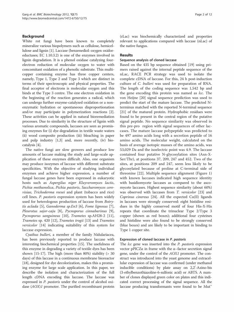
(methanol utilization phenotype). The clone pPICZα lcc-5,which showed deepest green color on the plates, waschosen for expression studies in liquid medium (InvitrogenBasal medium). Maximum laccase activity of 600 U-720 UL-1 was observed 3 days after initiation of induction by1.0% methanol. Effect of addition of copper sulfate at dif-ferent times, post induction, was investigated and max-imum laccase activity of ~7200 U L-1 was obtained at saltconcentration of 0.4 mM (Figure 2). This represented anincrease of about 12-fold over control cultures where nocopper was added. The PAGE-zymogram analysis of theconcentrated culture filtrate, carried out on guaiacol, con-firmed the expression of laccase in active form (Figure 3A,lanes 3,4). For equal volume of concentrated culture filtrateloaded on the gel, omission of SDS and β–mercaptoetha-nol in the loading buffer resulted in higher activity asjudged on the gel (Figure 3A, lanes 5,6 respectively).
Purification and characterization of rLacRecombinant laccase expressed in P. pastoris was puri-fied using ammonium sulfate precipitation, followed bychromatography on Sephadex G75 and Superdex G200columns kept in tandem. A summary of the purificationsteps is shown in Table 1. A final specific activity of 85U mg-1 was achieved representing a purification fold of~4. A total enzyme yield of 24% was obtained. A singleprotein band was detected on SDS-PAGE indicatingelectrophoretic homogeneity of the sample (Figure 3B,lane 3) and a relative molecular mass of ~60 kDa wasestimated. The mass of this purified rLac was slightlyhigher than the nLac (Figure 3B, lane 2) which has beenreported to be of ~58 kDa [15]. The laccase activity ofthe purified band was confirmed by zymogram analysisusing ABTS as a substrate, the oxidized radical of whichwas visualized as a green colored band (Figure 3C, lanes
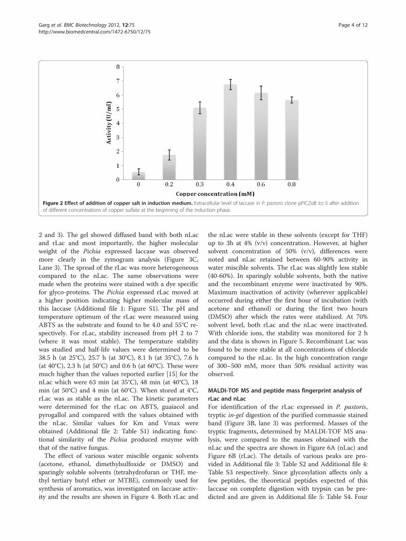
Figure 2 Effect of addition of copper salt in induction medium. Extracellular level of laccase in P. pastoris clone pPICZαB lcc-5 after additionof different concentrations of copper sulfate at the beginning of the induction phase.
Garg et al. BMC Biotechnology 2012, 12:75 Page 4 of 12http://www.biomedcentral.com/1472-6750/12/75
2 and 3). The gel showed diffused band with both nLacand rLac and most importantly, the higher molecularweight of the Pichia expressed laccase was observedmore clearly in the zymogram analysis (Figure 3C,Lane 3). The spread of the rLac was more heterogeneouscompared to the nLac. The same observations weremade when the proteins were stained with a dye specificfor glyco-proteins. The Pichia expressed rLac moved ata higher position indicating higher molecular mass ofthis laccase (Additional file 1: Figure S1). The pH andtemperature optimum of the rLac were measured usingABTS as the substrate and found to be 4.0 and 55°C re-spectively. For rLac, stability increased from pH 2 to 7(where it was most stable). The temperature stabilitywas studied and half-life values were determined to be38.5 h (at 25°C), 25.7 h (at 30°C), 8.1 h (at 35°C), 7.6 h(at 40°C), 2.3 h (at 50°C) and 0.6 h (at 60°C). These weremuch higher than the values reported earlier [15] for thenLac which were 63 min (at 35°C), 48 min (at 40°C), 18min (at 50°C) and 4 min (at 60°C). When stored at 4°C,rLac was as stable as the nLac. The kinetic parameterswere determined for the rLac on ABTS, guaiacol andpyrogallol and compared with the values obtained withthe nLac. Similar values for Km and Vmax wereobtained (Additional file 2: Table S1) indicating func-tional similarity of the Pichia produced enzyme withthat of the native fungus.The effect of various water miscible organic solvents
(acetone, ethanol, dimethylsulfoxide or DMSO) andsparingly soluble solvents (tetrahydrofuran or THF, me-thyl tertiary butyl ether or MTBE), commonly used forsynthesis of aromatics, was investigated on laccase activ-ity and the results are shown in Figure 4. Both rLac and
the nLac were stable in these solvents (except for THF)up to 3h at 4% (v/v) concentration. However, at highersolvent concentration of 50% (v/v), differences werenoted and nLac retained between 60-90% activity inwater miscible solvents. The rLac was slightly less stable(40-60%). In sparingly soluble solvents, both the nativeand the recombinant enzyme were inactivated by 90%.Maximum inactivation of activity (wherever applicable)occurred during either the first hour of incubation (withacetone and ethanol) or during the first two hours(DMSO) after which the rates were stabilized. At 70%solvent level, both rLac and the nLac were inactivated.With chloride ions, the stability was monitored for 2 hand the data is shown in Figure 5. Recombinant Lac wasfound to be more stable at all concentrations of chloridecompared to the nLac. In the high concentration rangeof 300–500 mM, more than 50% residual activity wasobserved.
MALDI-TOF MS and peptide mass fingerprint analysis ofrLac and nLacFor identification of the rLac expressed in P. pastoris,tryptic in-gel digestion of the purified commassie stainedband (Figure 3B, lane 3) was performed. Masses of thetryptic fragments, determined by MALDI-TOF MS ana-lysis, were compared to the masses obtained with thenLac and the spectra are shown in Figure 6A (nLac) andFigure 6B (rLac). The details of various peaks are pro-vided in Additional file 3: Table S2 and Additional file 4:Table S3 respectively. Since glycosylation affects only afew peptides, the theoretical peptides expected of thislaccase on complete digestion with trypsin can be pre-dicted and are given in Additional file 5: Table S4. Four

Figure 3 Expression, purification and zymogram analysis of purified laccase. (A) PAGE separation of proteins from culture supernatant ofP. pastoris pPICZαB lcc-5 and confirmation of laccase secretion by zymogram analysis using guaiacol as substrate. Lane 1, Pre-stained molecularweight markers. Lanes 3,4: 10, 20 μl respectively of the culture filtrate. Lane 5, 20 μl of the culture filtrate in loading buffer not containing SDS.Lane 6: 20 μl of the culture filtrate in loading buffer not containing β–mercaptoethanol. (B) SDS-PAGE analysis of the purified laccase. Lane 1,molecular weight markers. Lane 2, purified nLac (~5 μg). Lane 3, purified rLac from P. pastoris (~7 μg). (C) PAGE-Zymogram analysis using ABTS asa substrate. Lane 1, Pre-stained molecular weight markers. Lane 2, purified nLac. Lane 3, purified rLac.
Garg et al. BMC Biotechnology 2012, 12:75 Page 5 of 12http://www.biomedcentral.com/1472-6750/12/75
of the peptides in the rLac at m/z values of 1419.53,1528.45, 2098.71, and 2125.83 (Figure 6B) were identifiedand matched exactly with the peptides of m/z 1419.81,1528.72 + 1684.84 (with an additional D), 2099.10 (seeAdditional file 4: Table S3) and 2126.24 obtained from the
Table 1 Summary of purification of laccase secreted by P. pas
Purification Steps Total activity (U) Total protein (
Crude extract 370 18
Ammonium sulfate precipitation 138.24 3.6
Sephadex G75 + SuperdexG200 90 1.06
nLac (Table 2). This confirmed that rLac was the same asthe purified nLac. For rLac and the nLac, additional pep-tide fragments with m/z values of 2593.95 (Figure 6B) and2594.041, 2594.049 (Additional file 4: Table S3) and2132.1 (Additional file 3: Table S2), 2866.35 (Figure 6A)
toris pPICZαB lcc-5
mg) Specific activity (U/mg) Yield (%) Purification fold
20.5 100 1
38.4 37.3 ~1.9
84.9 24.3 ~4.0

Figure 4 Residual activity of laccase in the presence of organic solvents. Percent residual laccase activity after incubation of purified enzymefor 3 h in the presence of various solvents as indicated on X axis. Hundred % activity corresponds to 0.5 U in the total reaction mixture. Lightgrey: nLac, Dark grey: rLac.
Garg et al. BMC Biotechnology 2012, 12:75 Page 6 of 12http://www.biomedcentral.com/1472-6750/12/75
were detected in addition to several un-identified peptides.Glycomod tools [25] were used for calculating the theoret-ical mass of the potential glycosylated tryptic fragments(only 2 out of the 4 possible sites due to the presence ofPro at the carboxy-terminus) and their possible structures.Based on this, the peak observed at 2593.95 was identified
Figure 5 Residual activity of laccase in the presence of NaCl concentr2 h in the presence of different NaCl concentrations. Hundred % activity cogrey: nLac, Dark grey: rLac.
to be of the peptide 442 DAVNTGGAGDNVTIR 456 withthe assigned glycan structure of (Hex)4 (HexNac)2 (Sulph/Phos)1 . For the nLac, the tryptic fragment at m/z of 2132.1was concluded to be 442 DAVNTGGAGDNVTIR 456 withthe glycan structure of (Hex)1(HexNAc)2(NeuGc)1. Thedetails are provided in Table 2.
ation. Percent residual activity after incubation of purified enzyme forrresponds to ~1.0 U laccase activity in the total reaction volume. Light

A
B
32.07324.72726.48018.14470.99
Mass (m/z)
0
10
20
30
40
50
60
70
80
90
100
% I
nte
ns
ity
4700 Reflector Spec #1[BP = 1419.5, 5010]
1419.5310
1528.4595
2125.8379
2098.7163842.3719 1489.4830
1375.45671045.3931
2085.72711754.6260 3053.15012593.95211234.4613891.3176
Figure 6 MALDI-TOF mass spectra of peptide fragments of laccase. Mass fingerprint analysis of peptides arising from SDS-PAGE separatedand in situ trypsin digested purified nLac (A) and rLac (B).
Garg et al. BMC Biotechnology 2012, 12:75 Page 7 of 12http://www.biomedcentral.com/1472-6750/12/75
DiscussionIn the last few years laccases have been identified as im-portant enzymes for application in the environment sec-tor as well as for production of high value aromatics[4,26]. Almost all laccases are produced at low levels inthe native fungi. Multiple isozymes of laccase have alsobeen reported in numerous fungi making the study of
individual enzymes difficult. Cloning and expression ofgenes provides an opportunity to overproduce and studythese enzymes individually. Based on our previous studies[15,18], wherein a laccase was purified from C. bulleri andinvestigated for its application in degradation of textiledyes, the cDNA encoding this enzyme was isolated in thepresent study and expressed in P. pastoris. Although many
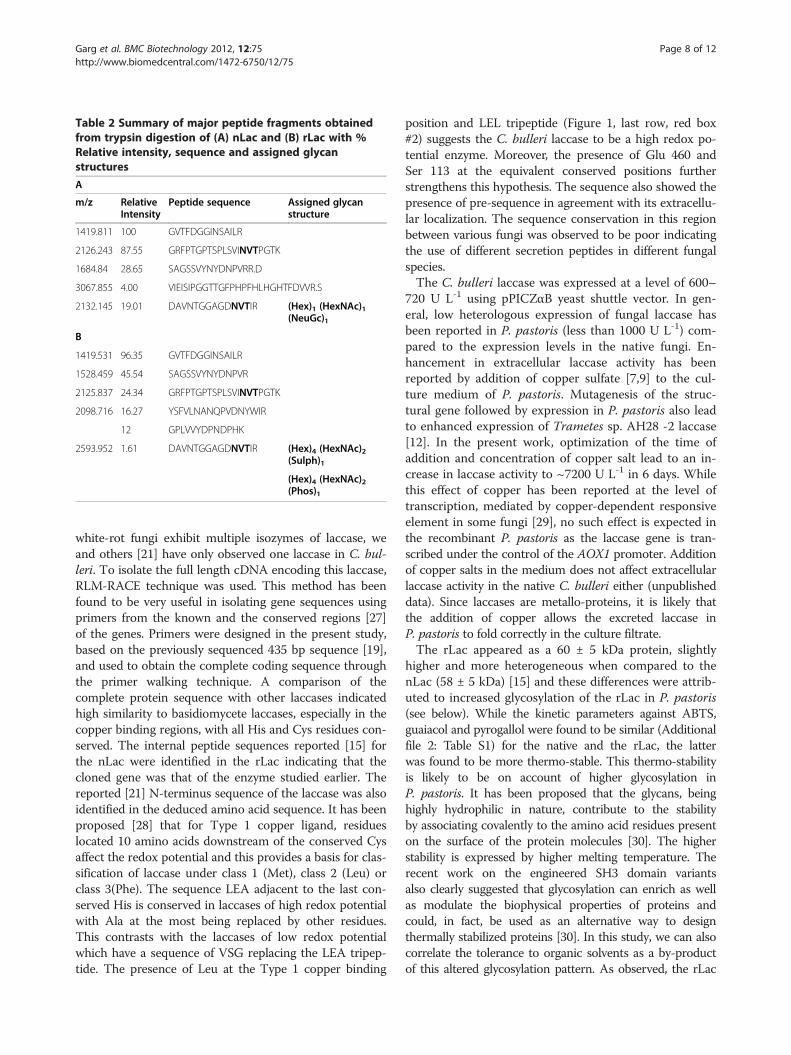
Table 2 Summary of major peptide fragments obtainedfrom trypsin digestion of (A) nLac and (B) rLac with %Relative intensity, sequence and assigned glycanstructures
A
m/z RelativeIntensity
Peptide sequence Assigned glycanstructure
1419.811 100 GVTFDGGINSAILR
2126.243 87.55 GRFPTGPTSPLSVINVTPGTK
1684.84 28.65 SAGSSVYNYDNPVRR.D
3067.855 4.00 VIEISIPGGTTGFPHPFHLHGHTFDVVR.S
2132.145 19.01 DAVNTGGAGDNVTIR (Hex)1 (HexNAc)1(NeuGc)1
B
1419.531 96.35 GVTFDGGINSAILR
1528.459 45.54 SAGSSVYNYDNPVR
2125.837 24.34 GRFPTGPTSPLSVINVTPGTK
2098.716 16.27 YSFVLNANQPVDNYWIR
12 GPLVVYDPNDPHK
2593.952 1.61 DAVNTGGAGDNVTIR (Hex)4 (HexNAc)2(Sulph)1
(Hex)4 (HexNAc)2(Phos)1
Garg et al. BMC Biotechnology 2012, 12:75 Page 8 of 12http://www.biomedcentral.com/1472-6750/12/75
white-rot fungi exhibit multiple isozymes of laccase, weand others [21] have only observed one laccase in C. bul-leri. To isolate the full length cDNA encoding this laccase,RLM-RACE technique was used. This method has beenfound to be very useful in isolating gene sequences usingprimers from the known and the conserved regions [27]of the genes. Primers were designed in the present study,based on the previously sequenced 435 bp sequence [19],and used to obtain the complete coding sequence throughthe primer walking technique. A comparison of thecomplete protein sequence with other laccases indicatedhigh similarity to basidiomycete laccases, especially in thecopper binding regions, with all His and Cys residues con-served. The internal peptide sequences reported [15] forthe nLac were identified in the rLac indicating that thecloned gene was that of the enzyme studied earlier. Thereported [21] N-terminus sequence of the laccase was alsoidentified in the deduced amino acid sequence. It has beenproposed [28] that for Type 1 copper ligand, residueslocated 10 amino acids downstream of the conserved Cysaffect the redox potential and this provides a basis for clas-sification of laccase under class 1 (Met), class 2 (Leu) orclass 3(Phe). The sequence LEA adjacent to the last con-served His is conserved in laccases of high redox potentialwith Ala at the most being replaced by other residues.This contrasts with the laccases of low redox potentialwhich have a sequence of VSG replacing the LEA tripep-tide. The presence of Leu at the Type 1 copper binding
position and LEL tripeptide (Figure 1, last row, red box#2) suggests the C. bulleri laccase to be a high redox po-tential enzyme. Moreover, the presence of Glu 460 andSer 113 at the equivalent conserved positions furtherstrengthens this hypothesis. The sequence also showed thepresence of pre-sequence in agreement with its extracellu-lar localization. The sequence conservation in this regionbetween various fungi was observed to be poor indicatingthe use of different secretion peptides in different fungalspecies.The C. bulleri laccase was expressed at a level of 600–
720 U L-1 using pPICZαB yeast shuttle vector. In gen-eral, low heterologous expression of fungal laccase hasbeen reported in P. pastoris (less than 1000 U L-1) com-pared to the expression levels in the native fungi. En-hancement in extracellular laccase activity has beenreported by addition of copper sulfate [7,9] to the cul-ture medium of P. pastoris. Mutagenesis of the struc-tural gene followed by expression in P. pastoris also leadto enhanced expression of Trametes sp. AH28 -2 laccase[12]. In the present work, optimization of the time ofaddition and concentration of copper salt lead to an in-crease in laccase activity to ~7200 U L-1 in 6 days. Whilethis effect of copper has been reported at the level oftranscription, mediated by copper-dependent responsiveelement in some fungi [29], no such effect is expected inthe recombinant P. pastoris as the laccase gene is tran-scribed under the control of the AOX1 promoter. Additionof copper salts in the medium does not affect extracellularlaccase activity in the native C. bulleri either (unpublisheddata). Since laccases are metallo-proteins, it is likely thatthe addition of copper allows the excreted laccase inP. pastoris to fold correctly in the culture filtrate.The rLac appeared as a 60 ± 5 kDa protein, slightly
higher and more heterogeneous when compared to thenLac (58 ± 5 kDa) [15] and these differences were attrib-uted to increased glycosylation of the rLac in P. pastoris(see below). While the kinetic parameters against ABTS,guaiacol and pyrogallol were found to be similar (Additionalfile 2: Table S1) for the native and the rLac, the latterwas found to be more thermo-stable. This thermo-stabilityis likely to be on account of higher glycosylation inP. pastoris. It has been proposed that the glycans, beinghighly hydrophilic in nature, contribute to the stabilityby associating covalently to the amino acid residues presenton the surface of the protein molecules [30]. The higherstability is expressed by higher melting temperature. Therecent work on the engineered SH3 domain variantsalso clearly suggested that glycosylation can enrich as wellas modulate the biophysical properties of proteins andcould, in fact, be used as an alternative way to designthermally stabilized proteins [30]. In this study, we can alsocorrelate the tolerance to organic solvents as a by-productof this altered glycosylation pattern. As observed, the rLac

Garg et al. BMC Biotechnology 2012, 12:75 Page 9 of 12http://www.biomedcentral.com/1472-6750/12/75
produced in P. pastoris exhibited higher tolerance towardsvarious water-miscible organic solvents compared to thenative laccases from T. versicolor and Pleurotus ostreatus[31]. Between 40-60% residual activity was observed at50% (v/v) in all these solvents after 3 h of incubation. Whilethese values were slightly lower than those observed forthe nLac (Figure 4), these are still high and useful for itsuse in organic synthesis work. Interestingly, both the rLacand the nLac were equally unstable in THF (solvent of ahigher log P value)which is likely to have distorted theenzyme hydration and distort the conformation leading toa drastic decrease in enzyme activity. It has been observedthat laccase structure, stability and activity are affected bywater miscible solvents through direct interaction with en-zyme and through its affect on water activity (aw) [32].Although Farnet et al. [33] have observed a high IC50 values(30-60%) of theMarasmius quercophilus laccase in differentsolvents but the enzyme was not incubated for longer timeperiods and thus their data cannot be compared to ourresults.Laccases are generally inhibited by chloride ions, an
important component in dye wastewaters, which limitsits use in treatment plants. Chloride ions directly affectthe conversion process through their intrinsic effects onrate constants mediated through availability of Type 2and Type 3 copper atoms in the active site [34]. Higherresistance to chloride ions (after 2 h incubation) wasobserved for the rLac of C. bulleri (Figure 5) when com-pared to the nLac. A chloride tolerant laccase havingIC50 of 1.5 M was recently reported [5] but again, theenzyme was not incubated for long time periods andhence cannot be compared to the laccase expressed inthis study.For many of the differences observed between the rLac
and the nLac, a detailed comparison of the trypsindigested peptide fragments was made. Several peptideswere found to be identical confirming the expression ofthe same laccase in P. pastoris, as reported previouslyfrom our group. Out of the 4 putative glycosylation sites,only 2 were likely to get glycosylated [20]. Differences inthe glycosylation patterns, leading to generation of aspectrum of different peptides, were observed. Softwaretools were used to identify these and the fragment withm/z of 2593.9521 (obtained from the rLac) was con-cluded to represent the aa sequence 442–456 withpossible glycan structure of (Hex)4 (HexNAc)2 (Sulph/Phos)1. The corresponding fragment from the nLacwas identified at 2132.1 m/z with an assigned struc-ture of (Hex)1(HexNAc)2(NeuGc)1. While theoretically,additional peptide (28 VISPDGFNRSAVLAGGTADNADFPGPLVTGNK 38) is predicted to undergo glycosylationand may indeed do so, this is not likely to be detected byMALDI-TOF MS, as the size of this exceeds the detectionlimits of the system.
ConclusionThe full length cDNA sequence of C. bulleri laccase isreported in this paper. The gene was efficiently expressedunder the control of the AOX1 promoter and secreted inthe culture supernatant of P. pastoris . Sequence analysisindicated this to code for a high redox laccase. Optimizationof the time of addition and concentration of copper saltsresulted in laccase activity of ~7200 U L-1. The laccase waspurified to homogeneity and found to be of a higher mol wtcompared to the nLac. An investigation of biochemicalproperties of the rLac indicated it to possess higher thermo-stability and tolerance towards chloride ions compared tothe nLac. A comparison of the peptide mass fingerprint datawith the nLac indicated presence of fragments, not observedin the nLac, which were attributed to different pattern ofglycosylation in the P. pastoris and which are likely to havecontributed to the observed differences in some biochemicalproperties. The data indicate usefulness of the rLac over thenLac in specific areas of applications.
MethodsOrganisms, plasmids and enzymesC. bulleri (Brodie) 195062 (common name: birds’ nest fun-gus) was from Canadian Type Culture Collection. The fun-gus was cultivated as described previously [15]. The Pichiavector pPICZαB and host P. pastoris X33 strain were fromInvitrogen. The yeast was maintained on YPD (1% yeastextract, 2% bacto-peptone, 2% glucose). Escherichia coliDH5α was from Technical University, Aachen, Germanyand TOP 10 cells were provided in the TOPO TA cloningkit for sequencing (Invitrogen). E. coli was grown in Luria-Bertani medium. Unless otherwise stated, the enzymesused to manipulate DNA or RNA were obtained fromNew England Biolabs, Promega or Fermentas.
OligonucleotidesThe oligonucleotides (Sigma-Aldrich) used in the studyare shown in Table 3.
RNA isolationC. bulleri cultures were grown in basal liquid medium[35] and induced with 2,6-dimethylaniline [15]. Fungalmycelium was collected 24 h after induction by filtra-tion, washed twice with sterile phosphate buffer (20mM, pH 7.0) and frozen in liquid nitrogen. Crushed fro-zen mycelium (100 mg) was used to isolate total RNAusing RNeasy Plant Mini Kit (Qiagen). The quality ofRNA was checked by running on agarose gel.
Determination of laccase nucleotide sequence by RNAligase mediated RACE-PCRGeneRacer™ RLM-RACE kit (Invitrogen) was used toobtain 50 and 30 ends of laccase cDNA. Two μg of thetotal RNA was treated with calf intestinal phosphatase

Table 3 List of oligonucleotides used in the study
Oligonucleotide Sequence
GeneRacer 50 Primer 50- CGA CTG GAG CAC GAG GACACT GA -30
GeneRacer 30 Primer 50- GCT GTC AAC GAT ACG CTACGT AAC G -50
GeneRacer Oligo dT Primer 50- GCT GTC AAC GAT ACG CTACGT AAC GGC ATG ACA GTG(T)24- 3
0
GeneRacer RNA Oligo sequence 50- CGA CUG GAG CAC GAG GACACU GAC AUG
For-gsp GAC UGA AGGAGU AGA AA -30
Rev-gsp 50- TAG CGC CGG AAG CAG CGTGTA CAA CTA -30
Oligo 2 50- GGT GTC TGG TGC ACC GGCATA TC -30
Oligo 3 50- ATT TCC CCG CGG TCA GGTGCC GGT TGG- 30
50- CCG CTG CAG CCA TTG GCCCAG TTT CGGA -30
Garg et al. BMC Biotechnology 2012, 12:75 Page 10 of 12http://www.biomedcentral.com/1472-6750/12/75
to remove phosphates from truncated and non-mRNA.The dephosphorylated RNA was given tobacco acid pyr-ophosphatase treatment to remove mRNA cap structure.Oligo (provided in the kit) ligated RNA was primed witholigo dT primer (Gene Racer Oligo dT Primer) and reversetranscription was carried out using Superscript II RT (Invi-trogen). GeneRacer 50 Primer, complimentary to the an-chor sequence and Rev-Gsp primer designed from the 435bp laccase sequence described previously [19] were used toamplify the 50end. Gene Racer 30 Primer, from the anchorattached to Oligo dT and For-Gsp designed from the 435bp laccase sequence described previously were used to ob-tain the 30end. The PCR product was sequenced (MWGDNA Sequencing Service, Germany) and complete laccasecDNA sequence deduced using primer walking technique.Sequences were aligned using Clustal V program.
Cloning and expression of laccase gene through yeastshuttle vectorTotal RNA was reverse transcribed using Oligo dT pri-mer with M-MuLV reverse transcriptase (New EnglandBiolabs). The von Heijne signal sequence prediction [20]was used to predict the start of the mature laccase. A1,491 bp fragment corresponding to the laccase cDNA(without the signal peptide encoding fragment) wasamplified using downstream Oligo 2 and upstream Oligo3 generating Sac II and Pst I sites respectively. The PCRproduct was cloned into pCR4-TOPO vector in E. coliTOP 10 cells as per instructions (Invitrogen). The pres-ence of the desired PCR product was verified by restric-tion enzyme digestion, agarose gel electrophoresis andsequencing. The recombinant plasmid was linearized
using SacI. The Easy select Pichia expression kit (Invi-trogen) was used for heterologous expression of the lac-case cDNA without its own signal peptide. The mediumrecipe, transformation and analysis of the recombinantswere carried out as per the kit manual. P. pastoris X33was transformed with Sac I linearized recombinantpPICZαB vector and the transformants were selected forZeocin resistance on YPD medium. Twenty or so trans-formants were screened on minimal methanol platessupplemented with 0.2 mM ABTS for development ofgreen color. One (pPIC lcc-5) of the recombinants(selected on the basis of development of intense greencolor in plate assay) was cultivated in 50 mL YPDmedium in 300 ml baffled flasks. At culture OD600 of 2–6, the cells were harvested by centrifugation and re-suspended in buffered complex methanol medium at anOD600 of 1.0. The culture was monitored for 6 days forproduction of extracellular laccase with the induction ofthe promoter being maintained by daily addition of 1%(v/v) methanol. For studying the effect of Copper ions,copper sulfate was added at a conc of 0.2, 0.3, 0.4, 0.6,0.8 mM at different time periods after transfer to the in-duction medium.
Purification and characterization of laccaseThe culture filtrate of recombinant X33 cells (75 ml)was concentrated using ammonium sulfate (100% satur-ation). The concentrated supernatant (2 ml) was sub-jected to gel filtration using Sephadex75 column (30 cmx 1 cm, Pharmacia) placed in tandem with a SuperdexG200 column (30 x 1 cm, Pharmacia) using AKTA FPLCsystem. Elution was carried out with 20 mM Tris-Cl buf-fer, pH 8.0 as mobile phase at a flow rate of 0.2 ml/min.Fractions of 2 ml were collected and the presence ofprotein monitored by measuring OD280. Alternate frac-tions of protein containing tubes were assayed for lac-case activity using ABTS as the substrate [35]. Thefractions showing laccase activity were pooled, dialyzedagainst distilled water and concentrated by lyophi-lization. The purity of the enzyme was checked on 15%SDS and activity was confirmed by zymogram analysis[15]. Laccase was also purified from the culture filtrateof C. bulleri to a specific activity of ~240 U mg-1 protein,as described previously [15].For biochemical characterization, active laccase was
reconstituted from the lyophilized powder by suspendingin distilled water to a final activity of 100 U mL-1. Thereconstituted enzyme was used to determine pH andtemperature optimum, pH and temperature stability, tol-erance to chloride ions and several organic solventsusing ABTS as the substrate. The experiments were per-formed either in a glass cuvette or in a Microtiter platereader (Infinite M200, Tecan) for multiple laccase mea-surements. Laccase activity was measured in a UV/VIS
Garg et al. BMC Biotechnology 2012, 12:75 Page 11 of 12http://www.biomedcentral.com/1472-6750/12/75
spectrophotometer (Uvikon 860) using a cuvette of 1 ml(coat thickness 1 cm) containing 50 mM Na citrate buf-fer (based on the observation that the pH optimum was4.0, see below), 10 μM ABTS and appropriately dilutedenzyme solution. Assay conditions were scaled down inmicrotitre plates (total volume 200 μl). Adequate units(~0.5-1.0 U) of laccase (aqueous solution of the lyophi-lized prep) were added and absorption was monitored at420 nm. The buffers used were 0.3 M Glycine HCl buf-fer (pH 2 and 3), 0.3 M Na citrate buffer (pH 4–6), 0.3M phosphate buffer (pH 7–8), 0.3 M Tris–HCl buffer(pH 9). All buffers were strong enough to buffer the cor-responding pH even after addition of the acidic ABTSsolution (checked via pH electrode). The dependence ofactivity on pH was determined for the rLac. Optimumtemperature was determined at pH 4.0. Temperaturestability was determined at pH 4.0 in temperatures ran-ging from room temperature (25°C) to 80°C. Kineticparameters (Km and Vmax) of the nLac and the rLacwere determined towards ABTS, Guaiacol and Pyrogal-lol. Spectrophotometric measurements of substrate oxi-dation by nLac and rLac were carried out in a 2 mlreaction volume containing the test substrate in 50 mMsodium citrate buffer (pH 4). All assays were carried outwith equal units of laccase activity.
Tolerance towards chloride ions and organic solventsTolerance to chloride ions was determined by incubatinglaccase solution (0.5-1.0 U) with varying concentrationsof NaCl for 2 h in a total volume of 1.5 ml. Reactionvials were stored at 4°C to rule out any effect caused dueto temperature. Aliquots (80 μl) were removed at regularintervals and laccase activity measured in a microtitreplate. Similarly, tolerance to organic solvents was mea-sured by incubating laccase with 1 ml of correspondingorganic solvent (at different concentrations, v/v) for 3 h.In case of acetone, ethanol, DMSO, THF, an organicsolvent/water mixture of 4, 50 and 70% (v/v) was used.Because of limited solubility of MTBE in water, only 4%(v/v) solution was tested. Organic solvent tolerance wasalso measured in a similar manner for the purified nLac.All enzyme activity measurements were done twice
and every value was measured three times. The variationwas between 5-7%.
Peptide mass fingerprint analysisThe purified (5–10 μg) rLac and nLac were run in 5lanes (for each protein) of 10%SDS-PAGE using a Mini-Protean Cell (Bio-rad). The proteins were stained withCoomassie blue as per standard protocols. The bandswere excised out of the gel and stored in autoclavedEppendorf tubes. The tryptic in-gel digestion and pep-tide finger printing was carried out using commercial
service provided by Vimta Labs Ltd on a Bruker Dal-tonics flexAnalysis system.
DNA sequencingThe DNA sequencing was done using MWG DNA sequen-cing service (Applied Biosystems 3730xl), Germany. Thecomplete nucleotide sequence of C. bulleri laccase reportedin this paper has been deposited in the GenBank databaseunder the Accession No. EU195884, version 2.
Additional files
Additional file 1: Figure S1. PAGE separated proteins stained withglycoprotein stain. Equal concentration (5μg) of nLac and the rLac wasloaded. The gels were stained with the Pierce glycoprotein staining kit.Lane 1: molecular weight marker, Lane 2: rLac, Lane 3: nLac, Lane 4: stdglycoprotein from the kit.
Additional file 2: Table S1. Kinetic parameters of the purified nLac andrLac. All the values represent means of duplicate measurements with asample mean deviation of lesser than 0.5%, stands for no activity.
Additional file 3: Table S2. Details of the peptide fragments generatedfrom trypsin digestion of purified nLac from Cyathus bulleri.
Additional file 4: Table S3. Details of the peptide fragments generatedfrom trypsin digestion of purified rLac from P. pastoris.
Additional file 5: Table S4. Theoretical tryptic fragments.
Competing interestsThe authors declare that they do not have competing interests.
Authors’ contributionsNG carried out the molecular biological studies leading to elucidation of thecomplete cDNA sequence and expression of the enzyme in P. pastoris. NBstandardized the procedure for purification of the rLac and performedextensive experiments on biochemical characterization. TK and MC purifiedthe rLac and the nLac, performed comparative analysis on solvent andchloride tolerance and analyzed the tryptic finger print data of the enzymes.MAS supervised the work in the Berlin lab. SM was the corresponding authorand supervised the overall study and contributed to the manuscriptorganization and writing. All authors have read and approved themanuscript.
AcknowledgementsThe author (SM) acknowledges the financial assistance received from Dept.of Biotechnology (GOI) to carry out the initial part of the project. Ms. NehaGarg gratefully acknowledges the scholarship received from DeutscherAkademischer Austausch Dienst (DAAD-IIT Masters Sandwich Program) tocarry out this work in Germany. Ms. Tenzin Kenzom and Dr. M. Chhabragratefully acknowledge the JRF scholarship received from CSIR, New Delhi.
Author details1Department of Biochemical Engineering and Biotechnology, Indian Instituteof Technology Delhi, Hauz-Khas, New Delhi 110016, India. 2Inst.Chemistry,Department Enzyme Technology (Sekr.TC4), TU Berlin, Str. Des 17. Juni 124 D,Berlin 10623, Germany. 3Centre of Excellence in Biologically Inspired SystemScience, Indian Institute of Technology Jodhpur, Jodhpur, Rajasthan 342011,India.
Received: 15 May 2012 Accepted: 8 October 2012Published: 23 October 2012
References1. Jurado M, Martinèz AT, Martinez MJ, Saparrat MCN: Application of white-
rot fungi in transformation, detoxification, or revalorization of agriculturewastes: role of laccase in the processes. In Comprehensive Biotechnology.6th edition. Edited by Moo-Young M: Academic Press; 2011:595–603.

Garg et al. BMC Biotechnology 2012, 12:75 Page 12 of 12http://www.biomedcentral.com/1472-6750/12/75
2. Thurston CF: The structure and function of fungal laccases. Microbiology1994, 140:19–26.
3. Rodriguez E, Nuero O, Guillen F, Martinez AT, Martinez MJ: Degradation ofphenolic and non-phenolic aromatic pollutants by four Pleurotus species:the role of laccase and versatile peroxidase. Soil Biol Biochem 2004,36:909–916.
4. Riva S: Laccases: blue enzymes for green chemistry. Trends Biotechnol2006, 24:219–226.
5. Roman K, Kitti M, Christoph G, Shima TK, Christoph S, Dietmar H, Roland L:A chloride tolerant laccase from the plant pathogen ascomycete Botrytisaclada expressed at high levels in Pichia pastoris. J Biotechnol 2012,157:304–314.
6. Rui Z, Li M, Fangfang F, Yangmin G, Xia W, Mulang J, Xiaoyu Z, Yang Y:Decolorization of different dyes by a newly isolated white-rot fungistrain Ganoderma sp.En3 and cloning and functional analysis of itslaccase gene. J Hazard Mater 2011, 192:855–873.
7. Liu W, Chao Y, Liu S, Bao H, Qian S: Molecular cloning andcharacterization of a laccase gene from the basidiomycete Fome lignosusand expression in Pichia pastoris. Appl Microbiol Biotechnol 2003,63:174–181.
8. Soden DM, O’Callaghan J, Dobson AD: Molecular cloning of a laccaseisozyme gene from Pleurotous sajor-caju and expression in theheterologous Pichia pastoris host. Microbiology 2002, 148:4003–4014.
9. Otterbein L, Record E, Longhi S, Asther M, Moukha S: Molecular cloning ofthe cDNA encoding laccase from Pycnoporus cinnabarinus I-937 andexpression in Pichia pastoris. Eur J Biochem 2000, 267:1619–1625.
10. Lu L, Zhao M, Liang SC, Zhao LY, Li DB, Zhang BB: Production andsynthetic dyes decolorization capacity of a recombinant laccase fromPichia pastoris. J Appl Microbiol 2009, 107:1149–1156.
11. Yuzhi H, Yazhong X, Hongmin Z, Wei F, Min Z, Jun W, Lijun W, Zengliang Y:Expression of a laccase cDNA from Trametes sp. AH28 -2 in Pichiapastoris and mutagenesis of transformants by nitrogen ion implantation.FEMS Microbiol Lett 2006, 258:96–101.
12. Hong YZ, Zhou HM, Tu XM, Li JF, Xiao YZ: Cloning of a laccase gene froma novel basidiomycete Trametessp. 420 and its heterologous expressionin Pichia pastoris. Curr Microbiol 2007, 54:260–265.
13. Colao MC, Lupino S, Garzillo AM, Buonocore V, Ruzzi M: Heterologousexpression of lcc1 gene from Trametes trogii in Pichia pastoris andcharacterization of the recombinant enzyme. Microb Cell Fact 2006,5:31–38.
14. Bohlin C, Jonsson LJ, Roth R, VanZyl WH: Hetrologous expression ofTrametes versicolor laccase in Pichia pastoris and Aspergillus niger. ApplBiochem Biotechnol 2006, 129–132:195–214.
15. Salony, Mishra S, Bisaria VS: Production and characterization of laccasefrom Cyathus bulleri and its use in decolorization of recalcitrant textiledyes. Appl Microbiol Biotechnol 646, 71:646–653.
16. Salony, Mishra S, Bisaria VS: Decolorization and detoxification of textiledyes and black liquor by laccase of Cyathus bulleri. J Sci Ind Res 2007,66:684–688.
17. Chhabra M, Sreekrishnan TR, Mishra S: Mediated assisted decolorizationand detoxification of textile dyes/dye mixture by Cyathus bulleri laccase.Appl Biochem Biotechnol 2008, 151:587–598.
18. Chhabra M, Sreekrishnan TR, Mishra S: Laccase/mediator assisteddegradation of triarylmethane dyes in a continuous membrane reactor. JBiotechnol 2009, 143:69–78.
19. Salony, Garg N, Baranwal R, Chhabra M, Mishra S, Chaudhuri TK, Bisaria VS:Laccase of Cyathus bulleri : structural, catalytic characterization andexpression in Escherichia coli. Biochim Biophys Acta 2008, 1784:259–268.
20. Von Heijne G: A new method for predicting signal sequence cleavagesites. Nucleic Acids Res 1986, 14:4683–4690.
21. Vasudev K, Dhawan S, Kapoor RK, Kuhad R: Biochemical characterizationand molecular evidence of a laccase from the bird’s nest fungus Cyathusbulleri. Fungal Genet Biol 2005, 42:684–693.
22. Andrei JP, Adina LM, Stefana MP, Raymond AD, Mark RW: Statisticalanalysis of the protein environment of x-glycosylation sites: implicationsfor occupancy, structure, and folding. Glycobiology 2004, 14:103–114.
23. Cassland P, Jonsson LJ: Characterization of a gene encoding Trametesversicolor laccaseA and improved heterologous expression inSaccharomyces cerevisiae by decreased cultivation temp. Appl MicrobiolBiotechnol 1999, 52:393–400.
24. Yaver DS, Overjero MDC, Xu F, Nelson BA, Brown KM, Halkier T, Bernauer S,Brown SH, Kauppinen SK: Molecular characterization of laccase genesfrom the basidiomycete Coprinus cinereus and heterologous expressionof the laccase lcc1. Appl Environ Microbiol 1999, 65:4943–4948.
25. Cooper CA, Gasteiger E, Packer N: GlycoMod:A software Tool fordetermining glycosylation compositions from mass spectrometric data.Proteomics 2001, 1:340–349.
26. Stentelaire C, Lesage-Messen L, Oddou J, Bernard O, Bastin G, Ceccaldi BC,Asther M: Design of a fungal bioprocess for vanillin production fromvanillic acid at scalable level by Pycnoporus cinnabarinus. J Bioeng 2000,89:223–230.
27. Piontek K, Antorini M, Choinowsk P: Crystal structure of a laccase from thefungus Trametes versicolor at 1.90 Ǻ resolution containing a fullcomplement of copper. J Biol Chem 2002, 40:37663–37669.
28. Canters GW, Gilardi G: Engineering type-1 copper sites in proteins. FEBSLett 1993, 325:39–48.
29. Alvarez JM, Canessa P, Mancilli RA, Polanco R, Santibanez PA, Vicuna R:Expression of genes encoding laccase and manganese-dependentperoxidase in the fungus Ceriporiopsis subvermispora is mediated by anACE1-like copper-fist transcription factor. Fungal Genet Biol 2009,46:104–111.
30. Shental-Bechor D, Levy Y: Effect of glycosylation on protein folding: aclose look at thermodynamic stabilization. Proc Natl Acad Sci USA 2008,105:8256–8261.
31. Keum YS, Li QK: Fungal laccase-catalysed degradation of hydroxylpolychlorinated biphenyls. Chemosphere 2004, 56:23–30.
32. Rodapiewicz-Novak J: Phenols oxidizing enzymes in water-restrictedmedia. Top Catal 2000, 11/12:419–434.
33. Farnet AM, Gil G, Ferre E: Effect of pollutants on laccase activities ofMarasmiun quercophilus, a white rot fungus isolated from aMediterranean schlerophyllous litter. Chemosphere 2008, 70:895–900.
34. Abadulla E, Tzanov T, Costa S, Robra K-H, Cavaco-Paulo A, Gubitz G:Decolorization and detoxification of textile dyes with a laccase fromTrametes hirsuta. Appl Environ Microbiol 2000, 66:3357–3362.
35. Eggert C, Lafayette PR, Temp U, Eriksson KEL, Dean JFD: Molecular analysisof a laccase gene from the white rot fungus Pycnoporous cinnabarinus.Appl Environ Microbiol 1998, 64:1766–1772.
doi:10.1186/1472-6750-12-75Cite this article as: Garg et al.: Cloning, sequence analysis, expression ofCyathus bulleri laccase in Pichia pastoris and characterization ofrecombinant laccase. BMC Biotechnology 2012 12:75.
Submit your next manuscript to BioMed Centraland take full advantage of:
• Convenient online submission
• Thorough peer review
• No space constraints or color figure charges
• Immediate publication on acceptance
• Inclusion in PubMed, CAS, Scopus and Google Scholar
• Research which is freely available for redistribution
Submit your manuscript at www.biomedcentral.com/submit