Embed Size (px)
Citation preview
UCR Stem Cell Core Facility Training Course in Human Embryonic Stem Cell Culture 2015
1
CMDB 211
Training Course in
Human Embryonic Stem Cell Culture
2015
Dr. Prue Talbot
Director
UCR Stem Cell Core
Dr. Iva Afrikanova
Academic Coordinator
UCR Stem Cell Core
UCR Stem Cell Core Facility Training Course in Human Embryonic Stem Cell Culture 2015
2
CMDB 211 Training Course in CMDB 211
Training Course in Human Embryonic Stem Cell Culture
2015
Contributors
Dr. Prue Talbot
Dr. Iva Afrikanova
Dr. Sabrina Lin
Dr. Sumeet Mathur
Jo-Hao Weng
Atena Zahedi
Daniel Nampe
Frank Harrington
UCR Stem Cell Core Facility Training Course in Human Embryonic Stem Cell Culture 2015
3
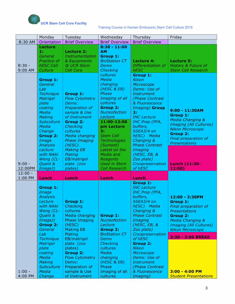
Monday Tuesday Wednesday Thursday Friday
8:30 AM Orientation Brief Overview Brief Overview Brief Overview
8:30 - 9:00 AM
Lecture 1: General Practice of hESC Cell Culture
Lecture 2: Instrumentation & Equipments @ UCR Stem Cell Core
8:30 - 11:00 AM Group 1: BioStation CT Demo Checking cultures Media changing (hESC & EB) Phase Imaging of all cultures Group 2: Nucleofection Lecture
Lecture 4: Differentiation of hESC
Lecture 5: History & Future of Stem Cell Research
9:00 - 12:00PM
Group 1: General Lab Technique Matrigel plate coating Media Making Subculture Media Change Group 2: Image Analysis Lecture with Nikki Weng (CL-Quant & ImageJ)
Group 1: Flow Cytometry Demo: Preparation of sample & Use of Instrument Group 2: Checking cultures Media changing Phase Imaging (hESC) Making EB Plating EB/matrigel plate (zoo plates)
Group 1: Nikon Microscope Demo: Use of Instrument (Phase Contrast & Fluorescence Imaging) Group 2: IHC Lecture IHC Prep (PFA, buffers, SSEA3/4 on hESC) Media Changing & Phase Contrast Imaging (hESC, EB, & Zoo plate) Cryopreservation of hESC
9:00 - 11:30AM Group 1: Media Changing & Imaging (All Cultures) Nikon Microscope Group 2: Final preparation of Presentations
11:00-12:00 pm Lecture 3: Stem Cell Technologies (Sumeet) Latest on the Media and Reagents Used in Stem Cell Research
Lunch (11:30-12:00)
12:00 - 1:00 PM Lunch Lunch Lunch Lunch
12:00 - 2:30PM Group 1: Final preparation of Presentations Group 2: Media Changing & Imaging (All Cultures) Nikon Microscope
1:00 - 4:00 PM
Group 1: Image Analysis Lecture with Nikki Weng (CL-Quant & ImageJ) Group 2: General Lab Technique Matrigel plate coating Media Making Subculture Media Change
Group 1: Checking cultures Media changing Phase Imaging (hESC) Making EB Plating EB/matrigel plate (zoo plates) Group 2: Flow Cytometry Demo: Preparation of sample & Use of Instrument
Group 1: Nucleofection Lecture Group 2: BioStation CT Demo Checking cultures Media changing (hESC & EB) Phase Imaging of all cultures
Group 1: IHC Lecture IHC Prep (PFA, buffers, SSEA3/4 on hESC) Media Changing & Phase Contrast Imaging (hESC, EB, & Zoo plate) Cryopreservation of hESC Group 2: Nikon Microscope Demo: Use of Instrument (Phase Contrast & Fluorescence Imaging)
2:30 - 3:00 BREAK
3:00 - 4:00 PM Student Presentations
UCR Stem Cell Core Facility Training Course in Human Embryonic Stem Cell Culture 2015
4
Chapter 1
Feeders and Substrates for Culturing Human Embryonic Stem Cells
Almost all available human ES cell lines commonly used in research are isolated from
blastocysts of embryos obtained by in vitro fertilization (IVF). These human ES cell lines were
first derived and cultured on mitotically inactivated feeder cells made from mouse embryonic
fibroblasts (MEF), or human foreskin fibroblast cells (for example, neonatal foreskin fibroblast
from ATCC, CRL-2429). Nevertheless, culture of human ES cells without mouse feeders is
critical for use of these cells in clinical applications. This would eliminate the risk of possible
cross-species and pathogenic contaminations from the animals.
Matrigel or Geltrex are mouse tumor-derived extracellular matrices ideal for human ES
cell culture, as they facilitate the attachment of human ES cells. The presence of matrix is
critical because it reduces selection pressure on the cells when they are freshly passaged. The
use of Matrigel or Geltrex has become the gold standard in maintaining human ES cells in
feeder-free system. Nevertheless, the use of these two substrates does not entail an animal-
free culture environment. The use of other substrates such as human recombinant laminin 521
(hrLN521, Biolamina, Product # LN521-03) and recombinant vitronectin XF (Stem Cell
Technologies) has also been established. In addition, a combination of laminin and E-cadherin
creates conditions for clonal expansion of pluripotent stem cells.
UCR Stem Cell Core Facility Training Course in Human Embryonic Stem Cell Culture 2015
5
Materials MEF Medium (10% FCS)
For 500 ml MEF medium: 1. 450 ml DMEM (high glucose)
(Invitrogen, 11965092; Lonza, 12-741 F) 2. 50 ml fetal calf serum (USA origin; Final concentration = 10%)
(Invitrogen, 16000044; HyClone, SH30070-03) 3. 5 ml 1X L-glutamine or 1X Glutamax I (100 X)
(Invitrogen 25030081; Invitrogen 35050061) 4. 5ml 1X Non Essential Amino Acid (NEAA; 100 X)
(Invitrogen, 11140050) 5. 5ml 1X sodium pyruvate (100 X)
(Invitrogen, 11360070) 0.25% Trypsin with EDTA (Invitrogen, 25200056) 1X PBS (without Ca2+, Mg2+) (Invitrogen, 10010023) Mitomycin C Medium
2mg of mitomycin C powder (Sigma, 128-K-0066) resuspended in 200ml of MEF media.
Filter-sterilized prior to use.
0.1% gelatin solution
0.1% gelatin (w/v) were prepared from lyophilized powder of Type A gelatin from Porcine (Sigma 128-K-0066) in distilled H2O. This solution must be autoclaved for dissolving gelatin power and to ensure sterility.
Coating:
BD Matrigel Basement Membrane Matrix
• Growth Factors reduced (BD Biosciences, 356231) • Regular (BD Biosciences, 356234)
Geltrex
• Growth factor reduced (Invitrogen, 12760021) • Regular, HESC-qualified (Invitrogen, 1048002)
UCR Stem Cell Core Facility Training Course in Human Embryonic Stem Cell Culture 2015
6
1.1 Derivation of mouse embryonic fibroblasts (MEF)
1. Perform a cervical dislocation on a 12.5-day to 13.5-day pregnant mouse.
CF-1 and MF-1 strains make good feeders; feeders derived from C57/BL6 or Black6 strain can be varied in quality.
2. Cover the abdomen with 70% ethanol. Cut through skin and peritoneum to expose the uterine horns, which are usually located beneath the intestines.
3. Remove the uterine horns and place in a Petri dish or a 50 ml tube containing PBS (without Ca2+/ Mg2+).
4. On a sterile Petri dish, remove the embryos from the embryonic sac by dissecting out
(‘pop out’) the embryos.
Discard the remaining placenta and other membranes. 5. Wash the embryos with PBS. Transfer the embryos to a new dish (prefilled with PBS).
Repeat this until the embryos become less bloody. 6. Decapitate (cut off the head) and eviscerate (remove the internal organs, if visible) the
embryos. Wash the remaining tissues with PBS. 7. Place the carcasses in a new Petri dish (prefilled with 5 ml trypsin) and then mince with
scalpel blades until tissues are in smaller pieces (smaller than 3 mm). 8. Incubate at 37oC for 10-20 minutes. 9. Transfer trypsinized embryo tissues into a new 15 ml centrifuge tube. Add 10 ml MEF
media. Triturate vigorously. 10. Pass the cell suspension through a syringe attached to a 25 G needle. Collect the pass
into a 15 ml centrifuge tube. 11. Allow large chunks to settle by gravity and then transfer supernatant to a T75 flask (or
T25 depending on the yield). Add 15 ml of MEF media + antibiotics. Triturate vigorusloy to obtain a homogenous cell suspension. This is MEF at Passage 0 (P0).
If yield is low, pellet from the large chunks can also be collected and cultured.
12. Incubate cells at 37oC overnight. 13. Change medium the next day to remove cellular debris. Grow the cells to 90%
confluency and then split the cells using trypsin (1:2 ratio into P1). MEF medium should be supplemented with antibiotics (Penicillin-Streptomycin; 1:100) and Gentamycin (1:1000) to avoid contamination.
UCR Stem Cell Core Facility Training Course in Human Embryonic Stem Cell Culture 2015
7
1.2 Passaging of mouse embryonic fibroblasts (MEF)
1. Passage MEF when they are 90% confluent by treatment with 0.25% trypsin.
2. Add 1.5 ml trypsin into a T75 flask. 3. Inactivate trypsin with MEF medium (9 ml MEF to 1 ml trypsin). Centrifuge at 1,200 rpm
for 3 mins. 4. Aspirate the supernatant, obtain cell pellet and gently flick the tube to disperse the cell
pellet. 5. Add MEF media and passage MEF at 1:2 or 1:3 dilution. Distribute the cells evenly onto
2 or 3 T75 flasks. There should be at least 15 ml of medium in a T75 flask. The use of antibiotics could be eliminated from P1.
6. MEF can be grown for 3 passages before they are used for embryonic stem cells feeders.
MEF-conditioned medium (CM): Seed irradiated or mitomycin C treated MEFs (P3) at ~55,000 cells/cm2 in MEF medium. After at least 4 h, exchange the MEF medium with ES medium (0.5 ml/cm2). Collect CM daily and supplement with an additional 4ng/ml of hbFGF before feeding hES cells. MEF CM medium can be collected daily from the same cells for 7-10 days. All collected CM can be filtered trough an ES cells certified filter and can be frozen for storage at -200C for one month and thawed for later use (reference: Nature 2001 Xu et al.).
1.3 Freezing of mouse embryonic fibroblasts (MEF)
1. MEF should be frozen at the earliest passage possible to ensure sufficient stock for future use. Maintain the remaining MEF for feeder cell derivation.
2. Trypsinize MEF. Inactivate trypsin with 9 ml of MEF medium. 3. Spin cells. Obtain pellet. Add 1.5 ml freezing medium (90% FBS and 10% DMSO) to
the pellet. Aliquot cell and freezing medium into 3 cryovials (500 µl in each cryovial). Label the cryovials clearly (eg MEF, P1, 1/3 T75, name and date)
1.4 Preparation of mouse embryonic fibroblast (MEF) feeder layers
1. Coat wells with 0.1% gelatin solution for at least 20 minutes.
2. Use MEF that are growing in log phase. MEF cells (P3) can be mitotically inactivated by either mitomycin C treatment or by irradiation.
UCR Stem Cell Core Facility Training Course in Human Embryonic Stem Cell Culture 2015
8
1.4.1 By mitomycin C treatment 1. Add MEF media with 10µg/ml mitomycin C to the cells for 1.5 hours. 2. Cells (on monolayer) are incubated in CO2 incubator at 37oC.
3. Aspirate mitomycin C solution well out. Wash the mitomycin C-treated cells 3 times with
PBS. 4. Harvest cells with 0.25% trypsin.
5. Resuspend the cells to a 10 ml tube and do a cell count.
6. Pellet the treated cells by centrifugation. Proceed to 1.4.3.
1.4.2 By irradiation 1. Trypsinize cells. Inactivate trypsin. Spin cells in a 15 ml tube. Obtain pellet.
2. Resuspend the pellet in 3 ml of MEF medium.
3. Place the tube in Faxitron irradiator. Irradiate cells at 60 kV or 75kV for 1 hour.
Irradiator has to be warmed up according to the manufacturer’s procedure (cycles of 30kV, 5 mins; 60kV, 5 mins; 90kV, 5 mins).
4. After 1 hour, centrifuge the irradiated cells. Proceed to 1.4.3.
1.4.3 Seeding MEF as Feeders
1. Seed MEF cells at 6x10³ cells per cm2 (1X 10⁵ cells per well of a 6-well plate; 2 X 10⁵ cells per T25 flask).
2. Feeders are ready to be used after overnight incubation. Good feeder layers should be
80% confluent with spaces available around the cells. Human ES cells attach to the space between the feeders and push the feeder cells when they proliferate and expand as colonies.

UCR Stem Cell Core Facility Training Course in Human Embryonic Stem Cell Culture 2015
9
1.5 Preparation of Matrigel or Geltrex
1.5.1 Aliquoting stock solution
1. Slowly thaw a vial of Matrigel or Geltrex at 4o C for at least overnight to avoid gel formation.
2. Gently mix thawed solution by swirling the bottle. 3. Aliquot solution into pre-chilled 1.7ml microcentrifuge tubes.
Aliquot must be done fast enough to avoid formation of gel.
4. Store at -20oC.
1.5.2 Coating plates with BD Matrigel or Geltrex LDEV-free hESC qualified
1. Slowly thaw 1 ml aliquot of Matrigel or Geltrex stock for at least overnight at 4o C. 2. Diluting subtrate:
For Matrigel:
Add 30 ml of cold DMEM; pipette to mix (1:30 dilution).
For Geltrex:
Add 100 ml of cold DMEM; pipette to mix (1:100 dilution).
3. Add 1 ml of diluted Matrigel to each well of a 6 well plate. 4. Incubate at 37oC for 1 hour or until needed.
Matrigel-coated plates can be kept in incubators as long as the solution is not dried up.
Alternatively, aspirate Matrigel or Geltrex from the wells after complete coating and keep the plates at 4 oC. Warm up plate in 37oC incubator before use.
5. Remove Matrigel or Geltrex and plate freshly split human ES cells on the coated plates.
UCR Stem Cell Core Facility Training Course in Human Embryonic Stem Cell Culture 2015
10
1.6 Coating plates with Vitronectin XF
Vitronectin XF manufactured by Stem Cell Technologies (SCT) is xeno-free alternative to Matrigel and Geltrex. The use of Vitronectin XF results in consistent cell growth of hESCs and iPSCs. It is currently our matrix of choice for growing human pluripotent stem cells at the Core.
Coating Culture Vessels with Vitronectin XF
Although SCT recommends the use of non-tissue culture treated vessels for the use of Vitronectin XF, we have used tissue culture treated vessels and have obtained satisfactory results. The following coating procedure is for tissue culture treated 6-well plates, if using other culturing vessels then adjustments should be made to the volumes needed to coat the specific culturing vessel.
1. Thaw Vitronectin XF at room temperature (15-25oC) or at 4oC overnight.
To avoid additional freeze-thaw cycles aliquot appropriate amounts of Vitronectin XF into 1.5ml micro-centrifuge tubes and store at -20oC or -80oC. Vitronectin XF is stable for two weeks at 4oC. Once is thawed, a final concentration of 10ug/ml is needed to coat culture vessels.
2. Dilute Vitronectin XF (250ug/ml) with 1xPBS (w/o Ca2+&Mg2+) to reach a final concentration of 10ug/ml (i.e. use 40 ul of Vitronectin XF per 1ml of PBS). Use an appropriate polypropylene conical tube (15ml or 50ml) to dilute the Vitronectin XF.
3. Gently mix the diluted Vitronectin XF inverting the tube 2-3 times and immediately use the diluted Vitronectin XF solution to coat tissue culture treated 6-well plates.
4. Add 1ml/well of the 6-well plate and gently rock and swirl the 6-well plate back and forth to spread the Vitronectin XF solution evenly across the surface.
Note: if using non-tissue culture-treated vessels for coating with Vitronectin XF make sure the surface is fully coated by the Vitronectin XF solution - additional rocking and swirling may be required in order to coat the hydrophobic surface.
5. Incubate at room temperature for at least 1 hour before use. Do not let the Vitronectin XF solution evaporate.
Note: if not used immediately, the culturing vessels can be stored at 4oC as long as evaporation of the Vitronectin XF does not occur. Six-well plates can be sealed with parafilm to prevent evaporation of the Vitronectin XF. Allow coated culture vessels to come to room temperature for 30 minutes before use.
6. Remove excess Vitronectin XF solution.
7. Add 1ml/well of mTeSR media - cells can then be added to each well at desired passage ratio.
UCR Stem Cell Core Facility Training Course in Human Embryonic Stem Cell Culture 2015
11
Chapter 2
Culture and Maintenance of Human Embryonic Stem Cells
Human embryonic stem (ES) cells can be grown on mouse embryonic fibroblast (MEF) feeder
layers (feeder-dependent) or on certain substrates (feeder-free cultures). If human ES cells
are grown on a substrate, human ES medium conditioned by MEF (conditioned medium, “CM”)
should be used in order to compensate for growth factors and components secreted by or
present in MEF feeder cells. Several other commercially available, feeder- and serum-free
maintenance media, such as STEMPRO (Invitrogen, A1000701) or mTeSR1 medium (Stem
Cell Technologies, 05850) have become increasingly popular amongst the choices of media
used to maintain the growth of human ES cells in a feeder-free system. Media free of animal
protein are also available such as mTeSR2 (derived from mTeSR1), TeSR-E8 (Stem Cell
Technologies) and L7 (Lonza Biosciences Inc).
UCR Stem Cell Core Facility Training Course in Human Embryonic Stem Cell Culture 2015
12
Materials:
Feeder-dependent culture
Human ES medium (20% KOSR)
For 500 ml human ES medium:
1. 400 ml Knockout DMEM/ F12 2. 100 ml Knockout SR (KOSR; 20%) 3. 5 ml 1X Glutamax I (100x) 4. 5 ml Non-Essential Amino Acid solution (NEAA) (100x) 5. 500 ul bFGF (Final concentration = 4 - 10 ng/ml) 6. 500 ul 2-mercaptoethanol (BME; 55mM; Final concentration = 1.5 nM)
It is advisable to filter-sterilize prior to use.
Store at 4oC and use within two weeks.
KO DMEM/F12 (Invitrogen 12660012; The use of DMEM/F12 will slow down the growth of the colonies) bFGF (Invitrogen PHG0263)
Feeder-free culture
(1) STEMPRO medium kit (Invitrogen, A1000701)
For 500 ml STEMPRO medium:
1. 454 ml Knockout DMEM/ F12 2. 10 ml STEMPRO human ES serum-free growth supplement (50X) 3. 5 ml 1X Glutamax I (100x) 4. 36 ml BSA (Bovine serum albumin, 25%) 5. 500 ul bFGF stock (Final concentration = 10 ng/ml) 6. 909 ul 2-mercaptoethanol (BME; 55mM; Final concentration = 55 nM)
It is advisable to filter-sterilize prior to use.
(2) mTESR 1 medium kit (Stem Cell Technologies, 05850)
For 500 ml mTESR medium:
1. 400 ml mTESR 1 basal medium 2. 100 ml 5X supplement (already include bFGF)
UCR Stem Cell Core Facility Training Course in Human Embryonic Stem Cell Culture 2015
13
Human recombinant bFGF Solubilize 10ug lyophilized powder in 5 ml PBS (w/o Ca++, Mg++) with 0.1% BSA. Aliquot and store at -20oC. Repeated thawing and freezing must be avoided. 2.1 Passaging of human ES cells on MEF feeders
1. Aspirate media from 6-well plate.
2. Add 1 ml of collagenase IV per well and incubate at 37oC for 3-5 minutes until the
edges of the colonies start to curl. Accutase can also be used (only 1 min incubation). Longer incubation is not advisable. However, a badly differentiated human ES cell culture can be incubated with collagenase IV for up to 30 minutes. Undifferentiated human ES cell colonies will detach, leaving behind feeders and any spontaneously differentiated cells on the original flask.
3. Gently roll the colonies off with glass beads. 4. Add 5 ml of DMEM and gently triturate. Transfer cell suspension to a 10 ml centrifuge
tube. 5. Spin at 800rpm for 3 min at RT. 6. Aspirate supernatant, leaving human ES cell pellet intact. 7. Remove medium from the fresh well of MEF feeders. 8. Gently flick human ES cell tube to disperse pellet. 9. Gently resuspend human ES cell pellet in an appropriate volume of human ES medium
(e.g. 3 ml for a 1:3 split into three wells of a 6-well plate (at 2 ml per well). Distribute between wells of feeders.
Add 2 ml human ES media per well for the first day of passaging. Gradually increase the volume of human ES media to ensure sufficient medium to support the growth of cells.
10. Carefully place the 6-well plate into CO2 incubator. After placing the plate down, swirl carefully to ensure an even distribution of cells across the flask.
11. Human ES cells are to be fed with fresh medium daily, if possible. Cells are grown until they reach 80-90% confluency before they are ready to be split again.
Cells from 1 well on a 6-well plate typically yield 1.5- 2 million cells.
UCR Stem Cell Core Facility Training Course in Human Embryonic Stem Cell Culture 2015
14
Collagenase IV solution (1 mg/ml) 1. Weigh out 50 mg of collagenase type IV (Invitrogen, 7104019)
2. Solubilize lyophilized powder in 50 ml DMEM/F12 3. Filter-sterilize with a 0.2micron cellulose acetate filter. Store at 4oC. Use within 2 weeks. 2.2 Passaging of human ES cells on Matrigel or Geltrex
The use of higher concentration of bFGF is required for feeder-free culture. We have found that cells are best to be passaged by accutase when cells are grown without feeders.
1. Precoat new wells with Matrigel or Geltrex at 37oC for 3 hours. 2. Add 1 ml of accutase per well and incubate at 37oC for 1-3 minutes until most cells
detach. 3. Gently roll the colonies off with glass beads. Alternatively, use 5 ml serological pipette
to gently scrape the colonies. 4. Add 5 ml of DMEM and gently triturate. Transfer cell suspension to a 10 ml centrifuge
tube. 5. Spin at 800rpm for 3 min at RT. 6. Aspirate supernatant from the tube, leaving human ES cell pellet intact. 7. Remove Matrigel or Geltrex from the wells. 8. Optional: Gently flick tube to disperse pellet. 9. Using a 1000 µl pipet tip, gently resuspend human ES cell pellet in an appropriate
volume of STEMPRO or mTESR 1. Distribute between wells of feeders. 10. Carefully place into CO2 incubator. Swirl the plate carefully to ensure an even
distribution of cells across the flask. 11. Feed cells daily until cells are ready to be split again (when cells reach 90%
confluency).
See Page 7 for procedure to prepare CM from MEF.
UCR Stem Cell Core Facility Training Course in Human Embryonic Stem Cell Culture 2015
15
2.3 Passaging of hESCs/iPSC on Vitronectin XF by using Lonza L7 hPSC Passaging Solution
L7 hPSC passaging solution is a chemically defined, non-animal origin, non-enzymatic cell detachment formulation based on a hypertonic sodium citrate solution manufactured by Lonza. L7 hPSC passaging solution gently dislodges hESC colonies from a substrate without the need of mechanical manipulation of cultures.
NOTE: The following passaging procedure is written for cells grown in tissue culture treated 6-well plates, if using other culturing vessels then adjustments should be made to the volumes needed for the specific culturing vessels.
1. Remove and discard cell culture medium from each well.
2. Add 1ml/well of 1xPBS and gently wash cells by rocking and swirling the plate back and forth.
3. Remove and discard 1xPBS.
4. Add 1ml/well of L7 hPSC Passaging Solution.
5. Incubate for 5 to 15 minutes at room temperature or for 3 to 10 minutes at 37oC – 3-5 minutes in the incubator has shown to be optimal on stem cells grown in the UCR Stem Cell Core: NOTE: Treatment times may vary and are dependent on culture confluence, cell line and the type of culture medium and matrix. Monitor cultures until an accurate treatment time has been determined.
6. Monitor culture under the microscope until the following characteristics are observed: a. Cells are rounding up and gaps begin to appear between groups of cells in the
colonies. b. Cells at the edge of the colonies begin to lift from the plate.
7. After incubation, remove and discard the L7 hPSC Passaging Solution.
***The majority of the cells should remain attached to the culture surface***.
Over-treatment of colonies can occur and can result in complete detachment of all cell colonies. If this occurs do the following:
Collect passaging solution + cells.
Centrifuge for 3 minutes at 800 rpm.
UCR Stem Cell Core Facility Training Course in Human Embryonic Stem Cell Culture 2015
16
Remove supernatant and re-suspend in appropriate amount of media and directly seed the cells onto coated culture vessels.
8. Tilt the plate and gently detach cellular colonies by rinsing the cell aggregates off the surface with fresh hPSC medium (Stem Cell Technologies mTeSR1 media (Catalog # 05850), or similar) NOTE: It is crucial to define the treatment time for each cell line being used. Cells should easily be rinsed off culture surface. Excessive pipetting will result in single cells and therefore reduced cell viability.
9. Collect the detached cell aggregates and seed directly at desired cell density, onto coated culture vessels with Stem Cell Technologies Vitronectin XF (Catalog# 07180), or similar).
10. At this point centrifugation is optional. If a cell pellet is desired perform the following: a. After collecting the detached cell aggregated in desired media centrifuge for 3
minutes at 800 rpm. b. Remove supernatant and re-suspend in appropriate amount of media and
directly seed the cells onto coated tissue culture vessels
2.4 Troubleshooting: Picking colonies
Human ES cell cultures are heterogeneous. It is not uncommon for spontaneously differentiated cells to grow in cultures. Differentiated human ES cell cultures can be rescued by several different methods. 1. Eliminate bad colonies:
Areas of spontaneous differentiation can be removed from the well by aspirating pipet and pipet tip.
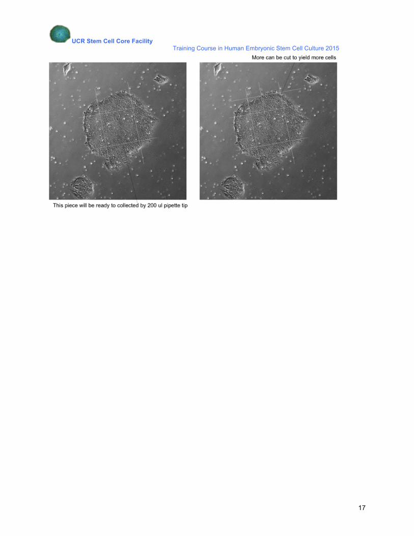
2. Picking good colonies: Undifferentiated colonies can be selected by either: (a) Pick colonies: 1. Pick colonies with a sterile 200 µl pipette tip 2. Transfer them to fresh feeder layer or substrate. (b) ‘Cut and Paste’:
1. ‘Cut’ one colony into 9 squares with 25G needle (2 lines vertically and horizontally).
2. ‘Paste’ the cut pieces of cells on new feeders or substrate
UCR Stem Cell Core Facility Training Course in Human Embryonic Stem Cell Culture 2015
17
UCR Stem Cell Core Facility Training Course in Human Embryonic Stem Cell Culture 2015
18
Chapter 3
Differentiation of Human Embryonic Stem Cells
Human ES cells differentiate spontaneously upon culturing in small aggregates termed
embryoid bodies (EBs) in non-adherent culture, or simply in an overgrown (over-confluent)
monolayer adherent culture. The formation of EBs recapitulate the three-dimensional
complexity of the embryo during gastrulation and usually results in the formation of precursor
cells representing the three germ layers of the embryo. Alternatively, when cells are allowed to
overgrow in monolayer cultures, provided that the medium is changed everyday (to avoid cell
death), the suboptimal culture condition will prompt the cells to differentiate. Cells can be
transferred to a gelatin-coated plate in a 1:3 dilution with appropriate medium for example,
DMEM with 10% FCS with BMP4 (Humanzyme, HZ-1046). Nevertheless, it should be
emphasized that cells in EB or confluent monolayer in vitro are differentiated in a disorganized
manner, as the correct order of developmental cues has been lost.
UCR Stem Cell Core Facility Training Course in Human Embryonic Stem Cell Culture 2015
19
Materials:
EB medium: Standard KnockOut human ES medium (with or without bFGF)
Mesoderm and Germ line cell differentiation medium:
HESC media (-FGF2) and 50 ng/ml BMP4
Neural differentiation medium: DMEM/F12 (Invitrogen 11965018) or Neurobasal medium supplemented with B27 (Invitrogen 7504044), N2 (Invitrogen 17502048) or retinoic acid.
Endoderm differentiation medium: RPMI-1640 (Invitrogen 11875093) Activin A (Humanzyme, HZ-1136)
Coating:
Poly-D-lysine (Sigma, P0296), 5 mg solubilized in 50 ml sterile H2O.
UCR Stem Cell Core Facility Training Course in Human Embryonic Stem Cell Culture 2015
20
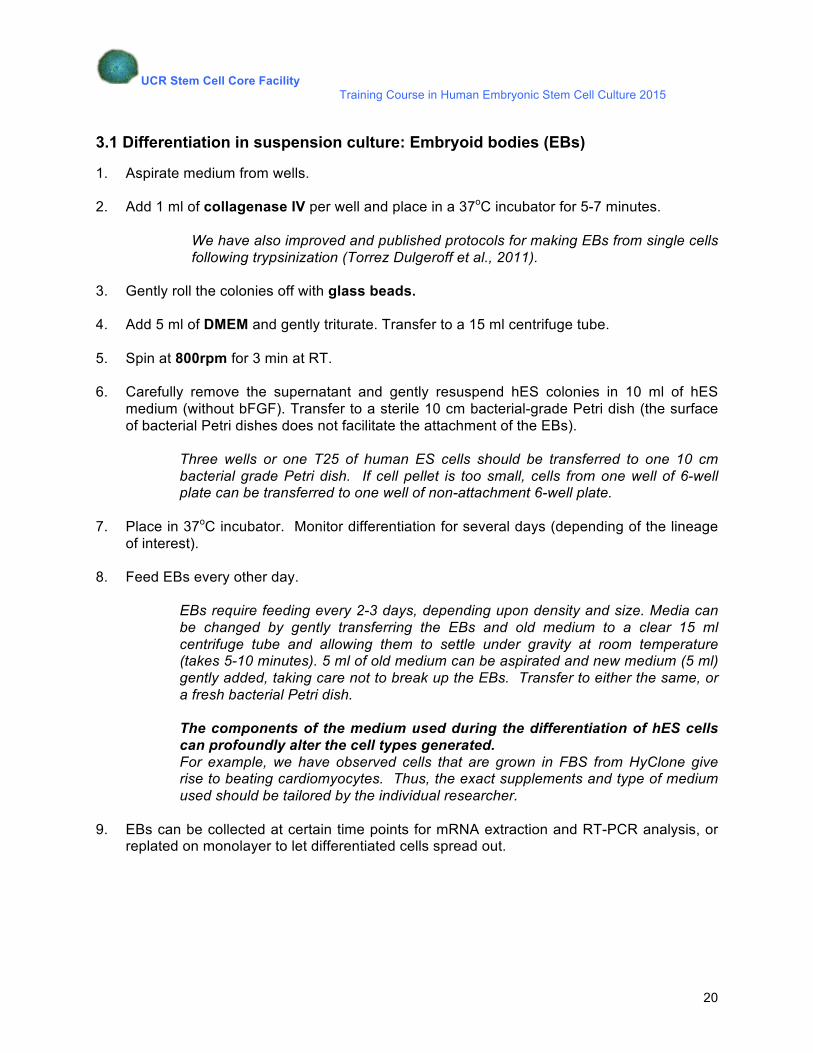
3.1 Differentiation in suspension culture: Embryoid bodies (EBs)
1. Aspirate medium from wells.
2. Add 1 ml of collagenase IV per well and place in a 37oC incubator for 5-7 minutes.
We have also improved and published protocols for making EBs from single cells following trypsinization (Torrez Dulgeroff et al., 2011).
3. Gently roll the colonies off with glass beads.
4. Add 5 ml of DMEM and gently triturate. Transfer to a 15 ml centrifuge tube.
5. Spin at 800rpm for 3 min at RT.
6. Carefully remove the supernatant and gently resuspend hES colonies in 10 ml of hES
medium (without bFGF). Transfer to a sterile 10 cm bacterial-grade Petri dish (the surface of bacterial Petri dishes does not facilitate the attachment of the EBs).
Three wells or one T25 of human ES cells should be transferred to one 10 cm bacterial grade Petri dish. If cell pellet is too small, cells from one well of 6-well plate can be transferred to one well of non-attachment 6-well plate.
7. Place in 37oC incubator. Monitor differentiation for several days (depending of the lineage
of interest).
8. Feed EBs every other day. EBs require feeding every 2-3 days, depending upon density and size. Media can be changed by gently transferring the EBs and old medium to a clear 15 ml centrifuge tube and allowing them to settle under gravity at room temperature (takes 5-10 minutes). 5 ml of old medium can be aspirated and new medium (5 ml) gently added, taking care not to break up the EBs. Transfer to either the same, or a fresh bacterial Petri dish.
The components of the medium used during the differentiation of hES cells can profoundly alter the cell types generated. For example, we have observed cells that are grown in FBS from HyClone give rise to beating cardiomyocytes. Thus, the exact supplements and type of medium used should be tailored by the individual researcher.
9. EBs can be collected at certain time points for mRNA extraction and RT-PCR analysis, or replated on monolayer to let differentiated cells spread out.
UCR Stem Cell Core Facility Training Course in Human Embryonic Stem Cell Culture 2015
21
3.2 Replating embryoid bodies in monolayer
1. Obtain pellet of EBs. Collect EBs from dish in a 10 ml centrifuge tube. Let it stand for 10 minutes.
2. Aspirate supernatant.
3. Add appropriate medium (eg MEF or neuro media) to the EBs and replate the EBs in precoated plate.
We have also published protocols for generating neural and retinal progenitors from replated EBs (Torrez Dulgeroff et al., 2011).
3.3 Induction of differentiation in monolayer culture
For monolayer differentiation, cells were induced to differentiate to different germ layers and
germline cells using specific growth factors and morphogens as follow.
For definitive endoderm differentiation, cells were treated with 100 ng/ml Activin A
(Humanzyme) and 25 ng/ ml Wnt3a (R& D Systems) in RPMI1640 media for 1 day and continue
to culture for 3-5 days with 100ng/ml Activin A in 0.2-2% FBS (Hyclone) from day 2-3 and day 3-
5, respectively.
For mesoderm and PGC differentiation, cells were incubated with 50ng/ml bone morphogenetic
protein-4 (BMP4) in HESC media (-FGF2) for 2 and 5 days respectively.
Cells were treated with neurobasal media (Invitrogen) supplemented with B27 (Invitrogen), N2
(GIBCO), 2 µM dorsomorphin (Compound C, Sigma Alrich) and 0.1 µM retinoic acid (RA, Sigma
Aldrich) for 5 days to induce neuroectoderm differentiation.
UCR Stem Cell Core Facility Training Course in Human Embryonic Stem Cell Culture 2015
22
Chapter 4
Labeling and Sorting of Human Embryonic Stem Cells
Human ES cells can be characterized based on the surface antigen marker expression analysis
on the cells. First introduced in the analyses of hematopoietic stem cells, it was an approach
adopted in early 1980s to study the surface marker expression of mouse embryonal carcinoma
(EC) and ES cells. Stage-specific embryonic antigen-1 (SSEA1) is widely accepted as a
standard marker for undifferentiated mouse EC, ES and also primordial germ cells (PGCs), from
which embryonic germ (EG) cells are derived from. However, human EC (e.g. NTERA2 cells)
and ES cells do not express SSEA1, but SSEA3, SSEA4 and a series of related sulfate
proteoglycans, notably TRA-1-60, TRA-1-81 and GCTM2. Strikingly, cells from inner cell mass
(ICM) of human embryos also express surface antigens in common to human ES cells,
suggesting that the differences between human and mouse ES cells are most likely attributed to
the differences in developmental stages of the embryos.
UCR Stem Cell Core Facility Training Course in Human Embryonic Stem Cell Culture 2015
23
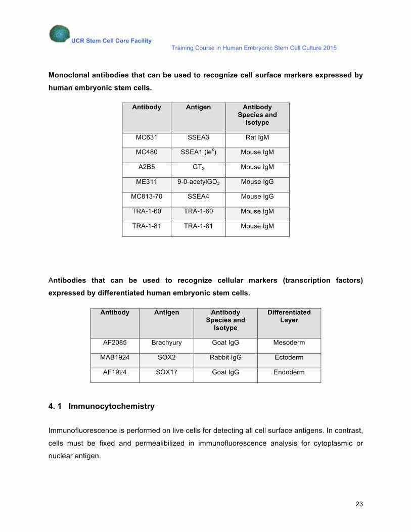
Monoclonal antibodies that can be used to recognize cell surface markers expressed by
human embryonic stem cells.
Antibody Antigen Antibody Species and
Isotype
MC631 SSEA3 Rat IgM
MC480 SSEA1 (lex) Mouse IgM
A2B5 GT3 Mouse IgM
ME311 9-0-acetylGD3 Mouse IgG
MC813-70 SSEA4 Mouse IgG
TRA-1-60 TRA-1-60 Mouse IgM
TRA-1-81 TRA-1-81 Mouse IgM
Antibodies that can be used to recognize cellular markers (transcription factors) expressed by differentiated human embryonic stem cells.
Antibody Antigen Antibody Species and
Isotype
Differentiated Layer
AF2085 Brachyury Goat IgG Mesoderm
MAB1924 SOX2 Rabbit IgG Ectoderm
AF1924 SOX17 Goat IgG Endoderm
4. 1 Immunocytochemistry
Immunofluorescence is performed on live cells for detecting all cell surface antigens. In contrast,
cells must be fixed and permealibilized in immunofluorescence analysis for cytoplasmic or
nuclear antigen.
UCR Stem Cell Core Facility Training Course in Human Embryonic Stem Cell Culture 2015
24
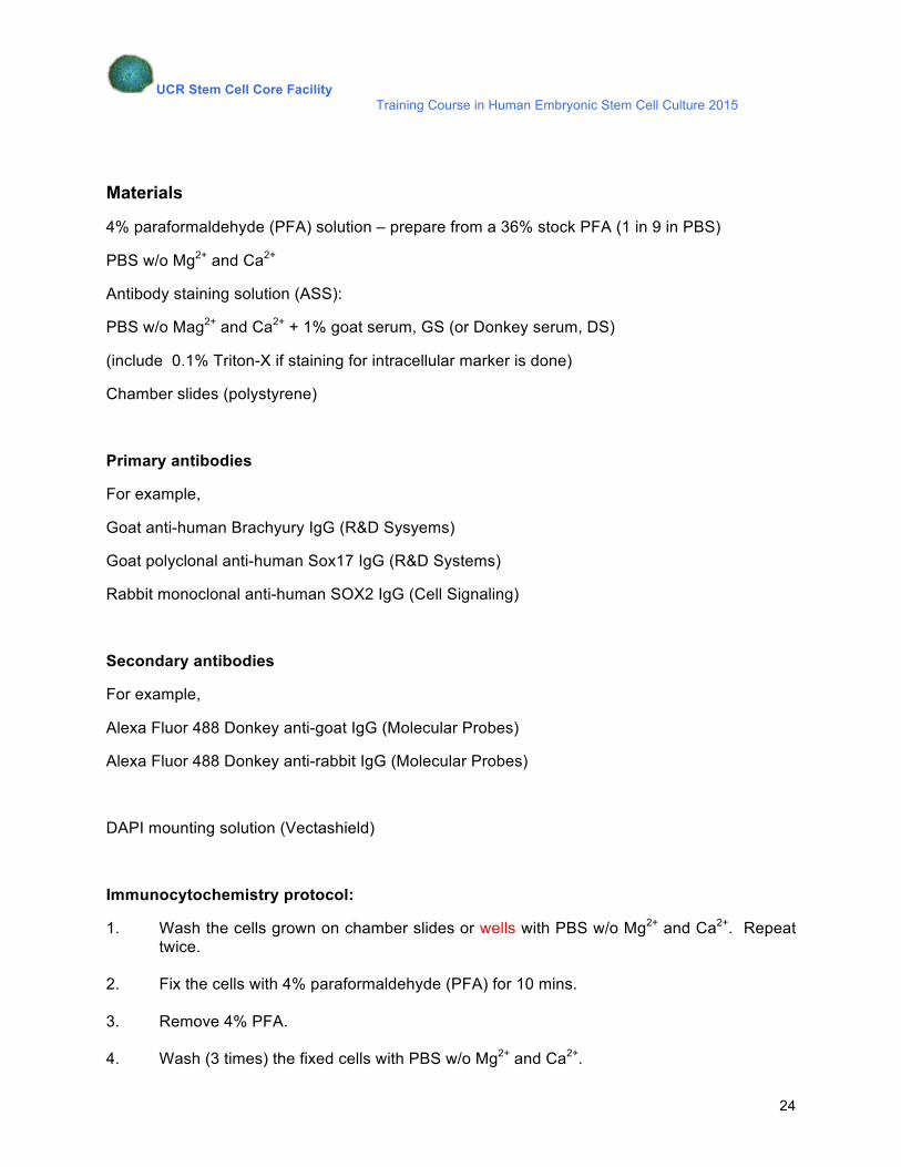
Materials
4% paraformaldehyde (PFA) solution – prepare from a 36% stock PFA (1 in 9 in PBS)
PBS w/o Mg2+ and Ca2+
Antibody staining solution (ASS):
PBS w/o Mag2+ and Ca2+ + 1% goat serum, GS (or Donkey serum, DS)
(include 0.1% Triton-X if staining for intracellular marker is done)
Chamber slides (polystyrene)
Primary antibodies
For example,
Goat anti-human Brachyury IgG (R&D Sysyems)
Goat polyclonal anti-human Sox17 IgG (R&D Systems)
Rabbit monoclonal anti-human SOX2 IgG (Cell Signaling)
Secondary antibodies
For example,
Alexa Fluor 488 Donkey anti-goat IgG (Molecular Probes)
Alexa Fluor 488 Donkey anti-rabbit IgG (Molecular Probes)
DAPI mounting solution (Vectashield)
Immunocytochemistry protocol:
1. Wash the cells grown on chamber slides or wells with PBS w/o Mg2+ and Ca2+. Repeat twice.
2. Fix the cells with 4% paraformaldehyde (PFA) for 10 mins.
3. Remove 4% PFA.
4. Wash (3 times) the fixed cells with PBS w/o Mg2+ and Ca2+.
UCR Stem Cell Core Facility Training Course in Human Embryonic Stem Cell Culture 2015
25
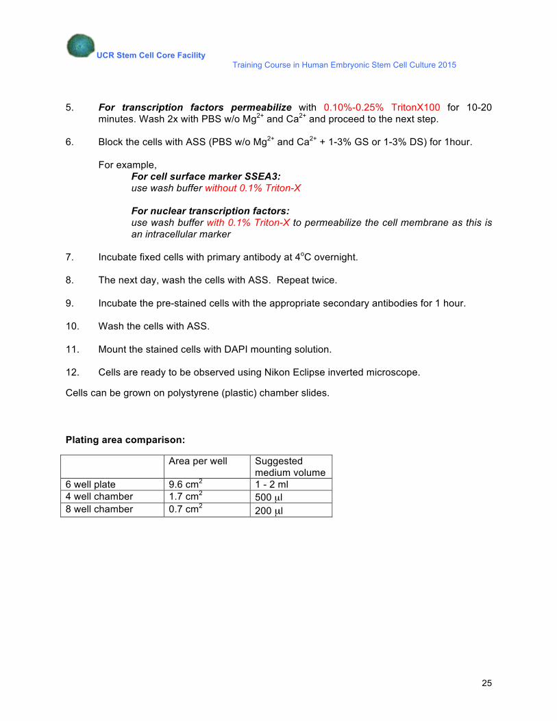
5. For transcription factors permeabilize with 0.10%-0.25% TritonX100 for 10-20
minutes. Wash 2x with PBS w/o Mg2+ and Ca2+ and proceed to the next step.
6. Block the cells with ASS (PBS w/o Mg2+ and Ca2+ + 1-3% GS or 1-3% DS) for 1hour.
For example, For cell surface marker SSEA3: use wash buffer without 0.1% Triton-X For nuclear transcription factors: use wash buffer with 0.1% Triton-X to permeabilize the cell membrane as this is an intracellular marker
7. Incubate fixed cells with primary antibody at 4oC overnight.
8. The next day, wash the cells with ASS. Repeat twice.
9. Incubate the pre-stained cells with the appropriate secondary antibodies for 1 hour.
10. Wash the cells with ASS.
11. Mount the stained cells with DAPI mounting solution.
12. Cells are ready to be observed using Nikon Eclipse inverted microscope.
Cells can be grown on polystyrene (plastic) chamber slides.
Plating area comparison:
Area per well Suggested medium volume
6 well plate 9.6 cm2 1 - 2 ml 4 well chamber 1.7 cm2 500 µl 8 well chamber 0.7 cm2 200 µl
UCR Stem Cell Core Facility Training Course in Human Embryonic Stem Cell Culture 2015
26
Chapter 5
Cryopreservation of Human Embryonic Stem Cells
It is critical to freeze and bank your human ES cells at the earliest passage number if possible.
Thus it is not advisable to throw excess cells away, but to freeze them. You will be able to
retrieve cells from early passage numbers if you encounter a problem with the current cell stock
(cell death, contamination, or just when the cells are getting too old, or exhibit karyotypic
changes). Nevertheless, poor survival of human ES cells after cell thawing is an obstacle to
research. Here we have revised an improved protocol to freeze your human ES cells that yield
higher survival and recovery rate upon thawing.
UCR Stem Cell Core Facility Training Course in Human Embryonic Stem Cell Culture 2015
27
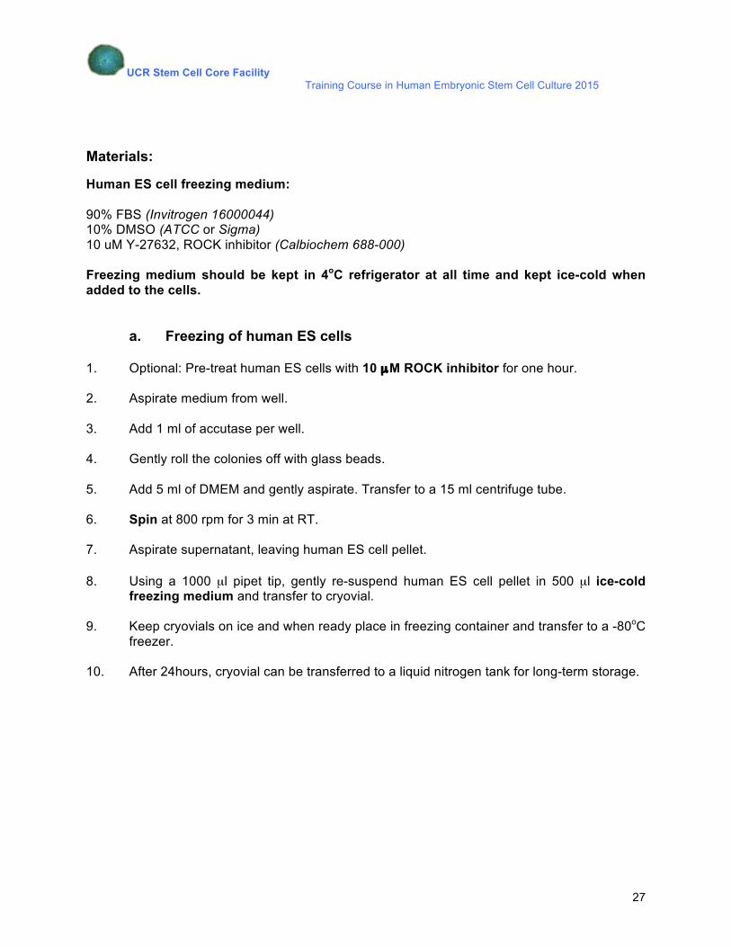
Materials:
Human ES cell freezing medium: 90% FBS (Invitrogen 16000044) 10% DMSO (ATCC or Sigma) 10 uM Y-27632, ROCK inhibitor (Calbiochem 688-000) Freezing medium should be kept in 4oC refrigerator at all time and kept ice-cold when added to the cells.
a. Freezing of human ES cells 1. Optional: Pre-treat human ES cells with 10 µM ROCK inhibitor for one hour.
2. Aspirate medium from well.
3. Add 1 ml of accutase per well. 4. Gently roll the colonies off with glass beads. 5. Add 5 ml of DMEM and gently aspirate. Transfer to a 15 ml centrifuge tube. 6. Spin at 800 rpm for 3 min at RT. 7. Aspirate supernatant, leaving human ES cell pellet. 8. Using a 1000 µl pipet tip, gently re-suspend human ES cell pellet in 500 µl ice-cold
freezing medium and transfer to cryovial. 9. Keep cryovials on ice and when ready place in freezing container and transfer to a -80oC
freezer. 10. After 24hours, cryovial can be transferred to a liquid nitrogen tank for long-term storage.
UCR Stem Cell Core Facility Training Course in Human Embryonic Stem Cell Culture 2015
28
b. Thawing of human ES cells
1. Remove cryovial from freezer and slightly loosen cap to allow trapped nitrogen to
escape.
2. Thaw by immersing the bottom half of the vial in a 37oC water bath and swirl the tube around.
Do not immerse the whole tube in the water bath, this could lead to contamination problems.
3. Using a 1000 µl pipet tip, take up 1 ml of prewarmed DMEM.
4. Gently add DMEM to the cells. 5. Transfer the content of the vial to a 15 ml centrifuge tube.
6. Gradually add 10 ml of prewarmed DMEM to the tube dropwise.
7. Spin down at 800 rpm for 3 mins.
8. Aspirate medium.
9. Resuspend human ES cell pellet in an appropriate volume of human ES medium or
mTESR and transfer to the flask with feeders or properly coated plate. 10. Carefully place well plate in CO2 incubator, maintaining an even distribution of cells
across the flask. We find that the best results are achieved when defrosted cultures are re-grown onto an area
less then that originally frozen (e.g. a cryovial containing 1 well of a 6-well plate would be
defrosted into 1 well of a 12-well plate). Additionally, the use of 10 µM ROCK inhibitor may
improve cell recovery after thawing.
UCR Stem Cell Core Facility Training Course in Human Embryonic Stem Cell Culture 2015
29
Chapter 6
Transfection of hESCs by using
Lonza Biosciences Nucleofection Technology
Dissociating Pluripotent Stem Cells with Lonza L13 hPSC Passaging Solution
1. Aseptically remove and discard the spent medium from the culture vessel.
2. Wash the attached cell layer with a calcium- and magnesium-free balanced salt solution, D-PBS. Add 1 ml of the wash solution per well of a 6-well plate. Evenly distribute the solution onto the culturing surface using the techniques instructed by the manufacturers of the vessels. Rinse by rocking the vessel back and forth several times. Aseptically remove and discard the wash solution being careful not to disturb the cell culture surface.
3. Add 1 ml of L13 Passaging Solution per well of a 6-well plate and evenly distribute to all the culturing surfaces.
4. Incubate at room temperature for five to fifteen minutes, or 37ºC for five to ten minutes, monitoring under a microscope for the following signs: • Cells rounding up and colonies appearing to be bright white in color • Gaps appearing among cells in a colony • Edges of colonies may beginning to shrink or lift from the plate • Some small colony fragments lifting off into solution
NOTE: Treatment times may vary with culture confluence, cell line, and type of culture medium and matrix. Monitoring during treatment is only required until an ideal treatment time can be determined for each culturing condition.
5. Aseptically remove and discard the L13 Passaging Solution leaving most of the cells still attached to the culture surface.
6. Add 1 ml of fresh D-PBS and evenly distribute to all the culturing surfaces. Tap and rock the vessels to dislodge the cells in the medium.
7. Collect the detached cell clusters into a sterile conical tube.
UCR Stem Cell Core Facility Training Course in Human Embryonic Stem Cell Culture 2015
30
Pluripotent Stem Cells Nucleofection with the Nucleofector 11B
• The PSC demo will consist of a targeted cell optimization, in which we will test up to 5 specific programs designed for human ES transfections and 2 specific human ES buffers. At the conclusion of the demo, we will have identified the single best parameter and buffer required for future down-stream transfections when using this cell type and therefore will not have to further optimize or go through this optimization process again.
• The demo will be accomplished using Lonza pmaxGFP Plasmid (comes with the kit). The Nucleofection process is substrate independent, so the observed/measured efficiency will not vary from substrate to substrate, as the yields are determined by the cell line/program & not the substrate itself. The pmaxGFP is a 3.5kb with a CMV promoter. It is not that this substrate is necessarily easy to transfect, but often times, construct design can have internal design challenges preventing the expression and this construct eliminates this/any variables. It is also modified to be brighter than a typical eGFP, which makes it easy tool to identify the proper parameter using a fluorescent microscope on day two. Look at cell morphology, efficiency and viability on day two will be used to determine qualitatively, but accurately, best condition.
• We are going to test two different buffers (Buffer 1 and Buffer 2) based on the recommendations from the Lonza optimized protocol (reference Lonza Protocols on the Lonza web site). The buffers do make a big difference. In addition to testing the two specific buffers, we are going to test 5 unique human ES specific programs per buffer & 2 no-pulse controls (one for each buffer). Essentially, 12 total transfections. Each participant will be responsible for one condition.
• We’ll need 8x10e5 cells per transfection, which should equate to approximately one well of a 6 well plate. The cells should be passaged 3 to 5 days before Nucleofection, and should be at 70 - 85% confluency on the day of the demo. For best viability results, cells should be treated with ROCK inhibitor for at least one hour prior to starting the dissociation and Nucleofection process.
• We don’t want to centrifuge the cells more than once in the nucleofection process, so when taking a cell count to determine the volume of culture media containing cells needed, we ask that you take a rough estimate and not spin/pellet in that process. The number of centrifuges & specific spin-speed at which we pellet is the most crucial step in the Nucleofection process. Each participant will take their tube of cells from the dissociation step above and spin the centrifuge tube at 90xg for 3 minutes, before re-suspending the pellet in the respected Nucleofector solutions.
• The buffers come in two components (solution & supplement). We will need to reconstitute the two components together to create the nucleofection buffer. Pre-mixed, each is good for 1 year at 4 degrees C. Once mixed, the buffer is good for 90 days.
UCR Stem Cell Core Facility Training Course in Human Embryonic Stem Cell Culture 2015
31
• After adding the supplemented nucleofection buffers, we will add the pmaxGFP construct. The amount of construct will vary depending on the type, but when using pmaxGFP, we will add 2ug per transfection, therefore a total of 12ug (12ul – as it is concentrated at 1ug/ul) for all the participants to share.
• Once pelleted, we will aspirate off the culture media and re-suspend in the respective Nucleofector buffers + suppliments + pmaxGFP. We will re-suspend each of the pellets in 100 ul of the prepared buffer and transfer the contents to a Nucleo-cuvette.
• Each participant will Nucleofect with one program, A-12, A-13, A-23, A27, or B-16.
• Once the Nucleofection is complete, you will IMMEDIATELY add warm media to the cuvette and transfer the contents to the appropriate destination well utilizing the supplied pipet.
• The plating density (post transfection) is the second most important aspect to consider and vital to recovery post transfection and overall cell health, therefore setting up the 24-well destination plates coated with matrix and warm media prior to getting started with the experiment will be the first step in the process. For best results, treat this Nucleofected cultures with ROCK Inhibitor overnight.
• Analyze the cultures for cell viability and GFP expression efficiency between 12 and 48 hours post Nucleofection.
UCR Stem Cell Core Facility Training Course in Human Embryonic Stem Cell Culture 2015
32
Appendix I
Cell Lab Quanta Demo
The Quanta SC is a flow cytometer that integrates 3 color fluorescence measurements plus a
measurement for side scatter detection. Two different light sources are provided; a mercury
lamp and the 488nm solid state laser.
The Quanta can easily calculate the concentration of samples as well as accurately measure
the volume of a cell because the Quanta is based upon the Coulter volume principle. The
combination of Coulter volume with fluorescence is a powerful method to interrogate and study
the various cellular processes using flow cytometry.
Quanta can
1. Quanta can accurately size a cell using Coulter Principle
2. Measure up to three fluorescent dyes simultaneously
3. Measure the concentration of sample
4. Measure the granularity of a sample through side scatter
You will see a demonstration for most of those features when using differentiated and non-
differentiated H9Oct4GFP cells and non-modified H9 cells for instrument settings control.
UCR Stem Cell Core Facility Training Course in Human Embryonic Stem Cell Culture 2015
33
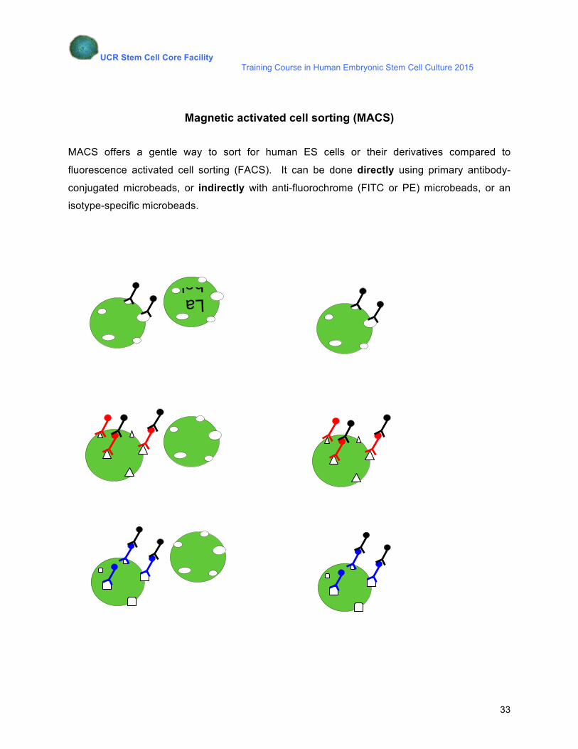
Magnetic activated cell sorting (MACS)
MACS offers a gentle way to sort for human ES cells or their derivatives compared to
fluorescence activated cell sorting (FACS). It can be done directly using primary antibody-
conjugated microbeads, or indirectly with anti-fluorochrome (FITC or PE) microbeads, or an
isotype-specific microbeads.
LabelD
II
UCR Stem Cell Core Facility Training Course in Human Embryonic Stem Cell Culture 2015
34
Materials:
CD184 (CXCR4)-PE, human
CD117 (A3C6E2)-PE, human
CD15-PE, human
Anti-PE MicroBeads
Ice-cold MACS buffer: PBS + 0.5% BSA, 2mM EDTA – to prevent cell clumping
Protocol:
1. Detach cells with 0.25% trypsin-EDTA – 1ml 0.25% trypsin-EDTA per well.
2. Inactivate trypsin 1:1 with FBS or 1:10 MEF media.
3. Collect cells in a 15ml tube and spin cells for 3min at 800rpm.
4. Obtain the cell pellet and re-suspend the cell pellet with 100ul of ice-cold wash buffer and transfer into a 1.5ml microcentrifuge tube.
5. Incubate cells with 10ul of primary-PE antibody (1:10 dilution) in ice-cold wash buffer for 1hr. Incubate in the refrigerator, and in the dark. Flick the tube every 15-30min.
6. Wash the cells with 10 ml ice-cold wash buffer. Spin cells. Obtain pellet.
7. Add 80ul ice-cold wash buffer and 20ul anti-PE microbeads to cell pellet. Incubate on ice for 1hr.
8. Wash the cells with 10 ml ice-cold wash buffer. Spin cells.
9. Obtain pellet. Add 1ml ice-cold wash buffer.
10. Sort the cells with a MACS Pro cell sorter.
11. The cells can be used for RNA extraction or further grown in culture. Gentamycin can be added to minimize the risk of contamination.
UCR Stem Cell Core Facility Training Course in Human Embryonic Stem Cell Culture 2015
35
Booklet Content
Schedule
Chapter 1: Feeders and Substrates for Culturing Human Embryonic Stem Cells
Chapter 2: Culture and Maintenance of Human Embryonic Stem Cells
2.1. Passaging of human ES cells on MEF feeders 2.2. Passaging of human ES cells on Matrigel or Geltrex
2.3. Passaging of hESCs/iPSC on Vitronectin XF
2.4. Troubleshooting: Picking colonies
Chapter 3: Differentiation of Human Embryonic Stem Cells
3.1. Differentiation in suspension culture: Embryoid bodies (EBs)
3.2. Replating embryoid bodies in monolayer
3.3. Induction of differentiation in monolayer culture
Chapter 4: Labeling and Sorting of Human Embryonic Stem Cells
4.1. Immunocytochemistry Chapter 5: Cryopreservation of Human Embryonic Stem Cells
Chapter 6: Transfection of hESCs by using Lonza Biosciences Nucleofection Technology
Appendix 1:
Cell Lab Quanta Demo
Magnetic activated cell sorting (MACS)