Embed Size (px)
Citation preview
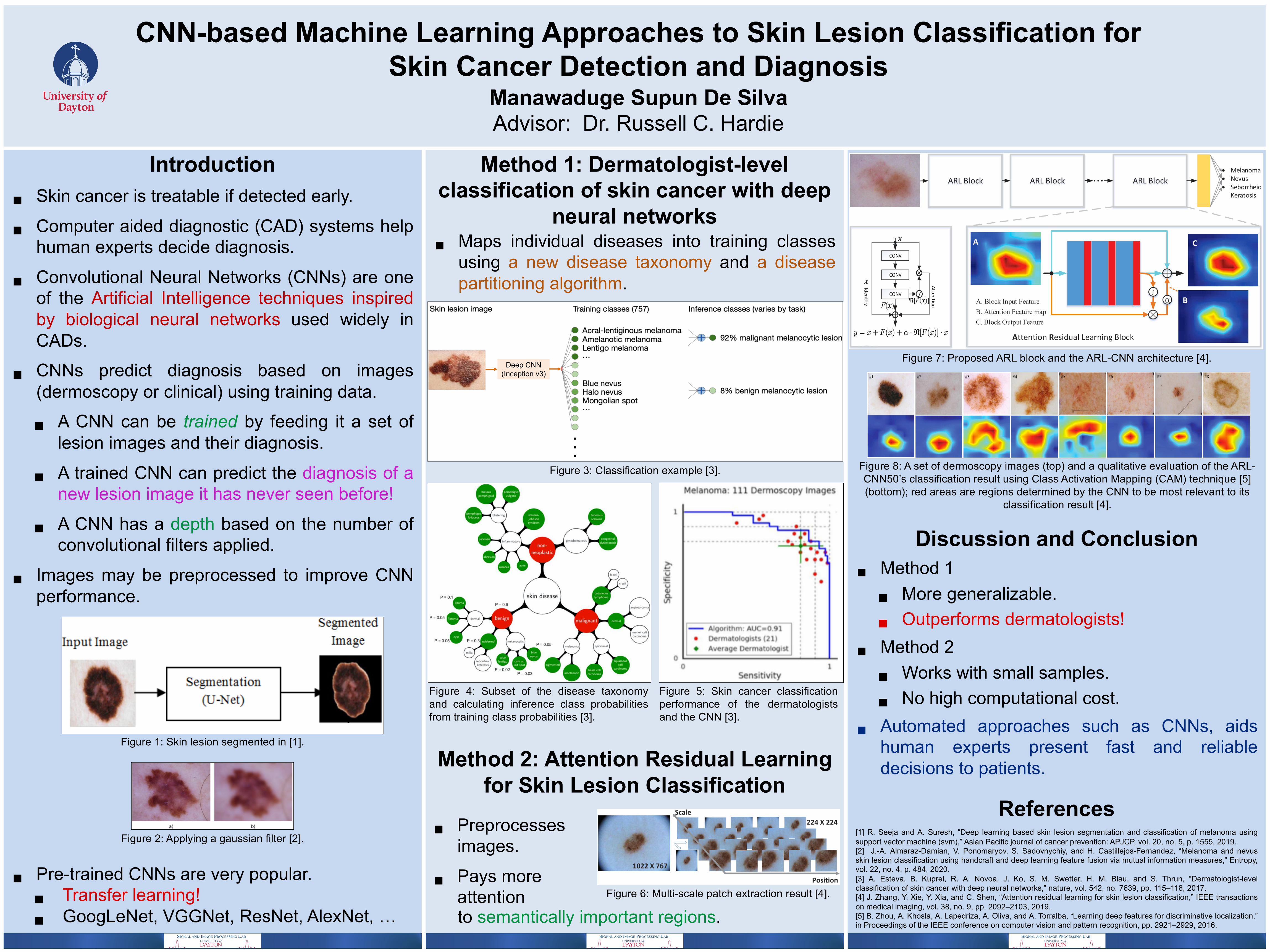
CNN-based Machine Learning Approaches to Skin Lesion Classification for Skin Cancer Detection and Diagnosis
Manawaduge Supun De SilvaAdvisor: Dr. Russell C. Hardie
Introduction§ Skin cancer is treatable if detected early.
§ Computer aided diagnostic (CAD) systems helphuman experts decide diagnosis.
§ Convolutional Neural Networks (CNNs) are oneof the Artificial Intelligence techniques inspiredby biological neural networks used widely inCADs.
§ CNNs predict diagnosis based on images(dermoscopy or clinical) using training data.
§ A CNN can be trained by feeding it a set oflesion images and their diagnosis.
§ A trained CNN can predict the diagnosis of anew lesion image it has never seen before!
§ A CNN has a depth based on the number ofconvolutional filters applied.
§ Images may be preprocessed to improve CNNperformance.
§ Pre-trained CNNs are very popular.§ Transfer learning!§ GoogLeNet, VGGNet, ResNet, AlexNet, …
Method 1: Dermatologist-level classification of skin cancer with deep
neural networks§ Maps individual diseases into training classes
using a new disease taxonomy and a diseasepartitioning algorithm.
Method 2: Attention Residual Learning for Skin Lesion Classification
Discussion and Conclusion§ Method 1
§ More generalizable.
§ Outperforms dermatologists!
§ Method 2
§ Works with small samples.
§ No high computational cost.
§ Automated approaches such as CNNs, aidshuman experts present fast and reliabledecisions to patients.
References[1] R. Seeja and A. Suresh, “Deep learning based skin lesion segmentation and classification of melanoma usingsupport vector machine (svm),” Asian Pacific journal of cancer prevention: APJCP, vol. 20, no. 5, p. 1555, 2019.[2] J.-A. Almaraz-Damian, V. Ponomaryov, S. Sadovnychiy, and H. Castillejos-Fernandez, “Melanoma and nevusskin lesion classification using handcraft and deep learning feature fusion via mutual information measures,” Entropy,vol. 22, no. 4, p. 484, 2020.[3] A. Esteva, B. Kuprel, R. A. Novoa, J. Ko, S. M. Swetter, H. M. Blau, and S. Thrun, “Dermatologist-levelclassification of skin cancer with deep neural networks,” nature, vol. 542, no. 7639, pp. 115–118, 2017.[4] J. Zhang, Y. Xie, Y. Xia, and C. Shen, “Attention residual learning for skin lesion classification,” IEEE transactionson medical imaging, vol. 38, no. 9, pp. 2092–2103, 2019.[5] B. Zhou, A. Khosla, A. Lapedriza, A. Oliva, and A. Torralba, “Learning deep features for discriminative localization,”in Proceedings of the IEEE conference on computer vision and pattern recognition, pp. 2921–2929, 2016.
Figure 2: Applying a gaussian filter [2].
Figure 1: Skin lesion segmented in [1].
Figure 4: Subset of the disease taxonomyand calculating inference class probabilitiesfrom training class probabilities [3].
Figure 3: Classification example [3].
Figure 5: Skin cancer classificationperformance of the dermatologistsand the CNN [3].
Figure 7: Proposed ARL block and the ARL-CNN architecture [4].Deep CNN
(Inception v3)
Figure 6: Multi-scale patch extraction result [4].
§ Preprocesses images.
§ Pays more attention
Figure 8: A set of dermoscopy images (top) and a qualitative evaluation of the ARL-CNN50’s classification result using Class Activation Mapping (CAM) technique [5] (bottom); red areas are regions determined by the CNN to be most relevant to its
classification result [4].
to semantically important regions.


![LI ET AL.: SEMI-SUPERVISED SKIN LESION SEGMENTATION …dermoscopy images [14,18]. For example, Jaisakthi et al. [14] proposed a semi-supervised skin lesion segmentation method using](https://img.pdfslide.net/doc/110x75/60658319b2024701434d8eca/li-et-al-semi-supervised-skin-lesion-segmentation-dermoscopy-images-1418-for.jpg)