Embed Size (px)
Citation preview

CNS – central nervous system
The spinal cord (medulla spinalis) The brain (cerebrum, encephalon) Division of the brain: The medulla oblongata(medulla oblongata) The pons (pons Varoli) The cerebellum (cerebellum) The midbrain (mesencephalon) The diencephalon (diencephalon) The cerebrum (telencephalon) The brain stem= The medulla oblongata, The pons, The cerebellum, The midbrain, The diencephalon

I. Nervus olfactorius – olfactory nerve
II. Nervus opticus – optic nerve
III.Nervus oculomotorius – oculomotor nerve
IV. Nervus trochlearis – trochlear nerve
V. Nervus trigeminus – trigeminal nerve
VI. Nervus abducens – abducens nerve
VII. Nervus facialis – facial nerve
VIII. Nervus vestibulocochlearis – vestibulocochlear nerve
IX. Nervus glosopharyngeus – glossopharyngeal nerve
X. Nervus vagus – vagus nerve
XI. Nervus accesorius – accessory nerve
XII. Nervus hypoglossus – hypoglossal nerve
The cranial nerves– nervi craniales


Spinal
cord Receptor
Thalamus
Brain stem
Sensory cortical areas Motor cortical area
Brain stem
Muscle
Cerebellum
Analyzer

The Medulla oblongata • within foramen magnum and on clivus • Continuation of the spinal cord (20–
25mm) • Extends from detachment of 1st pair of
the spinal nerves (or decussatio pyramisum) till the pons
Grooves: • betwen medulla oblongata and pons –
detachment of nerves (VI., VII., VIII.) • fissura mediana anterior • sulcus anterolateralis (XII.)
• sulcus posterolateralis (IX., X., XI.)
• sulcus medianus posterior The groove separate following structures Funiculus anterior – pyramis Funiculus lateralis - olive

Caudal: funiculi posteriores (parts - fasciculus gracilis and cuneatus) pass into inferior peduncles of cerebellum(pedunculi cerebellares inferiores)
Rostral: between divergent inferior and superior peduncles of cerebellum, medulla oblongata forms caudal part of floor of IV. brain ventricle
Posterior side of medulla oblongata
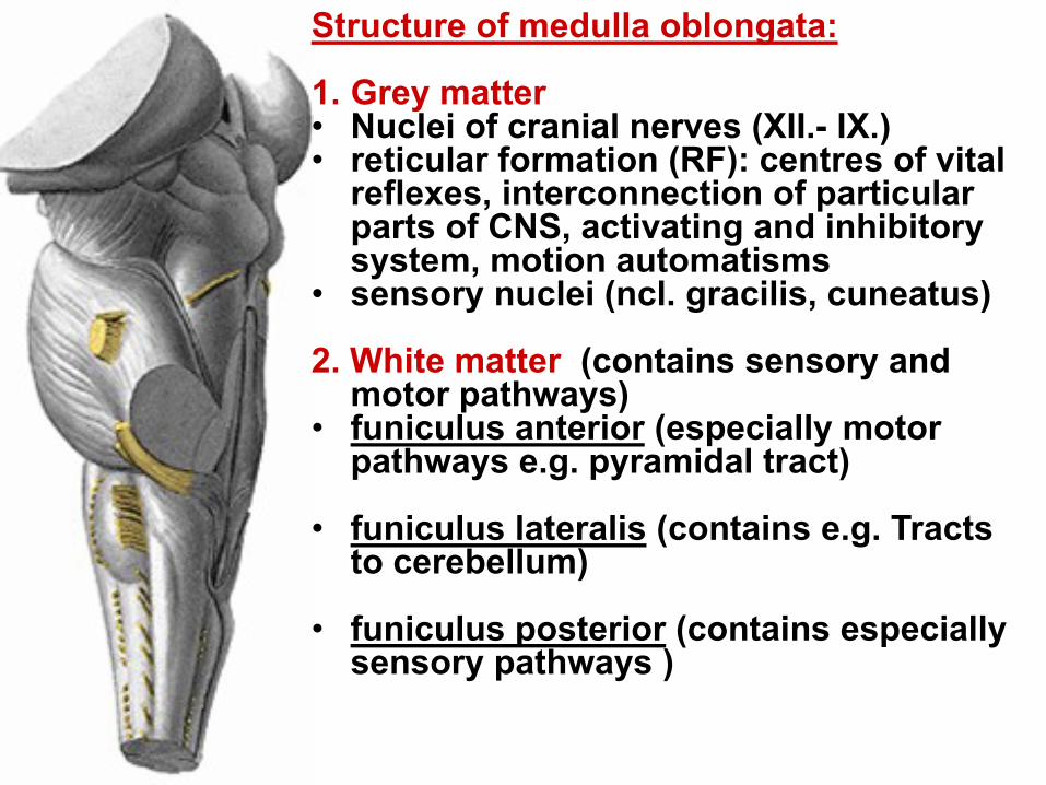
Structure of medulla oblongata: 1. Grey matter • Nuclei of cranial nerves (XII.- IX.) • reticular formation (RF): centres of vital
reflexes, interconnection of particular parts of CNS, activating and inhibitory system, motion automatisms
• sensory nuclei (ncl. gracilis, cuneatus) 2. White matter (contains sensory and
motor pathways) • funiculus anterior (especially motor
pathways e.g. pyramidal tract) • funiculus lateralis (contains e.g. Tracts
to cerebellum) • funiculus posterior (contains especially
sensory pathways )


The pons (Pons Varoli)
• Transverse rampart between medulla oblongata and midbrain (lenght circa 25 mm)
• In the median plane sulcus basilaris
passes (for a. basilaris) • In the groove between pons and
medulla oblongata, VI. – VIII. cranial nerves arise
• Pons passes laterally into middle
peduncles of cerebellum (pedunculi cerebellares medii)
• between pons and pedunculi
cerebellares medii, there is V. vranial nerve arising

Lateral side of the pons
• Middle peduncle (pedunculus
cerebellaris medius) with output
of n. V.
- especially motor pathways
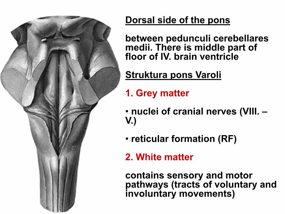
Dorsal side of the pons between pedunculi cerebellares medii. There is middle part of floor of IV. brain ventricle Struktura pons Varoli 1. Grey matter • nuclei of cranial nerves (VIII. – V.) • reticular formation (RF)
2. White matter contains sensory and motor pathways (tracts of voluntary and involuntary movements)


FORMATIO RETICULARIS • It belongs to the phylogenetically oldest structures of CNS
• It is interconnected with all sections of CNS
• Basic stereotypes (walk, sleep)
• It significantly influences wakefulness, tiredness and motivation
• It is located centrally and dorsally within the brain stem, especially within the pons
• ascending activation system → waking from sleep, maintaining wakefulness
• descending activation system
• Interruption of retikulární formace →blackout
• RF provides complex interconnection of cranial nerves
between each other and with other areas, it provides
vital reflexes since birth (blink, lachrymal, cough,
sucking, salivation, swallowing…)

Te midbrain (mesencephalon) • located between pons and
diencephalon Ventral side of the midbrain crura cerebri – two ramparts of
whote matter (motor pathways) detachment of III. cranial nerve
within groove between crura cerebri anf fossa interpeduncularis

Dorsal side of the midbrain corpora quadrigemina colliculi superiores (optical-motor reflexes) connection to the visual pathway colliculi inferiores (acoustic–motor reflexes) connection to the auditory pathway Pedunculi cerebellares superiores (superior peduncles of cerebellum) between them – roof of IV. brain ventricle Detachment of IV. cranial nerve– only crania nerve that arises from dorsal side of brain stem

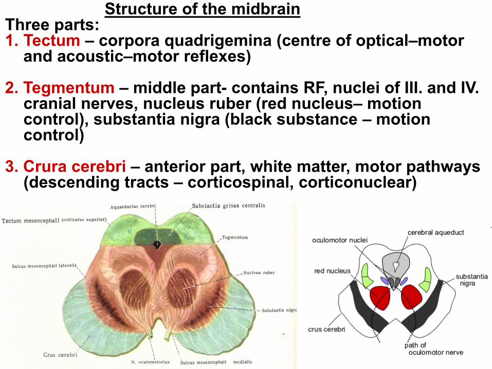
Structure of the midbrain Three parts: 1. Tectum – corpora quadrigemina (centre of optical–motor
and acoustic–motor reflexes) 2. Tegmentum – middle part- contains RF, nuclei of III. and IV.
cranial nerves, nucleus ruber (red nucleus– motion control), substantia nigra (black substance – motion control)
3. Crura cerebri – anterior part, white matter, motor pathways
(descending tracts – corticospinal, corticonuclear)

The midbrain (střední mozek)
source of III. and IV. cranial nerves
• centre of optical–motor and acoustic–motor reflexes, its
nuclei provide coordinated movements of eyes and head

Fossa rhomboidea Floor of IV. brain ventricle, rhombus shaped Nuclei of III. – XII. cranial nerves parts: 1. pars superior
between pedunculi cerebellares sup. covered with velum medullare superius
2. pars intermedia dorsal side of pons between pedunculi cerebellares med. covered with fastigium of cerebellum
3. pars inferior dorsal side of medulla obl. between pedunculi cerebellares inf. covered with velum medullare inferius

Fossa rhomboidea builds on: • rostrally on aquaeductus cerebri (channel between III. and IV. brain ventricle) • caudally on canalis centralis of the spinal cord


Spinal
cord Receptor
Thalamus
Brain stem
Sensory cortical areas Motor cortical area
Brain stem
Muscle
Cerebellum
Analyzer

The cerebellum
Functions: Control of muscle tension of striated muscles, it provides upright posture, balance, it coordinates and specifies movements
Is in paralel connected into systém of motor pathways
important centre of proprioception
It provides precise coordination of
movements
At failure: Muscle weaknes, unsure poise, uncoordinated walk on the wide base

Te cerebellum
• it lies within posterior
cranial fossa within fossae
cerebellares of occipital bone
• it touches dorsal side of
brain stem
• between cerebellum and
brain stem, there is IV. Brain
ventricle

The cerebellum is
interconnected with the
brain stem through three
peduncles:
1.Pedunculi cerebellares
superiores (with midbrain)
2.Pedunculi cerebellares
medii (with pons)
3.Pedunculi cerebellares
inferiores (with medulla
oblongata)

Structure of the cerebellum: 1. worm - vermis cerebelli middle part 2. cerebellar hemispheres
hemisheria cerebelli (lobus anterior, lobus posterior, floculus). On the surface of vermis and hemispheres, there are notches – sulci cerebelli, which separate particular threads - gyri cerebelli

Grey matter of the cerebellum: • cortex cerebelli – on the surface of hemispheres and
vermis • nuclei cerebelli – nuclei withincerebellum (ncll. fastigii,
ncl. emboliformis, ncl. globosus, ncl. dentatus) involved into motion control system
White matter of the cerebellum : • Below the cortex, it creates characteristic drawing - arbor
vitae (tree of life)

Distribution of the cerebellum: 1.vestibular cerebellum (archicerebellum) • crucial for maintaining balance (information from vestibular
apparatus)
• influence on motor nuclei within the spinal cord, which
controls ovement of axial muscles (erect posture)
• control of eyey movemet and their coordination with ead
movements
2. spinal cerebellum (palaeocerebellum) • Control of muscle tension and coordination of movements
– regulation of movements 3. cerebral cerebellum (neocerebellum)
• control of planning of movements
• control of voluntary movements in space and time


Spinal
cord Receptor
Thalamus
Brain stem
Sensory cortical areas Motor cortical area
Brain stem
Muscle
Cerebellum
Analyzer


The diencephalon
• builds on mesencephalon
• covered with cerebral
hemispheres
Distribution of diencephalon: Thalamencephalon (thalamus) dorsal part Hypothalamus - basal part (ventral part) (sulcus hypothalamicus – separates both parts)


Thalamencephalon:
1. thalamus – accumulation of grey matter on dorsal side of diencephalon (ovoid shape)
2. epithalamus – e.g. pineal gland, dorsal side of diencephalon
3. metathalamus - corpus geniculatum mediale and laterale
4. subthalamus – grey matter located below thalamus

THALAMUS
• Accumulation of grey matter (ovoid formation) (to its neurons come impulses from all sensory pathways
except olfactory tract)
• It contains a large number of nuclei • „gateway od consciousness“ – switching of all sensory
pathways and control feedback motor pathwaysinto cerebral cortex


Epithalamus • dorsally at roof of III. Brain ventricle
• corpus pineale (pineal gland) – endocrine gland


Metathalamus
• On the posterior side of thalamus
• corpus geniculatum
mediale connected with colliculus superior – part of auditory pathway
• corpus geniculatum laterale
connected with colliculus inferior – part off visual pathway

Subthalamus
• grey matter located ventrocaudylly from thalamus and
laterally from hypothalamus
• involved into involuntary movements

Hypothalamus
• Originated from motor plate • A part of hypothalamu is
hypophysis Functions: • visceral brain controls aktivity of
visceral organs through autonomous nerves and hormones of hypophysis (control centre of autonomous system)
• It coordinates neurohumoral
control (regulates functions of endocrine
system) • It is essential for preservation of
homeostasis


Nuclei of hypothalamus • A large number of nuclei (several
groups) Division from functional perspective: • secretory nuclei (at wall of III.
ventricle) neurosecretion – controlc aktivity of hypophysis
• nuclei that are parent to
parasympathicus (anterior group) • nuclei that are parent to sympathicus (middle group) • nuclei affecting the instinctive and
emotional behavior – serve limbic system (especially posterior group)

Hypophysis cerebri (pituitary gland, hypophysis)
• Endocrine gland, part of diencephalon, located at sella turcica of sphenoid bone
• Superior position to other endocrine glands

Hypophysis • adenohypophysis (lobus anterior) it produces e.g. somatotropic hormone and hormones affecting aktivity of other endocrine glands (gonadotropci, corticotropic…)
• pars media – produces melanocyte-stimulating hormone • neurohypophysis (lobus posterior) receives hormones (adiuretic hormone and oxytocin) from nuclei of hypothalamus through axonal flow