Embed Size (px)
Citation preview
CNS INFECTIONSOverview
Life-threatening problems with high associated mortality and morbidity
Presentation may be acute, subacute, or chronic Clinical findings determined by anatomic site(s) of
involvement, infecting pathogen, and host response Vulnerability of CNS to effects of inflammation &
edema mandates prompt diagnosis with appropriate therapy if consequences to be minimized

ACUTE CNS INFECTIONS
1. Bacterial meningitis***
2. Meningoencephalitis
3. Brain abscess
4. Subdural empyema
5. Epidural abscess
6. Septic venous sinus
thrombophlebitis
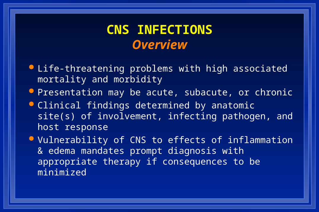
Routes of Entry
– Hematogenous
– Neighboring focus
– Anatomic defect
• congenital
• traumatic
• surgical
– Intraneural pathways
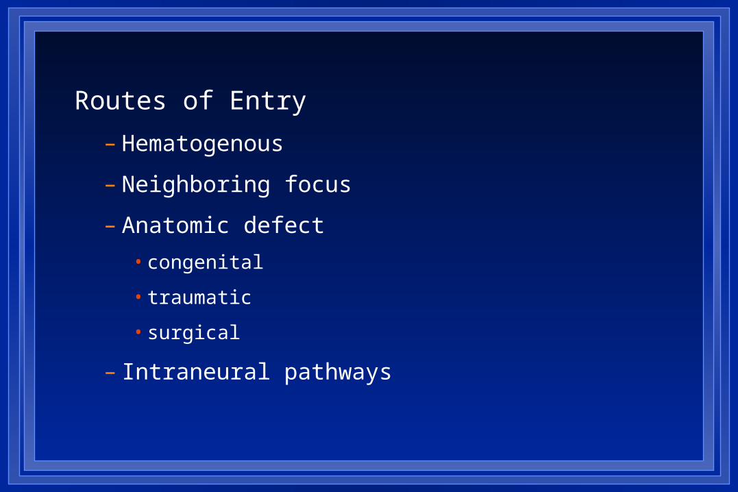
THE PATIENT WITH ACUTE CNS INFECTIONOverall Goals in Management
1. To promptly recognize the patient with an acute CNS infection syndrome
2. To rapidly initiate appropriate empiric therapy
3. To rapidly and specifically identify the etiologic agent, adjusting therapies as indicated
4. To optimize management of complicating features
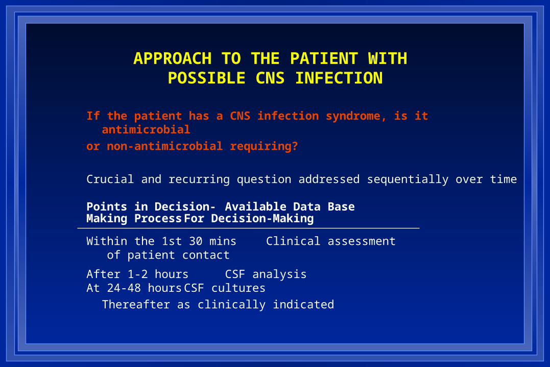
APPROACH TO THE PATIENT WITH POSSIBLE CNS INFECTION
If the patient has a CNS infection syndrome, is it antimicrobial
or non-antimicrobial requiring?
Crucial and recurring question addressed sequentially over time
Points in Decision- Available Data BaseMaking Process For Decision-Making
Within the 1st 30 mins Clinical assessment of patient contact
After 1-2 hours CSF analysisAt 24-48 hours CSF cultures
Thereafter as clinically indicated

APPROACH TO THE PATIENT WITH SUSPECTED MENINGITIS
Decision-Making Within the First 30 Minutes
Clinical Assessment
Mode of presentation Acute (< 24 hrs)
Subacute (< 7 days) Chronic (> 4 wks) Historical/physical exam clues
Clinical status of the patient
Integrity of host defenses
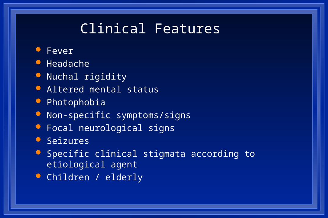
Clinical Features Fever Headache Nuchal rigidity Altered mental status Photophobia Non-specific symptoms/signs Focal neurological signs Seizures Specific clinical stigmata according to etiological agent Children / elderly
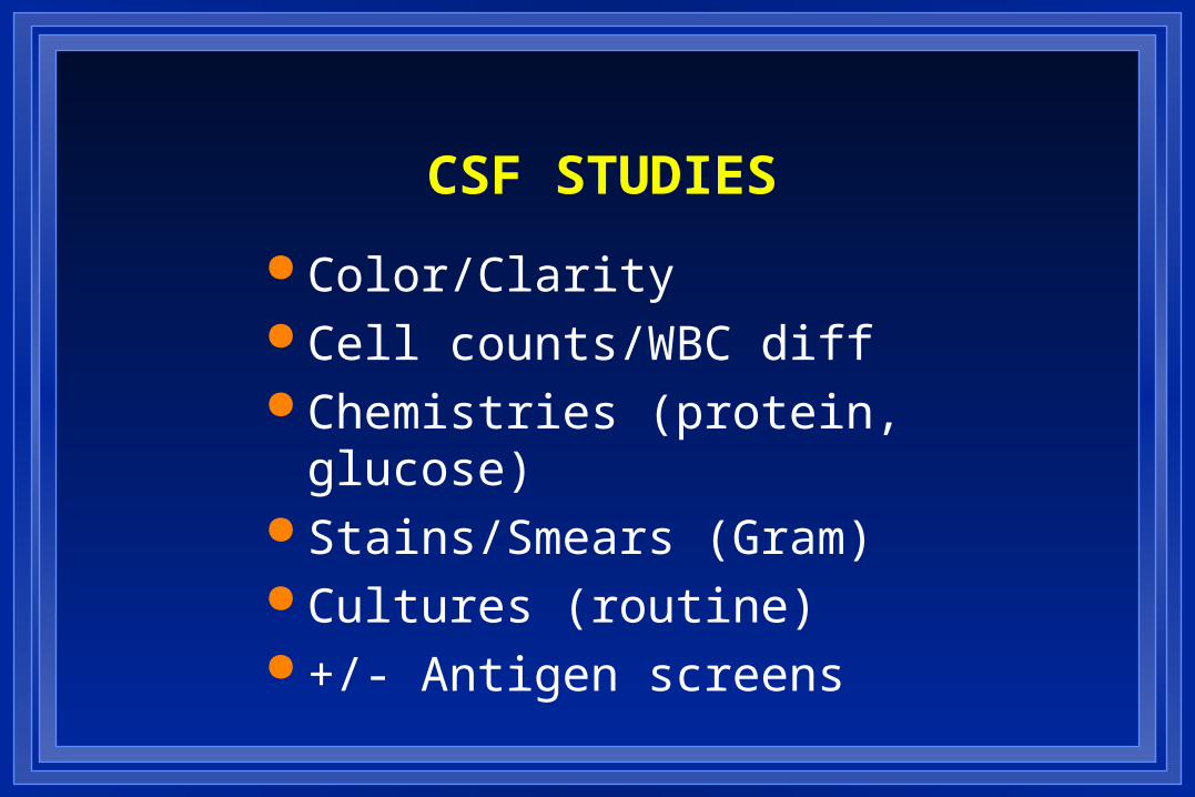
CSF STUDIES
Color/Clarity Cell counts/WBC diff Chemistries (protein, glucose) Stains/Smears (Gram) Cultures (routine) +/- Antigen screens
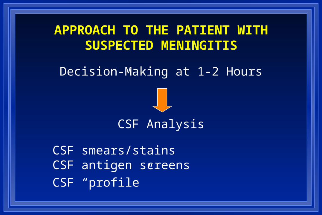
APPROACH TO THE PATIENT WITHSUSPECTED MENINGITIS
Decision-Making at 1-2 Hours
CSF Analysis
CSF smears/stainsCSF antigen screens
CSF “profile”
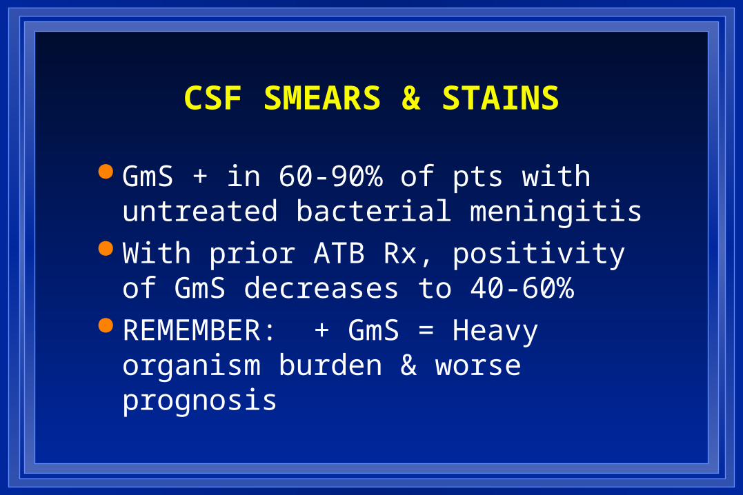
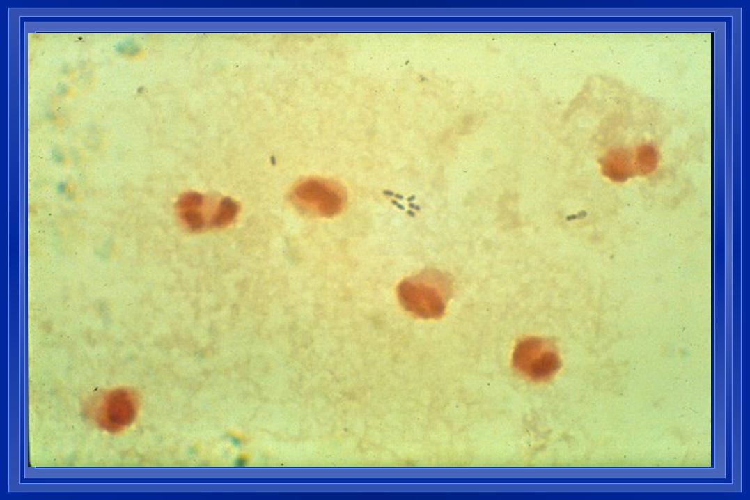
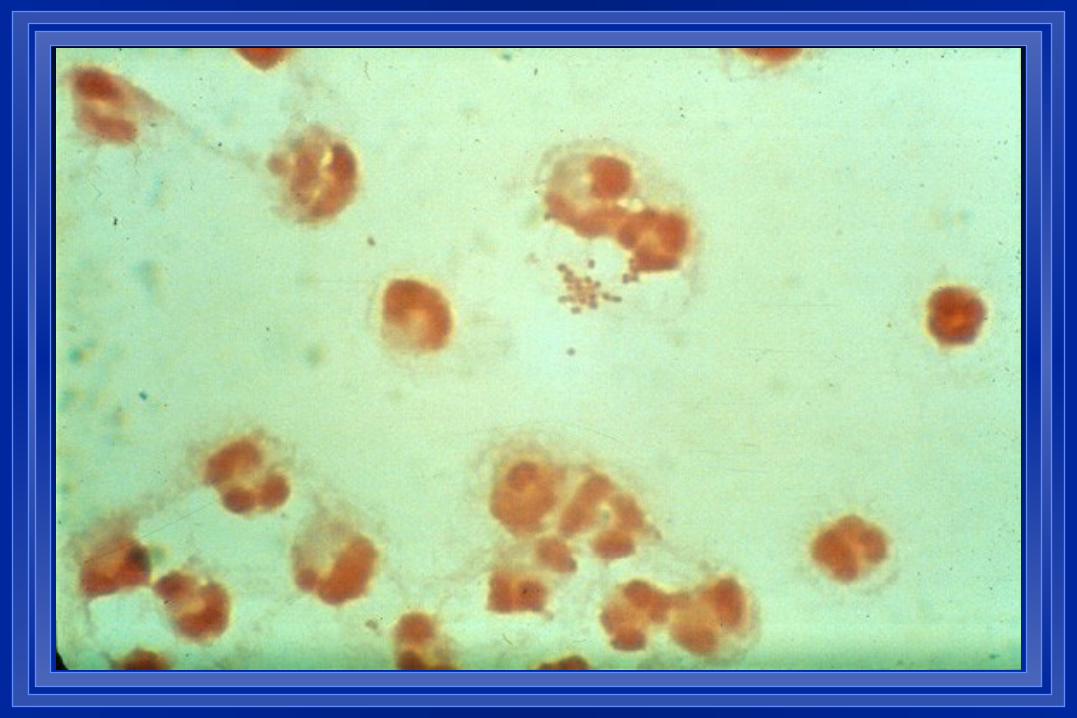
CSF SMEARS & STAINS
GmS + in 60-90% of pts with untreated bacterial meningitis
With prior ATB Rx, positivity of GmS decreases to 40-60%
REMEMBER: + GmS = Heavy organism burden & worse prognosis
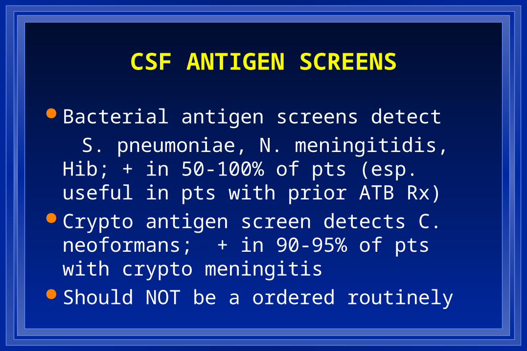
CSF ANTIGEN SCREENS
Bacterial antigen screens detect
S. pneumoniae, N. meningitidis, Hib; + in 50-100% of pts (esp. useful in pts with prior ATB Rx)
Crypto antigen screen detects C. neoformans; + in 90-95% of pts with crypto meningitis
Should NOT be a ordered routinely
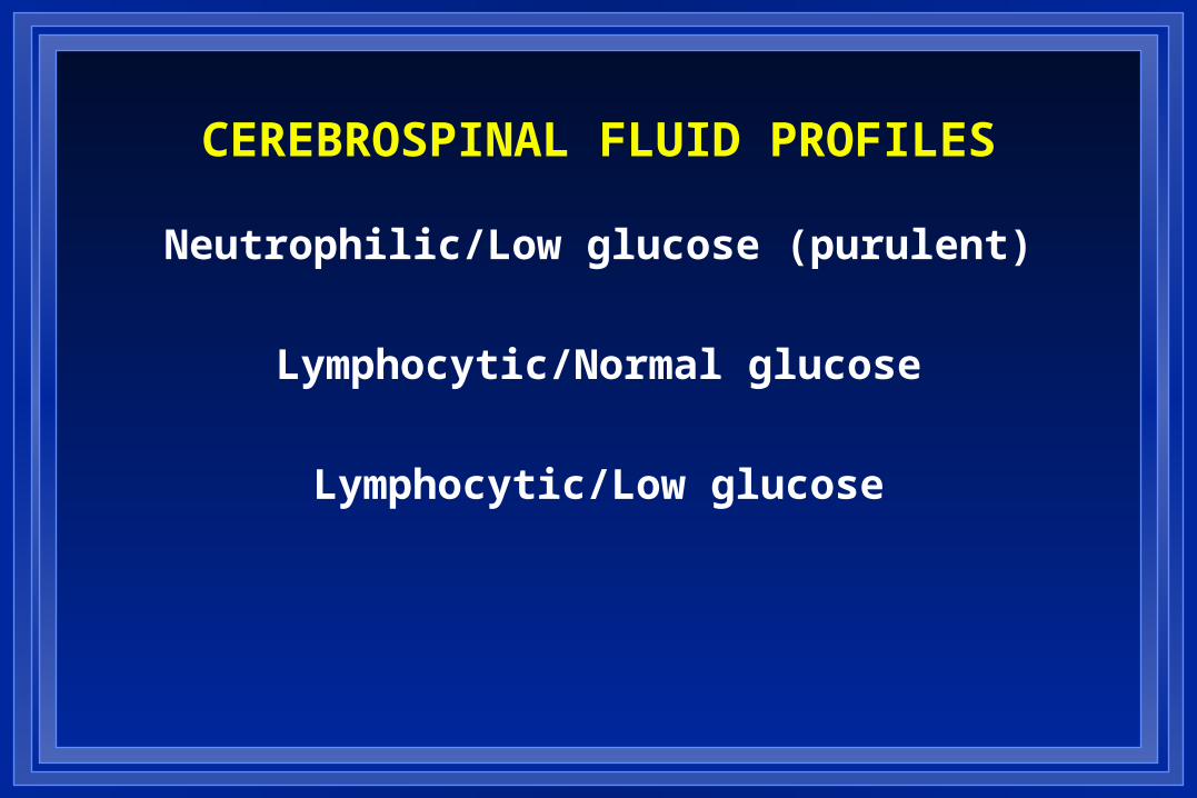
CEREBROSPINAL FLUID PROFILES
Neutrophilic/Low glucose (purulent)
Lymphocytic/Normal glucose
Lymphocytic/Low glucose

APPROACH TO THE PATIENT WITH SUSPECTED MENINGITIS
Decision-Making at 24-48 hours
CSF Culture Results
Culture positive Adjust therapy based upon specific organism and sensitivities
Culture negative Evaluate for “aseptic” meningitis syndrome
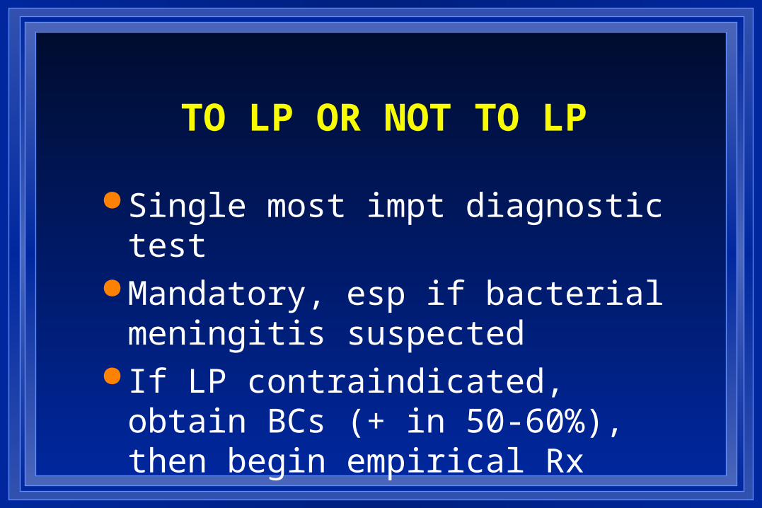
TO LP OR NOT TO LP
Single most impt diagnostic test Mandatory, esp if bacterial
meningitis suspected If LP contraindicated, obtain BCs (+
in 50-60%), then begin empirical Rx
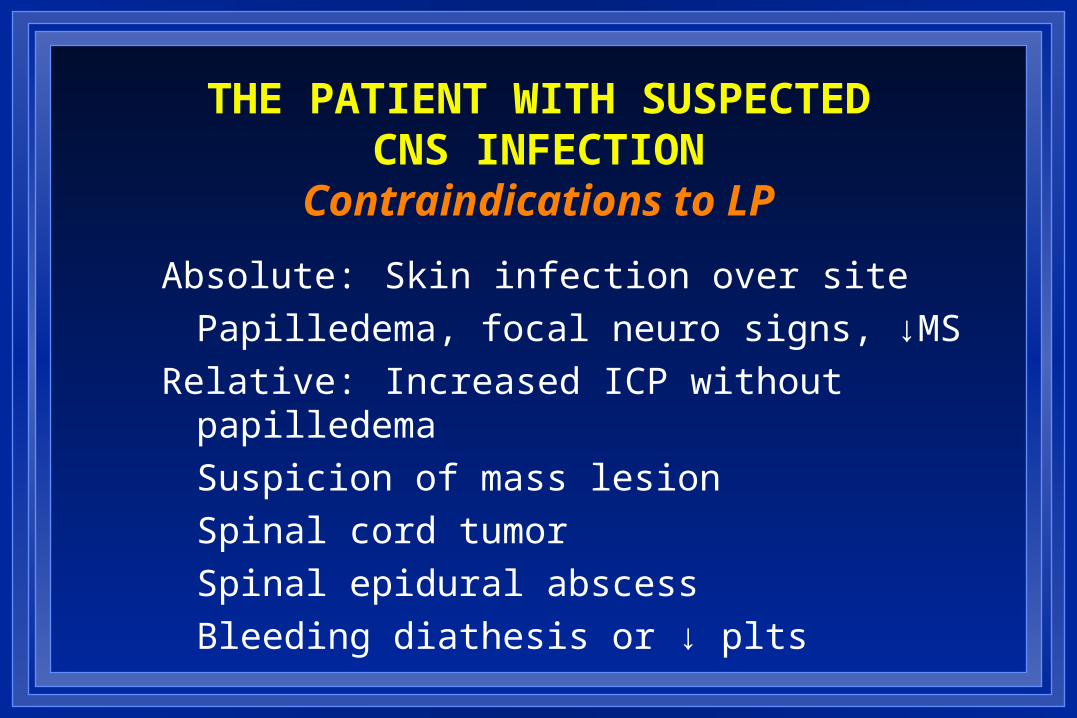
THE PATIENT WITH SUSPECTEDCNS INFECTION
Contraindications to LP
Absolute: Skin infection over site
Papilledema, focal neuro signs, ↓MS
Relative: Increased ICP without papilledema
Suspicion of mass lesion
Spinal cord tumor
Spinal epidural abscess
Bleeding diathesis or ↓ plts

CNS INFECTIONSCCT
Over-employed diagnostic modality Leads to unnecessary delays in Rx & added cost
Rarely indicated in pt with suspected acute meningitis
Mandatory in pt with possible focal infection Increased sensitivity with contrast
enhancement
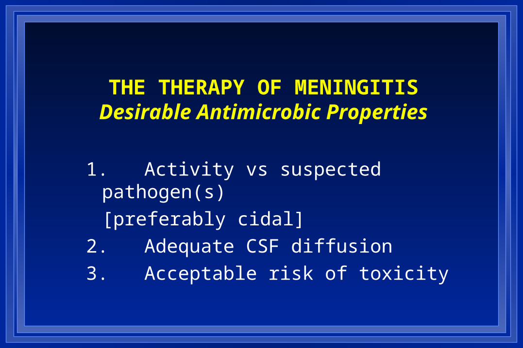
THE THERAPY OF MENINGITISDesirable Antimicrobic Properties
1. Activity vs suspected pathogen(s)
[preferably cidal]
2. Adequate CSF diffusion
3. Acceptable risk of toxicity
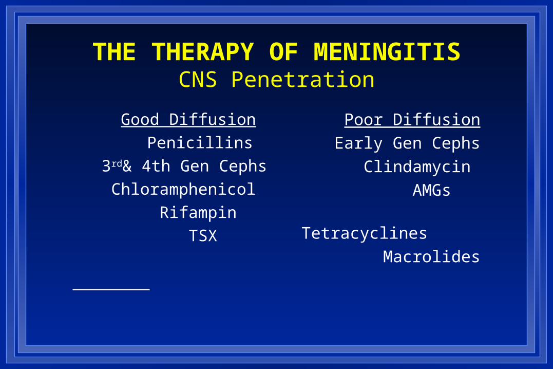
THE THERAPY OF MENINGITISCNS Penetration
Good Diffusion
Penicillins
3rd& 4th Gen Cephs
Chloramphenicol
Rifampin
TSX
Poor Diffusion
Early Gen Cephs
Clindamycin
AMGs
Tetracyclines
Macrolides
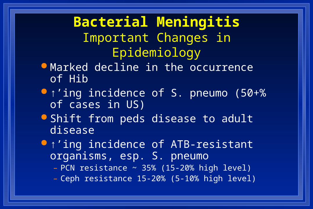
Bacterial MeningitisImportant Changes in Epidemiology
Marked decline in the occurrence of Hib ↑’ing incidence of S. pneumo (50+% of
cases in US) Shift from peds disease to adult disease ↑’ing incidence of ATB-resistant
organisms, esp. S. pneumo– PCN resistance ~ 35% (15-20% high level)– Ceph resistance 15-20% (5-10% high level)
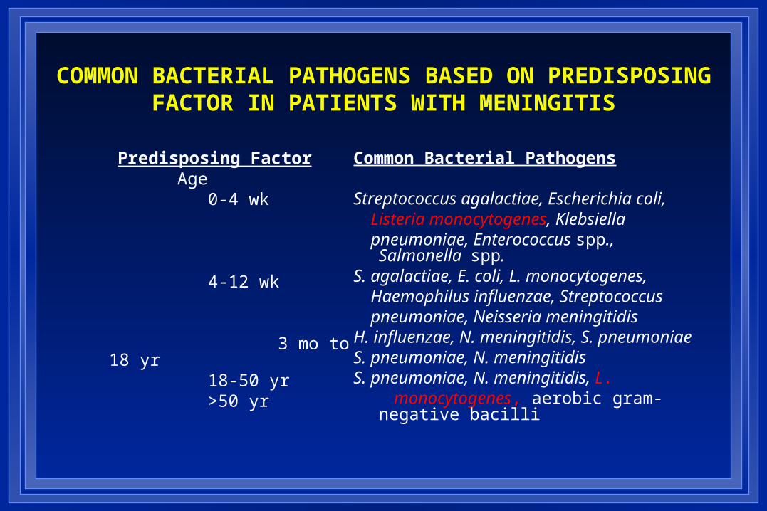
COMMON BACTERIAL PATHOGENS BASED ON PREDISPOSING FACTOR IN PATIENTS WITH MENINGITIS
Predisposing FactorAge 0-4 wk
4-12 wk
3 mo to 18 yr 18-50 yr >50 yr
Common Bacterial Pathogens
Streptococcus agalactiae, Escherichia coli, Listeria monocytogenes, Klebsiella pneumoniae, Enterococcus spp., Salmonella
spp.S. agalactiae, E. coli, L. monocytogenes, Haemophilus influenzae, Streptococcus pneumoniae, Neisseria meningitidisH. influenzae, N. meningitidis, S. pneumoniaeS. pneumoniae, N. meningitidisS. pneumoniae, N. meningitidis, L. monocytogenes, aerobic gram-negative bacilli
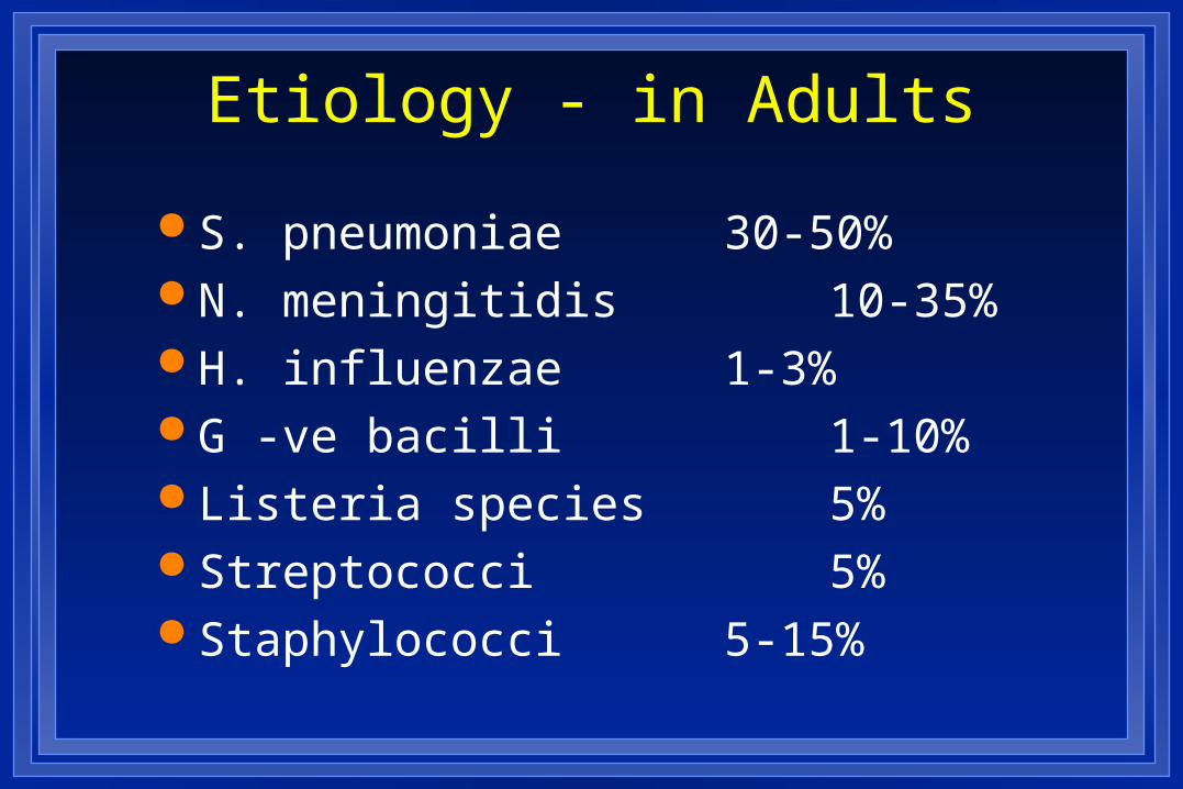
Etiology - in Adults
S. pneumoniae 30-50% N. meningitidis 10-35% H. influenzae 1-3% G -ve bacilli 1-10% Listeria species 5% Streptococci 5% Staphylococci 5-15%
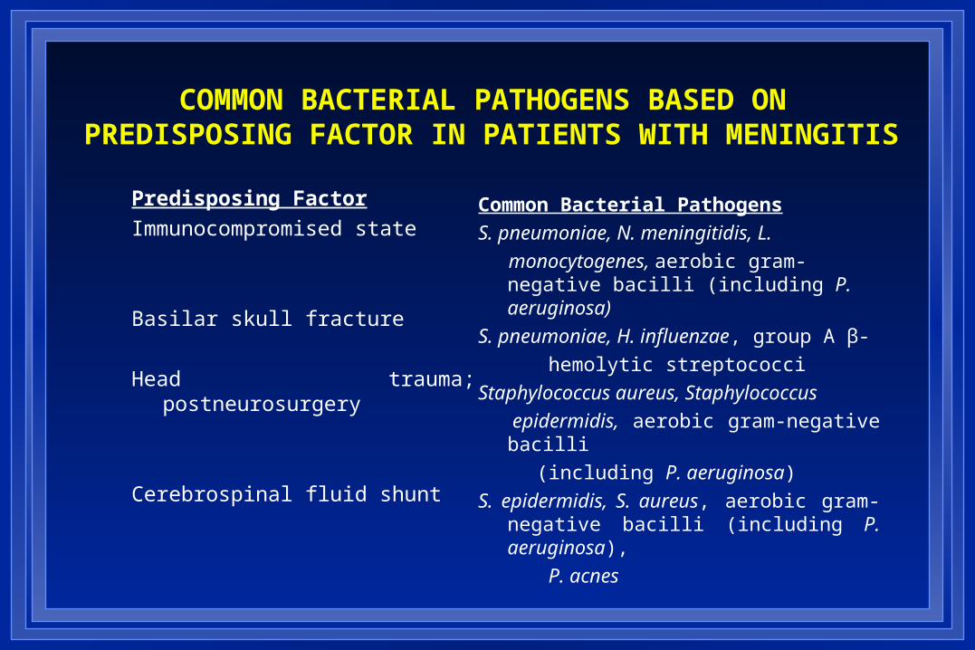
COMMON BACTERIAL PATHOGENS BASED ON PREDISPOSING FACTOR IN PATIENTS WITH MENINGITIS
Predisposing Factor
Immunocompromised state
Basilar skull fracture
Head trauma; postneurosurgery
Cerebrospinal fluid shunt
Common Bacterial Pathogens
S. pneumoniae, N. meningitidis, L.
monocytogenes, aerobic gram-negative bacilli (including P. aeruginosa)
S. pneumoniae, H. influenzae, group A β-
hemolytic streptococci
Staphylococcus aureus, Staphylococcus
epidermidis, aerobic gram-negative bacilli
(including P. aeruginosa)
S. epidermidis, S. aureus, aerobic gram- negative bacilli (including P. aeruginosa),
P. acnes
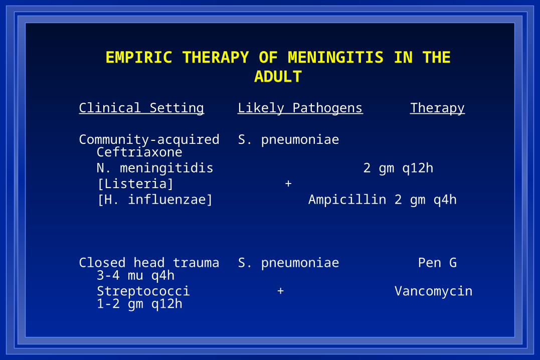
EMPIRIC THERAPY OF MENINGITIS IN THE ADULT
Clinical Setting Likely Pathogens Therapy
Community-acquired S. pneumoniae CeftriaxoneN. meningitidis 2 gm q12h[Listeria] +[H. influenzae] Ampicillin 2 gm
q4h
Closed head trauma S. pneumoniae Pen G 3-4 mu q4hStreptococci +
Vancomycin 1-2 gm q12h
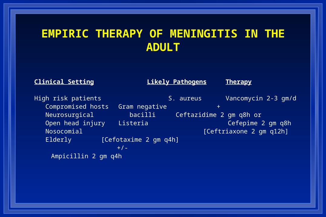
EMPIRIC THERAPY OF MENINGITIS IN THE ADULT
Clinical Setting Likely Pathogens Therapy
High risk patients S. aureus Vancomycin 2-3 gm/d Compromised hosts Gram negative + Neurosurgical bacilli Ceftazidime 2 gm q8h or Open head injury Listeria Cefepime 2 gm q8h Nosocomial [Ceftriaxone 2 gm q12h] Elderly [Cefotaxime 2 gm q4h] +/-
Ampicillin 2 gm q4h
Role of Steroids The addition of anti-inflammatory agents has been attempted
as an adjuvant in the treatment of meningitis
Early administration of corticosteroids for pediatric meningitis has shown no survival advantage, but there is a reduction in the incidence of severe neurologic complications and deafness
Less bilateral deafness late neurological sequelae in controls compared to children treated with steroids

VIRAL MENINGITIS/ENCEPHALITIS
Enteroviruses
Polioviruses
Coxsackieviruses
Echoviruses
Togaviruses Eastern equine
Western equine
Venezuelan equine
St. Louis
Powasson
California
West Nile
Herpesviruses
Herpes simplex
Varicella-zoster
Epstein Barr
Cytomegalovirus
Myxo/paramyxoviruses Influenza/parainfluenzae
Mumps
Measles
Miscellaneous Adenoviruses
LCM
Rabies
HIV
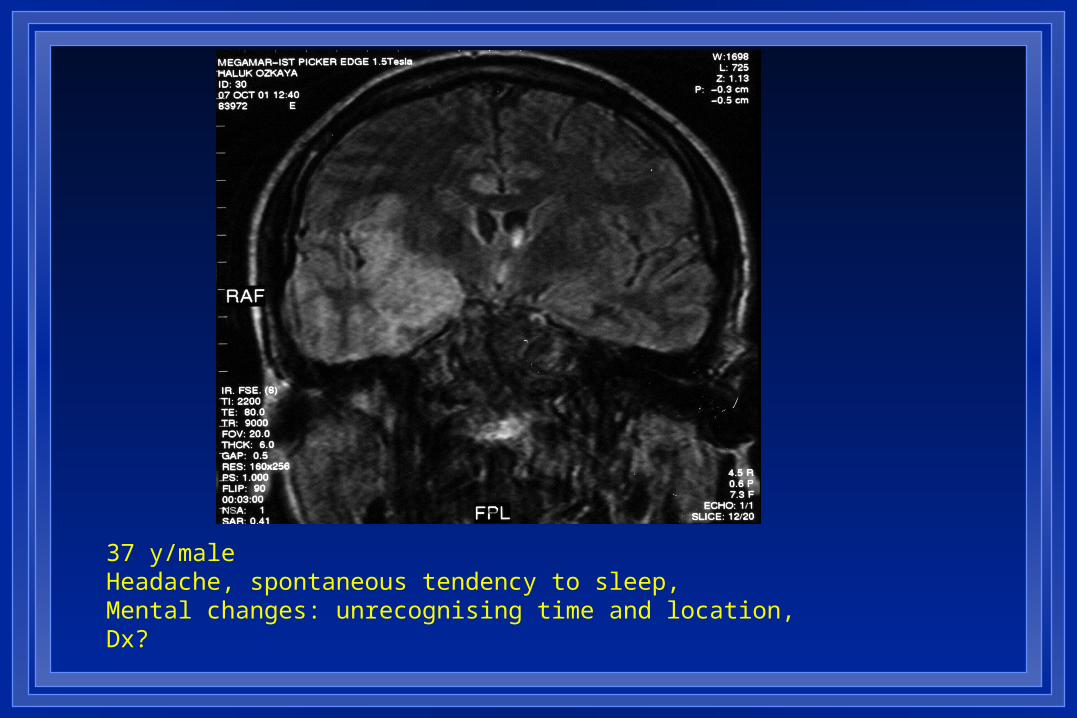
37 y/maleHeadache, spontaneous tendency to sleep, Mental changes: unrecognising time and location, Dx?
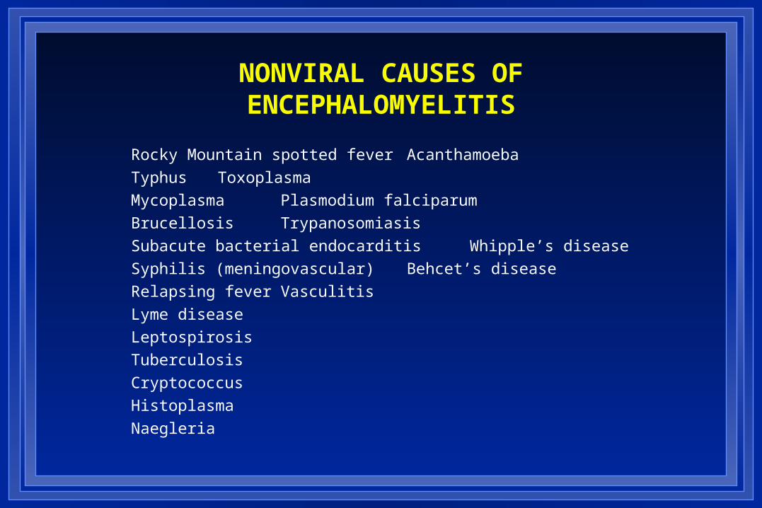
NONVIRAL CAUSES OF ENCEPHALOMYELITIS
Rocky Mountain spotted fever Acanthamoeba
Typhus Toxoplasma
Mycoplasma Plasmodium falciparum
Brucellosis Trypanosomiasis
Subacute bacterial endocarditis Whipple’s disease
Syphilis (meningovascular) Behcet’s disease
Relapsing fever Vasculitis
Lyme disease
Leptospirosis
Tuberculosis
Cryptococcus
Histoplasma
Naegleria

Cryptococcosis

Toxoplasmosis

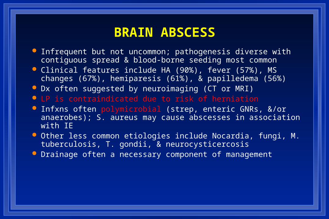
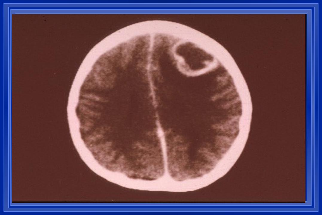
BRAIN ABSCESS Infrequent but not uncommon; pathogenesis diverse with
contiguous spread & blood-borne seeding most common Clinical features include HA (90%), fever (57%), MS changes
(67%), hemiparesis (61%), & papilledema (56%) Dx often suggested by neuroimaging (CT or MRI) LP is contraindicated due to risk of herniation Infxns often polymicrobial (strep, enteric GNRs, &/or
anaerobes); S. aureus may cause abscesses in association with IE
Other less common etiologies include Nocardia, fungi, M. tuberculosis, T. gondii, & neurocysticercosis
Drainage often a necessary component of management
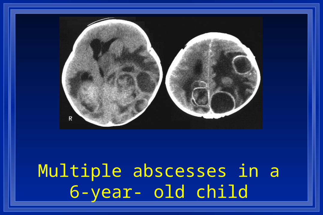
Multiple abscesses in a 6-year- old child
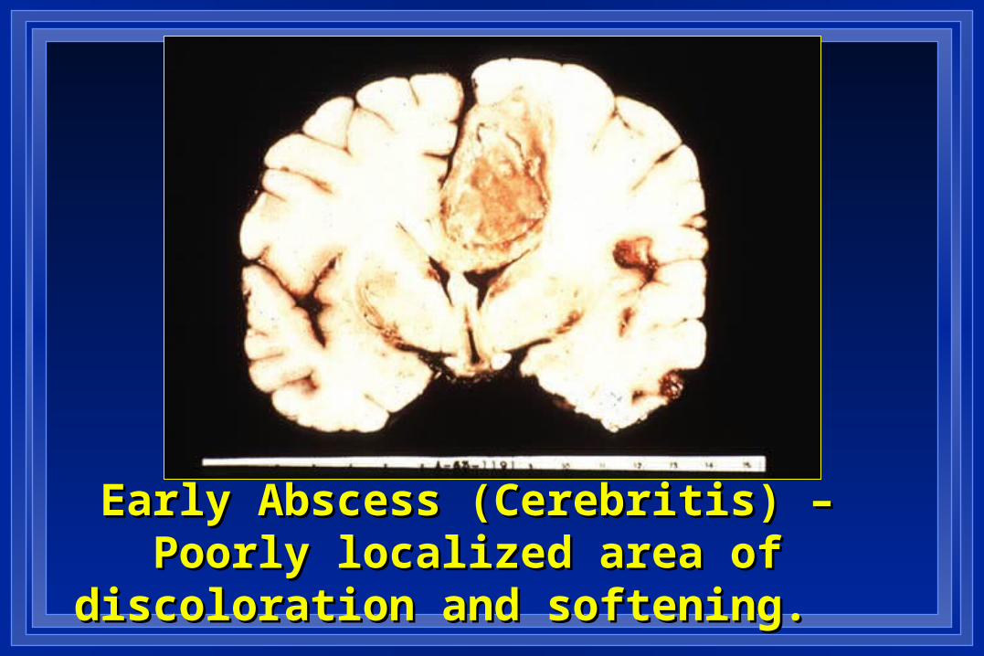
Early Abscess (Cerebritis) – Poorly Early Abscess (Cerebritis) – Poorly localized area of discoloration and localized area of discoloration and
softening. softening.
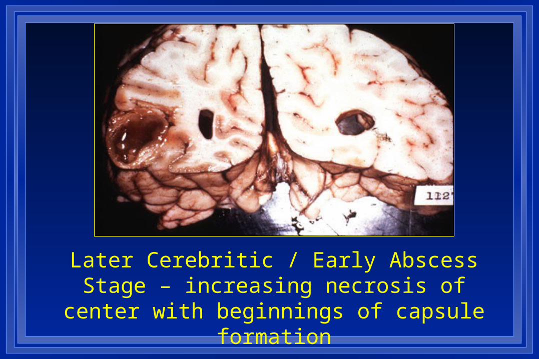
Later Cerebritic / Early Abscess Stage – increasing necrosis of center with beginnings of
capsule formation
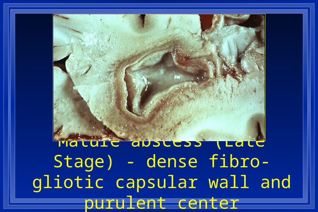
Mature abscess (Late Stage) - dense fibro-gliotic capsular wall
and purulent center

BRAIN ABSCESSEmpiric Therapy
Penicillin G 18-24 mu IV qd
Metronidazole 500 mg IV q6h
Add nafcillin 12 gm/d if staph suspected
(use vanc if MRSA a concern) Add cefotaxime, ceftriaxone, or ceftazidime if GNRs
suspected Substitute vanc 2-4 gm IV/d for pen G if DRSP
suspected
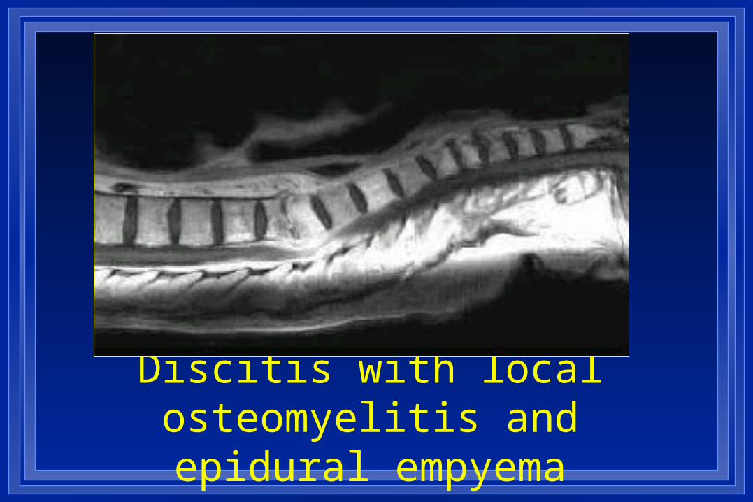
Discitis with local osteomyelitis and epidural empyema