Embed Size (px)
Citation preview
CNS Sequence
Eye and Ear Lab
March 18, 2009
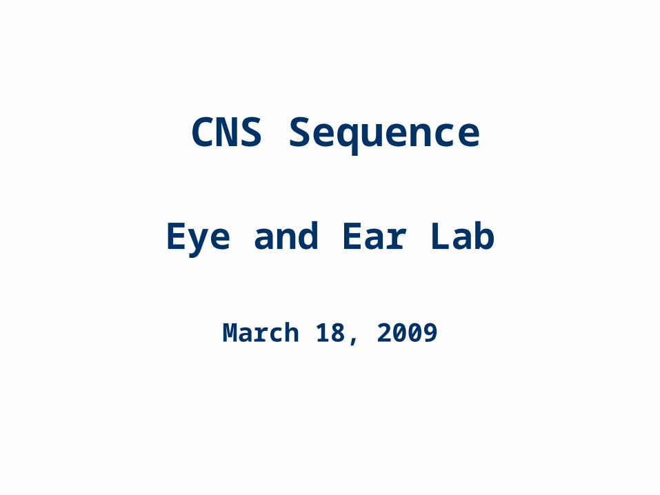
Eyelids: Netter pl. 76
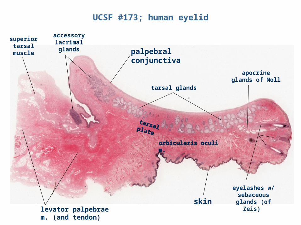
UCSF #173; human eyelid
skin
palpebral conjunctiva
apocrine glands of Moll
tarsal glands
orbicularis oculi orbicularis oculi m.m.
tarsal tarsal plateplate
accessory lacrimal glands
levator palpebrae m. (and tendon)
superior tarsal muscle
eyelashes w/ sebaceous glands (of
Zeis)
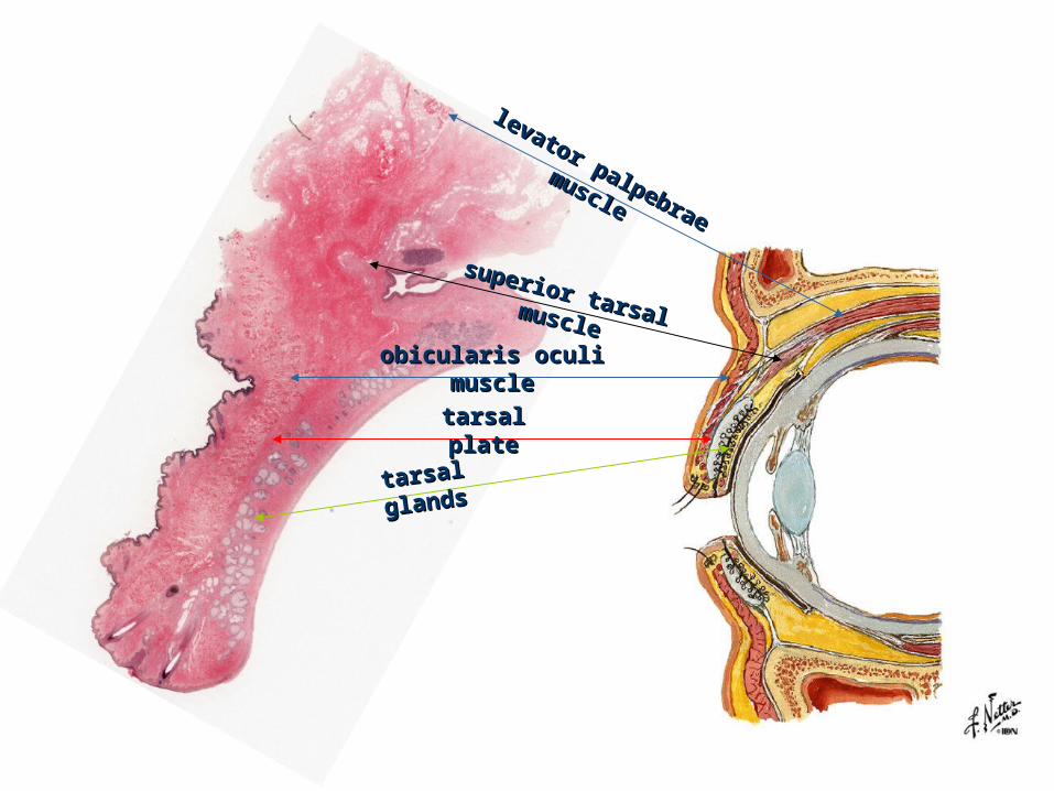
levator palpebrae
levator palpebrae
muscle
muscle
superior tarsal
superior tarsal musclemuscle
obicularis oculi obicularis oculi musclemuscle
tarsal tarsal plateplate
tarsal tarsal
glandsglands
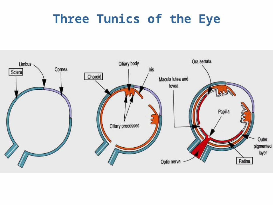
Three Tunics of the Eye
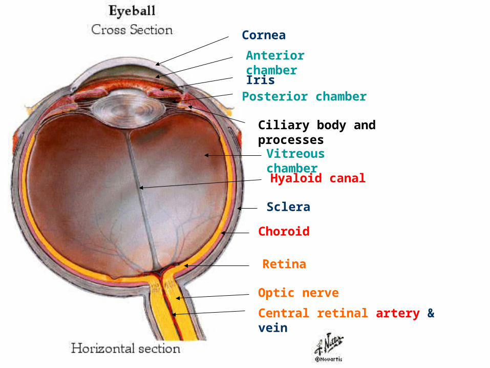
Cornea
Iris
Anterior chamber
Posterior chamber
Ciliary body and processes
Hyaloid canal
Optic nerve
Central retinal artery & vein
Retina
Choroid
Sclera
Vitreous chamber
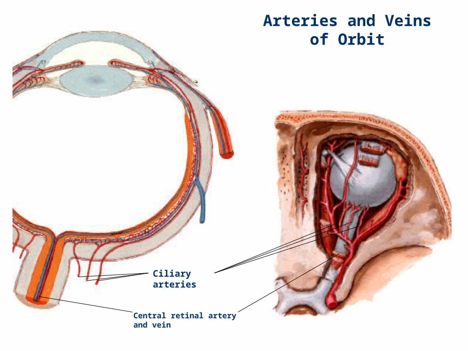
Arteries and Veins of Orbit
Ciliary arteries
Central retinal artery and vein
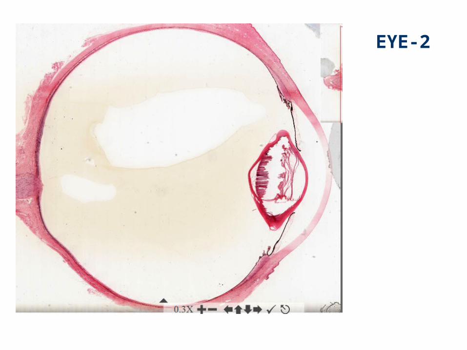
EYE-2
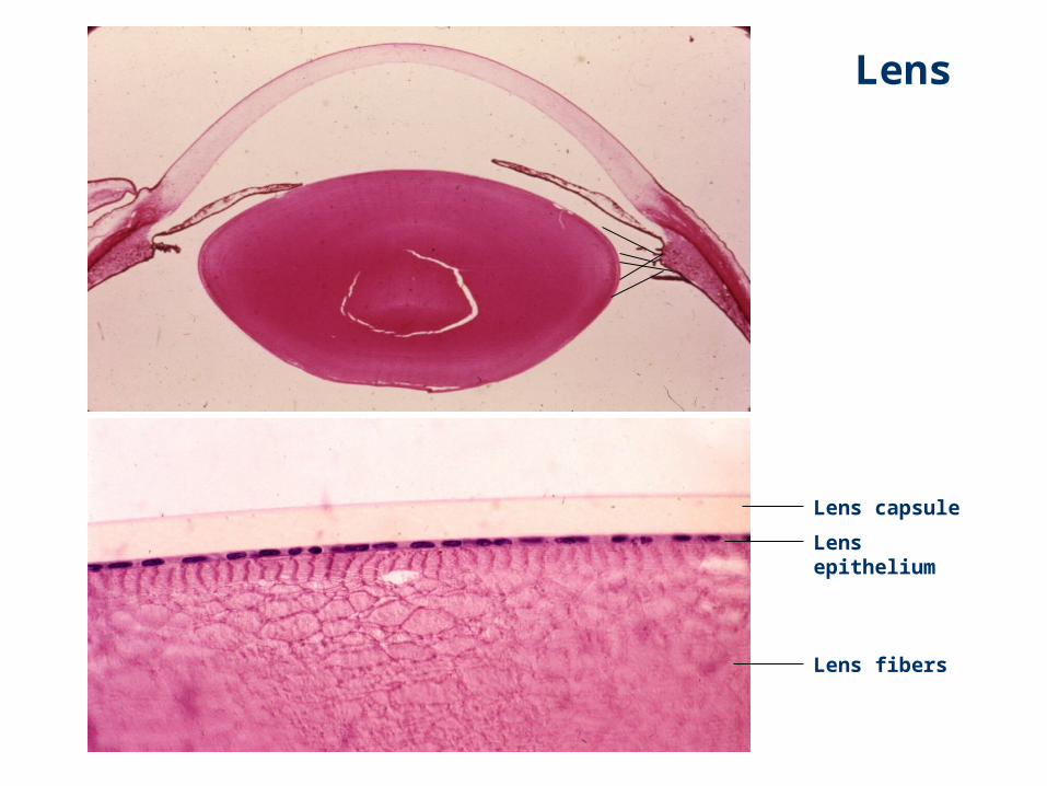
Lens
Lens capsule
Lens epithelium
Lens fibers
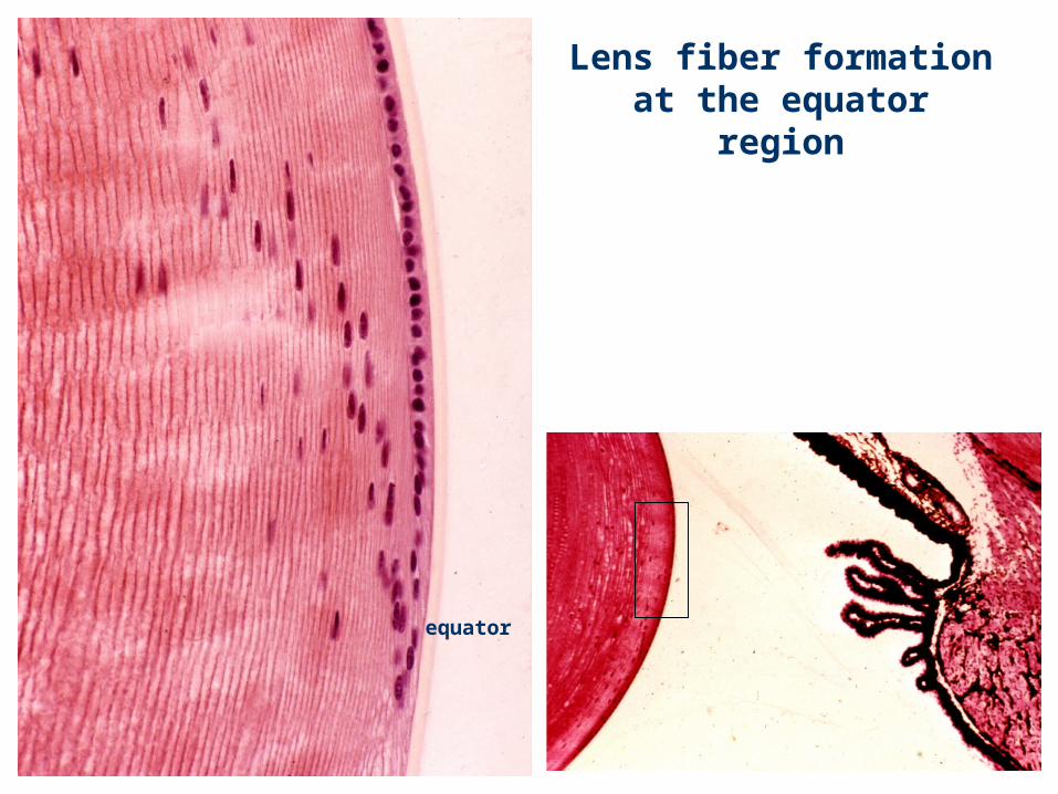
Lens fiber formation at the equator region
equator
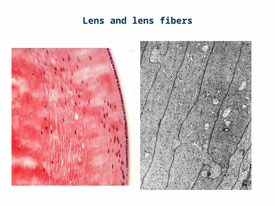
Lens and lens fibers
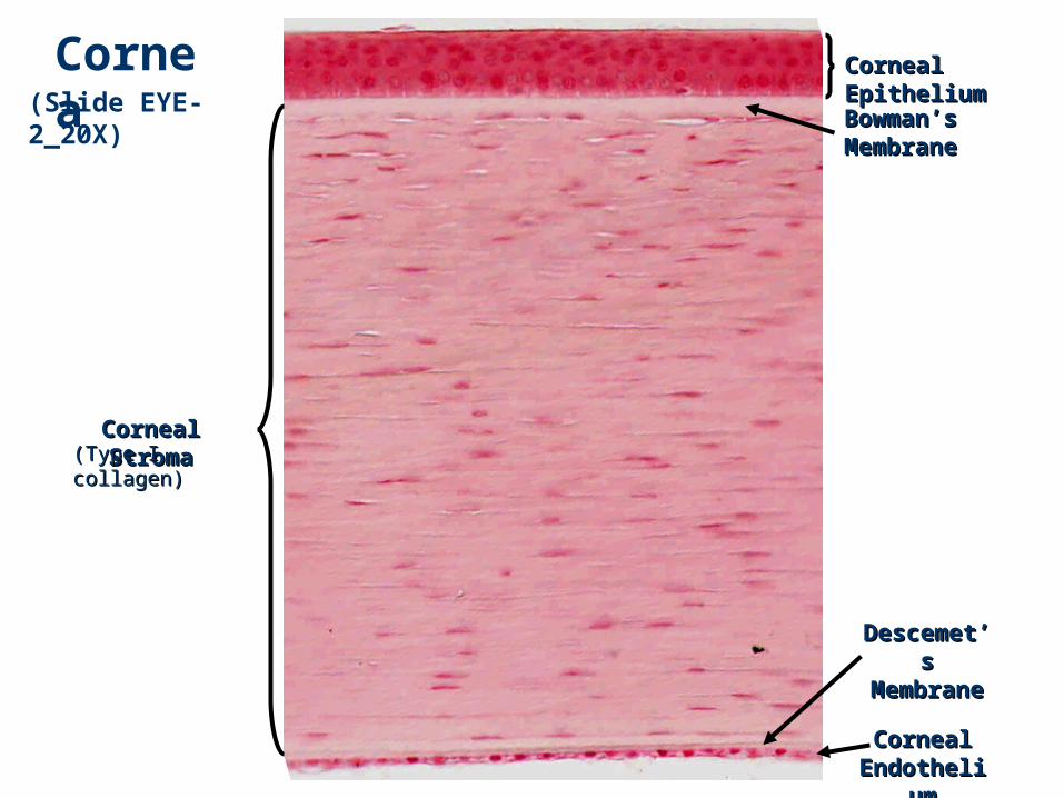
Corneal Corneal EpitheliumEpithelium
DescemeDescemet’s t’s
MembranMembranee
Corneal Corneal StromaStroma
Bowman’Bowman’s s
MembranMembranee
Corneal Corneal EndotheliEndotheli
umum
(Slide EYE-2_20X)
Cornea
(Type I (Type I collagen)collagen)
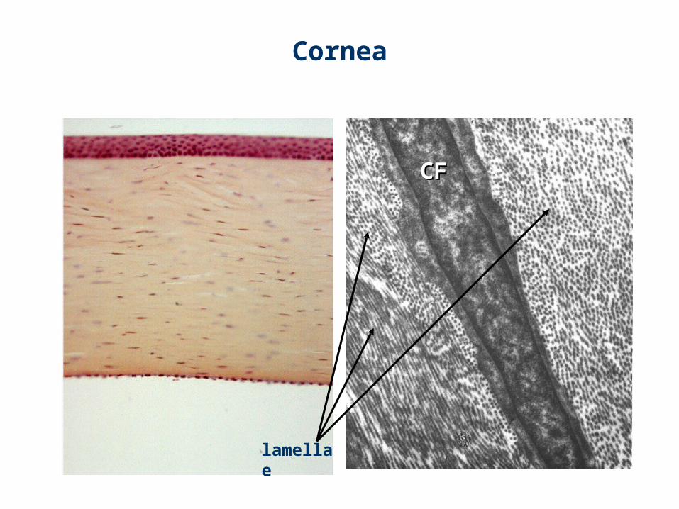
Cornea
CFCF
lamellae
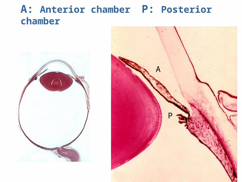
A: Anterior chamber P: Posterior chamber
A
P
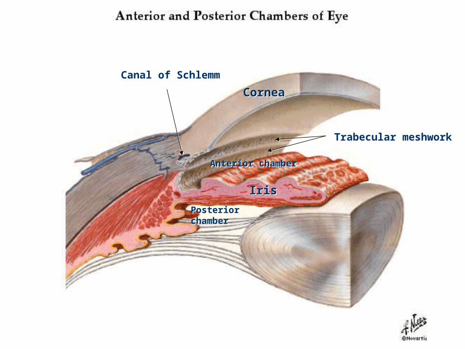
Anterior chamberAnterior chamber
IrisIris
Posterior chamberPosterior chamber
Trabecular meshworkTrabecular meshwork
Canal of SchlemmCanal of Schlemm
CorneaCornea
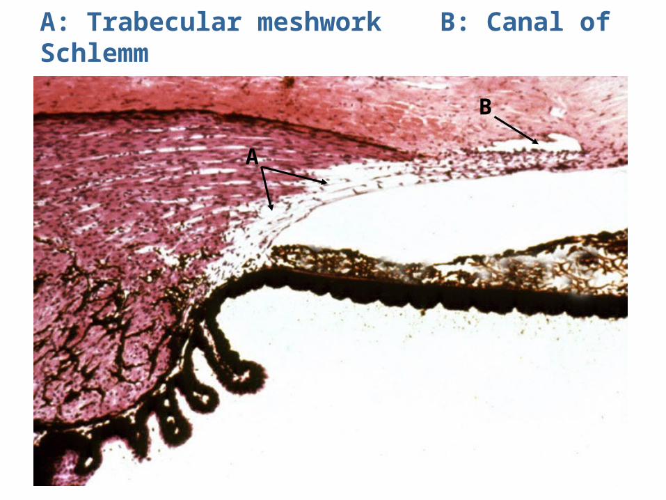
A: Trabecular meshwork B: Canal of Schlemm
A
B
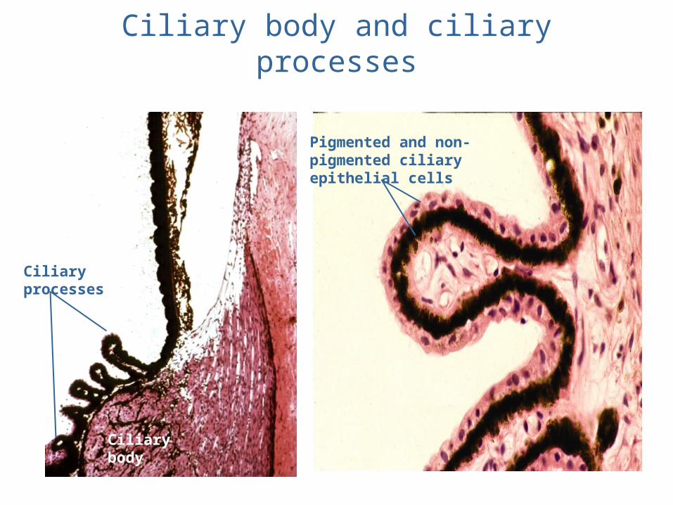
Ciliary body and ciliary processes
Ciliary processes
Ciliary body
Pigmented and non-pigmented ciliary epithelial cells
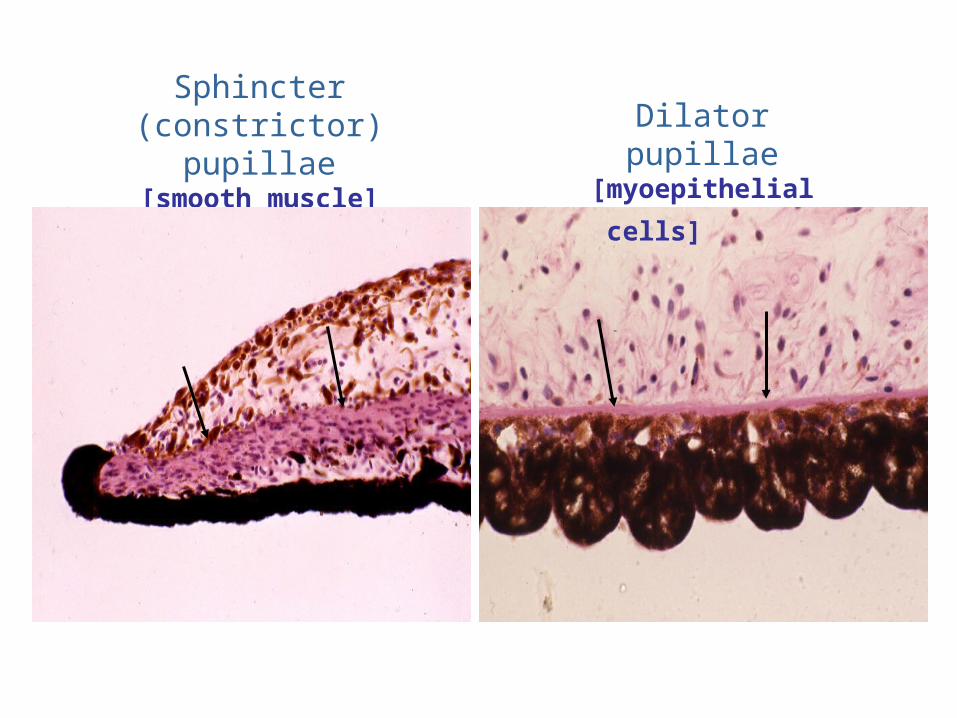
Sphincter (constrictor)pupillae
[smooth muscle]
Dilator pupillae [myoepithelial
cells]
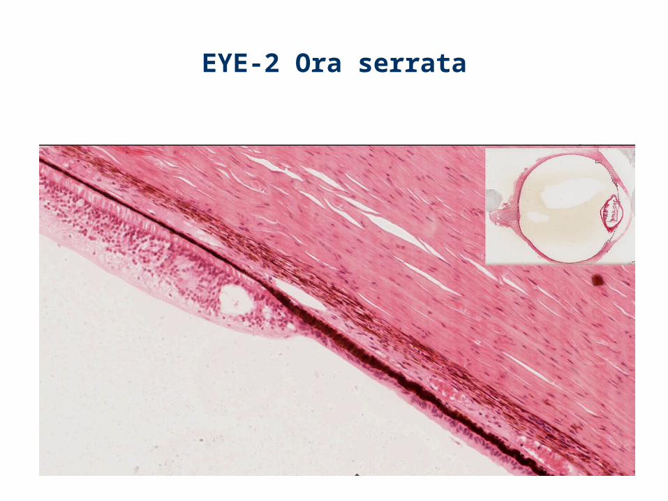
EYE-2 Ora serrata
Sclera
ChoroidBruch’s membrane
Choriocapillary layer
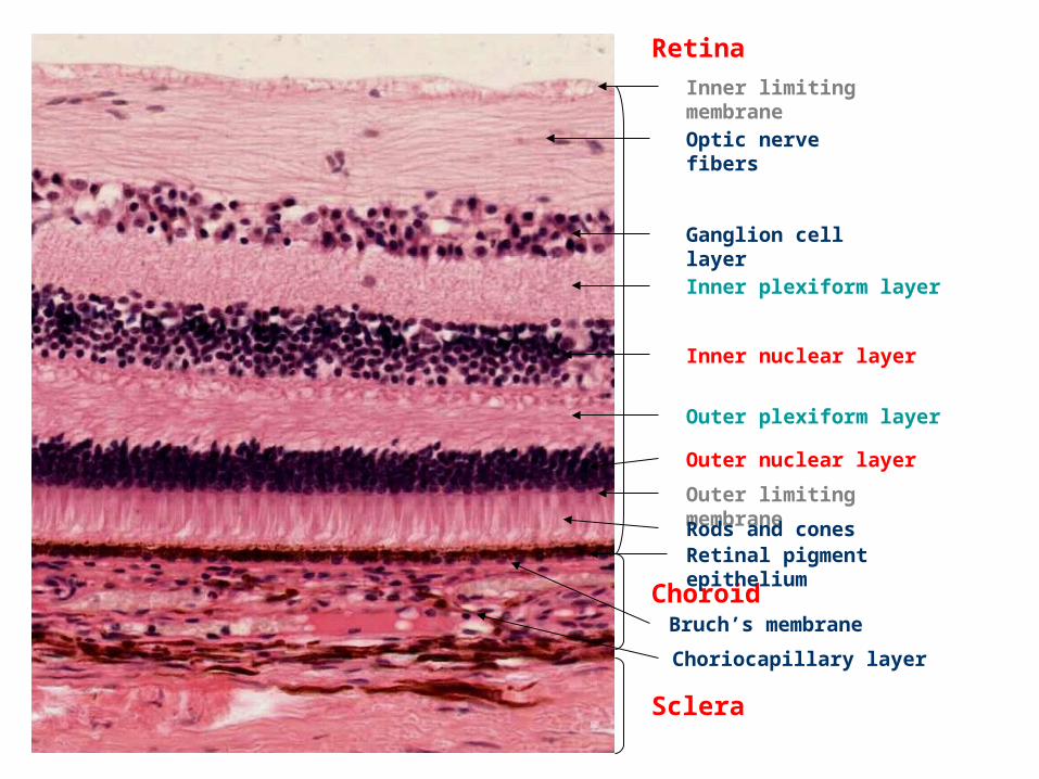
RetinaInner limiting membraneOptic nerve fibers
Ganglion cell layerInner plexiform layer
Inner nuclear layer
Outer plexiform layer
Outer nuclear layer
Outer limiting membraneRods and conesRetinal pigment epithelium
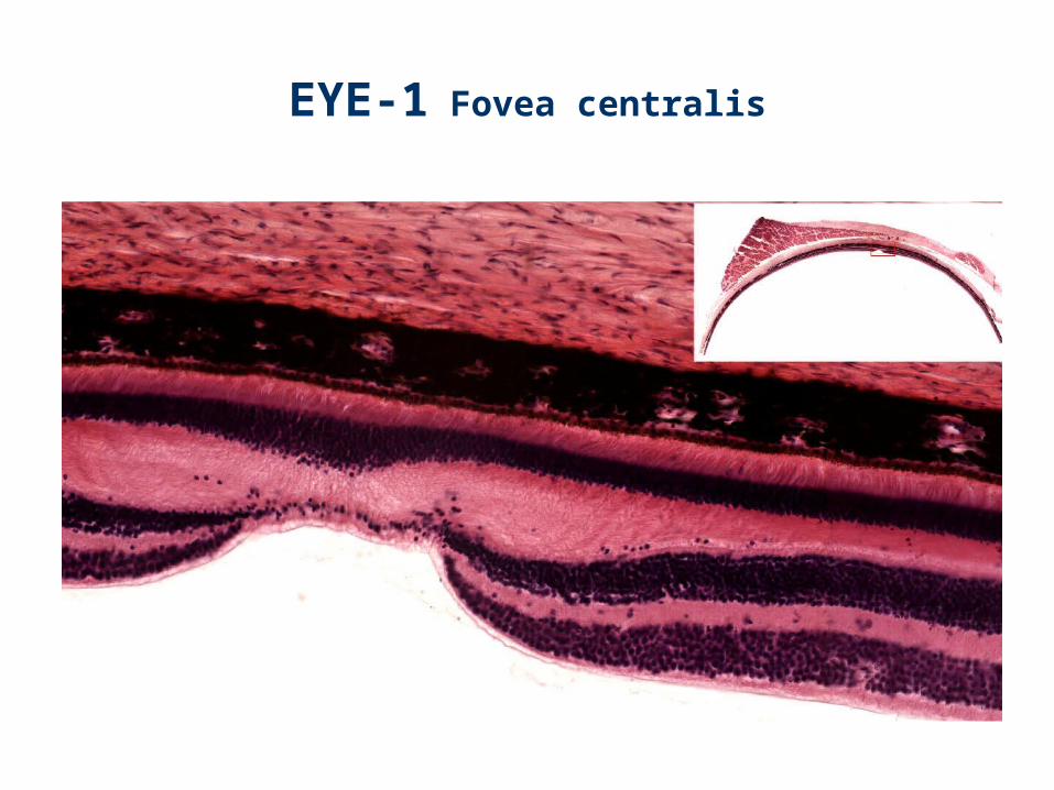
EYE-1 Fovea centralis
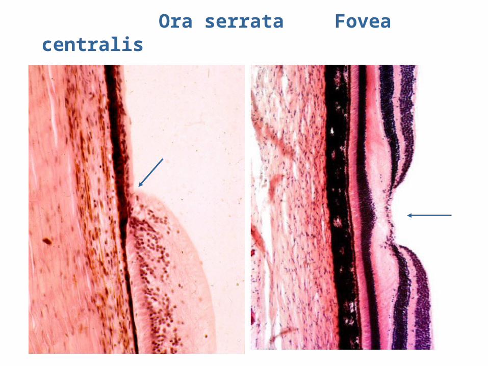
Ora serrata Fovea centralis
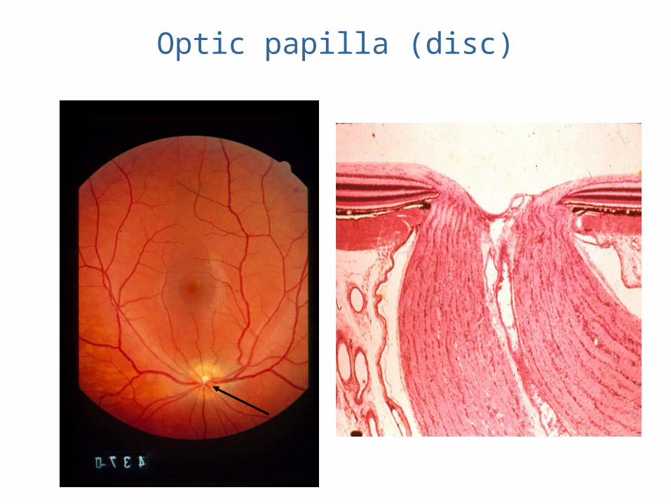
Optic papilla (disc)
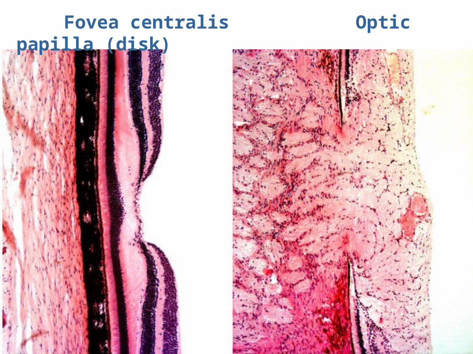
Fovea centralis Optic papilla (disk)
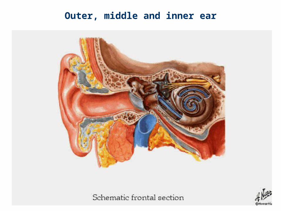
Outer, middle and inner ear
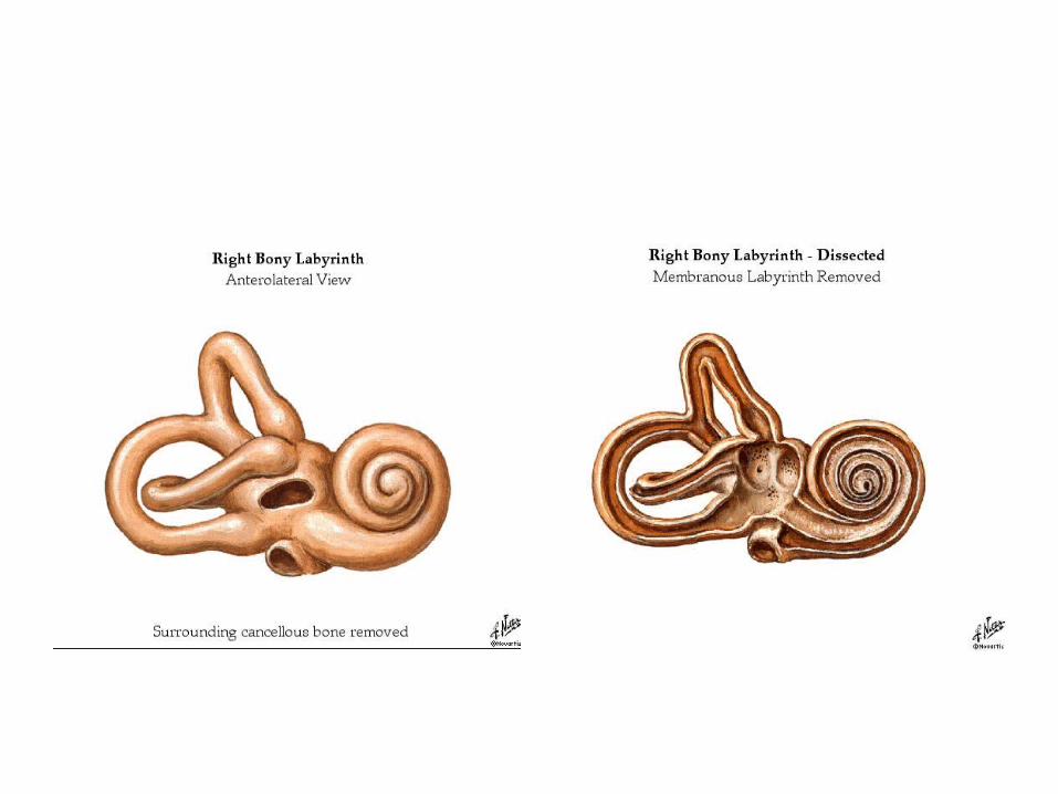
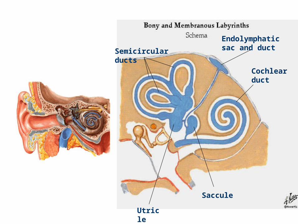
Semicircular ducts
Endolymphatic sac and duct
Cochlear duct
Saccule
Utricle
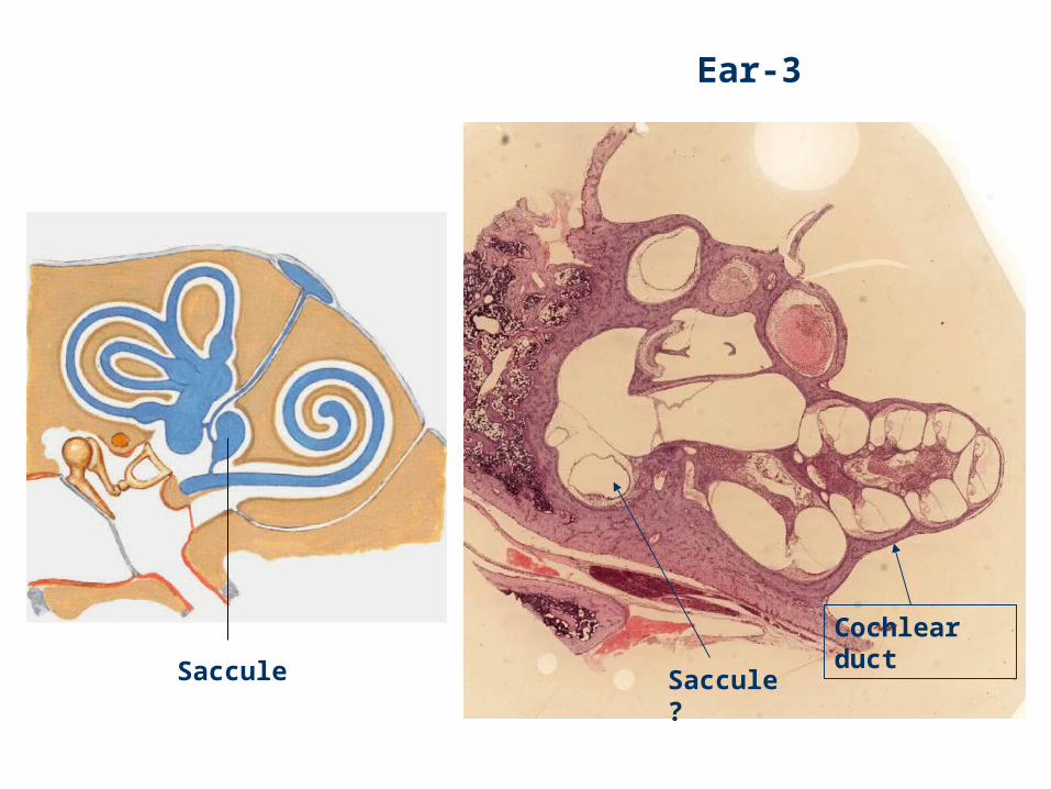
Ear-3
Saccule ?
Cochlear ductSaccule
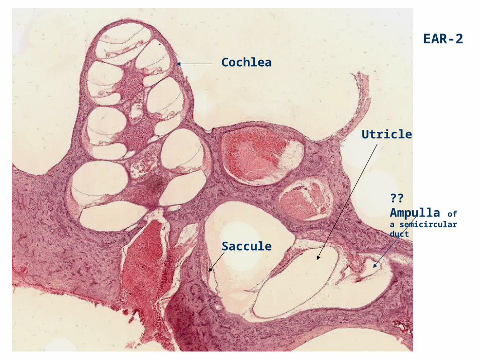
EAR-2
Cochlea
Utricle
?? Ampulla of a semicircular duct
Saccule
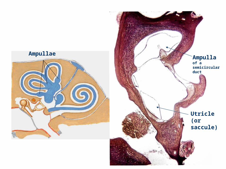
Ampulla of a semicircular duct
Utricle (or saccule)
Ampullae
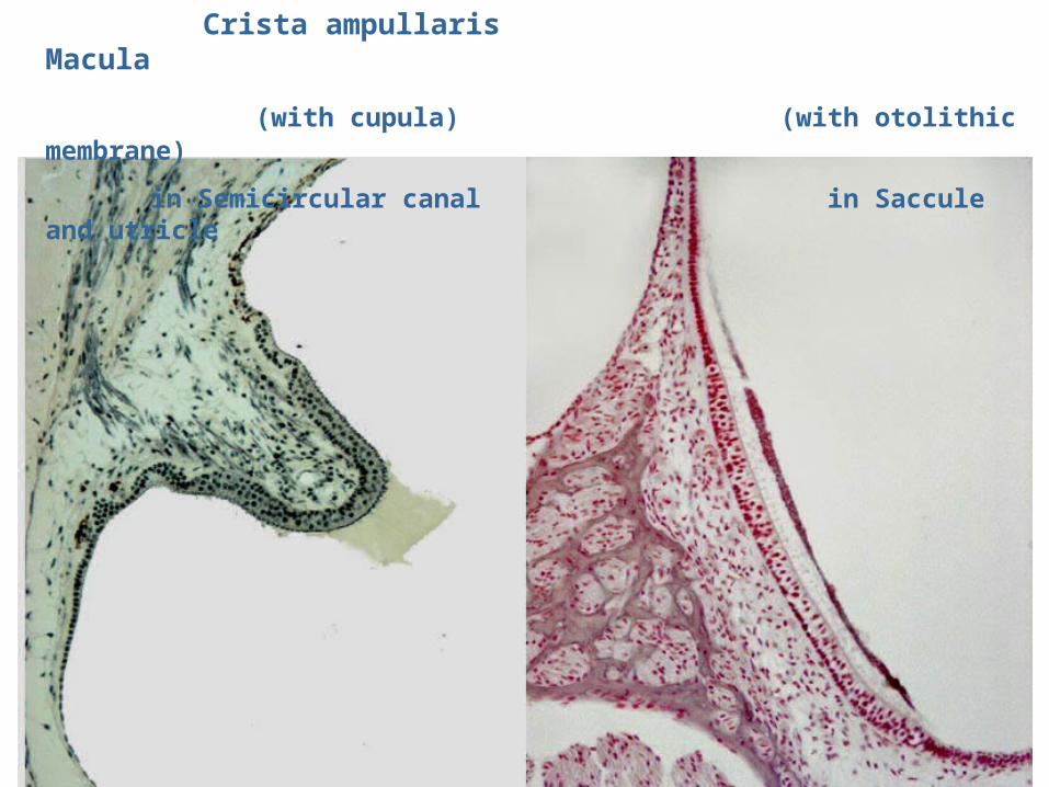
Crista ampullaris Macula
(with cupula) (with otolithic membrane)
in Semicircular canal in Saccule and utricle
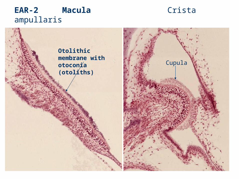
EAR-2 Macula Crista ampullaris
Otolithic membrane with otoconia (otoliths)
Cupula
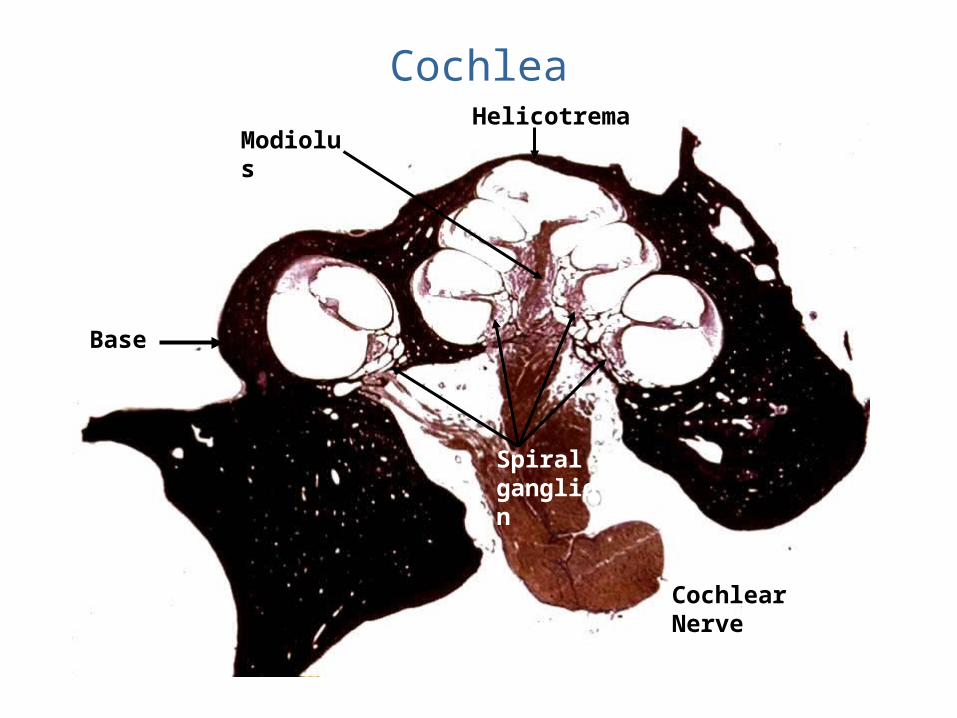
Cochlea
Base
Helicotrema
Cochlear Nerve
Modiolus
Spiral ganglion
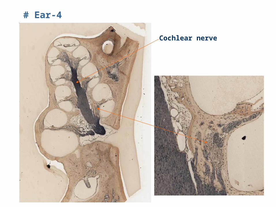
# Ear-4
Cochlear nerve
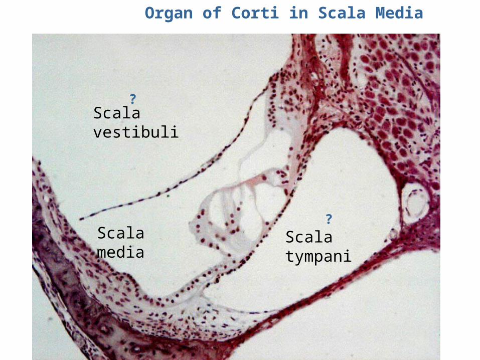
Organ of Corti in Scala Media
Scala vestibuli
Scala tympaniScala media
?
?
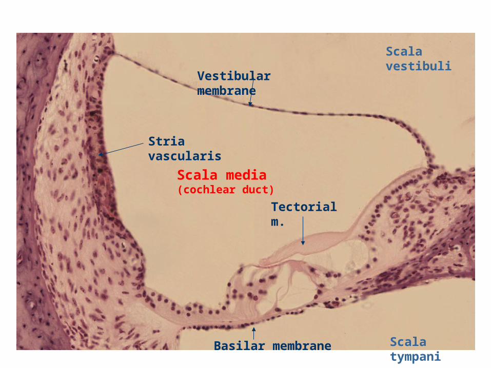
Stria vascularis
Scala media (cochlear duct)
Basilar membrane
Vestibular membrane
Tectorial m.
Scala vestibuli
Scala tympani
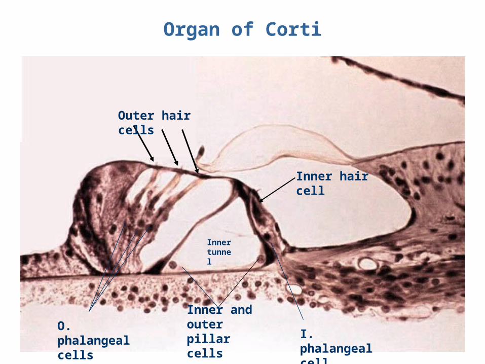
Organ of Corti
Outer hair cells
Inner hair cell
Inner and outer pillar cells
O. phalangeal cells
I. phalangeal cell
Inner tunnel
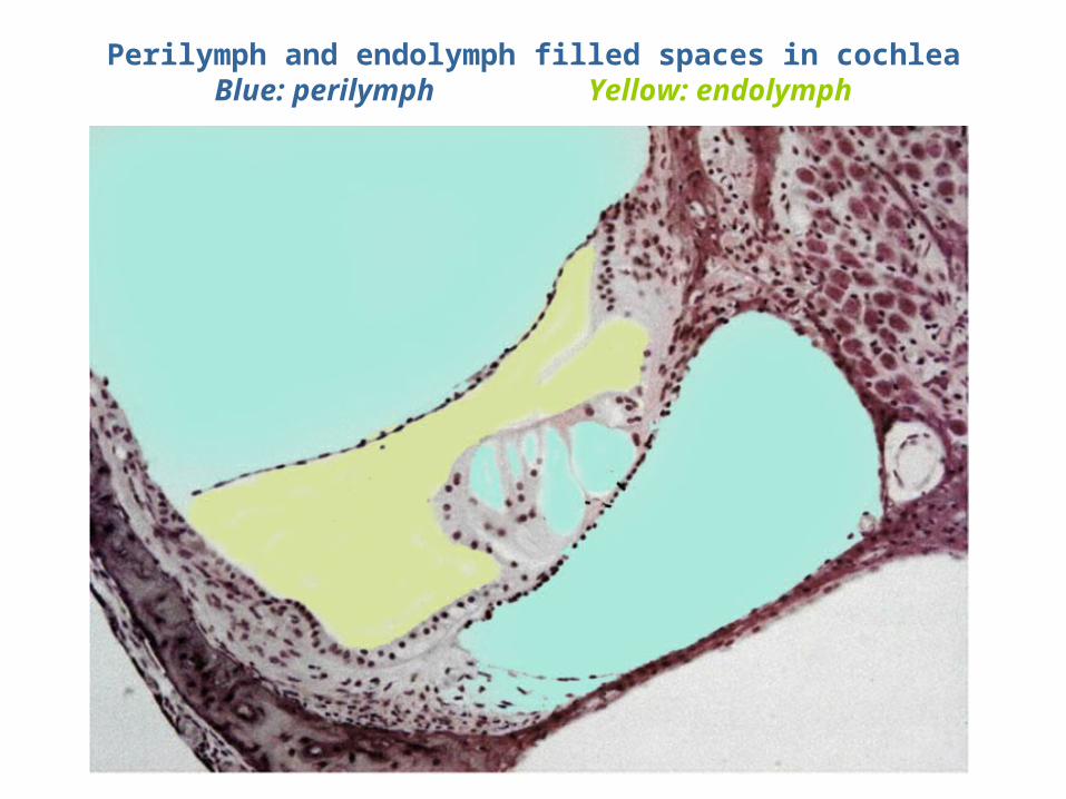
Perilymph and endolymph filled spaces in cochleaBlue: perilymph Yellow: endolymph
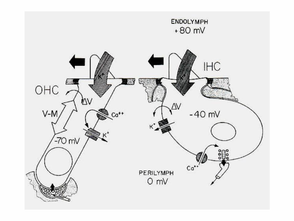
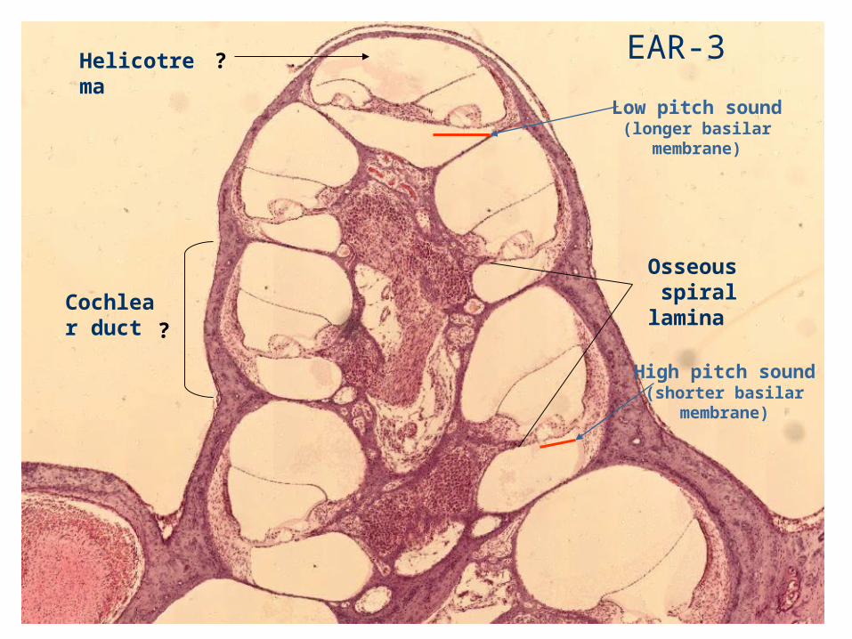
Helicotrema
Osseous spiral lamina
Cochlear duct
Low pitch sound(longer basilar
membrane)
High pitch sound (shorter basilar
membrane)
?
? EAR-3