Embed Size (px)
Citation preview
J Int Adv Otol 2017; 13(1): 47-52 • DOI: 10.5152/iao.2016.2181
Original Article
INTRODUCTIONIntratympanic gentamicin (ITG) is an effective treatment method for the vestibular symptoms of Meniere’s Disease (MD). Although gentamicin is mainly toxic to the vestibule, ototoxicity is an important concern and can be encountered in up to 34% of the treated patients depending on the dose regimen and number of injections [1]. Different ITG application protocols have been described recently in order to achieve complete or substantial vertigo control with minimal risk of ototoxicity [2-6]. Despite these efforts, ITG is not an ideal treatment option for patients with serviceable hearing and/or bilateral MD.
Intratympanic steroid (ITS) application has a role in the treatment of bilateral MD patients since most of these cases have underlying autoimmune etiology and respond well to steroids. Although ITS applications may seem more appropriate for patients with service-able hearing and bilateral MD, vertigo control rates are not as high as those obtained with ITG treatments [7, 8].
An ideal therapy for MD patients should provide both high vertigo control rates with hearing preservation. The aim of this study is to evaluate the effects of a combined gentamicin and dexamethasone intratympanic application on the inner ear.
MATERIALS and METHODSThe protocol of this study has been approved by the local ethical committee of the university’s animal care and use department (protocol number: 23/2014).
47
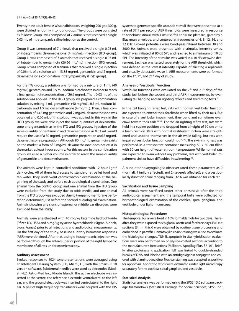
Cochlear and Vestibular Effects of Combined Intratympanic Gentamicin and Dexamethasone
OBJECTIVE: The aim of this study is to evaluate the effects of an intratympanic gentamicin-dexamethasone combination on the inner ear.
MATERIALS and METHODS: Twenty-six Wistar albino rats were divided into four groups: Group I (Control), group II (Intratympanic dexametha-sone; ITD), group III (Intratympanic gentamicin; ITG), and group IV (Intratympanic gentamicin and dexamethasone; ITGD). On the first day after basal auditory brainstem response (ABR) measurements, the ITG group received 0.03 mL of intratympanic gentamicin (26.7 mg/mL). Intratympan-ic injection of 0.06 mL of a solution containing 13.35 mg/mL gentamicin and 2 mg/mL dexamethasone was performed in the ITGD group. 0.03 mL of physiological intratympanic serum and dexamethasone (4 mg/mL) was applied in control and ITD groups, respectively. On the 7th day, ABR measurements were repeated and vestibular functions were evaluated. On the 21th day, ABR and vestibular tests were repeated, and the animals were sacrificed for histopathological investigation.
RESULTS: The ITG group’s hearing thresholds deteriorated in all frequencies. The ITGD group’s hearing thresholds were significantly better than the ITG group, except at 8 kHz on the 7th day and in all frequencies at the 21th day measurements. The vestibular function scores of the ITG and ITGD groups were higher than the controls. Apoptotic changes were seen in cochlea, spiral ganglion, and vestibule of the ITG group. Cochlear and vestibular structures were well preserved in the ITGD group, similar to the controls.
CONCLUSION: The ITGD combination led to a significant hearing preservation. Although in subjective vestibular tests, it seemed that vestibulo-toxicity was present in both ITG and ITGD groups the histopathological investigations revealed no signs of vestibulotoxicity in the ITGD group in contrast to the ITG group. Further studies using a combination of different concentrations of gentamicin and dexamethasone are needed.
KEYWORDS: Gentamicin, dexamethasone, intratympanic
Enis Alpin Güneri, Yüksel Olgun, Mustafa Aslıer, Daniele Nuti, Günay Kırkım, Serpil Mungan, Efsun Kolatan, Safiye Aktaş, Franco Trabalzini, Hülya Ellidokuz, Osman Yılmaz, Marco Mandala Department of Otorhinolaryngology, Dokuz Eylül University School of Medicine, İzmir, Turkey (EAG, YO, MA)Department of Otology and Skull Base, Azienda Ospedaliera Universiteria Senese, Siena, Italy (DN, FT, MM)Department of Otorhinolaryngology, Dokuz Eylül University School of Medicine Unit of Hearing Speech and Balance, İzmir, Turkey (GK, SM)Department of Laboratory of Animal Science, Dokuz Eylül University School of Medicine, İzmir, Turkey (EK, OY)Department of Basic Oncology, Dokuz Eylül University Institute of Oncology, İzmir, Turkey (SA) Department of Biostatistics, Dokuz Eylül University School of Medicine, İzmir, Turkey (HE)
Corresponding Address: Yüksel Olgun E-mail: [email protected]
Submitted: 24.01.2016 Revision Received: 31.08.2016 Accepted: 13.10.2016©Copyright 2017 by The European Academy of Otology and Neurotology and The Politzer Society - Available online at www.advancedotology.org
Twenty-nine adult female Wistar albino rats, weighing 200 g to 300 g, were divided randomly into four groups. The groups were consisted as follows: Group I was composed of 7 animals that received a single 0.03 mL of intratympanic saline injection as the control.
Group II was composed of 7 animals that received a single 0.03 mL of intratympanic dexamethasone (4 mg/mL) injection (ITD group). Group III was composed of 7 animals that received a single 0.03 mL of intratympanic gentamicin (26.66 mg/mL) injection (ITG group). Group IV was composed of 8 animals that received a single injection of 0.06 mL of a solution with 13.35 mg/mL gentamicin and 2 mg/mL dexamethasone combination intratympanically (ITGD group).
For the ITG group, a solution was formed by a mixture of 1 mL (40 mg/mL) gentamicin and 0.5 mL sodium bicarbonate in order to reach a final gentamicin concentration of 26.6 mg/mL. Then, 0.03 mL of this solution was applied. In the ITGD group, we prepared a combination solution by mixing 1 mL gentamicin (40 mg/mL), 0.5 mL sodium bi-carbonate, and 1.5 mL dexamethasone (4 mg/mL). Then, a final con-centration of 13.3 mg gentamicin and 2 mg/mL dexamethasone was obtained and 0.06 mL of this solution was applied. In this way, in the ITGD group, we were able inject the same quantities of dexametha-sone and gentamicin as in the ITD and ITG groups. Injection of the same quantity of gentamicin and dexamethasone in 0.03 mL would require the use of a 80 mg/mL gentamicin preparation and 8 mg/mL dexamethasone preparation. Although 80 mg/mL gentamicin exists on the market, a form of 8 mg/mL dexamethasone does not exist in the market, at least in our country. For this reason, in the combination group, we used a higher volume in order to reach the same quantity of gentamicin and dexamethasone.
The animals were kept in controlled conditions with 12 hour light/dark cycles. All of them had access to standard rat pellet food and tap water. They underwent otomicroscopic examination at the be-ginning of the study and before each audiological examination. One animal from the control group and one animal from the ITD group were excluded from the study due to otitis media, and one animal from the ITD group was excluded due to tympanic membrane perfo-ration determined just before the second audiological examination. Animals showing any signs of external or middle ear disorders were excluded from the study.
Animals were anesthetized with 40 mg/kg ketamine hydrochloride (Pfizer, NY, USA) and 5 mg/kg xylazine hydrochloride (Sigma-Aldrich, Lyon, France) prior to all injections and audiological measurements. On the first day of the study, baseline auditory brainstem responses (ABR) were obtained. After that, a single intratympanic injection was performed through the anterosuperior portion of the right tympanic membrane of all rats under otomicroscopy.
Auditory AssessmentEvoked responses to 1024 tone presentations were averaged using an Intelligent Hearing System (IHS, Miami, FL) with the Smart-EP 10 version software. Subdermal needles were used as electrodes (Mod-el F-E2, Astro-Med Inc., Rhode Island). The active electrode was in-serted at the vertex, the reference electrode ventrolateral to the left ear, and the ground electrode was inserted ventrolateral to the right ear. A pair of high frequency transducers were coupled with the IHS
system to generate specific acoustic stimuli that were presented at a rate of 37.1 per second. ABR thresholds were measured in response to toneburst stimuli with 1 ms rise/fall and 0 ms plateaus, gated by a Blackman envelope, and centered at frequencies of 4, 8, 12, 16, and 32 kHz. Evoked potentials were band-pass-filtered between 30 and 3000 Hz. Animals were presented with a stimulus intensity series, which was initiated at 80 dB SPL and reached to a minimum of 10 dB SPL. The intensity of the stimulus was varied in a 10 dB stepwise dec-rement. Each ear was tested separately for the ABR threshold, which was defined as the lowest intensity capable of eliciting a replicable and visually detectable wave II. ABR measurements were performed on the 1st, 7th, and 21th day of study.
Analyses of Vestibular FunctionVestibular functions were evaluated on the 7th and 21th days of the study, just before the second and third ABR measurements, by eval-uating tail hanging and air righting reflexes and swimming tests [9].
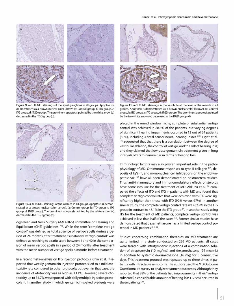
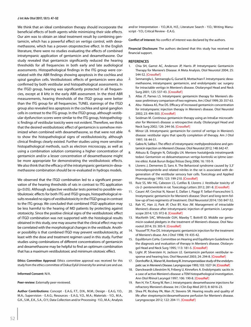
In the tail hanging reflex test, rats with normal vestibular function are expected to extend their forelimbs when lifted by their tails. But in case of a vestibular impairment, they bend and sometimes even crawl toward their tails [9, 10]. For the air righting reflex test, rats were held in a supine position and dropped from a height of 50 cm on to a foam cushion. Rats with normal vestibular function were straight-ened and unbend themselves in the air while falling, but rats with impaired vestibular function could not [9, 10]. The swimming test was performed in a transparent container measuring 50 x 50 cm filled with 30 cm height of water at room temperature. While normal rats are expected to swim without any problems, rats with vestibular im-pairment sink or have difficulties in swimming [9].
A blind otorinolaryngologist observer rated these parameters as 0 (normal), 1 (mildly affected), and 2 (severely affected); and a vestibu-lar dysfunction score ranging from 0 to 6 was obtained for each rat.
Sacrification and Tissue SamplingAll animals were sacrificed under ether anesthesia after the third audiological assessment and the temporal bulla were collected for histopathological examination of the cochlea, spiral ganglion, and vestibule under light microscopy.
Histopathological ProceduresThe temporal bulla were fixed in 10% formaldehyde for two days. There-after, they were exposed to 5% glacial acetic acid for three days. Full cut sections (3 mm thick) were obtained by routine tissue processing and embedded in paraffin. Hematoxylin eosin staining was used to evaluate the histological changes. TUNEL apoptosis in situ hybridization evalua-tions were also performed on polylysine-coated sections according to the manufacturer’s instructions (Millipore, ApopTag Plus, S7101). Brief-ly, after proteinase K application, TdT was linked to double-stranded breaks of DNA and labeled with an antidigoxigenin conjugate and col-ored with diaminobenzidine. Nuclear staining was accepted as positive for apoptosis. Apoptotic ratios were evaluated under light microscopy separately for the cochlea, spiral ganglion, and vestibule.
Statistical AnalysisStatistical analysis was performed using the SPSS 15.0 software pack-age for Windows (Statistical Package for Social Sciences; SPSS Inc.;
48
J Int Adv Otol 2017; 13(1): 47-52
Chicago, IL, USA). Results were presented as means and standard de-viations (SD). All data were analyzed by one-way analysis of variance (ANOVA) post-hoc Bonferroni test and p<0.05 was considered as a statistically significant difference between measurements.
RESULTS
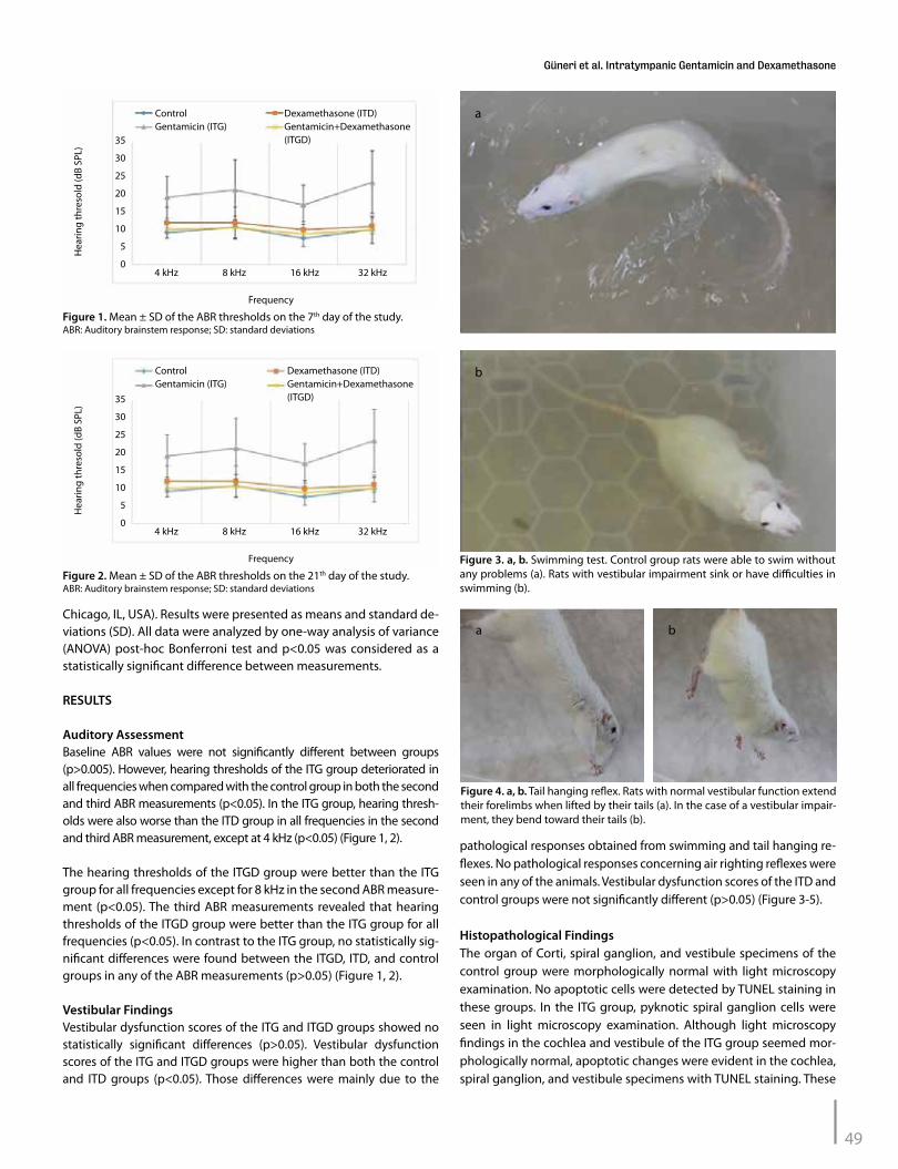
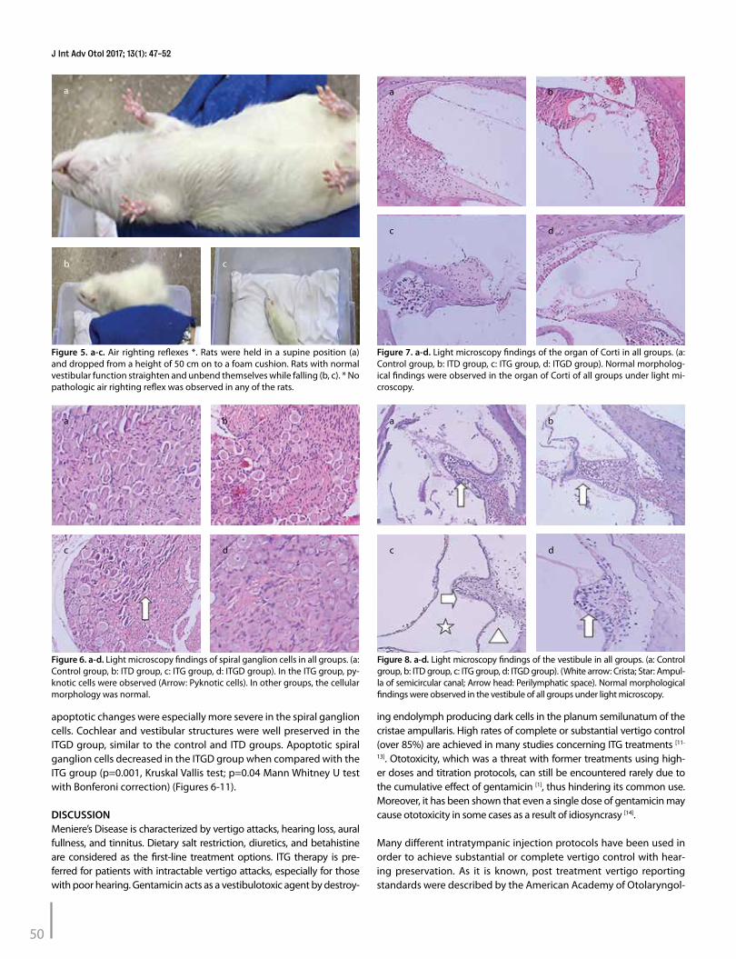
Auditory AssessmentBaseline ABR values were not significantly different between groups (p>0.005). However, hearing thresholds of the ITG group deteriorated in all frequencies when compared with the control group in both the second and third ABR measurements (p<0.05). In the ITG group, hearing thresh-olds were also worse than the ITD group in all frequencies in the second and third ABR measurement, except at 4 kHz (p<0.05) (Figure 1, 2).
The hearing thresholds of the ITGD group were better than the ITG group for all frequencies except for 8 kHz in the second ABR measure-ment (p<0.05). The third ABR measurements revealed that hearing thresholds of the ITGD group were better than the ITG group for all frequencies (p<0.05). In contrast to the ITG group, no statistically sig-nificant differences were found between the ITGD, ITD, and control groups in any of the ABR measurements (p>0.05) (Figure 1, 2).
Vestibular FindingsVestibular dysfunction scores of the ITG and ITGD groups showed no statistically significant differences (p>0.05). Vestibular dysfunction scores of the ITG and ITGD groups were higher than both the control and ITD groups (p<0.05). Those differences were mainly due to the
pathological responses obtained from swimming and tail hanging re-flexes. No pathological responses concerning air righting reflexes were seen in any of the animals. Vestibular dysfunction scores of the ITD and control groups were not significantly different (p>0.05) (Figure 3-5).
Histopathological FindingsThe organ of Corti, spiral ganglion, and vestibule specimens of the control group were morphologically normal with light microscopy examination. No apoptotic cells were detected by TUNEL staining in these groups. In the ITG group, pyknotic spiral ganglion cells were seen in light microscopy examination. Although light microscopy findings in the cochlea and vestibule of the ITG group seemed mor-phologically normal, apoptotic changes were evident in the cochlea, spiral ganglion, and vestibule specimens with TUNEL staining. These
49
Güneri et al. Intratympanic Gentamicin and Dexamethasone
Figure 1. Mean ± SD of the ABR thresholds on the 7th day of the study.ABR: Auditory brainstem response; SD: standard deviations
35
30
25
20
15
10
5
0
Dexamethasone (ITD)Gentamicin+Dexamethasone (ITGD)
ControlGentamicin (ITG)
Frequency
4 kHz 8 kHz 16 kHz 32 kHz
Hea
ring
thre
sold
(dB
SPL)
Figure 2. Mean ± SD of the ABR thresholds on the 21th day of the study.ABR: Auditory brainstem response; SD: standard deviations
35
30
25
20
15
10
5
0
Dexamethasone (ITD)Gentamicin+Dexamethasone (ITGD)
ControlGentamicin (ITG)
Frequency
4 kHz 8 kHz 16 kHz 32 kHz
Hea
ring
thre
sold
(dB
SPL)
Figure 3. a, b. Swimming test. Control group rats were able to swim without any problems (a). Rats with vestibular impairment sink or have difficulties in swimming (b).
a
b
Figure 4. a, b. Tail hanging reflex. Rats with normal vestibular function extend their forelimbs when lifted by their tails (a). In the case of a vestibular impair-ment, they bend toward their tails (b).
a b
apoptotic changes were especially more severe in the spiral ganglion cells. Cochlear and vestibular structures were well preserved in the ITGD group, similar to the control and ITD groups. Apoptotic spiral ganglion cells decreased in the ITGD group when compared with the ITG group (p=0.001, Kruskal Vallis test; p=0.04 Mann Whitney U test with Bonferoni correction) (Figures 6-11).
DISCUSSIONMeniere’s Disease is characterized by vertigo attacks, hearing loss, aural fullness, and tinnitus. Dietary salt restriction, diuretics, and betahistine are considered as the first-line treatment options. ITG therapy is pre-ferred for patients with intractable vertigo attacks, especially for those with poor hearing. Gentamicin acts as a vestibulotoxic agent by destroy-
ing endolymph producing dark cells in the planum semilunatum of the cristae ampullaris. High rates of complete or substantial vertigo control (over 85%) are achieved in many studies concerning ITG treatments [11-
13]. Ototoxicity, which was a threat with former treatments using high-er doses and titration protocols, can still be encountered rarely due to the cumulative effect of gentamicin [1], thus hindering its common use. Moreover, it has been shown that even a single dose of gentamicin may cause ototoxicity in some cases as a result of idiosyncrasy [14].
Many different intratympanic injection protocols have been used in order to achieve substantial or complete vertigo control with hear-ing preservation. As it is known, post treatment vertigo reporting standards were described by the American Academy of Otolaryngol-
50
J Int Adv Otol 2017; 13(1): 47-52
a
b c
Figure 5. a-c. Air righting reflexes *. Rats were held in a supine position (a) and dropped from a height of 50 cm on to a foam cushion. Rats with normal vestibular function straighten and unbend themselves while falling (b, c). * No pathologic air righting reflex was observed in any of the rats.
c
a
d
b
Figure 6. a-d. Light microscopy findings of spiral ganglion cells in all groups. (a: Control group, b: ITD group, c: ITG group, d: ITGD group). In the ITG group, py-knotic cells were observed (Arrow: Pyknotic cells). In other groups, the cellular morphology was normal.
c
a
d
b
Figure 7. a-d. Light microscopy findings of the organ of Corti in all groups. (a: Control group, b: ITD group, c: ITG group, d: ITGD group). Normal morpholog-ical findings were observed in the organ of Corti of all groups under light mi-croscopy.
c
a
d
b
Figure 8. a-d. Light microscopy findings of the vestibule in all groups. (a: Control group, b: ITD group, c: ITG group, d: ITGD group). (White arrow: Crista; Star: Ampul-la of semicircular canal; Arrow head: Perilymphatic space). Normal morphological findings were observed in the vestibule of all groups under light microscopy.
ogy-Head and Neck Surgery (AAO-HNS) committee on Hearing and Equilibrium (CHE) guidelines [15]. While the term “complete vertigo control’’ was defined as total absence of vertigo spells during a pe-riod of 24 months after treatment, ‘‘substantial vertigo control’’ was defined as reaching to a ratio score between 1 and 40 in the compar-ison of mean vertigo spells in a period of 24 months after treatment with the mean number of vertigo spells 6 months before treatment.
In a recent meta-analysis on ITG injection protocols, Chia et al. [1] re-ported that weekly gentamicin injection protocols led to a mild oto-toxicity rate compared to other protocols; but even in that case, the incidence of ototoxicity was as high as 13.1%. However, severe oto-toxicity up to 34.7% was reported with daily multiple injection proto-cols [1]. In another study in which gentamicin-soaked pledgets were
placed in the round window niche, complete or substantial vertigo control was achieved in 88.5% of the patients, but varying degrees of significant hearing impairments occurred in 12 out of 24 patients (50%), including 4 total sensorineural hearing losses [13]. Light et al. [16] suggested that that there is a correlation between the degree of vestibular ablation, the control of vertigo, and the risk of hearing loss; and they claimed that low dose gentamicin treatment given in long intervals offers minimum risk in terms of hearing loss.
Immunologic factors may also play an important role in the patho-physiology of MD. Otoimmune responses to type II collagen [17], de-posits of IgG [17], and mononuclear cell infiltrations on the endolym-pathic sac [18] have all been demonstrated on postmortem studies. Thus, anti-inflammatory and immunomodulatory effects of steroids have come into use for the treatment of MD. Akkuzu et al. [8] com-pared the effects of ITD and ITG in patients with MD and found that complete vertigo control rates that were achieved with ITG were sig-nificantly higher than those with ITD (92% versus 67%). In another similar study, the complete vertigo control rate was 82.9% in the ITG group in contrast to 48.1% in the ITD group [7]. In another study using ITS for the treatment of MD patients, complete vertigo control was achieved in less than half of the cases [19]. Former similar studies have demonstrated that dexamethasone has a limited vertigo control po-tential in MD patients [7, 8, 19].
Studies concerning combination therapies on MD treatment are quite limited. In a study conducted on 299 MD patients, all cases were treated with intratympanic injections of a combination solu-tion of streptomycin (10 mg/mL) and dexamethasone (24 mg/mL) in addition to systemic dexamethasone (16 mg) for 3 consecutive days. This treatment protocol was repeated up to three times in pa-tients with intractable symptoms. The authors used the MD Outcome Questionnaire survey to analyze treatment outcomes. Although they reported that 88% of the patients had improvements in their “vertigo subscore”, a considerable amount of hearing loss (17.9%) occurred in these patients [20].
51
Güneri et al. Intratympanic Gentamicin and Dexamethasone
c
a
d
b
Figure 9. a-d. TUNEL stainings of the spiral ganglions in all groups. Apoptosis is demonstrated as a brown nuclear color (arrow) (a: Control group, b: ITD group, c: ITG group, d: ITGD group). The prominent apoptosis pointed by the white arrow (d) decreased in the ITGD group (d).
c
a
d
b
Figure 11. a-d. TUNEL stainings in the vestibule at the level of the macula in all groups. Apoptosis is demonstrated as a brown nuclear color (arrows). (a: Control group, b: ITD group, c: ITG group, d: ITGD group). The prominent apoptosis pointed by the two white arrows (c) decreased in the ITGD group (d).
c
a
d
b
Figure 10. a-d. TUNEL stainings of the cochlea in all groups. Apoptosis is demon-strated as a brown nuclear color (arrow). (a: Control group, b: ITD group, c: ITG group, d: ITGD group). The prominent apoptosis pointed by the white arrows (c) decreased in the ITGD group (d).
We think that an ideal combination therapy should incorporate the beneficial effects of both agents while minimizing their side effects. Our aim was to obtain an ideal treatment result by combining gen-tamicin, which has a powerful effect on vertigo control, with dexa-methasone, which has a proven otoprotective effect. In the English literature, there were no studies evaluating the effects of combined intratympanic application of gentamicin and dexamethasone. Our study revealed that gentamicin significantly reduced the hearing thresholds for all frequencies in both early and late audiological assessments. Histopathological findings in the ITG group were cor-related with the ABR findings showing apoptosis in the cochlea and spiral ganglion cells. Vestibulotoxic effects of gentamicin were also confirmed by both vestibular and histopathological assessments. In the ITGD group, hearing was significantly protected in all frequen-cies, except at 8 kHz in the early ABR assessment. In the third ABR measurements, hearing thresholds of the ITGD group were better than the ITG group for all frequencies. TUNEL stainings of the ITGD group also revealed less apoptosis in the cochlea and spiral ganglion cells in contrast to the ITG group. In the ITGD group, although vestib-ular dysfunction scores were similar to the ITG group, histopatholog-ic findings of vestibular toxicity were not evident. Therefore, we think that the desired vestibulotoxic effect of gentamicin is somehow min-imized when combined with dexamethasone, so that were not able to show the histopathological signs of vestibulotoxicity, although clinical findings clearly existed. Further studies using more sensitive histopathological methods, such as electron microscopy, as well as using a combination solution containing a higher concentration of gentamicin and/or a lesser concentration of dexamethasone might be more appropriate for demonstrating the vestibulotoxic effects. We also think that the efficacy of the intratympanic gentamicin-dexa-methasone combination should be re-evaluated in hydrops models.
We observed that the ITGD combination led to a significant preser-vation of the hearing thresholds of rats in contrast to ITG application (p<0.05). Although subjective vestibular tests pointed to possible ves-tibulotoxic effects for both ITG and ITGD groups, histopathological re-sults revealed no signs of vestibulotoxicity in the ITGD group in contrast to the ITG group. We concluded that combined ITGD application may be less harmful to the hearing of MD patients by protecting against ototoxicity. Since the positive clinical signs of the vestibulotoxic effect of ITGD combination was not supported with the histological results obtained in this study, one may assume that the clinical effect may not be correlated with the morphological changes in the vestibule. Anoth-er possibility is that combined ITGD may prevent vestibulotoxicity, at least with the dose and treatment regimen used in this study. Further studies using combinations of different concentrations of gentamicin and dexamethasone may be helpful to find an optimum combination that has a maximum vestibulotoxic and minimum ototoxic effect.
Ethics Committee Approval: Ethics committee approval was received for this study from the ethics committee of Dokuz Eylül University for animal care and use.
Informed Consent: N/A.
Peer-review: Externally peer-reviewed.
Author Contributions: Concept - E.A.G, F.T., D.N., M.M.; Design - E.A.G, Y.O., M.A.; Supervision - E.A.G.; Resources - E.A.G, Y.O., M.A.; Materials - Y.O., M.A., G.K., S.M., E.K., S.A., O.Y.; Data Collection and/or Processing - Y.O., M.A.; Analysis
and/or Interpretation - Y.O.,M.A, H.E.; Literature Search - Y.O.; Writing Manu-script - Y.O.; Critical Review - E.A.G.
Conflict of Interest: No conflict of interest was declared by the authors.
Financial Disclosure: The authors declared that this study has received no financial support.
REFERENCES1. Chia SH, Gamst AC, Anderson JP, Harris JP. Intratympanic Gentamicin
Therapy for Meniere’s Disease: A Meta Analysis. Otol Neurotol 2004; 25: 544-52. [CrossRef]
2. Sennaroglu L, Sennaroglu G, Gursel B, Mottachian F. Intratympanic dexa-methasone, intratympanic gentamicin, and endolympatic sac surgery for intractable vertigo in Meniere’s disease. Otolaryngol Head and Neck Surg 2001; 125: 537-43. [CrossRef]
3. Atlas JT, Parnes LS. Intratympanic gentamicin therapy for Meniere’s dis-ease: preliminary comparison of two regimens. Am J Otol 1999; 20: 357-63.
4. Abu- Halawa AS, Poe DS. Efficacy of increased gentamicin concentration for intratympanic injection therapy in Meniere’s disease. Otol Neurotol 2002; 23: 494-503. [CrossRef]
5. Seidman M. Continous gentamicin therapy using an IntraEar microcath-eter for Meniere’s disease: a retrospective study. Otolaryngol Head and Neck Surg 2002; 126: 244-56. [CrossRef]
6. Minor LB. Intratympanic gentamicin for control of vertigo in Meniere’s disease: vestibular signs that specify completion of therapy. Am J Otol 1997; 18: 44-51.
7. Gabra N, Saliba I. The effect of intratympanic methylprednisolone and gen-tamicin injection on Meniere’s Disease. Otol Neurotol 2012; 148: 642-47.
8. Akkuzu B, Özgirgin N, Özlüoğlu LN. Meniere hastalığında intratimpanik tedavi: Gentamisin ve deksametazonun vertigo kontrolü ve işitme üzer-ine etkisi. Kulak Burun Boğaz İhtisas Derg 2006; 16: 193-9.
9. Llorens J, Dememes D, Sans A. The Behavioral syndrome caused by 3,3’ Iminodipropinitrile and related nitriles in the rat is associated with de-generation of the vestibular sensory hair cells. Toxicology and Applied Pharmacology 1993; 123: 199-210. [CrossRef]
10. Ruiz SS, Mir HG, Cabezon LS, Cutillas B, Llorens J. Vestibular toxicity of cis-2- pentenenitrile in rat. Toxicology Letters 2012; 281-8. [CrossRef]
11. Casani AP, Cerchiai N, Navari E, Dallan I, Piaggi P, Sellari-Franceschini S. Inratympanic gentamicin for Meniere’s disease: Short and long-term fol-low-up of two regiments of treatment. Otol Neurootol 2014; 150: 847-52.
12. Rah YC, Han JJ, Park JP, Choi BY, Koo JW. Management of intractable Meniere’s disease after intratympanic injection of gentamicin. Laryngo-scope 2014; 125: 972-8. [CrossRef]
13. MacKeith SAC, Whiteside OJH, Mawby T, Bottrill ID. Middle ear genta-micin-soaked pledgets in the treatment of Meniere’s disease. Otol Neu-rootol 2014; 35: 305-9. [CrossRef]
14. Youssef TF, Poe DS. Intratympanic gentamicin injection for the treatment of Meniere’s disase. Am J Otol 1998; 19: 435-42.
15. Equilibrium CoHa. Committee on Hearing and Equilibriym Guidelines for the diagnosis and evaluation of therapy in Meniere’s disease. Otolaryn-gol Head and Neck Surg 1995; 113: 181-5. [CrossRef]
16. Light JP, Silverstein H, Jackson LE. Gentamicin perfusion vestibular re-sponse and hearing loss. Otol Neurotol 2003; 24: 294-8. [CrossRef]
17. Dornhoffer JL, Waner M, Arenberg IK. Immunperoxidase study of the endolym-patic sac in Meniere’s Disease. Laryngoscope 1993; 103: 1027-34. [CrossRef]
18. Danckwardt-Lilieström N, Friberg U, Kinnefors A. Endolympatic sacitis in a case of active Meniere’s disease: a TEM histopathological investigation. Ann Otol Rhinol Laryngol 1997; 106: 190-8. [CrossRef]
19. Ren H, Yin T, Kong W, Ren J. Intratympanic dexamethasone injections for refractory Meniere’s disease. Int J Clin Exp Med 2015; 8: 6016-23.
20. Shea PF, Ricshey PA, Wan JY, Stevens SR. Hearing results and quality of life after streptomycin/dexamethasone perfusion for Meniere’s disease. Laryngoscope 2012; 122: 204-11. [CrossRef]
52
J Int Adv Otol 2017; 13(1): 47-52