Embed Size (px)
Citation preview
Coeliac DiseaseCoeliac DiseaseCoeliac DiseaseCoeliac Disease Maria Santangelo, aged 25, presents complaining of 3 months of worsening tiredness and malaise with a history of diarrhoea.
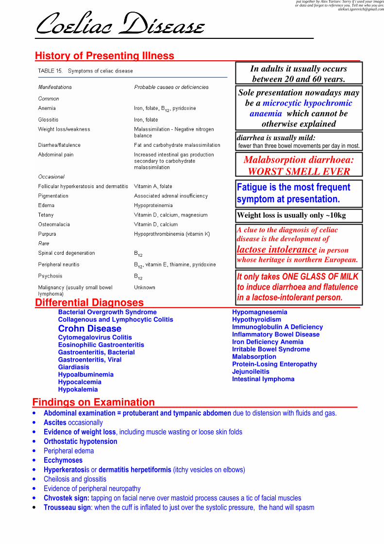
History of Presenting Illness
Differential Diagnoses Bacterial Overgrowth Syndrome Collagenous and Lymphocytic Colitis
Crohn Disease
Cytomegalovirus Colitis Eosinophilic Gastroenteritis Gastroenteritis, Bacterial Gastroenteritis, Viral Giardiasis Hypoalbuminemia Hypocalcemia Hypokalemia
Hypomagnesemia Hypothyroidism Immunoglobulin A Deficiency Inflammatory Bowel Disease Iron Deficiency Anemia Irritable Bowel Syndrome Malabsorption Protein-Losing Enteropathy Jejunoileitis Intestinal lymphoma
In adults it usually occurs
between 20 and 60 years.
Sole presentation nowadays may
be a microcytic hypochromic
anaemia which cannot be
otherwise explained diarrhea is usually mild: fewer than three bowel movements per day in most.
Fatigue is the most frequent symptom at presentation.
Weight loss is usually only ~10kg A clue to the diagnosis of celiac
disease is the development of
lactose intolerance in person
whose heritage is northern European. It only takes ONE GLASS OF MILK to induce diarrhoea and flatulence in a lactose-intolerant person.
Findings on Examination • Abdominal examination = protuberant and tympanic abdomen due to distension with fluids and gas.
• Ascites occasionally
• Evidence of weight loss, including muscle wasting or loose skin folds
• Orthostatic hypotension
• Peripheral edema
• Ecchymoses
• Hyperkeratosis or dermatitis herpetiformis (itchy vesicles on elbows)
• Cheilosis and glossitis
• Evidence of peripheral neuropathy
• Chvostek sign: tapping on facial nerve over mastoid process causes a tic of facial muscles
• Trousseau sign: when the cuff is inflated to just over the systolic pressure, the hand will spasm
Malabsorption diarrhoea:
WORST SMELL EVER
put together by Alex Yartsev: Sorry if i used your imagesor data and forgot to reference you. Tell me who you are.
Tests and Investigations Full Blood Count
Anemia is present in less than 50% of adult patients. Also want to make sure that the coagulopathy is due
to malnutrition and vitamin K depletion, not thrombocytopenia
Serum Ferritin.
iron deficiency is the most common laboratory abnormality.. Serum Biochemistry
Depletion of minerals (zinc, magnesium) and ions (potassium)
( occurs only with severe disease ) Malnutrition = decreased serum albumin.
RBC Folate
Folate deficiency is uncommon, but happens Hydrogen Breath test
the absorptive cell lesion also results in secondary lactase deficiency; thus, the H2-lactose breath test may be abnormal in celiac disease.
Stool Examination Steatorrhea can be confirmed by a 72-hour fecal fat study. It is usually mild (10-20 g/24 hours) and may be absent in some patients. Severity of steatorrhoea correlates with the extent of the intestinal lesion,
PLUS you look for ova, cysts, parasites, Leucocytes and BLOOD Barium Swallow radiography (IGNORE AS IT IS USELESS)
Barium studies of the small bowel may show dilation of the bowel and slight thickening of the mucosal folds. Intraluminal signs of malabsorption with flocculation, segmentation and clumping of the barium (features due to excess amount of fluid present within the lumen) are variable and not common. (The new barium suspensions now used have made this a rare finding.) Radiographic findings in celiac disease are not specific for this syndrome of malabsorption.
Serum Anti-Endomyseal Antibodies Anti-endomysial IgA antibody : antibodies against reticulin in monkey oesophageal smooth muscle..
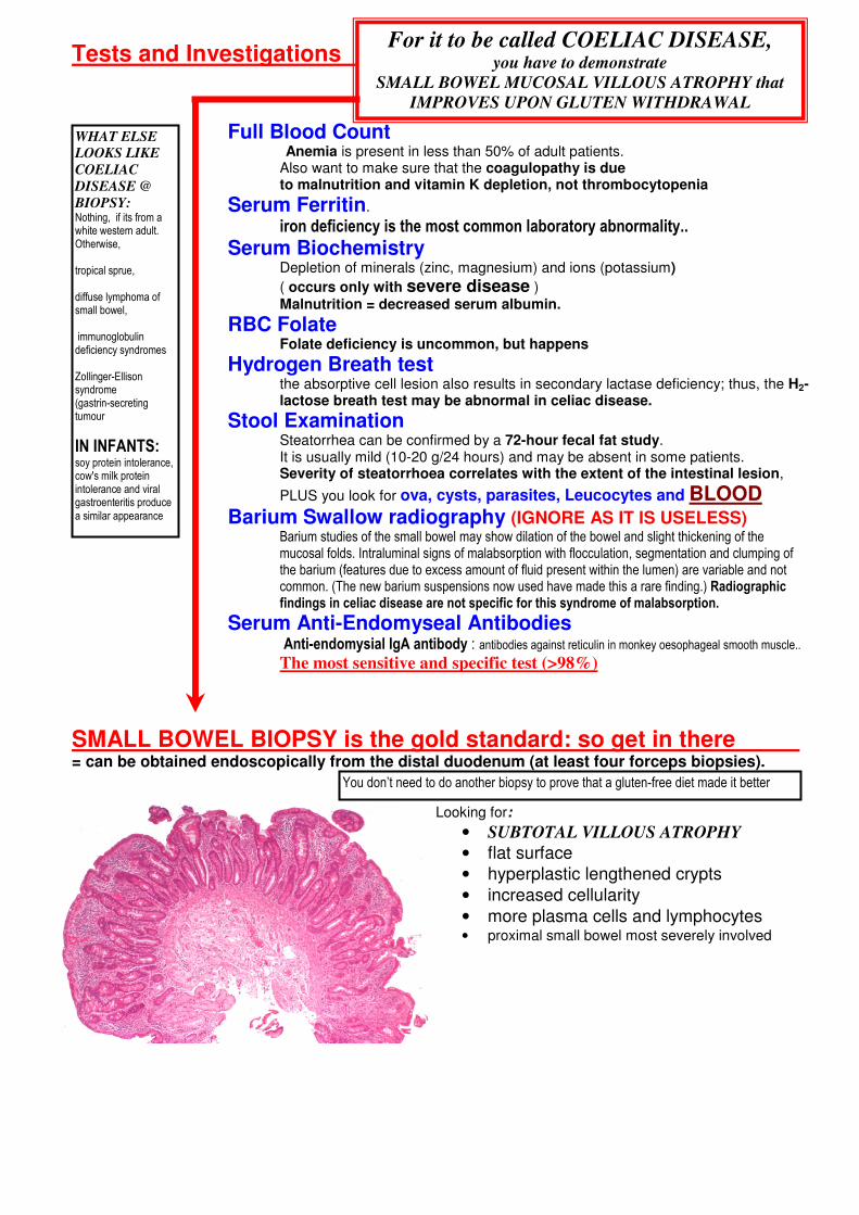
The most sensitive and specific test (>98%) SMALL BOWEL BIOPSY is the gold standard: so get in there = can be obtained endoscopically from the distal duodenum (at least four forceps biopsies).
Looking for:
• SUBTOTAL VILLOUS ATROPHY
• flat surface
• hyperplastic lengthened crypts
• increased cellularity
• more plasma cells and lymphocytes • proximal small bowel most severely involved
For it to be called COELIAC DISEASE, you have to demonstrate
SMALL BOWEL MUCOSAL VILLOUS ATROPHY that
IMPROVES UPON GLUTEN WITHDRAWAL
WHAT ELSE
LOOKS LIKE
COELIAC
DISEASE @
BIOPSY: Nothing, if its from a white western adult. Otherwise, tropical sprue, diffuse lymphoma of small bowel, immunoglobulin deficiency syndromes Zollinger-Ellison syndrome (gastrin-secreting tumour
IN INFANTS: soy protein intolerance, cow's milk protein intolerance and viral gastroenteritis produce a similar appearance
You don’t need to do another biopsy to prove that a gluten-free diet made it better
Management: STOP EATING GLUTEN! which requires avoiding wheat, rye, barley and oats
PLUS: may want to think about replacing + supplementing haematinics and other micronutrients
Nutritional approaches to GI disease Gluten is found in
- wheat (wheat flour is ~8% gluten) - rye and barley - wheaten cornflour, - malt - some thickeners - oats (contaminated with traces)
Gluten-free flours: - rice, - soy, - maize, - besan (chickpea) - potato - Arrowroot, - buckwheat, - lupin, - maize cornflour, - millet,
- modified maize starch, - polenta, - psyllium, - rice, - sago, - seeds, - sorghum
- tapioca
Prognosis: EXCELLENT provided gluten is withdrawn
Vast improvement within weeks if not days
Epidemiology Frequency:
• Internationally: Celiac sprue is prevalent in some European countries with temperate climates.
For example, the frequency of the disease is between 1 in 250 persons and 1 in 300 persons in
Italian and Irish populations. In comparison, the disease is rare in Africans or Asians.
Mortality/Morbidity:
• increased risk for lymphomas and adenocarcinomas of the intestinal tract.
• Untreated pregnant women are at risk of miscarriage
• There is risk of congenital malformation of the baby.
• celiac sprue @ childhood = FAILURE TO THRIVE and short stature
(!!30% have clubbing and 20% can have constipation. How very weird)
Race: Celiac sprue is most prevalent in Europeans and is rare in Africans and Asians.
Sex: Incidence of celiac sprue is slightly higher in females.
Age: The age distribution of patients with celiac disease is bimodal,
• first peak is at 8-12 months
• second peak in the third to fourth decades.
Commonest cause of malabsorption in the western world! 1:250 to 1:75
THIS DIET IS LIFE-LONG: Mention to the patient – persistent non-compliance may be associated
with small bowel malignancy
elastic properties of the wheat gluten protein permit the baking of leavened bread.
many foods on the market that are gluten-free
are not labelled as such
A person on a normal diet consumes about 10-14g of gluten per day.
Nutrient Absorption and Transport
Trace nutrients: Water soluble vitamins
Fat soluble vitamins
incorporated in bile salt micelles prior to absorption by the enterocytes usually in the upper jejunum.
Vitamin A is not as reliant on bile andpancreatic enzymes Vitamin D is absorbed in the upper jejunum presumably by diffusion. Vitamin E absorption is very dependent on adequate micelle formation Vitamin K requires both bile and pancreatic enzymes for absorption.
Trace metals
Most trace metals are absorbed by passive diffusion Iron bonds to a membrane receptor protein, and absorption may be facilitated by a cytosolic
apotransferrin. Iron absorption is impaired by certain foods, eg milk protein, tea and coffee, phytates,
and enhanced or impaired according to iron storage status.
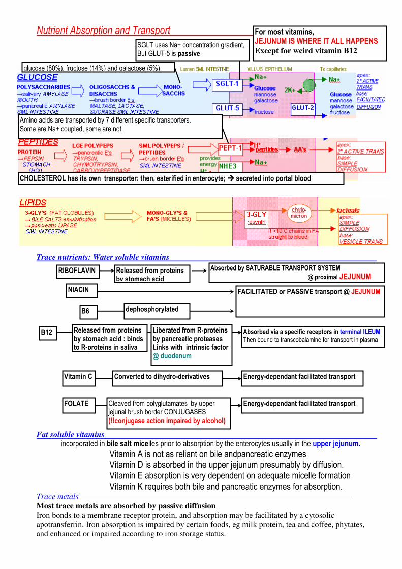
glucose (80%), fructose (14%) and galactose (5%).
SGLT uses Na+ concentration gradient, But GLUT-5 is passive
Amino acids are transported by 7 different specific transporters.
Some are Na+ coupled, some are not.
CHOLESTEROL has its own transporter: then, esterified in enterocyte; ���� secreted into portal blood
RIBOFLAVIN Absorbed by SATURABLE TRANSPORT SYSTEM
@ proximal JEJUNUM Released from proteins by stomach acid
NIACIN FACILITATED or PASSIVE transport @ JEJUNUM
B6 dephosphorylated
B12 Released from proteins by stomach acid : binds to R-proteins in saliva
Liberated from R-proteins by pancreatic proteases Links with intrinsic factor @ duodenum
Absorbed via a specific receptors in terminal ILEUM
Then bound to transcobalamine for transport in plasma
Vitamin C Converted to dihydro-derivatives Energy-dependant facilitated transport
FOLATE Cleaved from polyglutamates by upper jejunal brush border CONJUGASES (!!conjugase action impaired by alcohol)
Energy-dependant facilitated transport
For most vitamins, JEJUNUM IS WHERE IT ALL HAPPENS
Except for weird vitamin B12
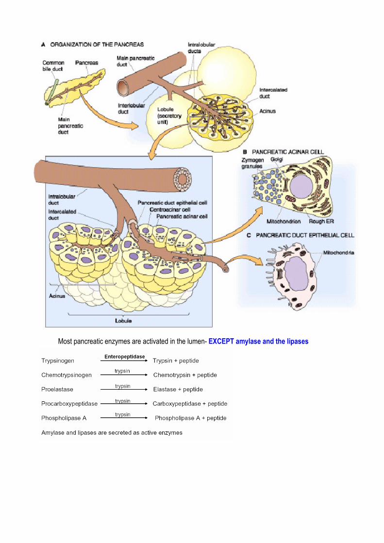
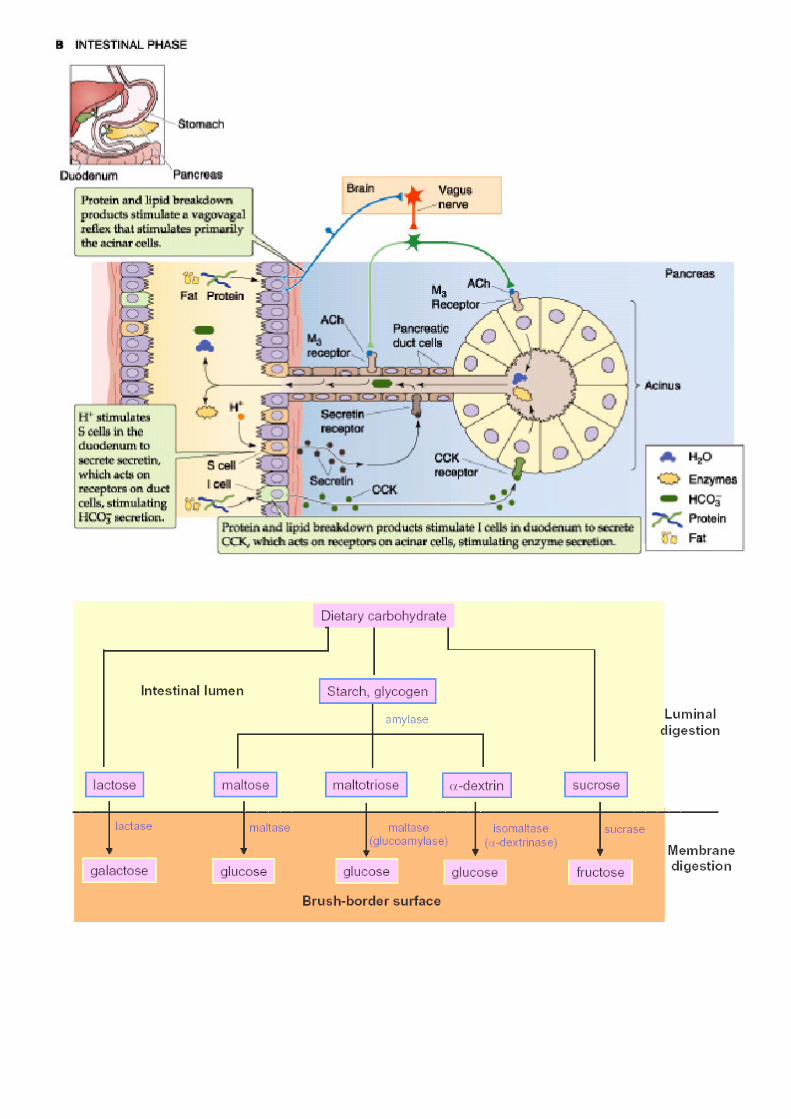
Most pancreatic enzymes are activated in the lumen- EXCEPT amylase and the lipases
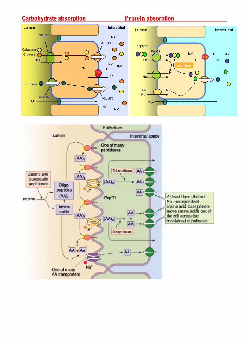
Carbohydrate absorption Protein absorption
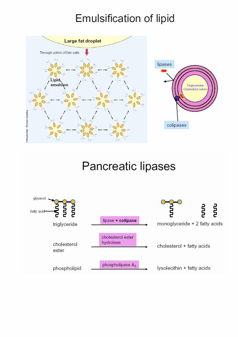
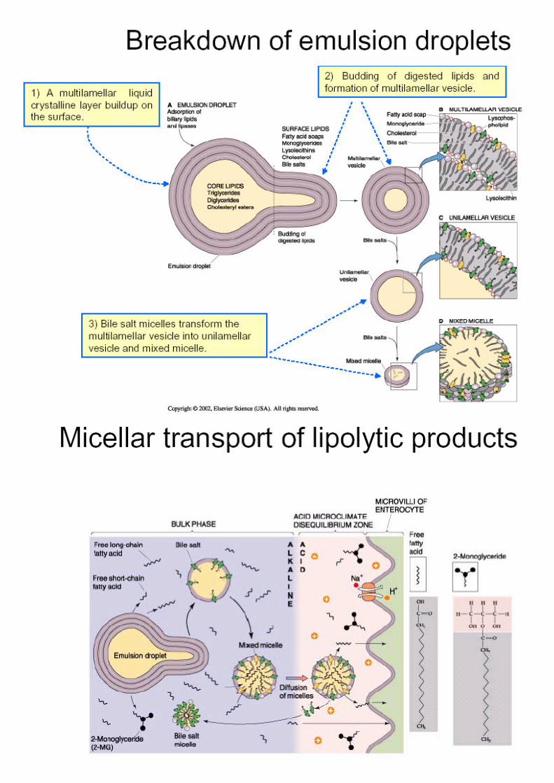
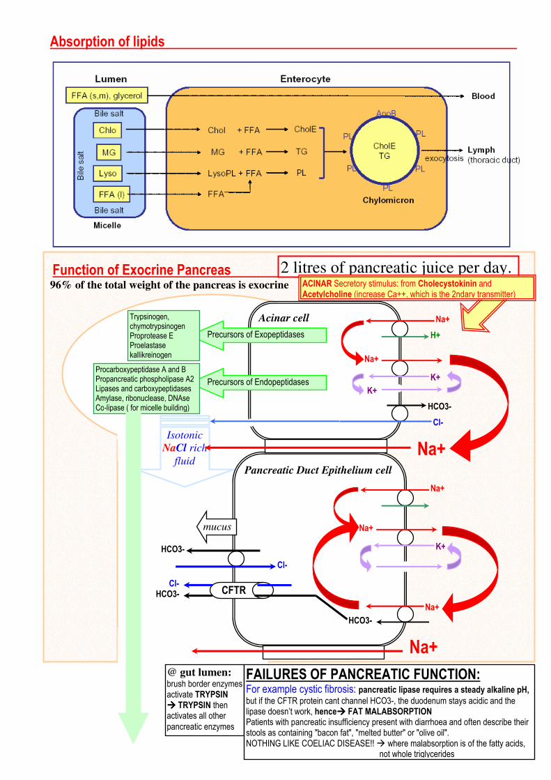
Absorption of lipids
Function of Exocrine Pancreas 96% of the total weight of the pancreas is exocrine
2 litres of pancreatic juice per day.
Acinar cell
Isotonic
NaCl rich
fluid Pancreatic Duct Epithelium cell
H+
Na+
K+
K+
Na+
Na+
HCO3-
Cl-
Precursors of Exopeptidases
Precursors of Endopeptidases
Trypsinogen, chymotrypsinogen Proprotease E Proelastase kallikreinogen
Procarboxypeptidase A and B Propancreatic phospholipase A2 Lipases and carboxypeptidases Amylase, ribonuclease, DNAse Co-lipase ( for micelle building)
@ gut lumen: brush border enzymes activate TRYPSIN ���� TRYPSIN then activates all other
pancreatic enzymes
ACINAR Secretory stimulus: from Cholecystokinin and Acetylcholine (increase Ca++, which is the 2ndary transmitter)
CFTR HCO3-
HCO3-
Cl-
Cl-
Na+
Na+
Na+
K+
HCO3-
Na+
FAILURES OF PANCREATIC FUNCTION: For example cystic fibrosis: pancreatic lipase requires a steady alkaline pH, but if the CFTR protein cant channel HCO3-, the duodenum stays acidic and the lipase doesn’t work, hence���� FAT MALABSORPTION Patients with pancreatic insufficiency present with diarrhoea and often describe their stools as containing "bacon fat", "melted butter" or "olive oil". NOTHING LIKE COELIAC DISEASE!! � where malabsorption is of the fatty acids,
not whole triglycerides
mucus
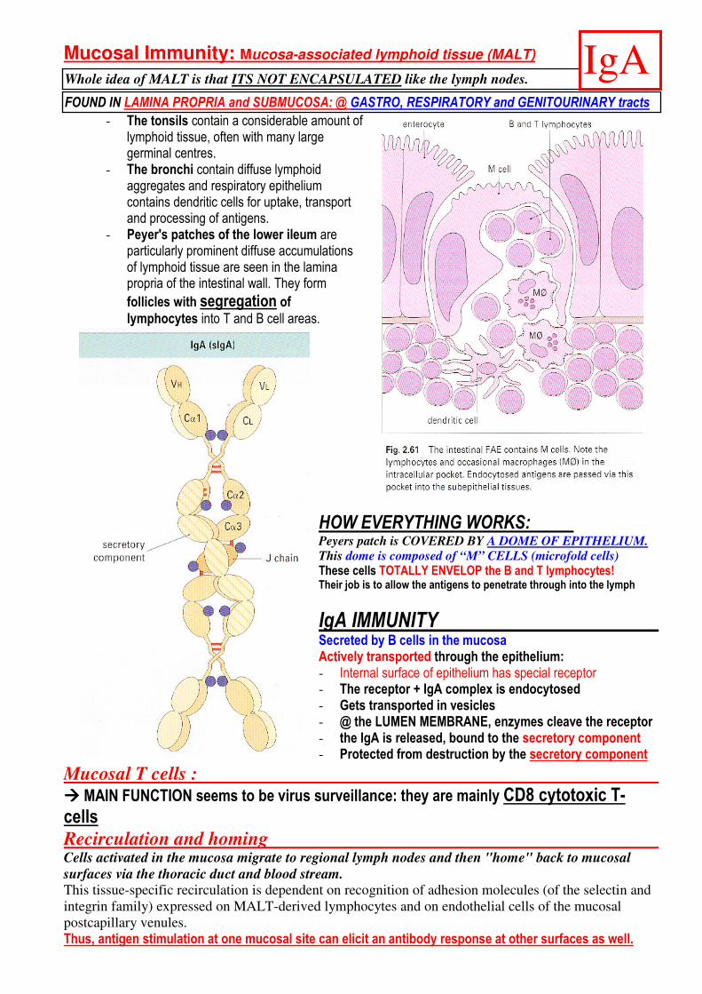
Mucosal Immunity: Mucosa-associated lymphoid tissue (MALT)
- The tonsils contain a considerable amount of lymphoid tissue, often with many large germinal centres.
- The bronchi contain diffuse lymphoid aggregates and respiratory epithelium contains dendritic cells for uptake, transport and processing of antigens.
- Peyer's patches of the lower ileum are particularly prominent diffuse accumulations of lymphoid tissue are seen in the lamina propria of the intestinal wall. They form
follicles with segregation of lymphocytes into T and B cell areas.
HOW EVERYTHING WORKS: Peyers patch is COVERED BY A DOME OF EPITHELIUM.
This dome is composed of “M” CELLS (microfold cells)
These cells TOTALLY ENVELOP the B and T lymphocytes! Their job is to allow the antigens to penetrate through into the lymph
IgA IMMUNITY Secreted by B cells in the mucosa
Actively transported through the epithelium: - Internal surface of epithelium has special receptor - The receptor + IgA complex is endocytosed - Gets transported in vesicles - @ the LUMEN MEMBRANE, enzymes cleave the receptor - the IgA is released, bound to the secretory component - Protected from destruction by the secretory component
Mucosal T cells :
���� MAIN FUNCTION seems to be virus surveillance: they are mainly CD8 cytotoxic T-cells Recirculation and homing Cells activated in the mucosa migrate to regional lymph nodes and then "home" back to mucosal
surfaces via the thoracic duct and blood stream. This tissue-specific recirculation is dependent on recognition of adhesion molecules (of the selectin and
integrin family) expressed on MALT-derived lymphocytes and on endothelial cells of the mucosal
postcapillary venules.
Thus, antigen stimulation at one mucosal site can elicit an antibody response at other surfaces as well.
Whole idea of MALT is that ITS NOT ENCAPSULATED like the lymph nodes.
FOUND IN LAMINA PROPRIA and SUBMUCOSA: @ GASTRO, RESPIRATORY and GENITOURINARY tracts
IgA
Overview of Spectrum of non-infectious diarrhoea : worth ignoring
Inflammatory bowel disease is the term used to describe ulcerative colitis and Crohn's disease. These are
chronic, relapsing conditions of unknown aetiology affecting the gastrointestinal tract. Onset can be at
any age, but with a peak from late teens to mid-30s.
Aetiology The cause of either condition is unknown. Factors implicated in Crohn's disease include smoking,
genetic factors and intraluminal bacteria. Mucosal inflammation with activation of the immune system is
a feature of both diseases.
Ulcerative colitis Pathology: affects only the large intestine. Always involves the rectum and usually a variable length of
colon proximal to this in continuity. The inflammation is confined to the mucosa. Neutrophils prominent
in the inflammatory infiltrate.
Clinical features: typically presents with diarrhoea containing blood and mucus. Patients with proctitis
may have bleeding only. Pain is a feature of severe disease. Toxic megacolon and perforation may
result. Extraintestinal manifestation can develop, affecting joints, skin, eyes and liver. The risk of
colorectal cancer is increased in patients with extensive disease after 7-10 years.
Diagnosis: sigmoidoscopy or colonoscopy, with biopsy. Barium enema occasionally used as the
alternative. Infectious causes of colitis must be excluded by stool microscopy and culture.
Treatment: In active disease depends upon the extent of involvement and degree of inflammation.
Corticosteroids are used rectally for left-sided disease, orally if more extensive, more active or
unresponsive, and intravenously in severe cases. 5-aminosalicylic acid (5-ASA) compounds,
sulphasalazine, mesalazine and olsalazine, of benefit in mild-moderate disease, but their main role is in
maintenance therapy. Immunosuppressants (azathioprine, 6-mercaptopurine) are used for resistant
disease or for their steroid-sparing effect. Cyclosporine may have a limited role in severe colitis where
surgery is not possible.
Maintenance therapy with 5-ASA compounds reduces the relapse rate by 50% or more. Treatment
should be continued indefinitely. Azathioprine/6-MP can also be used for maintenance.
Surgery, usually ileal pouch-anal anastomosis, is required in approximately 20% of patients. Indications
include severe active disease, chronic unresponsive disease, cancer risk or overt cancer.
Crohn's disease Pathology: can involve any part of the gastrointestinal tract, most commonly the ileum, colon or both.
Discontinuous involvement, with characteristic "skip" lesions. The inflammation is transmural, leading
to wall thickening. Fistulae may develop and are specific for Crohn's disease. Histologically there is a
mononuclear infiltrate with the characteristic granulomas present in 70%.
Clinical features: In small bowel disease the main symptom is pain. Patients with colitis develop
diarrhoea, but with bleeding in only 50%. Fistulae may develop between loops of intestine or between
intestine and skin or other organs. Systemic symptoms such as fatigue, fever and weight loss are also
common in Crohn's disease. Perianal complications occur in 25%. Extraintestinal manifestations may
develop as in ulcerative colitis.
Diagnosis: a high index of suspicion is required. Colonoscopy with biopsy and small bowel radiology
are generally used. Other investigations depend on the site of involvement.
Treatment: active disease is treated with corticosteroids. Azathioprine or 6-MP are also used as in
ulcerative colitis. Oral mesalazine in high dose may be of benefit in ileal inflammation. In resistant
disease methotrexate has been tried. Cessation of smoking is essential because of its adverse effect on
the course of the disease. New treatments include broad-spectrum antibiotics and anti-tumour necrosis
factor (infliximab).
Maintenance therapy with oral mesalazine is used in ileal disease. Immunosuppressants can also be used
in selected patients.
Surgery is needed in up to 80% of patients, especially those with small bowel involvement. This is
mostly segmental resection of the most inflamed areas, with a conservative approach. Strictureplasty is
performed on small bowel strictures. Surgery is not curative, with approximately 50% of patients
requiring a subsequent operation.
Prognosis: although these are chronic diseases, most patients can lead productive lives. Only a small
proportion are disabled by the condition. Death from inflammatory bowel disease is rare.
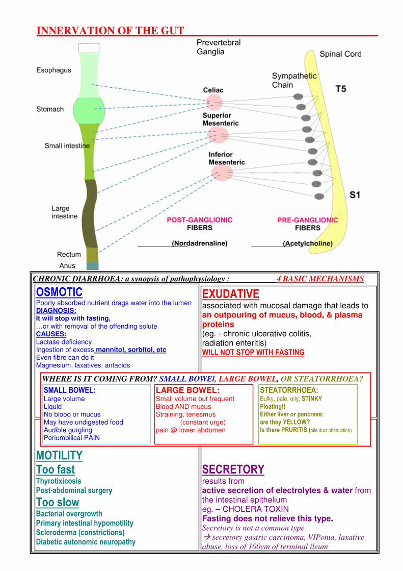
INNERVATION OF THE GUT
CHRONIC DIARRHOEA: a synopsis of pathophysiology : 4 BASIC MECHANISMS OSMOTIC Poorly absorbed nutrient drags water into the lumen DIAGNOSIS: It will stop with fasting, …or with removal of the offending solute CAUSES: Lactase deficiency Ingestion of excess mannitol, sorbitol, etc Even fibre can do it Magnesium, laxatives, antacids
EXUDATIVE associated with mucosal damage that leads to an outpouring of mucus, blood, & plasma proteins (eg. - chronic ulcerative colitis, radiation enteritis) WILL NOT STOP WITH FASTING
SECRETORY results from active secretion of electrolytes & water from the intestinal epithelium eg. – CHOLERA TOXIN Fasting does not relieve this type. Secretory is not a common type.
� secretory gastric carcinoma, VIPoma, laxative
abuse, loss of 100cm of terminal ileum
MOTILITY Too fast Thyrotixicosis Post-abdominal surgery
Too slow Bacterial overgrowth Primary intestinal hypomotility Scleroderma (constrictions) Diabetic autonomic neuropathy
WHERE IS IT COMING FROM? SMALL BOWEl, LARGE BOWEL, OR STEATORRHOEA? SMALL BOWEL: Large volume Liquid No blood or mucus May have undigested food Audible gurgling Periumbilical PAIN
LARGE BOWEL: Small volume but frequent Blood AND mucus Straining, tenesmus
(constant urge) pain @ lower abdomen
STEATORRHOEA: Bulky, pale, oily, STINKY Floating!! Either liver or pancreas: are they YELLOW? Is there PRURITIS (bile duct obstruction)
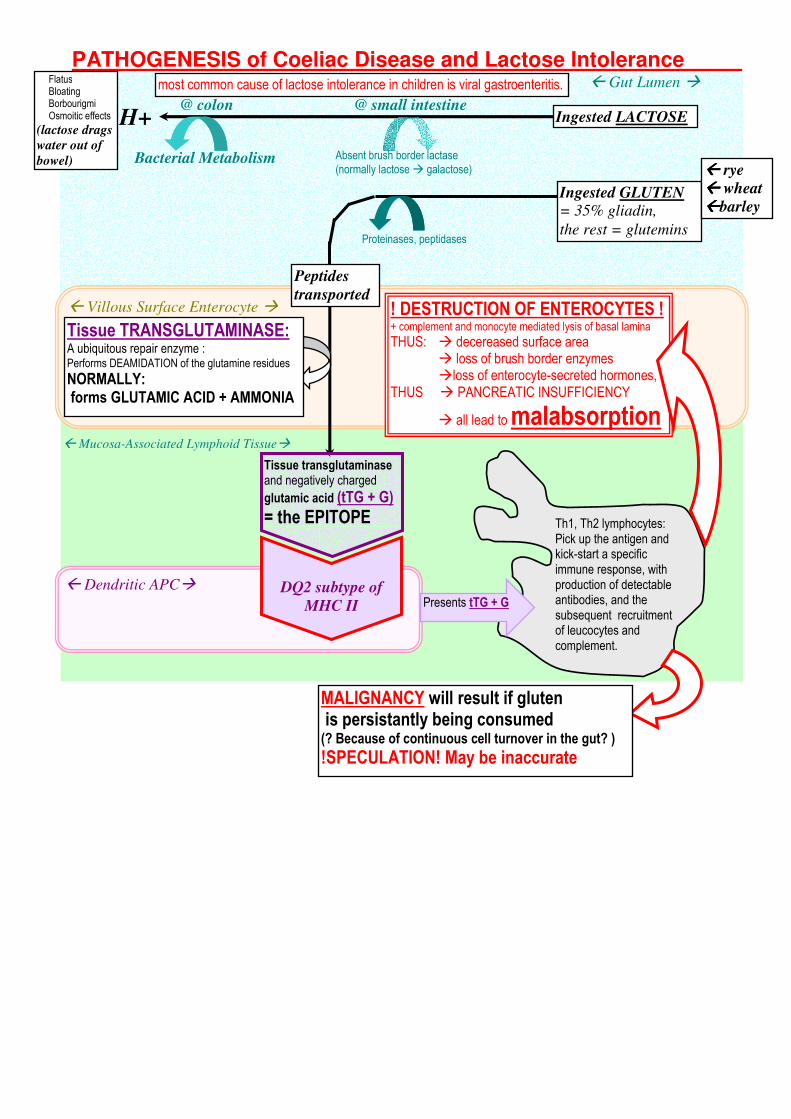
Gut Lumen �
Mucosa-Associated Lymphoid Tissue�
Dendritic APC�
PATHOGENESIS of Coeliac Disease and Lactose Intolerance
Villous Surface Enterocyte �
Ingested LACTOSE
Ingested GLUTEN = 35% gliadin,
the rest = glutemins
rye
wheat
barley
@ small intestine @ colon
Absent brush border lactase
(normally lactose � galactose) Bacterial Metabolism
H+
Flatus Bloating Borbourigmi Osmoitic effects
(lactose drags
water out of
bowel)
Proteinases, peptidases
Peptides
transported
Tissue TRANSGLUTAMINASE: A ubiquitous repair enzyme : Performs DEAMIDATION of the glutamine residues
NORMALLY: forms GLUTAMIC ACID + AMMONIA
Tissue transglutaminase and negatively charged
glutamic acid (tTG + G)
= the EPITOPE
DQ2 subtype of
MHC II Presents tTG + G
Th1, Th2 lymphocytes: Pick up the antigen and kick-start a specific immune response, with production of detectable antibodies, and the subsequent recruitment of leucocytes and complement.
! DESTRUCTION OF ENTEROCYTES ! + complement and monocyte mediated lysis of basal lamina
THUS: � decereased surface area � loss of brush border enzymes �loss of enterocyte-secreted hormones,
THUS � PANCREATIC INSUFFICIENCY
� all lead to malabsorption
most common cause of lactose intolerance in children is viral gastroenteritis.
MALIGNANCY will result if gluten is persistantly being consumed (? Because of continuous cell turnover in the gut? )
!SPECULATION! May be inaccurate