Embed Size (px)
Citation preview
Gut, 1974, 15, 260-267
Collagen proline hydroxylase activity and 35Ssulphate uptake in human liver biopsiesJ. O'D. McGEE, R. S. PATRICK, MARION C. RODGER, ANDCAROLYN M. LUTY
From the University Department ofPathology, Royal Infirmary, Glasgow
SUMMARY In an effort to assess connective tissue biosynthetic activity in human liver disease,collagen proline hydroxylase (a key enzyme in collagen biosynthesis) and the uptake of 35S sulphate(a precursor of sulphated mucopolysaccharides) were measured in hepatic tissue obtained mainly bypercutaneous biopsy.A procedure is described for the quantitation of collagen proline hydroxylase in cryostat sections
which allows for the simultaneous histopathological examination of the liver specimen. A three toeightfold increase in the activity of this enzyme was found in four cirrhotic livers compared with themean value of four normal livers and two biopsies from patients with Gilbert's syndrome. Elevatedhydroxylase levels were found also in five patients with hepatic dysfunction but without cirrhosis(four alcoholics and one patient with persistent hepatitis associated with serum smooth muscleantibody). It is suggested that the hepatic level of collagen proline hydroxylase may be a usefulquantitative index of fibroblastic activity in human liver disease.
Autoradiographic studies of radioactive sulphate uptake in biopsy specimens from patients withchronic liver disease showed an exaggerated incorporation of isotope not only at sites of establishedfibrogenesis but also in the walls of sinusoids throughout the liver.
Active hepatic fibrogenesis compared with passivestromal condensation may be involved in the pro-gression of some liver diseases to cirrhosis. Thisimplies an increase in the biosynthesis within theliver of the connective tissue components, collagenand mucopolysaccharide (Galambos, 1966; Popperand Udenfriend, 1970). Morphological examinationof liver can determine the presence or absence ofhepatic fibrosis, but it is impossible by this means toassess the amount of collagen or mucopolysac-charide being produced at any given point in time.Similarly, it is not feasible to use the standard iso-topic procedures for measuring collagen and muco-polysaccharide synthesis in percutaneous needlebiopsies of liver because of the limited amount oftissue available.
It has been reported that the activity of collagenproline hydroxylasel, the enzyme responsible forhydroxyproline biosynthesis in collagen, generally
'Collagen proline hydroxylase has also been referred to as prolyl-hydroxylase, protocollagen proline hydroxylase, and peptidyl prolinehydroxylase.Received for publication 19 December 1973.
reflects the rate of collagen synthesis in a variety oftissues (see Grant and Prockop, 1972, for review).It has also been shown that the uptake of 35S sul-phate determined autoradiographically reflects therate of sulphated mucopolysaccharide synthesis inexperimental liver disease (McGee and Patrick,1969a and b). Since it seemed possible that thisenzyme and 35S sulphate uptake could be measuredin needle biopsies of liver taken for diagnostic pur-poses, the present investigation was undertaken witha small number of normal specimens and with otherspecimens from a variety of hepatic diseases. Thetechniques used allow for simultaneous histopatho-logical examination of the biopsy material.
Materials and Methods
3,4 3H-proline (specific activity 38Ci per mole) waspurchased from New England Nuclear Enterprises,Frankfurt, Germany; 35S sulphate (specific activity100 mCi per mole) from the Radiochemical Centre,Amersham, England; minimum essential mediumfrom Biocult, Paisley, Scotland; Triton X-100 and
260
on 7 May 2018 by guest. P
rotected by copyright.http://gut.bm
j.com/
Gut: first published as 10.1136/gut.15.4.260 on 1 A
pril 1974. Dow
nloaded from

Collagen proline hydroxylase activity and 35S sulphate uptake in human liver biopsies
ferrous ammonium sulphate from British DrugHouses, Liverpool, England; all other reagentsfrom Sigma, London, England.
Most of the liver biopsies were carried out by thepercutaneous route using a Menghini needle anda few by open surgical operation.
MEASUREMENT OF COLLAGEN PROLINEHYDROXYLASE ACTIVITYEntire needle biopsy specimens were frozen im-mediately onto a metal cutting block on top of alayer of partially frozen 0.15 M NaCl by immersingthe base of the chuck in a mixture of ethanol andsolid CO2. Surgical wedge biopsy specimens weretrimmed to exclude the edges of excision and theliver capsule before freezing in the same manner. Atotal of seven or 12 sections were cut from theneedle and surgical specimens respectively in a cryo-stat maintained at - 15°C. The first and last sections(8 ,um thick) were processed for histological exami-nation to determine the purity of the samples usedfor assay; those contaminated with other tissues,such as skeletal muscle or large pieces of blood clot,were excluded from the study. The intervening un-thawed sections (20 ,tm thick) were used for themeasurement of collagen proline hydroxylase acti-vity. These were transferred by a cold needle to a1 ml glass-teflon homogenizer which was stored inthe cryostat on ice. The sections were homogenizedat 4°C in 0.5 ml of 0.05 M Tris-HCI, pH 7.2, con-taining 025 M sucrose, 10-5 M ethylenediamine-tetraacetic acid, 10-3 M dithiothreitol, 0.1 % TritonX-100, and 50 gg/ml of phenylmethylsulphonefluoride. Aliquots of the homogenate were assayedin duplicate for enzyme activity by the tritium releasemethod using a 3,43H-proline-labelled substrate(Hutton, Tappel, and Udenfriend, 1966); theseassays were performed immediately since the acti-vity of the enzyme rapidly deteriorates in crude liverhomogenates. The reaction mixture for the assaycontained the following reagents in a final volume of1 ml: 50 ,gmoles of Tris - HCl (pH 7.2); 5 ,umoles ofascorbate; 1 ,umole of ferrous ammonium sulphate;2 mg of heat-denatured bovine serum albumin;0.4 mg of catalase; 1 ,umole of x-ketoglutarate; 0-1,umole of dithiothreitol; 0.1 ml of 3H-proline-labelledsubstrate; 0 05-0.20 ml of liver homogenate asenzyme. The complete mixture was incubated at30°C and the reaction terminated after 30 minutesby the addition of 0.1 ml of 50% trichloroacetic acid.The concentration of Fe++, ascorbate, and a-keto-glutarate is 10 times higher than that usually em-ployed (McGee, Langness, and Udenfriend, 1971)in order to increase the sensitivity of the assay. It hasbeen shown that the tritium released into water inthis assay is proportional to the formation of hy-
droxyproline (Hutton et al, 1966). Separate aliquotsof the homogenate were taken for protein estimationby the method of Lowry, Rosebrough, Farr, andRandall (1951) using bovine serum albumin as astandard. The collagen proline hydroxylase activityof each biopsy specimen was expressed as counts perminute (cpm) released from the tritium-labelled sub-strate per milligram of tissue protein in the homo-genate. Tissue protein was chosen as the referencepoint for enzyme activity rather than tissue masssince it is impracticable on a routine basis to weighcryostat sections. Variation in protein concentrationof the homogenates did not affect the linearity of theenzyme assay (see results).The tissue remaining after sections had been taken
for the hydroxylase assay was processed routinelyfor histopathological examination. It was entirelyadequate in size and preservation for diagnosticpurposes.
35S SULPHATE UPTAKEImmediately after the biopsy specimen was taken asmall piece (3 mm x 1 mm) was cut from each endand placed in 5 ml of HEPES-buffered minimumessential medium, pH 7.2, containing 10-3M ascor-bate, 10-6M ferric nitrate, and 250 ,uCi of 35S sul-phate in a sterile universal container. The tissue wasincubated at 37°C for 90 minutes in a 95% 02, 5%C02 atmosphere. After incubation the medium wasdecanted, the tissue washed three times with 10%buffered formalin, pH 7.0, and fixed in the samesolution for 16 hours. Thereafter it was processedroutinely to paraffin and autoradiographs were pre-pared using Ilford G5 nuclear track emulsion. Theautoradiographs were exposed for six weeks, devel-oped by routine methods, and then stained withneutral red.
Results
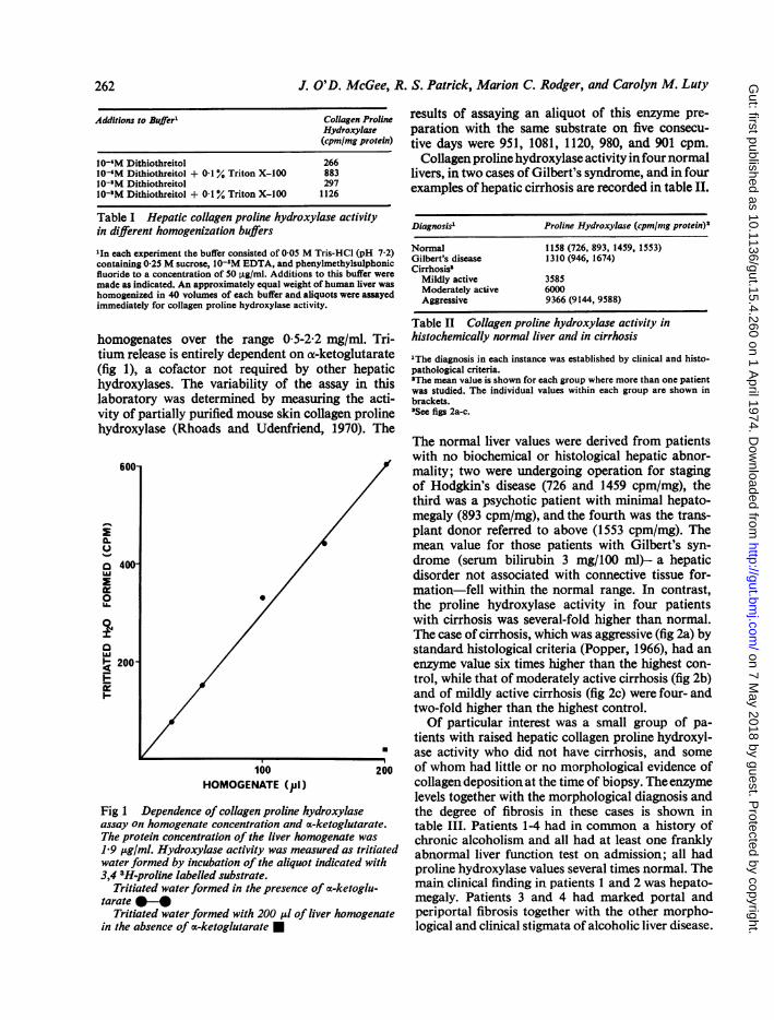
The procedure used for the measurement of collagenproline hydroxylase activity in human liver wasworked out initially on a large sample of tissueobtained from a renal transplant donor immediatelyafter death. After excision the liver was immersed inliquid nitrogen and stored at - 80°C. Of the varioushomogenization conditions used for the measure-ment of hepatic collagen proline hydroxylase it wasfound that 0-05 M Tris-HCl (pH 7-2) containing0-1 % Triton and 10-3 dithiothreitol yielded optimumenzyme activity (table I). This buffer was usedthroughout the study. As shown in fig 1 the releaseof tritium from 3,4 3H-proline-labelled substrate isdirectly proportional to the amount of liver homo-genate. The linearity of the reaction was not affectedby changes in the protein concentration of the
261
on 7 May 2018 by guest. P
rotected by copyright.http://gut.bm
j.com/
Gut: first published as 10.1136/gut.15.4.260 on 1 A
pril 1974. Dow
nloaded from
J. O'D. McGee, R. S. Patrick, Marion C. Rodger, and Carolyn M. Luty
Additions to Buffer' Collagen ProlineHydroxylase(cpm/mg protein)
10-'M Dithiothreitol 26610-'M Dithiothreitol + 0-1% Triton X-100 88310-'M Dithiothreitol 29710-'M Dithiothreitol + 01 % Triton X-100 1126
Table I Hepatic collagen proline hydroxylase activityin different homogenization buffers
'In each experiment the buffer consisted of 0.05 M Tris-HCI (pH 7.2)containing 0.25 M sucrose, 10-'M EDTA, and phenylmethylsulphonicfluoride to a concentration of 50 ,tg/ml. Additions to this buffer weremade as indicated. An approximately equal weight ofhuman liver washomogenized in 40 volumes of each buffer and aliquots were assayedimmediately for collagen proline hydroxylase activity.
homogenates over the range 0-5-2.2 mg/ml. Tri-tium release is entirely dependent on a-ketoglutarate(fig 1), a cofactor not required by other hepatichydroxylases. The variability of the assay in thislaboratory was determined by measuring the acti-vity of partially purified mouse skin collagen prolinehydroxylase (Rhoads and Udenfriend, 1970). The
aCL
U-
a 400-
0
200
E
0
100 200HOMOGENATE (Pj)
Fig 1 Dependence of collagen proline hydroxylaseassay on homogenate concentration and oc-ketoglutarate.The protein concentration of the liver homogenate was
1.9 ,ug/ml. Hydroxylase activity was measured as tritiatedwater formed by incubation of the aliquot indicated with3,4 3H-proline labelled substrate.
Tritiated water formed in the presence of oc-ketoglu-tarate *-*
Tritiated water formed with 200 ,ul of liver homogenatein the absence of a-ketoglutarate E
results of assaying an aliquot of this enzyme pre-paration with the same substrate on five consecu-tive days were 951, 1081, 1120, 980, and 901 cpm.Collagen proline hydroxylase activity in four normal
livers, in two cases of Gilbert's syndrome, and in fourexamples of hepatic cirrhosis are recorded in table II.
Diagnosis' Protine Hydroxylase (cpm/mg protein)*
Normal 1158 (726, 893, 1459, 1553)Gilbert's disease 1310 (946, 1674)Cirrhosis'
Mildly active 3585Moderately active 6000Aggressive 9366 (9144, 9588)
Table II Collagen proline hydroxylase activity inhistochemically normal liver and in cirrhosis
'The diagnosis in each instance was established by clinical and histo-pathological criteria.'The mean value is shown for each group where more than one patientwas studied. The individual values within each group are shown inbrackets.'See figs 2a-c.
The normal liver values were derived from patientswith no biochemical or histological hepatic abnor-mality; two were undergoing operation for stagingof Hodgkin's disease (726 and 1459 cpm/mg), thethird was a psychotic patient with minimal hepato-megaly (893 cpm/mg), and the fourth was the trans-plant donor referred to above (1553 cpm/mg). Themean value for those patients with Gilbert's syn-drome (serum bilirubin 3 mg/100 ml)- a hepaticdisorder not associated with connective tissue for-mation-fell within the normal range. In contrast,the proline hydroxylase activity in four patientswith cirrhosis was several-fold higher than normal.The case of cirrhosis, which was aggressive (fig 2a) bystandard histological criteria (Popper, 1966), had anenzyme value six times higher than the highest con-trol, while that of moderately active cirrhosis (fig 2b)and of mildly active cirrhosis (fig 2c) were four- andtwo-fold higher than the highest control.Of particular interest was a small group of pa-
tients with raised hepatic collagen proline hydroxyl-ase activity who did not have cirrhosis, and someof whom had little or no morphological evidence ofcollagendepositionat the time of biopsy. Theenzymelevels together with the morphological diagnosis andthe degree of fibrosis in these cases is shown intable III. Patients 1-4 had in common a history ofchronic alcoholism and all had at least one franklyabnormal liver function test on admission; all hadproline hydroxylase values several times normal. Themain clinical finding in patients 1 and 2 was hepato-megaly. Patients 3 and 4 had marked portal andperiportal fibrosis together with the other morpho-logical and clinical stigmata of alcoholic liver disease.
262
on 7 May 2018 by guest. P
rotected by copyright.http://gut.bm
j.com/
Gut: first published as 10.1136/gut.15.4.260 on 1 A
pril 1974. Dow
nloaded from

Collagen proline hydroxylase activity and 35S sulphate uptake in human liver biopsies
4
Fig 2a Fig 2bFig 2 Light micrographs of (a) aggressive, (b) moderately active, and (c) mildly active cirrhosis. In (a) thereis marked chronic inflammatory cell infiltration offibrous septa, piecemeal necrosis atdfine (immature) collagenfibre deposition (arrows) at the periphery of regenerative nodules. In (b) there is inflammatory cell infiltration of thesepta with slight erosion of the edge of regenerative nodules. The collagen proline hydroxylase activity found in thesebiopsies expressed as cpm/mg protein in the liver homogenate was (a) 9144, (b) 6000, and (c) 3585. (a) Haematoxylinand Van Gieson's stain, x 225; (b and c) haematoxylin and eosin, x 140.
Fig 2c In (c)the fibrous septaconsist mainlyof thick (mature)collagen bundlesand there is littleinflammatorycell infiltrate.
263
on 7 May 2018 by guest. P
rotected by copyright.http://gut.bm
j.com/
Gut: first published as 10.1136/gut.15.4.260 on 1 A
pril 1974. Dow
nloaded from
J. O'D. McGee, R. S. Patrick, Marion C. Rodger, and Carolyn M. Luty
Patient Diagnosis Fibrosis' Collagen ProlineHydroxylase(cpm/nmg protein)
1 Fatty change Nil 29662 Intrahepatic cholestasis Nil 30273 Alcoholic hepatitis + + 29814 Alcoholic hepatitis + + 42555 Persistent hepatitis Nil 5166
Table III Collagen proline hydroxylase activity innon-cirrhotic liver disease
'Fibrosis was assessed morphologically. Patients 3 and 4 had moderateportal and periportal fibrosis.
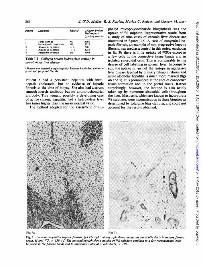
Patient 5 had a persistent hepatitis with intra-hepatic cholestasis, but no evidence of hepaticfibrosis at the time of biopsy. She also had a serumsmooth muscle antibody but no antimitochondrialantibody. This woman, possibly a developing caseof active chronic hepatitis, had a hydroxylase levelfive times higher than the mean normal value.The method adopted for the assessment of sul-
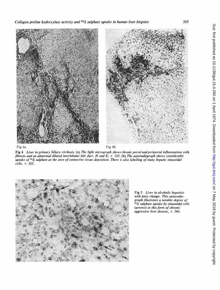
phated mucopolysaccharide biosynthesis was theuptake of 35S sulphate. Representative results froma study of nine cases of chronic liver disease areillustrated in figures 3-5. A case of congenital he-patic fibrosis, an example of non-progressive hepaticfibrosis, was used as a control in this series. As shownin fig 3b there is little uptake of 35S04 except ina few cells in the connective tissue bands and inisolated sinusoidal cells. This is comparable to thedegree of cell labelling in normal liver. In compari-son, the uptake in vitro of the isotope in aggressiveliver disease typified by primary biliary cirrhosis andacute alcoholic hepatitis is much more marked (figs4b and 5). It is pronounced at the sites of connectivetissue formation and in the portal tracts. Rathersurprisingly, however, the isotope is also avidlytaken up by numerous sinusoidal cells throughoutthe liver. Mast cells, which are known to incorporate35S sulphate, were inconspicuous in these biopsies asdetermined by toluidine blue staining, and could notaccount for the results obtained.
Fig 3a Fig 3bFig 3 Liver in congenital hepatic fibrosis. (a) The light micrograph shows numerous small bile ducts in mature fibroussepta. H and VG, x 159. (b) The autoradiograph shows uptake of 35S sulphate confined to a few mesenchymal cells(arrows) in the fibrous bands and to mucinous material in bile ducts, x 159.
264
on 7 May 2018 by guest. P
rotected by copyright.http://gut.bm
j.com/
Gut: first published as 10.1136/gut.15.4.260 on 1 A
pril 1974. Dow
nloaded from
Collagen proline hydroxylase activity and 35S sulphate uptake in human liver biopsies
Fig 4a Fig 4b
Fig 4 Liver in primary biliary cirrhosis. (a) The light micrograph showsfibrosis and an abnormal dilated interlobular bile duct. H and E, x 105.uptake of 35S sulphate at the sites ofconnective tissue deposition. There i
cells, x 105.
..
....,i,,X,.E~~ ~~~i,ig4 5% Lie in alohli hptitis
;4t
A.&grap ilutaesantbe ereo
'* CC
S.,~~~~~~~~~~~~~~W
chronic portal andperiportal inflamimation wtith(b) The autoradiograph shows considerablesalso labelling ofmany hepatic sinusoidal
Fig 5 Liver in alcoholic hepatitis*with fatty change. This autoradio-
graph illustrates a notable degree of35S sulphate uptake by sinusoidal cells
X (arrows) in thisform of chronicaggressive liver disease, x 560.
265
on 7 May 2018 by guest. P
rotected by copyright.http://gut.bm
j.com/
Gut: first published as 10.1136/gut.15.4.260 on 1 A
pril 1974. Dow
nloaded from
J. O'D. McGee, R. S. Patrick, Marion C. Rodger, and Carolyn M. Luty
Discussion
Collagen proline hydroxylase, an enzyme specific tothe collagen pathway, catalyses the biosynthesis ofhydroxyproline, an amino acid found only in col-lagen and to a very limited extent in elastin, byhydroxylating certain prolyl residues in growingcollagen oc chains (Miller and Udenfriend, 1970). Itdoes not hydroxylate free proline (Hutton andUdenfriend, 1966) but only peptidyl proline occur-ring in sequences of the general structure -X-Pro-Gly- where X can be one of several amino acids(McGee, Rhoads, and Udenfriend, 1971); the bulkof the collagen molecule is composed of repeating-X-Pro-Gly- triplets. Collagen proline hydroxylaserequires as cofactors for its action 02, Fe++, ox-keto-glutarate, and a reducing reagent such as ascorbate.It is the prototype of a new class of oxygenaseswhich stoichiometrically decarboxylate ox-ketogluta-rate during the hydroxylation process (Udenfriend,1970). In this respect it differs from the many otherhepatic hydroxylases. The fact that the enzymeactivity assessed in this study was totally dependentin the presence of a-ketoglutarate indicates that onlycollagen proline hydroxylase activity was measured.
Several observations suggest that the tissue levelof collagen proline hydroxylase is in general an indi-cator of fibroblastic biosynthetic activity. In physio-logical conditions where collagen production is ele-vated, as in the developing embryo (Mussini, Hutton,and Udenfriend, 1967) and in the pregnant uterus(Halme and Jaaskelainen, 1970), activity is increased.High enzyme levels have also been found in othercollagen formative conditions such as healingwounds and granulomas (Mussini et al, 1967), devel-oping experimental cirrhosis (Takeuchi and Prockop,1969), alcoholic liver disease in animals, (Feinmanand Lieber, 1972), and experimental pulmonaryfibrosis (Halme, Uitto, Kahanpaa, Karhunen, andLinay, 1970). In acuteliver disease induced in mice ithas been shownthat thereis infact a roughparallelismbetween hydroxylase activity and the rate of collagensynthesis in liver (McGee, O'Hare, and Patrick, 1973).On the basis of this evidence it is reasonable to as-sume that the high levels of collagen proline hydrox-ylase found in human cirrhosis in the present studyare probably a manifestation of a general increase infibroblastic biosynthetic activity. This interpreta-tion of the results in the cirrhotic patients is in keep-ing with the finding that the hydroxylase level inindividual cirrhotics showed a good positive corre-lation with the degree of aggressive activity in eachcase assessed morphologically; collagen formationat the periphery of regenerating nodules is a promi-nent feature of progressive liver disease (Popper,1966). The measurement of the hepatic level of this
enzyme may be, therefore, a useful quantitative indexof progression of the cirrhotic process in individualpatients. It may also prove to be helpful in assessingthe effect of therapeutic agents on collagen metab-olism in chronic liver disease.The increase in collagen proline hydroxylase
activity in the alcoholic patients and in the case ofpersistent hepatitis without noteworthy fibrosisrequires discussion. If, as argued above, this obser-vation is a reflection of a general increase in collagenbiosynthetic activity, it has to be postulated thatcollagen degradative mechanisms at the time ofbiopsy were capable of preventing a net morpho-logical increase in collagen deposition. In experi-mental hepatic fibrosis collagen proline hydroxylaseactivity begins to increase before fibrosis (McGeeand Patrick, 1972; McGee et al, 1973) or cirrhosis(Takeuchi and Prockop, 1969) are morphologicallyevident. If the latter experimental observations areapplicable to human liver disease the measurementof hepatic collagen proline hydroxylase activity maybe of value in predicting which types of liver damagemay eventually progress to chronic liver disease andcirrhosis. The group of patients under discussien isbeing enlarged and followed up therefore to deter-mine whether this is so.
Until recently it has been difficult to understandwhy collagen proline hydroxylase should rise inconditions characterized by increased collagen for-mation since it has not been shown that this enzymeis rate limiting in collagen biosynthesis. The recentfinding that proline hydroxylation is required for theassembly of a thermally stable triple helical collagenmolecule (Jimenez, Harsch, and Rosenbloom, 1973;Berg and Prockop, 1973), however, indicates thatelevated hydroxylase levels in cirrhosis and othercollagen formative conditions is biologically neces-sary for the production of collagen molecules whichwill not spontaneously denature at body tempera-ture.The results of the 35S sulphate investigation dem-
onstrate that there is a very marked increase insulphated mucopolysaccharide synthesis in activechronic liver diseases. Rather surprisingly it wasfound that this increase was not confined to the areasof connective tissue formation in the portal tractsbut was also prominent within hepatic sinusoids. Itwould seem therefore that connective tissue biosyn-thesis is increased throughout the entire liver inchronic progressive liver disease. Although auto-radiography is a useful procedure for determiningthe sites of intrahepatic fibrogenesis it suffers fromthe disadvantage that it is not easily made quantita-tive and the results are not available for a period ofweeks. The results of collagen proline hydroxylasemeasurements, however, are quantitative and are
266
on 7 May 2018 by guest. P
rotected by copyright.http://gut.bm
j.com/
Gut: first published as 10.1136/gut.15.4.260 on 1 A
pril 1974. Dow
nloaded from

Collagen proline hydroxylase activity and 35S sulphate uptake in human liver biopsies 267
available about 2-5 hours after the biopsy has beentaken, while the tissue remaining is entirely adequatefor histopathological diagnosis.
We are grateful to Dr R. I. Russell and ProfessorL. H. Blumgart of Glasgow Royal Infirmary fortheir willing cooperation in providing the liverbiopsies used in this study. The work was supportedin part by a grant from the Medical Council onAlcoholism.
References
Berg, R. A., and Prockop, I). J. (1973). The thermal transition of anonhydroxylated form of collagen. Evidence for a role forhydroxyproline in stabilizing the triple-helix of collagen.Biochem. biophys. Res. Commun., 52, 115-120.
Feinman, L., and Leiber, C. S. (1972). Hepatic collagen metabolism:effect of alcohol consumption in rats and baboons. Science(N. Y.), 176, 795.
Galambos, J. T. (1966). Acid mucopolysaccharides and cirrhosis ofthe liver. Gastroenterology, 51, 65-74.
Grant, M. E., and Prockop, D. J. (1972). The biosynthesis of collagen.New Engi. J. Med., 286, 194-300.
Halme, J., and Jaaskeliinen, M. (1970). Protocollagen proline hydrox-ylase of the mouse uterus during pregnancy and post-partuminvolution. Biochem. J., 116, 367-369.
Halme, J., Uitto, J., KahanpAf, K., Karhunen, P., and Lindy, S.(1970). Protocollagen proline hydroxylase activity in experi-mental pulmonary fibrosis of rats. J. Lab. clin. Med., 75, 535-541.
Hutton J. J., Jr., Tappel, A. L., and Udenfriend, S. (1966). A rapidassay for collagen proline hydroxylase. Analyt. Biochem., 16,384-394.
Hutton, J. J., Jr., and Udenfriend, S. (1966). Soluble collagen prolinehydroxylase and its substrates in several animal tissues. Procnat. Acad. Sci. (Wash.), 56, 198-202.
Jimenez, S., Harsch, M., and Rosenbloom, J. (1973). Hydroxyprolinestabilizes the triple helix of chick tendon collagen. Biochem.biophys. Res. Commun., 52, 106-115.
Lowry, 0. H., Rosebrough, N. J., Farr, A. L., and Randall, R. J.
(1951). Protein measurement with the Folin phenol reagent.J. biol. Chem., 193, 265-275.
McGee, J. O.'D., Langness, U., and Udenfriend, S. (1971). Immuno-logical evidence for an inactive precursor of collagen prolinehydroxylase in cultured fibroblasts. Proc. nat. Acad. Sci. (Wash.)68, 1585-1589.
McGee, J. O'D., O'Hare, R. P., and Patrick, R. S. (1973). Stimulationof the collagen biosynthetic pathway by factors isolated fromexperimentally injured iliver. Nature [new Biol.], 243, 121-123.
McGee, J. O'D., ani Patrick, R. S. (1969a). The synthesis of sul-phated mucopolysaccharide in mouse liver following carbontetrachloride injury. I. Autoradiographic studies. Brit. J. exp.Path., 50, 521-526.
McGee, J. O'D., and Patrick, R. S. (1169b). The synthesis of sul-phated mucopolysaccharide in (mouse liver following carbontetrachloride injury. II. Quantitation and partial characteriza-tion of extracted mucopolysaccharide. Brit. J. exp. Path., 50,527-532.
McGee, J. O'D., and Patrick, R. S. (1972). The role of perisinusoidalcells in hepatic fibrogenesis. Lab. Invest., 26, 429-440.
McGee, J. O'D., Rhoads, R. E., and Udenfriend, S. (1971). The sub-strate recognition site of collagen proline hydroxylase: thehydroxylation of -x-Pro-Gly-sequences in bradykinin analogsand other peptides. Arch. Biochem., 144, 343-351.
Miller, R. L., and Udenfriend, S. (1970). Hydroxylation of prolineresidues in collagen nascent chains. Arch. Biochem., 139,104-113.
Mussini, E., Hutton, J. J., Jr., and Udenfriend, S. (1967). Collagenproline hydroxylase in wound healing, granuloma formation,scurvy and growth. Science (N. Y.), 157, 927-929.
Popper, H. (1966). What is chronic hepatitis? Gastroenterology, 50,444448.
Popper, H., and Udenfriend, S. (1970). Hepatic fibrosis: correlationof biochemical and morphologic investigations. Amer. J. Med.,49, 707-721.
Rhoads, R. E., and Udenfriend, S. (1970). Purification and propertiesof collagen proline hydroxylase from newborn rat skin. Arch.Biochem., 139, 329-339.
Takeuchi, T., and Prockop, D. J. (1969). Protocollagen proline hyd-roxylase in normal liver and in hepatic fibrosis. Gastroenterol-ogy, 56, 744-750.
Udenfriend, S. (1970). Biosynthesis of hydroxyproline in collagen. InChemistry and Molecular Biology of the Intercellular Matrix,edited by E. A. Balazs, Vol. I, pp. 371-384. Academic Press,London and New York.
on 7 May 2018 by guest. P
rotected by copyright.http://gut.bm
j.com/
Gut: first published as 10.1136/gut.15.4.260 on 1 A
pril 1974. Dow
nloaded from