Embed Size (px)
Citation preview
University of Rhode Island University of Rhode Island
DigitalCommons@URI DigitalCommons@URI
Open Access Master's Theses
1983
COLLAGENASE AND ELASTASE ACTIVITIES IN HUMAN AND COLLAGENASE AND ELASTASE ACTIVITIES IN HUMAN AND
MURINE CANCER CELLS AND THEIR MODULATION BY MURINE CANCER CELLS AND THEIR MODULATION BY
DIMETHYLFORMAMIDE DIMETHYLFORMAMIDE
David Ray Olsen University of Rhode Island
Follow this and additional works at: https://digitalcommons.uri.edu/theses
Recommended Citation Recommended Citation Olsen, David Ray, "COLLAGENASE AND ELASTASE ACTIVITIES IN HUMAN AND MURINE CANCER CELLS AND THEIR MODULATION BY DIMETHYLFORMAMIDE" (1983). Open Access Master's Theses. Paper 213. https://digitalcommons.uri.edu/theses/213
This Thesis is brought to you for free and open access by DigitalCommons@URI. It has been accepted for inclusion in Open Access Master's Theses by an authorized administrator of DigitalCommons@URI. For more information, please contact [email protected].
COLLAGENASE AND ELASTASE ACTIVITIES
IN HUMAN AND MURINE CANCER CELLS
AND THEIR MODULATION BY DIMETHYLFORMAMIDE
BY
DAVID RAY OLSEN
A THESIS SUBMITTED IN PARTIAL FULFILLMENT
OF THE REQUIREMENTS FOR THE DEGREE OF
MASTER OF SCIENCE
IN
PHARMACOLOGY AND TOXICOLOGY
UNIVERSITY OF RHODE ISLAND
1983
MASTER OF SCIENCE THESIS
APPROVED:
Thesis Committee
Major Professor
OF
DAVID RAY OLSEN
/ /
/ • l / .r
Dean of the Graduate School
UNIVERSITY OF RHODE ISLAND
1983
ABSTRACT
Olsen, David R.; M.S., University of Rhode Island,
1983. Collagenase and Elastase Activities in Human and
Murine Cancer Cells and Their Modulation by Dimethyl
formamide. Major Professor: Dr. Clinton O. Chichester.
The transformation from carcinoma in situ to in
vasive carcinoma occurs when tumor cells traverse extra
cellular matracies allowing them to move into paren
chymal tissues. Tumor invasion may be aided by the
secretion of collagen and elastin degrading proteases
from tumor and tumor-associated cells. In this study
~ the production of Type I and Type V collagen degrading
activities and elastolytic activities by DLD-1 human
colon carcinoma cells, B16-F10 murine melanoma cells,
and normal human dermal fibroblasts was examined. DLD-1
cells and normal fibroblasts produced similarly high
levels of collagenolytic activity. DLD-1 cells also
produced high levels of elastinolytic activity; this
activity was found exclusively in the extracellular
medium. DLD-1 cells and normal fibroblasts produced
more collagen and elastin degrading activity than did
B16-F10 melanoma cells, a cell line characterized as
highly metastatic. The Type I and Type V collagenolytic
ii

activities from DLD-1 cells were separated and characterized
using DEAE cellulose chromatography and gel filtration
chromatography. The Type I collagenolytic activity appeared
to be an anionic protein at pH 8.3, and two forms of the
enzyme were detected, one with a molecular weight of 60,000
and the other with a molecular weight of 35,000 daltons.
The Type V collageno-ytic activity was a cationic protein
at pH 8.3 and two forms of this activity were detected, one
with a molecular weight of 80,000 and the second with a
molecular weight of 54,000 daltons.
The effect of the polar solvent, dimethylformamide
(DMF) on the production of collagen and elastin degrading
activities was studied using cultured DLD-1 cells. DMF
treated cells produced significantly higher levels of
Type I collagenolytic, Type V collagenolytic and cell
associated elastase activities than did control cells.
DMF treatment had no significant effect on media elastase
levels. Treatment of DLD-1 cells with DMF elevated the
cellular levels of protein and RNA while these same cells
had less DNA per cell. DMF treated cells were also able
to synthesize protein at a significantly faster rate than
control cells. This increased rate of protein synthesis
may account for part of the increased production of con
nective tissue degrading enzymes seen in DMF treated cells.
iii
However, the effect of DMF on enzyme production was still
present when the cells were treated with cycloheximide.
The failure of cycloheximide to prevent the DMF induced
increase in enzyme activity suggests that the effect of
DMF is not entirely dependent on protein synthesis. These
data suggest that DLD-1 hu.~an colon carcinoma cells pro
duce at least two different collagen degrading enzymes
and that DMF treatment may facilitate the invasive move
ment of cancer cells during the matastatic process by
increasing the secretion of connective tissue degrading
enzymes.
iv
ACKNOWLEDGEMENTS
The author wishes to thank Dr. Clinton Chichester
for his guidance, encouragement, and dedicated support
throughout the research and writing of this thesis.
I would like to thank my parents for their endless
encouragement, advice, and support. Lastly, I wish to
extend a special thank you to Ellen Austin.
v

TABLE OF CONTENTS
ABSTRACT . . . .
ACKNOWLEDGEMENTS .
TABLE OF CONTENTS
LIST OF TABLES .
LIST OF FIGURES
INTRODUCTION • . .
LITERATURE SURVEY
Metastasis Invasion . . . . . . . . . . . . . . Modulation of Tumor Cell Growth ... Collagen Heterogenity . •......... Mammalian Coll agenase . • . . . • . . Regulation of Collagenase Activity .. . Collagenase Inhibitors .......... . Ela st in . . . . . . . . . . . . . . . Elastase . . . . . . Elastase Inhibitors .
EXPERIMENTAL .
. - . .
PAGE
ii
v
vi
viii
ix
1
4
4 6 7 9
15 22 24 26 27 29
31
Materials • . . . . . . . . . • . . . . . 31 Cell Culture . . . . . . . . . 31 Preparation of Culture Media for Enzyme
Assays . • . . . . . . . . . . . . • . . . . 33 Preparation of Cells for Elastase, Protein,
and DNA Assays . . . . . . . . . . • • . Dimethylf ormamide Treatment . . • . . • . Measurement of Protein Synthesis Protein Determination •....... DNA Determination .... Purification of Collagens ...•.... 14c-Acetylation of Type I and Type V Collagens • . . . • . . . • . . . . . .
Type I Collagenase Assay . . . . . . . . Preparation of Type V Collagen Degrading
Activity . . . . . . . . . . . . ... Type V Collagenase Assay . . • . . . . . . . Elastase Assay . . . . . . . • . Ion Exchange Chromatography .... . Gel Filtration Chromatography ....... . Statistical Analysis . • . . . . • . • .
vi
34 34 34 36 36 37
39 40
41 42 43 44 45 46
RESULTS
DISCUSSION .
CONCLUSIONS
REFERENCES .
TABLE OF CONTENTS
. . . . . . . . . . . . .
. . . . . . . . . . . .
vii
PAGE
47
82
98
100
TABLE
1
2
3
4
5
6
7
8
9
10
LIST OF TABLES
Levels of Type I Collagenase, Type V Collagenase, Cellular Elastase, and Media Elastase Produced by DLD-1 Colon Carcinoma Cells, B16-F10 Melanoma Cells, and Normal Dermal Fibroblasts • • • • • .
Inhibition of Type I and Type V Collagen Degrading Activities by Protease Inhibitors . • • . • • • . . . • . .
Effect of DMF on Type I Collagenase, Type V Collagenase, Cellular Elastase, and Media Elastase Production by DLD-1 Colon Carcinoma Cells • . • . . • • • • .
Effect of DMF on Protein, DNA, and RNA Content of DLD-1 Colon Carcinoma Cells
Effect of DMF on the Incorporation of 3H-glycine Into Cellular Protein of DLD-1 Colon Carcinoma Cells . . . . •
Effect of DMF on the Incorporation of 3H-gylcine Into Media Protein of DLD-1 Colon Carcinoma Cells . • • • • . • . . .
Effect of Cycloheximide on Type I Collagenase Production by Control and DMF Treated DLD-1 Colon Carcinoma Ce 11 s • • • • • • • • . • • • • • • •
Effect of Cycloheximide on Type V Collagenase Production by Control and DMF Treated DLD-1 Colon Carcinoma Ce 11 s • • • • • • • • • • • • • • • • • •
Effect of Cycloheximide on Cellular Elastase Production by Control and DMF Treated DLD-1 Colon Carcinoma Cells . . •
Effect of Cyclobeximide on Media Elastase Production by Control and DMF Treated DLD-1 Colon Carcinoma Cells • • • . • • •
viii
PAGE
56
68
69
72
73
75
76
78
79
81
FIGURE
1
2
3
LIST OF FIGURES
Diagram of The Purification Procedure Used to Isolate Type V Collagen •........
SDS Polyacrylamide Slab Gel Electrophoresis of Type V Collagen Purification.
Solubility of Native and Denatured Radiolabeled Type V Collagen in Dioxane/Methanol/Water ...........•.....•
4 Solubility of Radiolabeled Type V Collagen Degradation Products Formed By Three Collagenases in Dioxane/Methanol/
PAGE
38
49
51
Water................................... 53
5 Degradation of Radiolabeled Type V Collagen By Alveolar Macrophage Collagenase as a Function of Time •......
6 DEAE Cellulose Chromatography of Concentrated DLD-1 Colon Carcinoma Cell
55
Serum-Free Media ........................ 59
7 Ultrogel AcA44 Gel Filtration Chromato-graphy of Pool I ....................... . 62
8 Ultrogel AcA44 Gel Filtration Chromato-graphy of Pool I I ••..........•.......... 63
9 Ultrogel AcA44 Gel Filtration Chromato-graphy of Pool I I I •.•.•.........•....... 65
ix
1
INTRODUCTION
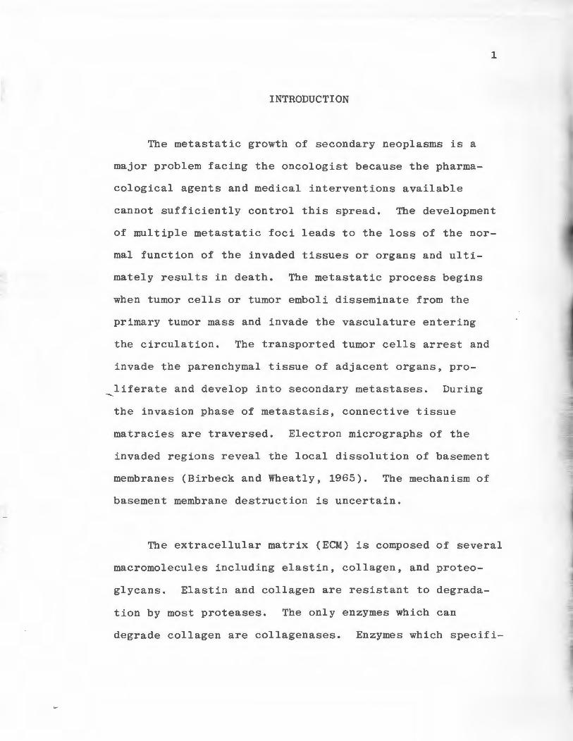
The metastatic growth of secondary neoplasms is a
major problem facing the oncologist because the pharma
cological agents and medical interventions available
cannot sufficiently control this spread. The development
of multiple metastatic foci leads to the loss of the nor
mal function of the invaded tissues or organs and ulti
mately results in death. The metastatic process begins
when tumor cells or tumor emboli disseminate from the
primary tumor mass and invade the vasculature entering
the circulation. The transported tumor cells arrest and
invade the parenchymal tissue of adjacent organs, pro
liferate and develop into secondary metastases. During
the invasion phase of metastasis, connective tissue
matracies are traversed. Electron micrographs of the
invaded regions reveal the local dissolution of basement
membranes (Birbeck and Wheatly, 1965). The mechanism of
basement membrane destruction is uncertain.
The extracellular matrix (ECM) is composed of several
macromolecules including elastin, collagen, and proteo
glycans. Elastin and collagen are resistant to degrada
tion by most proteases. The only enzymes which can
degrade collagen are collagenases. Enzymes which specifi-
2
cally degrade elastin are termed elastases.
Elastase activity is associated with several tumor
associated cells and tumor cell lines (Mainardi et al,
1980a; Kao et al, 1982). Collagenases are also synthesized
by several tumor and tumor associated cells (Horwitz et al,
1977; Mainardi et al, 1980a,b; O'Grady et al, 1982) and
recently collagenases specific for the different types of
collagen have been identified in the medium of certain
tumor cell lines (Liotta et al, 1981a,b; Salo et al, 1983).
During invasion, elastases and collagenases may be involved
in the local destruction of basement membranes and stromal
tissues.
In this study Types I and V collagen degrading acti
vities and elastase activities were measured in several
tumor cell lines. The collagenolytic activities which
were secreted by cultured DLD-1 human colon carcinoma
cells were analyzed by ion exchange chromatography and gel
filtration chromatography to determine if different
enzymes were responsible for the degradation of Types I
and V collagen. The results of these studies may indicate
that destruction of ECM components by invading tumor cells
results from the action of more than one specific degrada
tive enzyme.

3
A novel attempt to control the spread of cancer has
been the use of biological inducers as chemotherapeutic
agents. Biological inducers are compounds which induce
malignant cancer cells to differentiate into benign cells
(Sachs, 1981). One such compound, dimethylformamide (DMF)
has induced a human colon carcinama cell line, DLD-1, to
differentiate in vitro and these treated cells have a
reduced tumorigenic capacity in vivo (Dexter et al, 1979).
This study will also examine the effects of DMF treatment
on the levels of connective tissue degrading proteases in
the DLD-1 cell line. The reduction in tumorigenicity
seen with DMF may be paralleled by a decrease in the cell's
ability to invade host tissues.
4
LITERATURE SURVEY
Metastasis
Metastasis is a multistep process in which cells
derived from a primary tumor detach and are transported
to a distant site where they form a secondary tumor
(Poste and Fidler, 1980). The metastatic process is
initiated when cells or clumps of cells dissociate from
the primary tumor and enter blood or lymph vessels. The
tumor cells interact with host cells as they are trans-
ported by the circulation or lymph. During this process
some of the tumor cells may be destroyed by host defense
mechanisms (Old et al, 1961). ~ ~ ~
The circulation of tumor cells
stops when they encounter a vessel which is too small
to allow passage (Ziedman, 1961) or when they attach to
vascular endothelium due to cell-cell interactions
(Winkelhade and Nicolson, 1976). The cells may exit
the vessel at this point, invade surrounding tissue, pro-
liferate, and develop into a secondary tumor. Alterna-
tively the arrested cells may be engulfed and destroyed
by host macrophages and lymphocytes (Vose, 1980; Hibbs
et al, 1977).
5
The intravenous injection of mice with syngeneic tumor
cells is an experimental model of metastasis. This model
is employed to study various aspects of the metastatic
process including the organ distribution of blood borne
tumor cells (Fidler, 1973). The growth of tumors in
specific organs following intravenous injection of cells
has been demonstrated with several cell lines (Kinsey,
1960; Fidler and Nicoloson, 1976; Nicoloson and Winkel-
hade, 1975; Brunson et al, 1978). Cell types which meta-
stasize to specific organs and variants of the same parent
line which have different capacities to form tumor nodules
following intravenous injection exhibit difference in
their cell surface components (Dobrassy et al, 1981;
Brunson et al, 1978; Raz et al, 1980). Cell lines which ~ ~ ~ ~
are characterized as "highly metastatic" following intra-
venous injection have greater quantities of cell surface
sialic acid, increased levels of sialyltransferase and
other cellular glycosidase activities (Dobrassy et al,
1981). Sialic acid is a component of cell surface glyco-
proteins involved in cell-cell and cell-substratum
adhesion. Others have used the intravenous injection
model with different tumor cell lines and noted random
growth of metastases (Proctor, 1976). Tumor nodules
arose in organs where capillary beds were first encountered.
Models of metastasis which employ intravenously
injected cells are inadequate because they do not allow
the examination of the entire metastatic process. They
bypass the first step of metastasis when cells break off
the primary tumor and invade the vasculature. In the
future better models will further our understanding of
the pathogenesis of metastasis.
Invasion
During invasion tumor cells traverse stromal tissue
and blood vessels to infiltrate the parenchyma of host
organs. The invading cells then proliferate to eventu-
6
~ally form secondary tumors. The mechanisms involved in
the invasive movement of tumor cells is not completely
understood. Invasion may occur as the result of mechan
ical pressures exerted by rapidly proliferating tumor
cells (Eaves, 1973). The fast growing tumors create
regions of increased pressur~ and invasion occurs at
points where the host tissues were weakest. Infiltra
tion of host tissues may also result from tumor cell
products acting on stromal tissues and blood vessels
(Fidler et al, 1978). Tumor cells synthesize and secrete
proteases capable of degrading the host ECM (Dresden et
al, 1972; Hashimoto et al, 1973). Areas where the matrix
......_
is weakened will then be invaded by proliferating tumor
cells. Invasion of target organs by malignant tumor
cells in vitro can be inhibited when the incubation
medium contains several types of protease inhibitors
(Latner et al, 1973).
7
Stromal tissues and blood vessels are partially
composed of the structural proteins collagen and elastin,
these proteins are degraded by specific enzymes called
collagenases and elastases. Analysis of the media from
cultured tumor cells has confirmed the presence of colla
genase and elastase activities (Liotta et al, 1979; Kao
et al, 1982; O'Grady et al, 1982). These enzymes may aid
invading tumor cells but this has not been proven. In-
vasion most likely results from a combination of mechan
ical forces exerted by proliferating tumor cells and de
gradative enzymes secreted by tumor and tumor associated
cells.
Modulation of Tumor Cell Growth
Tumor cells established in vitro are less differ
entiated than the normal cell-type from which they
originated (Market, 1968). The decreased extent of
differentiation characteristic of malignant cells is
8
thought to result from an imbalance between the number of
genes which express and suppress normal growth and dif-
ferentiation (Rabinowitz and Sachs, 1970). In vitro induc-
tion of differentiation of tumor cells has been achieved
using several compounds. Dimethylsulfoxide has induced
erythroleukemia cells to differentiate resulting in their
synthesis of hemoglobin (Friend et al, 1971). Cyclic
adenosine 3~, 5~ monophosphate and dibutyryl cyclic
adenosine monophosphate stimulated cellular differentia-
tion leading to increased melanin synthesis, cellular
enlargement and increased dendrite formation in B16 mel-
anoma cells (Kreider et al, 1973). Hexamethylene bisace
tamide induced morphological changes (cell elongation) and
maturational changes in a malignant mesenchymal cell line
(Rabson et al, 1977). The agent caused a shift from the
synthesis of Type III collagen to the synthesis of Type I
collagen. Dimethylformamide (DMF) has induced morpholo-
gical changes and reduced the tumorigenicity of rhabdo-
myosarcoma cells (Dexter, 1977). DMF has also induced
the loss of tumorigenicity and clonogenicity of the human
colon carcinoma cell line DLD-1 (Dexter et al, 1979).
DLD-1 cells treated in vitro with DMF have exhibited a
decreased expression of tumor associated antigens and an
increase in the amount of normal colonic surface antigen
(Hager et al, 1980). DMF alters several in vitro growth
.....
9
characteristics of DLD-1 cells causing their doubling time
to increase and their saturation density to decrease
(Dexter et al, 1979). These data suggest that DMF has
induced a malignant cell type to differentiate and mature
into a cell-type with benign phenotypes. The effect of
biological inducers such as DMF on the invasive capacity
of tumor cells has not been studied, these experiments
may provide information on the modulation of the meta
static potential of tumor cells.
Collagen Heterogenity
Collagens are a group of triple-helical, inflexible,
rod-shaped proteins found in mammalian connective tissues .
Collagens have a 20% imino acid (proline and hydroxypro
line) content, a 30% glycine content and are glycosylated
(Burgeson, 1982). The precursor subunits of collagen,
procollagen alpha (pro°') chains undergo several post
transla t ional modifications. The first modification is
the enzymatic hydroxylation of specific prolyl and lysyl
residues (Prockop and Juva, 1965). Carbohydrate moieties
are then added to some of the hydroxylysyl residues by
galactosyl and glucosyltransferases (Spiro and Spiro, 1971).
These events are followed by intra- and interchain di
sulfide bond fomration and packaging of proo{ chains into

10
a triple helix (Ditto and Prockop, 1973). Triple helical
procollagen molecules are packaged into secretory gran
ules in the Golgi complex and are ready for export (Wein
stock and Leblond, 1974). Extracellularly procollagen
molecules are modified by procollagen peptidases (Lapiere
et al, 1971). These enzymes cleave off non-helical pep
tides from the NH 2 and COOH terminal ends of procollagen
to form collagen.
In the extracellular matrix collagen fibers aggre
gate to form orderly fibrils via parallel lengthwise
interactions (Prockop et al, 1979). The fibrils are
stabilized by intra- and intermolecular covalent cross
links formed following oxidative deamination of lysine
and hydroxylysine residues by the enzyme lysyl oxidase
(Pinell and Martin, 1968). Crosslinks are formed when
enzymatically produced aldehydes react with lysine or
hydroxylysine residues (Siegel and Martin, 1970). Cross
link formation increases the collagen fibrils resistance
to degradation by collagenase (Vater et al, 1979).
Presently five major collagen types and several minor
collagenous proteins have been identified (Burgeson, 1982).
The major collagens, designated Types I, II, III, IV, and
V, differ in subunit composition, carbohydrate content,
11
tissue distribution and arrangement in the extracellular
matrix (Bornstein and Sage, 1980). The minor collagenous
proteins which have been partially characterized include
the E and F chains of human hyaline cartilage (Burgeson
and Hollister, 1979), the HMW and LMW collagenous frag
ments of chick hyaline cartilage (Reese et al, 1979), EC
collagen synthesized by cultured endothelial cells (Sage
et al, 1980) and a collagenous fragment isolated from
placental tissue (Furuto and Miller, 1980).
Types I, II, and III collagen, collectively called
the interstitial collagens, are the major structural com
ponents of connective tissues. The interstitial collagens
~are composed of three subunit polypeptide chains called
alpha (q ) chains. Alpha chains from Types I, I I, and I I I
collagen differ in amino acid composition and size,
additionally the three~ chains of Type I collagen are
not similar. Type I collagen is composed of two different
subunits designatedo< l(I) 2 o\2(I). Type II collagen is
composed of three idential a'. chains represented by the
formulao< l(II) 3 • Type III collagen has a molecular
configuration of P'( 1 (I I I) 3 (Bornstein and Sage, 198 0).
12
The basement membrane collagens Types IV and V make up
a minor portion of the total collagenous fraction of the
extracellular matrix. Type IV collagen has been localized
to the basement membranes of most tissues (Bornstein and
Sage, 1980). Type IV collagen is composed of three o{ chains
which are biochemically distinct from those of the inter
stitial collagens. Two different O'\ chains have been identified
and are designated o( l(IV) and~ 2(IV) (Sage and Bornstein,
1979). Type IV collagen o( chains contain more 4-hydroxypro
line than 3-hydroxyproline which is present in interstitial
collagens. They contain more carbohydrate residues than
interstitial collagens and these chains are stabilized by
disulfide bonds (Sage et al, 1979). The IV collagen cl;
chains are wound into a triple helix and form fibers which
are different from other collagenous fibers. Type IV colla-
gen fibers are composed of four distinct regions; at one end
there is a non-collagenous globular peptide called NCl, the
second region is the major triple helical domain which con
tains several regions where the triple helix is interrupted.
The third region is another non-collagenous domain called
NC2, and the other end of the fiber is a collagenous peptide
called the 7S domain (Timpl et al, 1981; Schuppan et al,
1980; Glanville and Rauter, 1981). Type IV collagen fibers
do not appear to be processed by procollagen peptidases
13
(Dehm and Kefalides, 1978). These fibers exhibit a speci
fic orientation in basement membranes. The globular
heads (NCl) of two fibers are connected in a non-covalent
manner while the 78 domains of four fibers from aggre
gates (Timpl et al, 1981). The association of four 78
domains yields a structure termed 78 collagen (Risteli
et al, 1980). 7S collagen is more stable than other
collagenous proteins due to extensive disulfide bonding
between subunits and it is partially resistant to diges
tion by bacterial collagenase.
Type V collagen, discovered in 1976 by two groups of
investigators (Chung et al, 1976; Burgeson et al, 1976)
represents approximately 5% of the total collagenous
fraction of tissues. Immunofluorenscence has localized
Type V collagen in basement membranes and on the peri
cellular surface of cells (Madri and Furthmayr, 1980; Gay
et al, 1981). This type of collagen has been isolated
from several tissues including vasculature, skin, synovium,
gingiva, liver, tendon, placenta, bone, lung, and muscle
(Bornstein and Sage, 1980). The exact function of Type V
collagen is not known, it may be involved in cell migra
tion (Stenn et al, 1979) and may play a role in stabilizing
cytoskeletal architecture.
14
The physical properties of Type V collagen are simi-
lar to the other collegens. Four different Type V colla
gen o\ chains have been isolated and are designated
o( l(V),o\ 2(V), P°\3(V), and d\4(V) (Sage and Bornstein,
1979; Fessler et al, 1983). Presently, the exact molecular
configuration of Type V collagen is not established. Most
preparations are composed of two~ l(V) chains and one
~ 2(V) chain, the ~3(V) orC"\4(V) chains may be present
depending on the tissue source (Rhodes and Miller, 1978;
Bentz et al, 1978; Sage and Bornstein, 1979; Fessler et al,
1983). Type V collagen is glycosylated like the other
collagens, it's carbohydrate content is intermediate
between the interstitial and basement membran e collagens
(Burgeson et al, 1976). It is secreted into the extra---.. --cellular matrix in a procollagen form and undergoes limited
proteolysis by procollagen peptidases (Kumamoto and
Fessler, 1981). The globular extension peptides which
remain intact are larger than those of Types I, II, and
III collagen but smaller than those of Type IV collagen.
The triple helical region of Type V collagen is also larger
than the triple helicies of Types I, II, and III collagen
(Burgeson et al, 1976). The configuration of Type V coJla-
gen fibers in the extracellular matrix is not firmly
established. The fibers appear to interact forming
parallel longitudinal arrays of fibrils similar to those
formed by interstital collagens (Bentz et al, 1978).
The thermal stability of the Type V collagen triple
helix appears to be similar to the helicies of other
collagens, all have mid-point melting temperatures (TM)
of 37°C (Rhodes and Miller, 1978). The Type V collagen
15
melting curve shows a slight inflection at 33°C-35°C which
suggests the existence of two distinct molecules, one
being less heat stable than other collagenous molecules.
This unstable species may allow catabolism of the mole-
cule by proteases which do not degrade triple helical
collagen but can degrade denatured collagen. Type V
collagen is degraded by thrombin, trypsin, chymotrypsin
and elastase at temperatures above 34°C (Sage et al, 1981).
At these temperatures the triple helix of Type V collagen
begins to denature making the molecule more susceptible to
degradation.
Mammaliam Collagenase
The catabolism of collagen is initiated by the enzyme
collagenase. Collagenases are a family of enzymes which
play an important role in tissue remodeling (Mainardi et
al, 1980a) and certain pathological states such as
rheumatoid arthritis (Evanson et al, 1968). They are
......_
16
the only enzymes capable of initiating the breakdown of
mature triple helical collagen (Gross et al, 1974). Collagen
ases act by making one break in all threeo( chains in the
triple helical region of the molecule. The cleavage site
in Type I collagen has been localized to glycine residue
778 and isoleucine residue 779 which occur three quarters
of the distance down the length of the<?\ chains from the
amino terminal end (Gross et al, 1974). Two digestion
fragments are formed as a result of enzymatic cleavage,
the TCA and TCB fragments. The TCA fragment represents
75% of the o'\ chain and the TCB fragment is 25%. The pro
duction of specific reaction products at neutral pH and
temperature which supports a triple helical conformation
of the substrate is one method of identifying a protease
as a true collagenase (Harris and Vater, 1982). These
specific reaction products were first identified by
sodium dodecyl sulfate polyacrylamide gel electrophoresis
(SDS-PAGE) following incubation of Type I collagen with
tadpole tailfin collagenase (Gross and Nagai, 1965).
Following the initial cleavage by collagenase the
collagen triple helix denatures under physiological con-
ditions, the TCA and TCB fragments are not thermally
stable at temperatures above 34°C (Sakai and Gross, 1967).
The denatured molecule is now susceptible to degradation
17
by non-specific proteinases (Harper, 1980). Denatured
collagen fragments are degraded into smaller peptides by
enzymes called gelatinases (Vaes et al, 1978). Collagenase
isolated from fibroblasts can degrade denatured as well as
native collagen; however, the rate of proteolysis of native
substrate is faster (Welgus et al, 1982).
The study of collagen catabolism has led to the dis
covery of substrate specific collagenases. Collagen
Types I, II, and III are degraded by classic or inter
stitial collagenase (Harper, 1980). This enzyme does not
degrade Types IV and V collagen (Sage et al, 1979; Liotta
et al, 1981a; Woolley et al, 1978). Interstitial colla
genases have been isolated from several cell types in
cluding fibroblasts (Bauer et al, 1975), macrophages
(Mainardi et al, 1980a), polymorphonuclear leukocytes
(PMN) (Horwitz, 1977), eosinophils (Hibbs et al, 1982),
bone cells (Puzas and Brand, 1979), rheumatoid synovial
cells (Woolley et al 1975a) and several tumor cell lines
(Wolf et al, 1982; O'Grady et al, 1982; Liotta et al,
1979). All collagenases studied share several properties
including: (1) a pH optimum in the range of neutrality;
(2) require Ca2+ for maximal activity; (3) are inhibited
by ethylenediamine tetracetic acid (EDTA), a metal ion
~
18
chelator and thus are characterized as metalloproteinases;
(4) produce TCA and TCB reaction products under non
denaturing conditions; and (5) they are synthesized in a
latent form requiring activation for maximal activity.
Collagenases which degrade interstitial collagen have
been purified from several sources using a variety of
techniques. A range of molecular weights from 30,000 to
150,000 daltons have been reported for the different
purified collagenases (Nagai, 1973). Collagenase in tissue
culture medium or tumor homogenates is usually purified by
ammonium sulfate fractionation followed by ion exchange
chromatography and/or affinity chromatography. These pro
cedures are followed by gel filtration chromatography and
molecular weight determination on SDS-PAGE.
Collagenase isolated from the medium of cultured
human skin fibroblasts is the best characterized form of
the enzyme. Fibroblast collagenase appears to have both
cationic and anionic regions, it bi~ds to anionic resins
(Woolley et al, 1973), and cationic resins (Stricklin et
al, 1977) at similar pH and salt concentrations. Fibro
blasts secrete collagenase in a latent procollagenase form
(Bauer et al, 1975), two different procollagenases with
molecular weights of 60,000 and 55,000 daltons have been
identified (Stricklin et al, 1977). Procollagenase does
not bind to collagen substrates, upon activation the
enzyme binds tightly to its substrate (Stricklin et al,
1978). The two forms of procollagenase can be activated
by limited tryptic digestion to give two active species
19
of enzyme with molecular weights of 50,000 and 45,000
daltons (Stricklin et al, 1977). Fibroblast procollagenase
can also be activated by organomercurial compounds, this
reaction proceeds without an initial decrease in enzyme
molecular weight (Stricklin et al, 1983). Lower molecular
weight species are formed, however, their production is
not coincident with the increase in enzyme activity
(Stricklin et al, 1983). Autocatalytic cleavage is re-
~sponsible for the eventual decrease in enzyme molecular
weight (Stricklin et al, 1977).
Collagenases which degrade interstitial collagen have
been purified to homogeneity from several sources. Colla
genase from rabbit v2 ascites cell carcinomas and Walker
256 carcinoma cells have been purified to homogeneity and
molecular weights (MW) of 34,000 and 42,000 daltons,
respectively, were reported (McCroskery et al, 1975; Wolf
and Wirl, 1982). Other collagenases which have been
isolated in pure form include human rheumatoid colla
genese MW 33,000 daltons (Woolley et al, 1975a), porcine
--.
20
synovial collagenase MW 44,000 daltons (Cawston and Tyler,
1979), neutrophil collagenase MW 70,000 daltons (Christ
ner et al, 1982), and guinea pig skin collagenases MW 40, 000
and 130,000 daltons (Huang and Abramson, 1975). Fiedler
Nagy et al (1977) have isolated collagenase from human
fibroblast cultures. They reported a series of peaks of
enzymatic activity following gel filtration chromato
graphy which corresponded to MW of 40,000 to 150,000
daltons. They found that the enzyme was present in a
tightly bound complex with a hydroxyproline containing
peptide, most likely a fragment of collagen. Complexes
of collagenase and different sized collagenous fragments
were responsible for the numerous peaks of enzyme activity.
These types of complexes may also account for the dif-
ferences in MW values reported for different purified
collagenases.
Collagenases which specifically degrade Type IV
collagen have been isolated from human leukocytes (Ditto
et al, 1980) and from the medium of a cultured metastatic
murine sarcoma cell line (Liotta et al, 1979). Type IV
collagenase from sarcoma cells was later purified (Salo
et al, 1983). The isolated activity had a MW of 160,000
daltons, if Triton X-100 was added to the preparation and
rechromatographed its MW decreased to 70,000 daltons.
This indicated the enzyme has hydrophobic properties and
the large molecular weight form may be an aggregate of
enzyme molecules. SDS-PAGE further separated the activ
ity into two distinct components with MW of 68,000 and
62,000 daltons. The Type IV collagenase was secreted in
a latent form, required Ca2+ for activity and produced
specific reaction products.
The degradation of Type IV collagen by PMN elastase
has been reported (Mainardi et al, 1980b). PMN's also
contain a collagenase which degrades interstitial colla
gens (Horwitz et al, 1977). The PMN Type IV collagenase
described by Uitto et al (1980) is different from the
interstitial collagenase, however, it may be an elas
tase-like protease similar to that reported by Mainardi
21
et al (1980b). It has several properties which are
characteristic of elastases including inhibition by phenyl
methylsulfonyl floride (PMSF), an inhibitor specific for
serine proteases (Fahrney and Gold, 1963).
Type V collagen specific degrading activities have
been detected in the medium of cultured normal and mali-
gnant macrophages (Mainardi et al, 1980a; Liotta et al,
198lb). Mainardi separated normal macrophage Type I and
Type V collagenases using DEAE cellulose chromatography.
-.....
22
The Type V collagenase was characterized as a neutral pro
tease inhibited by EDTA but not PMSF. Liotta et al (1981b)
characterized a Type V collagen degrading activity synthe
sized by malignant macrophages. This activity was secreted
in a latent form, was inhibited by EDTA but not PMSF, pro
duced specific reaction products and had a MW of 80,000
daltons as determined by gel filtration chromatography.
Regulation of Collagenase Activity
Collagenases are secreted by most cells in a latent
form, requiring activation for maximal activit y (Bauer et
al, 1975). The exact nature of latent collagenases has
not been established. The enzyme may be secreted as an
enzyme-inhibitor complex (Sellers et al, 1977 ) or in
zymogen form (Stricklin et al, 1977). Activation of
latent collagenases usually proceeds with a d ecrease in
molecular weight of 10,000 to 20,000 daltons (Stricklin
et al, 1977; Sellers et al, 1977; Wolf and Wirl, 1982).
Limited proteolysis of latent collagenase results in
increased enzymatic activity. In vitro several proteases
catalyze this activation process, included are trypsin
(Bauer et al, 1975), plasmin (Vaes et al, 1975), mast
cell proteinase (Birkedal-Hansen et al, 1976), endogenous
serine protease (Woessner, 1977), tadpole proteinase
23
(Harper et al, 1971), lysozomal protease (Eeckhout and
Vaes, 1977) and metalloproteinases (Horwitz et al, 1976).
Collagenase can be activated in vitro by several non-
proteolytic mechanisms. Incubation of latent enzyme with
chaotropic ions such as I and SCN-, organomercurial com-
pounds (Stricklin et al, 1983) and disulfide containing
compounds (McCartney et al, 1980) all produce a more active
species of collagenase. These compounds act by dissociating
enzyme-inhibitor complexes or by disrupting the configura-
tion of the native zymogen enzyme (Stricklin et al, 1983).
These processes are followed by autocatalytic intramole-
cular activation, and the active enzyme formed is of lower
molecular weight (Bauer et al, 1975; Stricklin et al, 1977).
The production of collagenases is regulated by the
capacity of the cell to synthesize the protein (Valle and
Bauer, 1977) and by the degree to which the cell is stimu-
lated by surrounding cells. Johnson-Muller et al (1978)
showed that normal epithelial cells stimulate the release
of collagenase from stromal cells via the action of a
soluble factor. Macrophages and T-lymphocytes also
secrete a soluble factor which stimulates release of
collagenase from synovial cells (Dayer et al, 1979).
24
Secretion of collagenases by fibroblasts is increased when
the fibroblasts are cocultured with B-16 melanoma and A-10
adenocarcinoma cells, two epithelial-like tumor cell lines
(Biswas, 1982). Rat mammary adenocarcinoma cells, also an
epitelial-like tumor cell secrete plasminogen activator CPA).
PA catalyzes the formation of plasmin, this protease can
stimulate collagenase activity in vivo by activating latent
collagenases present in stromal tissues ( 0' Grad_y et al, 1980).
Cell-cell interactions may be important during invasion, such
interactions can result in increased levels of collagen de-
grading enzymes in the extracellular matrix. These enzymes
may facilitate the breakdown of connective tissue barriers
encountered by invading tumor cells.
Collagenase Inhibitors
Collagenases are inhibited by both tissue specific
inhibitors and by several components of serum. Normal
serum has been fractionated by isoelectric focusing and
three inhibitory zones have been identified according to
their electrophoretic mobility. They are the'7( , ~ ,
and 0 zones (Broth et al, 1981). The major inhibitory
component of serum, found in the ~-zone is o( 2 macro
globulin. c;;:{ 2 macroglobulin is a non-specific inhibitor of
most endopeptidases (Seifter et al, 1970). Also present
in this fraction are antithrombim III and ~l anti trypsin,
the latter has only minor collagenase inhibitory
25
capacity (Woolley et al, 1975b). The second fraction of
serum, the ~ -zone contains a low molecular weight protein
named ,f> 1- anticollagenase which specifically inhibits
collagenase but not other proteases (Woolley et al, 1976).
The ~ electrophoretic zone of serum contains a cationic
protein which inhibits collagenase. This protein may be
the Clq component of complement which has a collagen-like
structure. It inhibits collagenase by acting as a com-
petitive substrate (Nagai et al, 1978).
Several tissue specific collagenase inhibitors have
been identified. Keuttner et al (1976) characterized a
low molecular weight (11,000 daltons) cationic protein
from bovine aorta and cartilage which inhibit e d colla--....
genases under physiological conditions. Two groups have
isolated and characterized a 28,000 dalton protein from
amniotic fluid which inhibits collagenolytic activity
(Murphy et al, 1981a; Aggeler et al, 1981). Additional
tissue specific collagenase inhibitors have been isolated
from human synovium (Murphy et al, 198lb), rabbit bone
cells in culture (Cawston et al, 1981), cultured human
skin fibroblasts (Welgus et al, 1979), and porcine gingival
explants (Pettigrew et al, 1981).

...__
26
Elastin
Elastin is a structural protein found in the extra
cellular matracies of several tissues including lung, skin,
blood vessels, cartilage, and breast (Werb et al, 1982).
Elastin provides these tissues with tensile strength and
elasticity (Anwar et al, 1977). Alterations in elastin
metabolism are associated with several disease states
(Sandberg et al, 1981). Desmoplasia, the excessive deposi
tion of stromal connective tissue is often associated with
the growth of invasive breast carcinomas (Barsky et al,
1982). Accumulation of newly synthesized elastin is a
common feature associated with the desmoplastic response
(Lundmark, 1972) .
The primary structure of elastin is unique in that it
contains large quantities of non-polar amino acids (Keeley
et al, 1974). Tropoelastin, the subunit of mature elastin
contains 11 repeating sequences of the pentapeptide val-pro
gly-val-gly, a second hexapeptide pro-gly-val-gly-val-ala
found in a trypsin-sensitive region of tropoelastin repeats
itself 6 times (Foster et al, 1973). Tropoelastin also has
a high lysine content, these residues are present in pairs
separated by 1 to 3 alanine residues and preceded by 1 to 8
alanine residues (Sandberg et al, 1972). Tropoelastin
subunits are crosslinked to form elastin following the
oxidative deamination of specific lysine residues by the
27
enzyme lysyl oxidase (Pinell and Martin, 1968). The
enzymatically formed reactive aldehyde groups of adjacent
subunits spontaneously react to form covalent bonds. The
crosslinking residues desmosine and isodesmosine are
formed when three deaminated lysine residues and one
epsilon amino group of a fourth lysine from tropoelastin
monomers are joined (Lent et al, 1969). Crosslinks
stabilize the protein and make it resistant to proteo
lysis (Stone et al, 1982). Elastin fibers in the extra-
cellular matrix consist of two components: (1) a micro-
~f ibrillar component which is very susceptible to degrada
tion and (2) an amorphous component which is resistant to
proteolysis (Werb et al, 1982).
Elastase
Mature elastin is degraded by several enzymes known
as elastases. Elastolytic activity has been detected in
the ~-cells of the pancreas (Marshall et al, 1969),
PMN's (Ohlsson and Olsson, 1974), macrophages (Banda and
Werb, 1981), platelets (Hornebeck et al, 1980), smooth
muscle cells and fibroblasts (Boudillon et al, 1980),
-.....
human breast carcinoma cells (Kao et al, 1982) and meta
static human tumor cells (Jones and DeClerck, 1980).
These elastolytic activities cleave the elastin molecule
28
in different sites, are inhibited by different compounds,
and have different cellular locations. Elastin has
multiple cleavage sites and is degraded to several low
molecular weight peptides by elastases (Werb et al, 1982).
The cleavage site for pancreatic, macrophage, and PMN
elastases have been identified. Pancreatic elastase
cleaves peptide bonds in elastin which are carboxyl to
glycine, valine and alanine residues (Barrett et al, 1980).
This elastase is inhibited by PMSF and classified as a
serine protease (Fahrney and Gold, 1963). Macrophage
elastase only cleaves peptide bonds on the amino side of
leucine residues (Banda and Werb, 1981). Macrophage
elastase is characterized as a metal-dependent proteinase,
its activity is blocked by EDTA (White et al, 1980).
Elastase from PMN's cleaves elastin at sites which are
carboxyl to alanine and valine residues (Barrett et al,
1980). This enzyme is inhibited by PMSF and is a serine
protease. Fibroblast elastase has been characterized as
a metal-dependent protease (Bourdillon et al, 1980). The
elastolytic activity associated with cultured human breast
carcinoma cells is inhibited by both EDTA and PMSF, these
cells may contain more than one protease which degrades
elastin substrates (Kao et al, 1982).
29
Pancreatic elastase is secreted from the -.E3-cells in
an inactive form and is activated by trypsin in the
intestine (Barrett et al, 1980). PMN elastase has been
localized inside the cell in azurophil granules (Werb et
al, 1982). Elastase from macrophages and human breast
carcinoma cells is found in the medium when these cells
are maintained in culture (Banda and Werb, 1981; Kao et al,
1982). The elastolytic activity of metastatic human tumor
cells was not detected in the medium or inside the cells
and was present only when the cells were attached to a
suitable substrate (Jones and DeClerk, 1980). Smooth ---
muscle cell and fibroblast elastases are not secreted into
the growth medium but these activities can be extracted fran
cell sonicates using Triton X-100, suggesting they are mem
brane bound (Hornebeck et al, 1980).
Elastase Inhibitors
The activity of elastases are limited by the presence
of several natural inhibitors (Werb et al, 1982). The two
major elastase inhibitors are found in the serum, they are
30
cA 1 antitrypsin and °' 2 macroglobulin (Ohlsson and Olsson,
1974). Pancreatic and PMN elastases are more susceptible
to inhibition by c)\ 1 antitrypsin than ~ 2 macroglobulin.
Macrophage elastase is primarily inhibited by c::\ 2 macro
globulin. Macrophage elastase also inactivates ~ 1
anti
trypsin by degrading it, freeing other bound inactivated
elastases (Banda et al, 1980). Imbalances in the ratio of
elastases to inhibitors where elastases arein excess are
thought to be involved in disease states such as chronic
obstructive pulmonary disease (Eriksson, 1979), rheumatoid
arthritis (Cox and Huber, 1976), and emphysema (Stone et
al, 1982).
EXPERIMENTAL
Materials
All reagents used in this study were of analytical
grade. Reagents used included [ 3H]-sodium borohydride,
specific activity 100 mCi/mmol, [2-3H]-glycine, specific
activity 44 Ci/mmol, [1-14c]-acetic anhydride, specific
activity 10 mCi/mmol and Atomlight liquid scintilation
fluid, purchased from New England Nuclear Corporation.
Trypsin, 2X crystallized was purchased from Worthington
Biochemical Corporation. Soybean trypsin inhibitor type
II-S, pepsin, 2X crystallized and bacterial collagenase
Type VII were from Sigma Chemical Company. Acrylamide,
N', N'-methylene bisacrylamide, sodium dodecyl sulfate,
N, N, N, N tetramethylenediamine, ammonium preoxysulfate
and CoomassieBrilliant Blue were obtained from Eastman
Kodak. Ultrogel AcA44 was purchased from LKB and
diethylaminoethyl cellulose (DE-52) was from Whatman.
All tissue culture reagents were obtained from Grand
Island Biological Company.
Cell Culture
31
The cell lines used in this study were obtained from
the Roger Williams General Hospital Cancer Center and
32
included: DLD-1 a human colon carcinoma (Dexter et al, 1979),
Bl6-Fl0 a murine melanoma (Fidler, 1973), and normal human
dermal fibroblasts. DLD-1 cells were grown in RPMI 1640
medium supplemented with 10/. heat inactivated fetal calf
serum (FCS), 100 U penicillin/ml, 100 µg streptomycin/ml,
2.5 µg Fungizone/ml, 20 µg gentamycin/ml, 60 µg Tylocine/ml
and buffered with 0.075% NaHC03 , 10 µM Hepes and 10 µM Tri
cine. Fibroblasts were grown in Dulbecco's Modified Eagle
(DME) medium supple~ented with 10% FCS, 3.5 mg glucose/ml,
100 U pencillin/ml and 100 µg streptomycin/ml. B16-F10
cells were grown in Minimum Essential Medium supplemented
with 10% FCS, 100 U pencillin/ml, 100 µg streptomycin/ml,
2.5 µg Fungizone/ml, 200 µg gentamycin/ml, 20 mM gultamine,
~ 1 mM non-essential amino acids, 1 mM sodium pyruvate and
buttered with 0.1% NaHC03 and 2 µM Hepes.
Cells were maintained in a humidified environment at
37°C containing 5% co2 and 95% air in 100 x 20 mm tissue
culture dishes (Falcon 3003). Confluent monolayers were
subcultured by removing the medium by suction, washing the
cell layer with isotonic phosphate buffered saline (PBS) pH
7.0 and incubating the cultures in PBS which contained 0.25%
trypsin, 0.2 mg glucose/ml and 0.2 mg EDTA/ml. Detached
cells were centrifuged for 5 minutes at 500 Xg and
resuspended in fresh medium. Each dish of cells was re
plated into four 100 x 20 mm tissue culture dishes.
Preparation of Culture Media for Enzyme Assays
33
Media from cell cultures were assayed for Types I and
V collagen degrading activities and elastase activity. The
secreted enzymes were precipitated and concentrated by ammon
ium sulfate fractionation using the method of Liotta et al
(1981). The medium of late log phase cultures was decanted
and the cell layers were washed three times with PBS. Fresh
serum-free media (10 mls/100 x 20 mm plate) was added and
cultures were incubated for 24 hours. The medium from three
100 x 20 mm plates was pooled and centrifuged for 5 minutes
at 900 Xg to remove cells and debris. Medium proteins were
precipiated by addition of crystalline ammonium sulfate to
60% of saturation and centrigured for 60 minutes at 27,000
Xg at 4°C. Precipitates were resuspended in 2 ml of 0.05M
Tris-HCl pH 7.5 containing O.OlM CaC12 and dialyzed against
10 liters of this buffer for at least 16 hours at 4°C. Ali
quots of the dialysate were activated by incubation with
trypsin (10 ~g/ml) at a 4:1 dialysate to trypsin ratio for
5 minutes at 37°C. The reaction was halted by addition of
a five molar excess of soybean trypsin inhibitor, samples
were cooled to 4°C, and then assayed for enzymatic activity.
34
Preparation of Cells for Elastase, Protein and DNA Assays
Cell layers were harvested by incubation with PBS con-
taining 1 mM EDTA at 37°C. Detached cells were collected
and washed three times with 0.05M Tris-HCl pH 7.5 containing
0.9/. NaCl and 0.01 M CaC1 2 . The cells were resuspended in
1 ml of idalysis buffer which contained 0.1/. Triton X-100
and sonicated three times for 10 seconds at 50 watts. Ali-
quots of the sonicate were used for elastase assays and
determination of protein and DNA content.
Dirnethylformamide (DMF) Treatment
DMF was added to RPMI culture medium to give a final
concentration of 0.8/. (V/V). DLD-1 colon carcinama cells
were grown in this media and only cells which had been sub-
cultured 3-5 times were used. This dose of DMF is not
toxic to this cell line and the cells displayed the mor-
phologic changes described by Dexter et al (1979) after
3 passages.
Measurement of Protein Synthesis
The effect of DMF treatment on the capacity of DLD-1
colon carcinoma cells to synthesize and secrete protein was
--.
35
studied by measuring the incorporation of 3H-glycine into
trichloracetic acid (TCA) precipitable cellular and media
protein. Control and DMF treated DLD-1 cells were plated
into 100 x 20 mm culture dishes at a density of 1 x 10⁶
cells/plate in 10 ml of media. The experiments were per-
formed when the cultures were in the late log phase of
growth. The medium was removed and replaced with 10 ml
of fresh medium supplemented with 1% FCS and 5 µCi/ml
2- 3H-glycine. Incorporation of 3H-glycine into protein
was measured after 3 and 6 hours of incubation. At these
times the media were collected and the cells were immediately
washed 3 times with PBS containing 5 µg cycloheximide/ml.
The cells were detached with 0.25% trypsin-PBS, counted and
sonicated in PBS containing 5 µg cycloheximide/ml 3 times
for 10 seconds at 50 watts. Medium and cell proteins were
precipiated by addition of ice cold TCA to a final concen
tration of 10%. Protein precipitates were washed three
times with 5 ml of ice cold 10% TCA then hydrolyzed in 1 ml
of Digestol (Yorktown Research) for 3 hours at 60°C. The
hydrosylates were mixed with 10 ml of Econofluor liquid
scintilation fluid and the radioactivity determined. Ali-
quots of the cell sonicates were removed prior to precipi
tation and used for protein and DNA determinations.

36
Protein Determination
All cell and medium enzyme values in this study were
normalized using total intracellular protein values. Pro
tein content was determined by the method of Lowry et al
(1951); bovine serum albumin was used as a standard. An
aliquot of 0.01 ml of the cell sonicate was used in each
determination.
DNA Determination
The DNA content of cell mornoayers was determined using
the fluorometric assay of Prasad et al (1972). Bovine
thymus DNA was used as a standard. In a typical assay 0.1
ml of cell sonicate was diluted with 0.4 mls of PBS.
Ethidium bromide (10 µg) was added to each assay, this com
pound intercalates nucleic acids making them fluorscent.
Any RNA present was digested by addition of 200 µg of
ribonuclease. Fluorescence was measured at an excitation
wavelength of 360 nm and an emission wavelength of 590 nm.
The fluorescence observed before addition of ribonuclease
minus that observed after its addition was used to calcu
late the a.mount of DNA present. The decrease in fluore
scence following ribonuclease treatment represents the
RNA content.
37
Purification of Collagens
Type I collagen previously purified in this laboratory
by the method of Fuji and Kuhn (1975) was labeled with 14c-
acetic anhydride and used as a Type I collagenase substrate.
Type V collagen was purified by the method of Rhodes and
Miller (1978). Human placentas were collected and stored
forzen at -20°C until sufficient quantities were accumulated.
Placentas were thawed, the amnionic membranes were removed
and used in subsequent steps. All of the following procedures
were carried out at 4°C. The membranes were extracted for 2
days in 0.9% NaCl and for 2 days in 0.5 M acetic acid, both
extractions were performed in the presence of protease in-
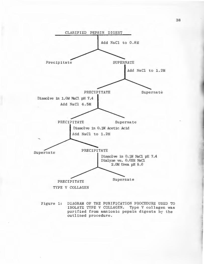
~ hi bi tors. The purification procedure is outlined in Figure 1.
Amnions were minced and centrfuged and the wet weight was
determined. The minced tissues were suspended in 0.5 M ace
tic acid at a ratio of 20 grams wet tissue/100 ml of acetic
acid. Pepsin, 1500 units/ml, was added to the suspension,
stirred for 24 hours then centrifuged for 45 minutes at
30,000 Xg. The supernatant fluid was collected and crystal
line NaCl was added to a final concentration of 0.8 M. The
solution was stirred for 24 hours, then centrifuged for 45
minutes at 30,000 Xg, and the pellet was discarded. The
supernatant NaCl concentration was increased to 1.2 M by
the addition of crystalline NaCl, and the solution was
CLARIFIED PEPSIN DIGEST
Add NaCl to O.BM
Precipitate SUPERNATE
Add NaCl to l.2M
PRECIPITATE
Dissolve in 1. Oili NaCl pH 7. 4
Supernate
Add NaCl 4.5M
PRECIPITATE Supernate
Dissolve in O.lM Acetic Acid
Add NaCl to 1.2M
Supernate
Dissolve in O~lM NaCl pH 7.4 Dialyze vs. 0.02M NaCl
PRECIPITATE
TYPE V COLLAGEN
2.0M Urea pH 8.6
Supernate
Figure 1: DIAGRAM OF THE PURIFICATION PaOCEDURE USED TO ISOLATE TYPE V COLLAGEN. Type V collagen was purified from amnionic pepsin digests by the outlined procedure.
38
39
stirred for 24 hours. The precipitate which formed was col
lected by centrifugation and resuspended in 0.05 M Tris-HCl
pH 7.4 containing 1 M NaCl. The Type V collagen in this
solution was precipitated by adjusting the NaCl concentration
to 4.5 M. The precipiate collected by centrifugation was
dissolved in 0.1 M acetic acid and the collagen was repre-
cipitated by adding NaCl to 1.2 M. The precipitate formed
was collected and resuspended in 0.05 M Tris-HCl pH 7.4
containing 0.1 M NaCl and was dialyzed against 0.01 M Tris-
HCl pH 8.6 containing 0.02 M NaCl and 2 M urea. The pre-
cipitate formed during dialysis was collected and dialyzed
against 0.05M acetic acid and lyophilized. Aliquots of eaeh
purification step were analyzed by SDS-PAGE by the method
of Neville (1971) using 6% polyacrylamide slab gels.
14c-Acetylation of Types I and V Collegens
Types I and V collagens were labeled with 14c-acetic
anhydride. The labeling procedure utilizes an acetylation
reaction which adds a 14c-acetate group to the epsilon
amino groups of lysine residues in the triple helical
region of the collagen molecule (Gisslow and McBride 1975).
Purified collagen was suspended in 0.01% acetic acid at a
concentration of 2 mg/ml; a typical reaction employed a
40
total of 200-300 mg of collagen. Prior to addition of the
acetylating agent, the pH of the collagen solution was ad
justed to 8 by the slow addition of 1 M K2HP04
. 1-14c-acetic
anhydride, 1 mCi in 1.5 ml of benzene, was added dropwise
over the course of 2 hours, the pH was maintained at 8 during
the course of the reaction by addition of 1N NaOH. After
this time the solution was acidified with glacial acetic
acid to pH 4, dialyzed exhaustively against deionized water
and lyophilized. A 2 mg sample of the dried protein was
hydrolyzed in 0.5 mls of Digestol at 60°C for 3 hours and
the specific activity of the substrate was determine. Ace-
tylation of Type I collagen yielded a substrate with a
specific activity of 1.41 x 10⁶ dpm/mg; the Type V sub
~strate had a specific activity of 1.12 x 10⁶ dpm/mg. All
substrates were stored at -20°C.
Type I Collagenase Assay
Type I collagenase activity was measured according to
Lindblad and Fuller (1982). Lyophilized 14C-labeled Type I
collagen was suspended in 0.01% acetic acid at a concentra-
tion of 2 mg/ml. Immediately prior to use, this solution
was diluted with 0.1 M Tris-HCl pH 7.6 containing 0.4 M
NaCl and 0.01 M Cac1 2 so that 0.05 ml contained approxi
mately 20,000 counts per minute (cpm). In a typical assay,
41
0.2 ml of activated culture medium was mixed with 0.1 ml of
0.05 M Tris-HCl pH 7.5 containing 0.005 M CaC1 2 . Each
assay contained 0.05 ml of the diluted substrate solution
and was incubated at 35°C for 1 hour. The assay was termin
ated by addition of 0.1 ml of 0.1 M EDTA containing 150 µg
of carrier Type I collagen. This mixture was incubated for
an additional 30 minutes to assure complete denaturation of
degraded substrate, and then cooled to 15°C for 5 minutes.
Native substrate was precipitated by the addition of 0.8 ml
of a 4:1 (V/V) dioxane/methanol solution. This mixture was
centrifuged for 25 minutes at 6000 Xg and 0.8 ml of the
supernatant was mixed with 5 ml of Atomlight to determine
soluble radioactivity.
Preparation of a Type V Collagen Degrading Activit y
Type V collagen degrading activity was obtained from
in vivo activated alveolar macrophages maintained in cul
ture using the method of Mainardi et al (1980a). An adult
female albino rabbit (3 kg) was injected with 0.2 ml of
Freunds complete adjuvant in the ear vein. Two weeks later
the rabbit was anesthetized and its lungs were surgically
removed. Alveolar macrophages were washed out of the lungs
by lavage using sterile PBS, the lavage treatment was re
peated eight times using a total of 200 ml of PBS. The
42
collected cells were washed three times in DME media con-
taining 100 U penicillin/ml and 100 µg streptomycin/ml.
Cells were plates out in 100 x 20 mm culture dishes at
densities of 1 x 10⁷ cells/dish in 10 ml of DME media supple
mented with 10% FCS, 100 U penicillin/ml, 100 µg streptomycin/
ml and 2.5 µg Fungizone/ml. Cells which adhered to the plates
after 24 hours of growth in serum containing medium were used
for the remainder of the procedure. Plates were maintained
at 37°C in a humidified environment containing 5% co2 and 95%
air. Each plate contained 10 ml of serum-free DME media
which was collected and replaced with fresh media every 2
days for 2 weeks. The collected media was concentrated to
10 ml by pressure dialysis using an Amicon PM-10 ultrafil-
tration membrane. A 0.05 ml aliquot of this preparation
degraded approximately 4 µg of Type V collagen substrate in
5 hours at 30°C pH 7.5.
Type V Collagenase Assay
Type V collagen degrading activity was measured using
an improved assay which utilized a soluble substrate, non
denaturing conditions, and dioxane/methanol which preci
pitates native substrate molecules. Lyophilized 14c
labeled Type V collagen was suspended in 0.01% acetic
43
acid at a concentration of 1 mg/ml. Immediately prior to
use, this solution was diluted with 0.1 M Tris-HCl pH 7.6
containing 0.2 M NaCl and 0.01 M CaC1 2 so that 0.05 ml con
tained 20,000 cpms (20 µg substrate). Activated culture
medium or other enzyme solutions were mixed with 0.05 M
Tris-HCl pH 7.5 containing 0.005 M CaC1 2 in a final reaction
volume of 0.3 ml. A volume of 0.05 ml of the diluted sub-
strate solution was added to each assay tube and incubated
at 30°C for 12-24 hours. The assay was terminated by cooling
the samples to 15°C for 5 minutes and adding 0.1 ml of ice
cold dioxane/methanol (4:1 V/V). Precipitated native sub
strate was separated from degraded substrate by centrifuga
tion at 6000 Xg for 25 minutes at 4°C. A 0.2 ml aliquot of
~the supernatant was mixed with 5 ml of Atomlight to deter-
mine soluble radioactivity.
Elastase Assay
Insoluble elastin powder, purchased from Sigma Chemical
Company, was labeled using 3H-sodium borohydride by the
method of Stone et al (1982). The lyophilized labeled
elastin was suspended in 0.05 M Tris-HCl pH 7.5 containing
0.005 M CaC12
at a concentration of 1 mg/ml; 0.25 ml of
this suspension was added to 1 ml microcentrifuge tubes.
Aliquots of the cell sonicates (0.1 to 0.5 ml) and 0.05
........
44
M Tris-HCl pH 7.5 containing 0.005 M CaC1 2 were added to the
microcentrifuge tubes in a final reaction volume of 1.0 ml.
The reaction mixtures were incubated at 37°C for 24 hours
without agitation, cooled on ice, and centrfuged in a Fischer
Model 59 microcentrifuge for 2 minutes at 7000 Xg. A 0.2 ml
aliquot of the supernatant was removed and mixed with 5 ml
of Atomlight to determine soluble radioactivity.
Ion Exchange Chromatography
DEAE cellulose ion exchange chromatography was utilized
to separate Types I and V collagen degrading activities
secreted by DLD-1 colon carcinoma cells. DEAE cellulose
(Whatman DE-52) was suspended in 0.05 M Tris-HCl pH 8.3
containing 0.005 M CaC1 2 and 0.02% NaN 3 , packed into a
column 1.6 x 13 cm and allowed to equilibrate at 4°C by
washing with several volumes of buffer. Pooled serum-free
media from 900 100 x 20 mm culture dishes of late log phase
DLD-1 cells were concentrated by pressure dialysis using
an Amicon PM-10 ultrafiltration membrane and dialyzed
against 10 liters of column buffer. The sample, approxi
mately 100 ml, was trypsin activated as previously de
scribed and centrifuged for 10 minutes at 8000 Xg to
remove any precipitate which had formed during concen
tration and dialysis. The supernatant was then applied to
45
the column at a flow rate of 25 ml/hour and the column
was washed with buffer until absorbance at 280 nm of the
effluent returned to baseline. Bound proteins were
eluted in a linear gradient from 0.0 to 1.0 M NaCl in
column buffer. The gradient was run over the course of
16 hours using an Ultrograd Gradient Mixer (LKB Bromma,
Sweden). 'Ihe total gradient volume was 480 mls. Six ml
fractions were collected and monitored for absorbance at
280 nm and assayed for Type I and V collagenase activity.
Gel Filtration Chromatography
Molecular weights of Type I and V collagenolytic
~activities were determined using Ultrogel AcA44 gel
filtration medium. The column material was packed into
a column 1.6 x 90 cm at 4°C and equilibrated in 0.05 M
Tris-HCl pH 7.6 containing 0.01 M CaC12 and 0.02% NaN3 .
The flow rate of the column was kept constant at 8 ml/
hour. The column was calibrated using the globular
protein standards bovine serum albumin, ovalbumin,
carbonic anhydrase and ribonuclease, 1 mg of each
standard was chromatographed separately. Blue Dextran
2000 was used to determine the void volume (V ). Fraco
tions of 2 ml were collected and the absorbance at 280
run was monitored. Peaks of Type I or TypP V collagenolytic
activity from the DE-52 chromatogram were concentrated to
46
1 ml by pressure dialysis using an Amicon PM-10 ultra-
filtration membrane. These samples were applied to the
column. Fractions of 2 ml were collected, the absorbance
monitored at 280 nm was monitored, .and the fractions were
assayed for Ty..pes I and V collagenase activity.
Statistical Analysis
The statistical methods used in this study were
obtained from the statistics textbook, "Introduction to
Applied Statistics", (Lentner, M., 1976).
1. Mean (X) = EX. /n l
n = sample size
X. = ith sample value l
2. Standard Deviation( s) ~ Exi - X) 2 I (n - 1 )! I
3. Unpaired Student t test = t = (Xl x2)
[i: 8~1;2 n = sample size of 1 group
degrees of freedom = 2(n-1)
47
RESULTS
Purification of Type V Collagen
Collagenous proteins present in an insoluble form in
the extracellular matracies of many tissues can be
extracted in a soluble form by limited digestion of the
tissue with pepsin. This procedure was employed to
solubilize Type V collagen from human amnionic membranes.
Pepsin soluble collagens can be separated by differential
precipitation with NaCl. This procedure takes advantage of
solubility differences between the five genetically dis
tinct collagen types. Interstitial collagen Types I and
~III in the amnionic pepsin digest were precipitated at
low ionic strength (0.8M NaCl). Type V collagen preci
pitated when the ionic strength of the solution was
raised to 1.2M NaCl. This precipitate was suspended in
neutral salt buffer to inactivate any residual pepsin
and the Type V collagen was reprecipitated by the addition
of NaCl to 4.5 M. The 4.5 M NaCl precipitate was suspended
in neutral salt buffer and dialyzed against a buffer of
low ionic strength. This step separates Type V collagen
from low molecular weight protein fragments which are
soluble in low ionic strength solutions.
The precipitate formed during dialysis contained
purified Type V collagen. The purification began with
745 grams of amnionic membranes (wet weight) and pro
duced 350 mg of Type V collagen, a yield of 0.05%. The
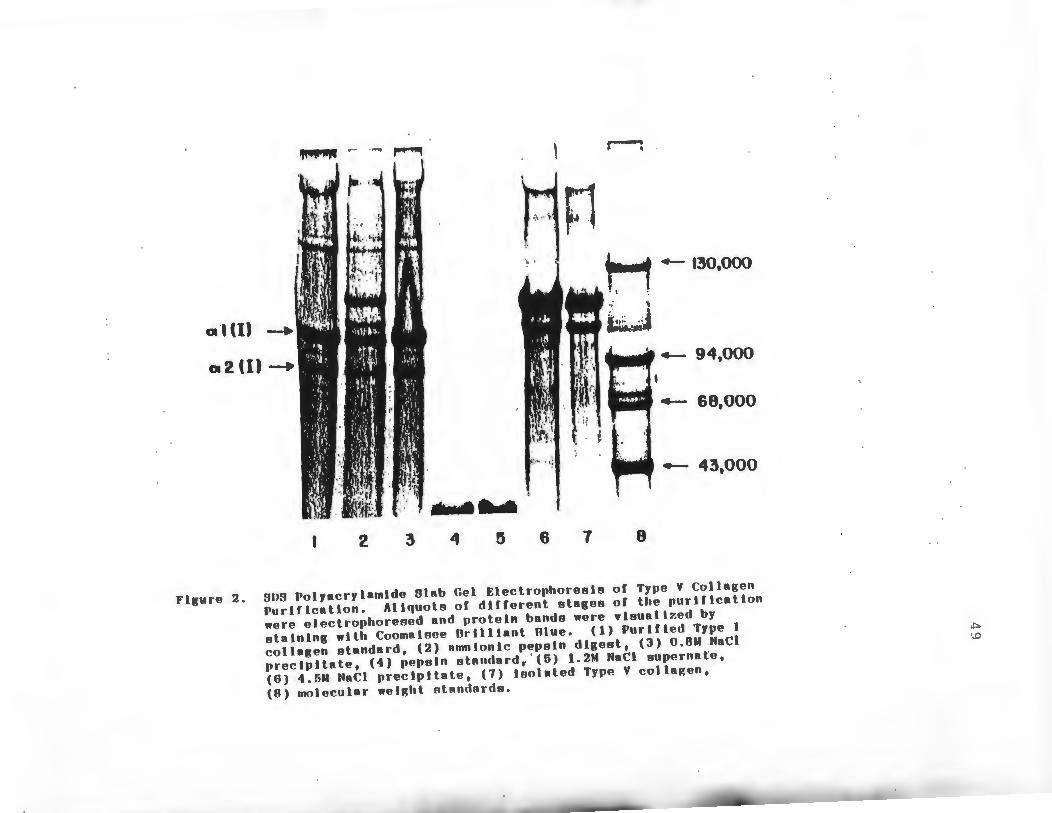
purity of the preparation was monitored by SDS-PAGE at
various stages of the purification as shown in Figure 2.
The electrophoretic pattern of purified Type I collagen
is shown for comparison (Lane 1). The whole pepsin
digest was found to contain both interstitial and Type V
collagens (Lane 2). The precipitate which formed at
0.8M NaCl contained predominately Type I collagen and
was devoid of Type V collagen (Lane 3). Pepsin added
at the start of the purification was separated from
~Type V collagen when the NaCl concentration was raised
to 1.2M. Type V collagen precipitates at this ionic
strength and the pepsin remains soluble (Lane 5). The
Type V collagen which was purified by pepsin digestion
and differential NaCl precipitation consisted of two~
chains,o\ l(V) and<1'\2(V) present in an approximate 2:1
ratio (Lane 7). The molecular weights of these J\chains
were estimated to be 117,000 and 105,500 using molecular
weight standards (Lane 8).
48
"""""' .- ..... ,,.._... ~
; .... ~' r.1 \1
.._ 130,000
a·•ul _. a2(1)-. .. '4-- 94,000
4- 60,000
.__ 43,000
2 3 4 ~ 6 7 8
Figure 2. SOS Polyacrylamlde Slab Gel Electrophoresis of Type V Collagen Purtflcatlon. Aliquots of different stages of the purtflcatton were electrophoresed and protein bands were vleuallzed by stalnlng wtth Coomatsee Orllltant Olue. (1) Purlfled Type I collagen standard, (2) amntonlc pepeln digest, (3) 0.8M NaCl prectpltate, (4) pepsin ~tandard, ' (5) 1.2M NaCl supernate, (6) 4.5M NaCl precipitate, (7) Isolated Type V colla~en, (8) molecular weight standards.
""" \ 0
50
Type V Collagenase Assay
An assay for measuring Type V collagen degrading
activity was developed. Purified Type V collagen was
labeled with 14c-acetic anhydride. This compound acetylates
the epsilon amino groups of lysine residues in collagen.
The labeled Type V collagen had a specific activity of
6 1.12 x 10 dpm/mg. The assay was performed at an incuba-
tion temperature of 30°C. This is several degrees below
the point at which the Type V collagen triple helix
begins to denature (Rhodes and Miller, 1978). The pre-
cipitating solvent dioxane/methanol (4:1, v/v) was used
to separate native substrate molecules from degraded sub-
strate molecules. This solvent has been used previously
in Type I collagenase assays (Lindblad and Fuller, 1982).
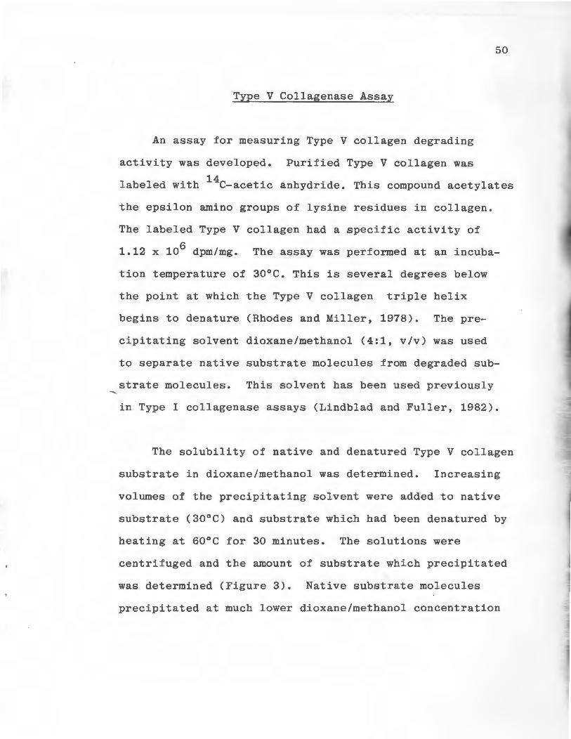
The solubility of native and denatured Type V collagen
substrate in dioxane/methanol was determined. Increasing
volumes of the precipitating solvent were added to native
substrate (30°C) and substrate which had been denatured by
heating at 60°C for 30 minutes. The solutions were
centrifuged and the amount of substrate which precipitated
was determined (Figure 3). Native substrate molecules
precipitated at much lower dioxane/methanol concentration

.......
I
51
0 0 DENATURED
• • NATIVE
70
-..I c:( ~ 60
~ ~ • - S> ~ ~
> ~ u c:( 0 0 c:(
.30 a: UJ _J al ::> zo _J 0 (/)
10
10 20 30 60
OIOXANE:METHANOL (%)
Figure 3. Solubility of Native and Denatured Radiolabeled Type V Collagen in Dioxane/Methanol/Water. Increasing volumes of a dioxane/methanol solution (4:1, v/v) were added to native (30°C) and heat denatured (60°C for 30 mi ns.) 14c-Type V collagen, centrifuged and soluble radioactivity was determined.
-....._
than denatured substrate molecules. The difference in
their solubilities was greatest between 23%-46 % dioxane/
methanol.
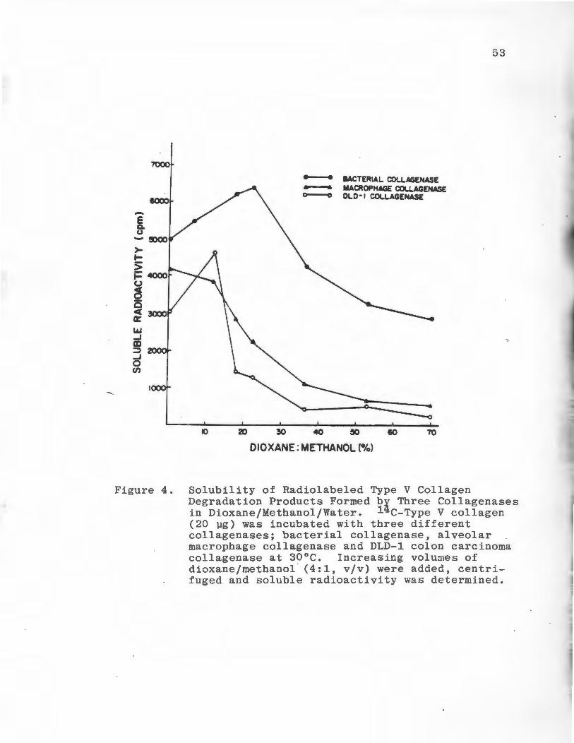
The solubility of enzymatically produced Type V
collagen reaction products in dioxane/methanol were
determined. Native substrate was degraded by three
different collagenases, Clostridium histolytium colla
genase. (E.C. 3.4.24.3), rabbit alveolar macrophage
collagenase, and collagenase obtained from the media
of cultured DLD-1 colon carcinoma cells. The enzyme
substrate solutions were incubated at 30°C for six hours
then increasing volumes of dioxane/methanol were added.
Following centriguration, the solubility of the reaction
products were determined (Figure 4). When the substrate
52
was degraded by bacterial collagenase soluble radio
activity was highest at 29% dioxane/methanol. Following
incubation with alveolar macrophage and DLD-1 collagenases
soluble radioactivity was highest at 13% dioxane/methanol.
To optimize the detection of enzyme activity while pre
cipitating the majority of the native substrate molecules
23% dioxane/methanol was chosen as the standard concen
tration to be used in the assay. At this concentration
70% of the native substrate is precipitated and appreci-
---
53
7'000
• • BACTERIAL COLI.AGENASE - MACROPHAGE COl.l.AGENASE 0
IOOO o DLD-t COLLAGENASE
E Q. u
- SXX> >-~
> ~~ (.)
g 0 c( er LAJ ...J en :'.) ...J 0 Cf)
IO 20 30 70
OIOXANE: METHANOL(%)
Figure 4. Solubility of Radiolabeled Type V Collagen Degradation Products Formed by Three Collagenases in Dioxane/Methanol/Water. 14 c-Type V collagen (20 µg) was incubated with three different collagenases; bacterial collagenase, alveolar macrophage collagenase and DLD-1 colon carcinoma collagenase at 30°C. Increasing volumes of dioxane/methanol · (4:1, v/v) were added, centrifuged and soluble radioactivity was determined.
54
able amounts of enzymatic activity can be detected.
As shown on Figure 5,degradation of the substrate by
alveolar macrophage collagenase was linear with time.
Approximately 20% of the added substrate (20 µg) had been
degraded after five hours of incubation at 30°C and pH 7.5.
Levels of Collagenolytic and Elastinolytic Activities
In Normal Fibroblasts, DLD-1, and B16-F10 Cells
Late log phase cultures of DLD-1 colon carcinoma
cells, B16-F10 murine melanoma cells, and normal dermal
fibroblasts were maintained in serum-free media for 24
hours. The cells and media were processed for enzyme
assays, protein and DNA determinations as described in
the Experimental Section. DLD-1 cells and normal fibro
blasts produced comparable levels of Type I collagenolytic
activity (Table 1). B16-Fl0 cells produced approximately
half as much activity. Normal fibroblasts produced the
highest level of Type V collagenolytic activity (0.48 ±
0.14 µg collagen degraded/mg protein/hr), DLD-1 cells
produced about half this amount of activity while Bl6-Fl0
cells only degraded 0.13 ± 0.06 µg collagen/mg protein/hr.
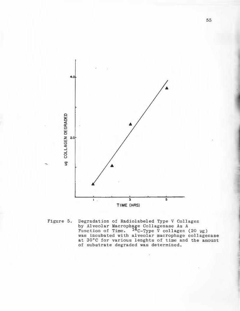
55
4.0
0 w 0 cf a: (,!) w 0
z 2.0 w (,!)
cf ..J ..J 0 u O> ~
I . 3
TIME (HRS)
Figure 5. Degradation of Radiolabeled Type V Collagen by Alveolar Macrophage Collagenase As A Function of Time. 14c-Type V collagen (20 µg) was incubated with alveolar macrophage collagenase at 30°C for various lenghts of time and the amount of substrate degraded was determined.
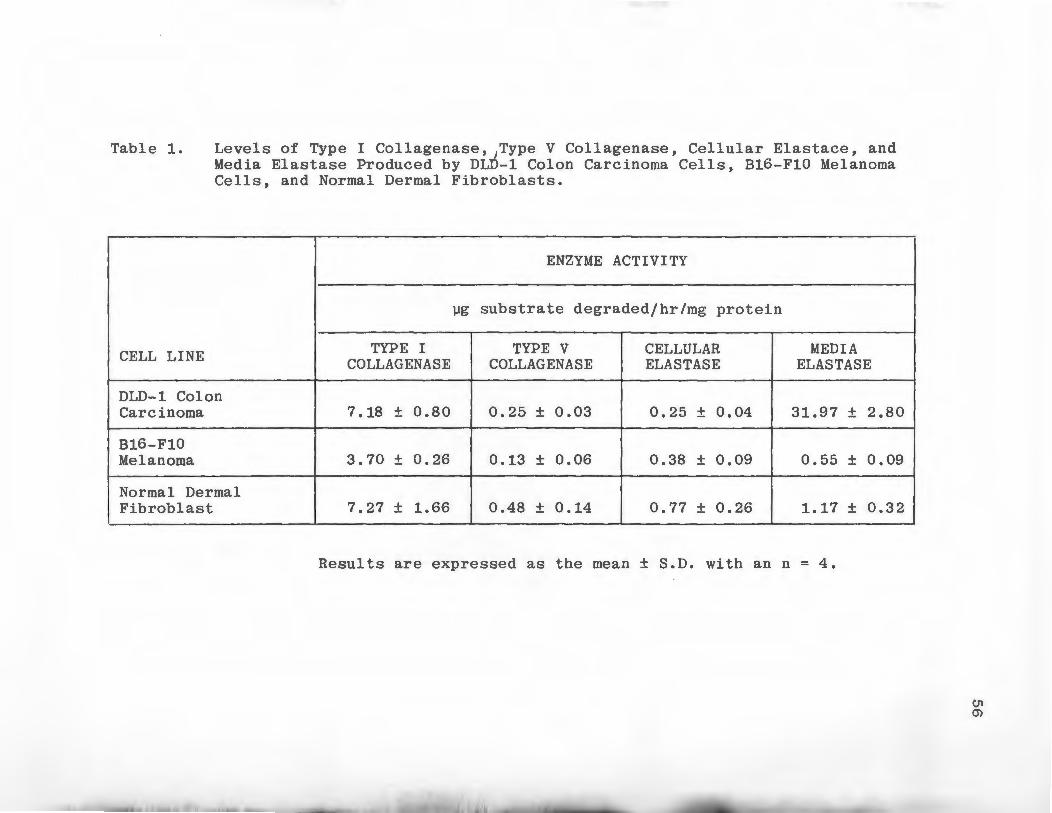
Table 1.
CELL LINE
Levels of Type I Collagenase,~Type V Collagenase, Cellular Elastace, and Media Elastase Produced by DLD-1 Colon Carcinoma Cells, B16-F10 Melanoma Cells, and Normal Dermal Fibroblasts.
ENZYME ACTIVITY
µg substrate degraded/hr/mg protein
TYPE I TYPE V CELLULAR MEDIA COLLAGENASE COLLAGEN ASE ELASTASE ELASTASE
DLD-1 Colon Carcinoma 7 .18 ± 0 .80 0.25 ± 0.03 0.25 ± 0.04 31.97 ± 2.80
B16-F10 Melanoma 3.70 ± 0.26 0.13 ± 0.06 0.38 ± 0.09 0.55 ± 0.09
Normal Dermal Fibroblast 7.27 ± 1.66 0.48 ± 0.14 0.77 ± 0.26 1.17 ± 0.32
Results are expressed as the mean ± S.D. with an n = 4.
C.11 (j)

57
The Triton X-100 cell extracts and culture media from
the three cell lines were assayed for elastin degrading
activity. DLD-1 cells produced the highest levels of
elastinolytic activity of the three cell lines, this
activity was found predominately in the media (>99% of the
total). Fibroblasts contained the second highest level of
elastinolytic activity, approximately 40% of the activity
was cell-associated and 60% was in the media. The elastin
degrading activity from Bl6-F10 cells was distributed
between the cellular compartment and the media in the same
ratio as the fibroblast activity.
DEAE Ion Exchange Chromatography
Media containing DLD-1 collagen degrading activities
was collected and several attempts were made to separate
Type I and Type V collagen degrading activities using gel
filtration chromatography. These attempts proved to be
unsuccessful, each chromatogram revealed numerous peaks
of Type I and Type V collagen degrading activity and
adequate resolution of the two activities was not possi
ble. DEAE cellulose ion exchange chromatography was then
employed to separate Type I and Type V collagen degrading
58
activities. DEAE cellulose chromatography was used to
separate these two activities produced by cultured macro
phages (Mainardi et al, 1980a). Serum-free media from 900
plates of late log phase DLD-1 co-on carcinoma cells
was used as the source of collagenase activity. As seen
in Figure 6 approximately half of the media proteins bound
to the DE-52 column and half passed through the column.
The fractions containing the protein which did not bind to
the resin were analyzed and only Type V collagen degrading
activity was detected. The proteins which bound to the
resin were eluted with a 400 ml linear NaCl gradient
(0-0.3M). This was found to be insufficient to elute all
the proteins, a second gradient of 0.3M to 1.0M NaCl was
used to elute the remainder of the proteins. The first
peak of protein eluted between 0.2M and 0.3M NaCl, these
fractions were found to contain only Type I collagen
degrading activity. The proteins which eluted in the
second gradient between 0.3M and 0.5M NaCl were analyzed
for collagenase activity and both Type I and Type V
collagen degrading activity were detected.
Gel Filtration Chromatography
The three peaks of collagen degrading activity gen
erated by DEAE cellulose chromatography were further
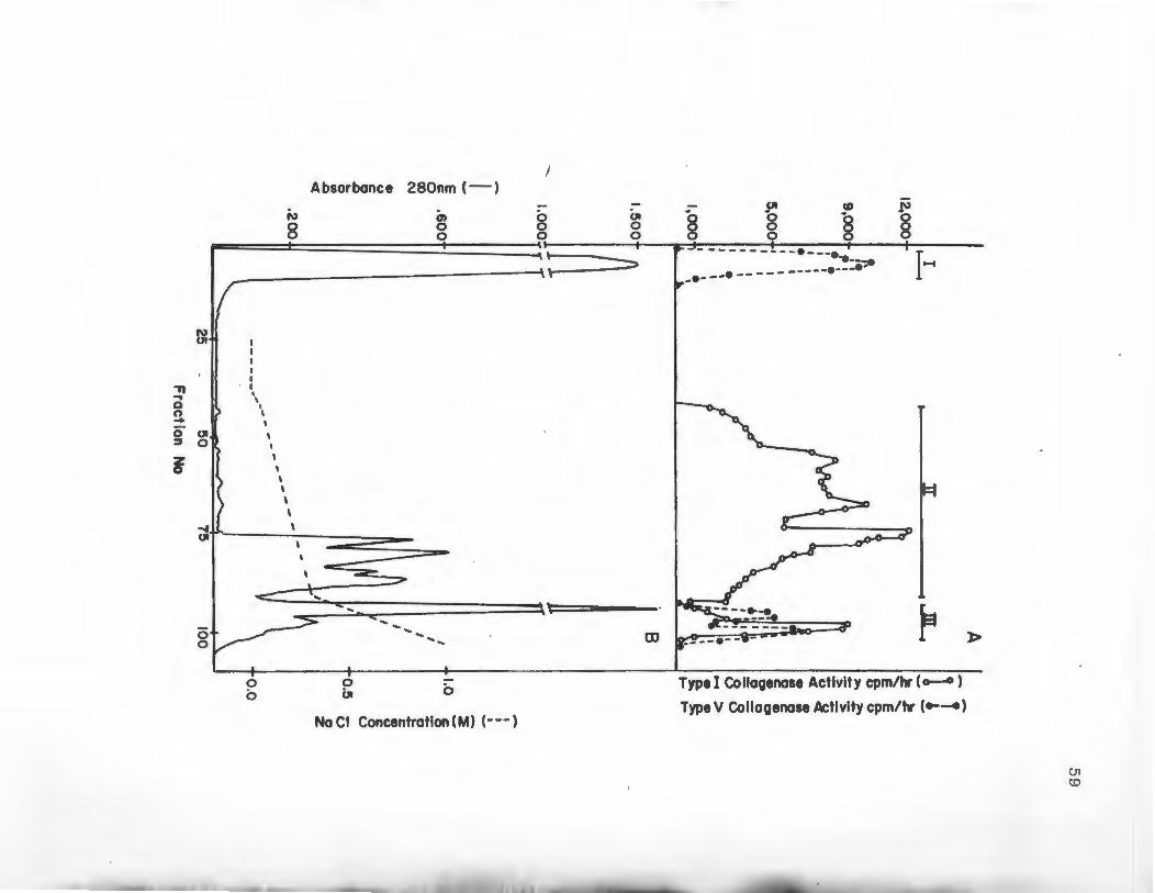
~
~ ~ 0 UI ~ 0
f
0 0
I Absorbance 280nm (-)
--~~~~~~~~~~~~~----\' ==:>
' I ' I
I
I I
I I I I I I
....... ., < .... ., \~====-----
q::: <
0 0
0 bi
~ .....................
0
Na Cl Concentration (M) (---)
CD
~l=t
r Type I Collagenose Activity cpm/hr (~)
Type V Collagenose Activity cpm/hr (e-~)
)>
CJl (.0
60
Figure 6. DEAE Cellulose Chromatography of Concentrated DLD-1 Colon Carcinoma Cell Serum-Free Media. Serum-free media from 900 100 x 20 mm culture dishes of late log phase DLD-1 cells was concentrated and applied to a 1.5 x 13 cm column of DE-52 equilibrated in 0.05M Tris~Cl pH 8.3 containing 0.005M CaCl2 and 0.02 % NaN3 . The bound proteins were eluted in two NaCl gradients, one from 0 to 0.3M NaCl and the second gradient from 0.3M to 1.0M. Six ml fractions were collected and monitored for absorbance at 280 nm (Panel B) and analyzed for Type I and Type V collagen degrading activity (Panel A). The fractions were combined to form three pools according to enzyme activity (I, II, III) as indicated.
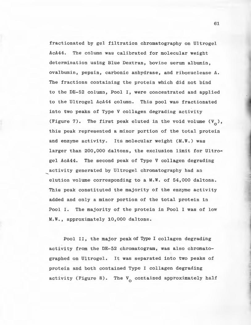
........
61
fractionated by gel filtration chromatography on Ultrogel
AcA44. The column was calibrated for molecular weight
determination using Blue Dextran, bovine serum albumin,
ovalbumin, pepsin, carbonic anhydrase, and ribonuclease A.
The fractions containing the protein which did not bind
to the DE-52 column, Pool I, were concentrated and applied
to the Ultrogel AcA44 column. This pool was fractionated
into two peaks of Type V collagen degrading activity
(Figure 7). The first peak eluted in the void volume (V0
),
this peak represented a minor portion of the total protein
and enzyme activity. Its molecular weight (M.W.) was
larger than 200,000 daltons, the exclusion limit for Ultro~
gel AcA44. The second peak of Type V collagen degrading
activity generated by Ultrogel chromatography had an
elution volume corresponding to a M.W. of 54,000 daltons.
This peak constituted the majority of the enzyme activity
added and only a minor portion of the total protein in
Pool I. The majority of the protein in Pool I was of low
M.W., approximately 10,000 daltons.
Pool II, the major peak of Type I collagen degrading
activity from the DE-52 chromatogram, was also chromato-
graphed on Ultrogel. It was separated into two peaks of
protein and both contained Type I collagen degrading
activity (Figure 8). The V contained approximately half 0
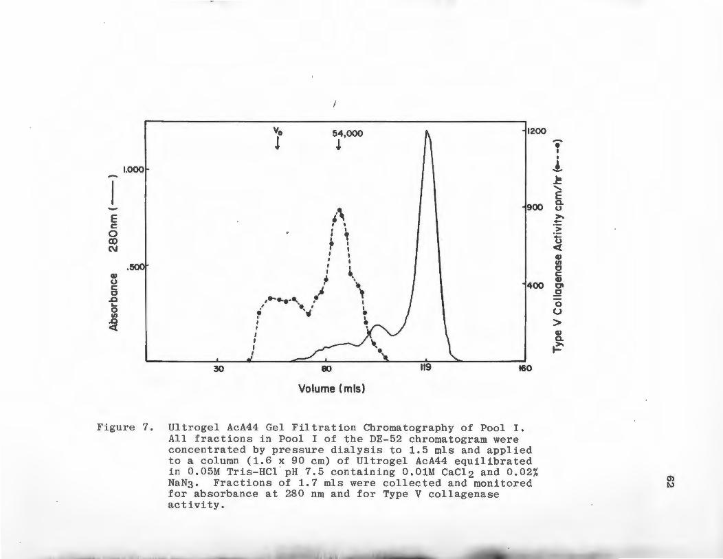
-E c: 0 Q) C\J
Q) 0 c: c
L> ~
0 en
L> <(
l.000
.5001
30
Vo
l
I
54,000
!
,, I I . ' : ,
• I I I : I I I
~ •. I '
/.......... ,i ·~ . •. , : " I I I I I
80
Volume (mis)
1200
900
400
119 160
Figure 7. Ultrogel AcA44 Gel Filtration Chromatography of Pool I. All fractions in Pool I of the DE-52 chromatogram were concentrated by pressure dialysis to 1.5 mls and applied to a column (1.6 x 90 cm) of Ultrogel AcA44 equilibrated in 0.05M Tris-HCl . pH 7.5 containing O.OlM CaCl2 and 0.02% NaN3. Fractions of 1.7 mls were collected and monitored for absorbance at 280 nm and for Type V collagenase activity.
-• I
' .! ~
' E Q. 0 >--> 0 <( Q) en c c: Q) Cl .2 0 u > Q) Q.
~
O> t.)
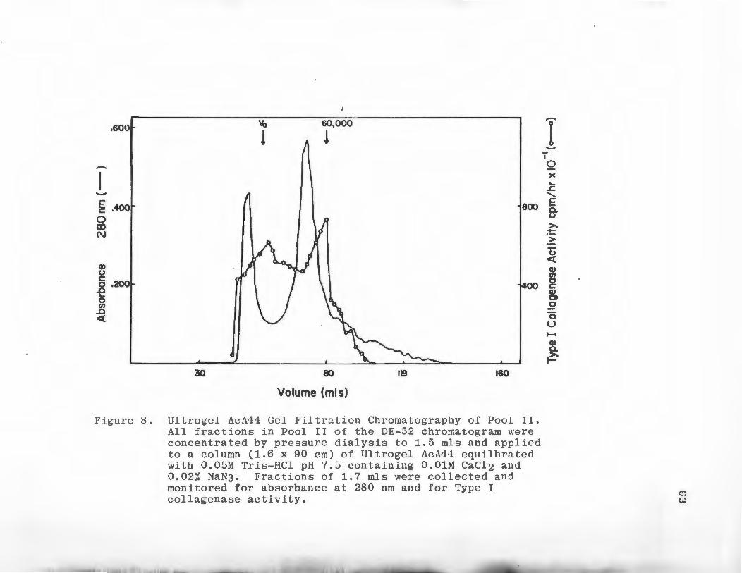
.600
E .400 0 (X) C\J
G> u c: 0 .200 ..c L. 0 en
..c <{
30
~
l
I 60,000
!
80
Volume (mis)
800
119 160
Figure 8. Ultrogel AcA44 Gel Filtration Chromatography of Pool II. All fractions in Pool II of the DE-52 chromatogram were concentrated by pressure dialysis to 1.5 mls and applied to a column (1.6 x 90 cm) of Ultrogel AcA44 equilbrated with 0.05M Tris-HCl pH 7.5 containing O.OlM CaCl2 and 0.02% NaN3. Fractions of 1.7 mls were collected and monitored for absorbance at 280 nm and for Type I collagenase activity.
-I ..... I
0 )(
L. ..s::. ........ E fr >. ~ > -u <{
~ 0 c: Q) Cl 0
0 u ......
Q) a. ~
(j)
w
.....
64
of the total enzyme activity and about 30% of the total
protein applied to the column. The second peak of enzy-
matic activity eluted in the 60,000 dalton M.W. range.
Pool III which eluted from the DEAE cellulose column
during the 0.3M to 1.0M NaCl gradient, was fractioned into
three peaks of enzyme activity by chromatography on Ultro-
gel AcA44 (Figure 9). The V fractions from this chromao
togram contained the majority of the protein that was
loaded on the column. Both Type I and Type V collagen
degrading activities were detected in these fractions.
The second of the three peaks generated contained only
Type V collagen degrading activity and had a M.W. of
approximately 80,000 daltons. The final peak which eluted
in the 35,000 dalton M.W. range contained Type I co l lagen
degrading activity.
Inhibition of Types I and V Collagen Degrading Activities
Concentrated serum-free media from DLD-1 colon cari-
noma cells were used as the source of Type I and Type V
collagen degrading activities. Fetal calf serum, EDTA,
PMSF, dithiothreital (DTT), and N-ethylmaleimide (NEM)
were tested for their ability to inhibit DLD-1 collagen
degrading activities using the standard enzyme assays
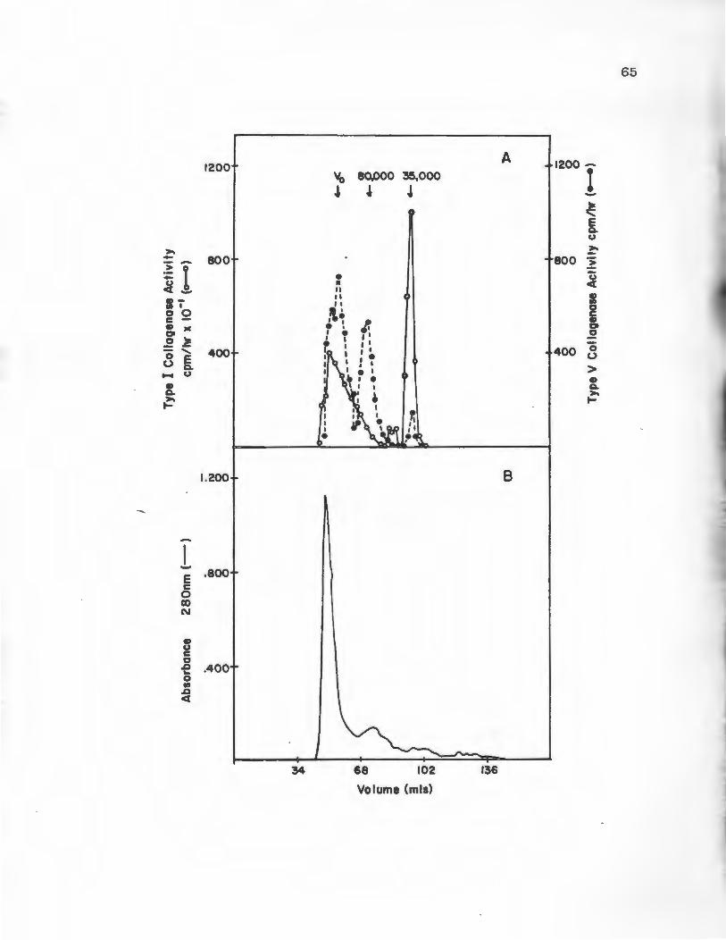
>. -~I <( --
11 0 0 c -~ JC 0 ~
...... 8 [ - u ., ~
~
l E c 0 Cl) N
., u c 0
.D ... 0 .,,
.D <(
1200
800
400
1.200
.800
.400
V0 80,000 35,000
~ ~ J
• II II II 11
,\• . : ~ I e •,I I I
• I I I I. I I I
I I~ I I
IJ ~ ~ \ I I
~
68 102
Volume (mis)
A
B
136
1200 I
800
--
> -u <(
I ~ ., 0 ~
400 ° u > ., ~ >. ~
65
Figure 9. Ultrogel AcA44 Gel Filtration Chromatography of Pool III. All fractions in Pool III of the DE-52 chromatogram were concentrated by pressure dialysis to 1.5 mls and applied to a column (1.6 x 90 cm) of Ultrogel AcA44 equilibrated with 0.05M Tris-HCl pH 7.5 containing O.OlM CaCl2 and 0.02 % NaN3. Fractions of 1.7 mls were collected and monitored for Type I and Type V collagenase activity (Panel A) and absorbance at 280 nm (Panel B).
66
67
(Table 2). Both Type I and Type V collagen degrading
activities were inhibited by serum and EDTA, the metallo
proteinase inhibitor. The serine protease inhibitor PMSF
had no effect on Type V collagen degrading activity and
reduced the Type I collagen degrading activity by only 18%.
DTT, which reduces disulfide bonds and maintains sulfhydryl
groups in a reduced state, blocked 41% and 32% of Type I
and Type V collagen degrading activities, respectively.
NEM, which reacts with free sulfhydryl groups, had no effect
on either enzyme activity.
Effects of DMF on the Production of Type I and Type V
Collagenase, Cellular, and Media Elastase Activities
By DLD-1 Colon Carcinoma Cells
The effect of DMF on the production Type I and Type V
collagenase and elastase activities by DLD-1 cells was
determined using late log phase cultures. The production
of these enzymatic activities was measured over a 24 hour
period. Enzyme activity was normalized using total cellu
lar protein and DNA values (Table 3). DMF caused a 20%
increase in Type I collagenase activity over control
cultures when the activity was expressed per mg of protein.
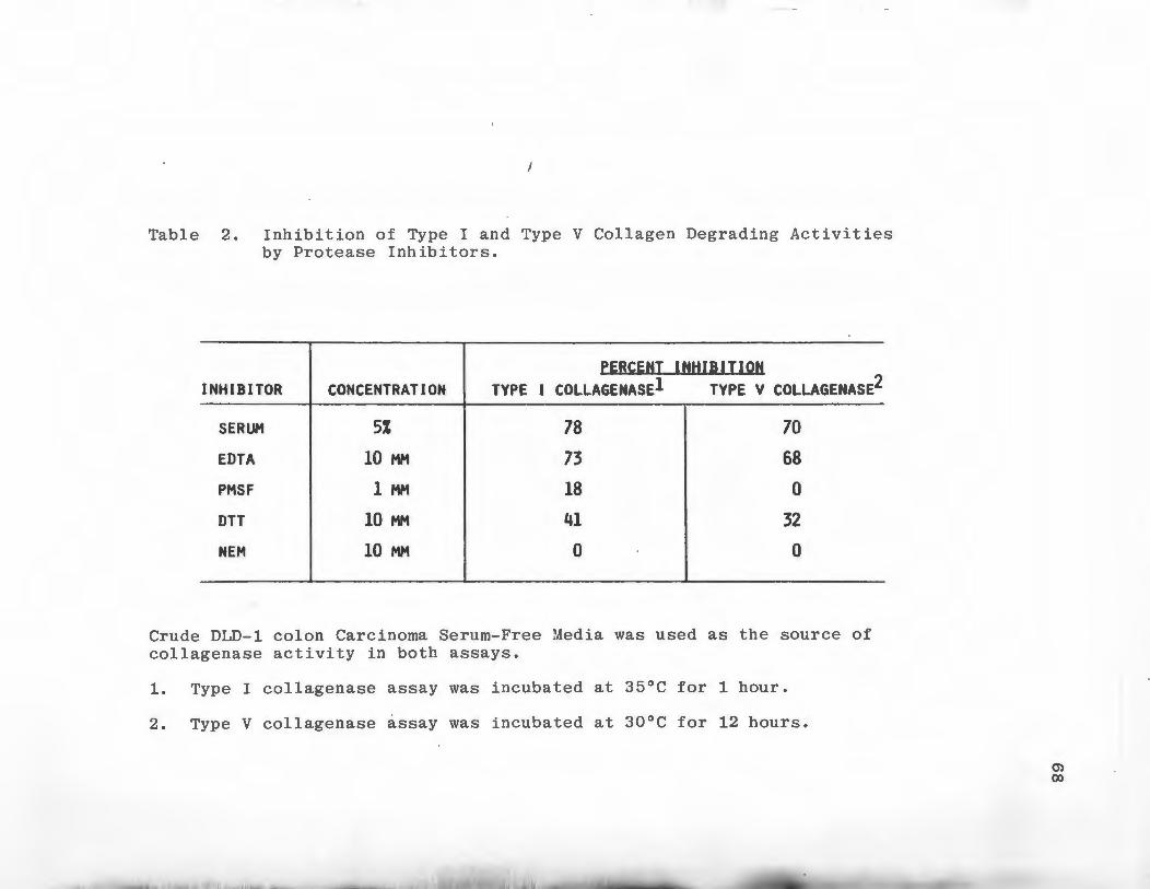
J
Table 2. Inhibition of Type I and Type V Collagen Degrading Activities by Protease Inhibitors.
PERCENT INHIBITION INHIBITOR CONCENTRATION TYPE I COLLAGENASEl TYPE V COLLAGENASE2
SERUM 5% 78 70
EDTA 10 MM 73 68
PHSF 1 HM 18 0
DTT 10 HM 41 32
NEM 10 HM 0 0
Crude DLD-1 colon Carcinoma Serum-Free Media was used as the source of collagenase activity in both assays.
1. Type I collagenase assay was incubated at 35°C for 1 hour.
2. Type V collagenase assay was incubated at 30°C for 12 hours.
m 00
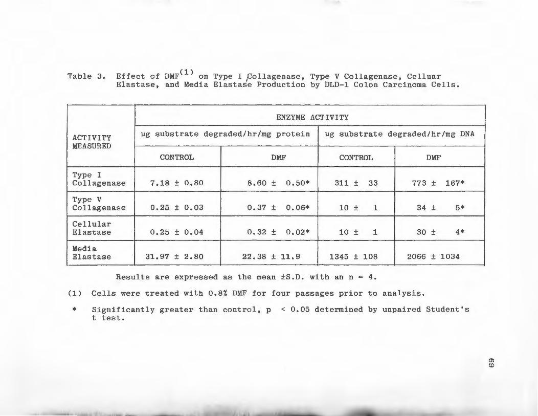
(1) Table 3. Effect of DMF on Type I pollagenase, Type V Collagenase, Celluar
Elastase, and Media Elastase Production by DLD-1 Colon Carcinoma Cells.
ENZYME ACTIVITY
ACTIVITY µg substrate degraded/hr/mg protein µg substrate degraded/hr/mg DNA
MEASURED
CONTROL DMF CONTROL DMF
Type I Collagenase 7.18 ± 0.80 8.60 ± 0.50* 311 ± 33 773 ± 167*
Type V Collagenase 0.25 ± 0.03 0.37 ± 0.06* 10 ± 1 34 ± 5*
Cellular Elastase 0.25 ± 0.04 0.32 ± 0.02* 10 ± 1 30 ± 4*
Media Elastase 31. 97 ± 2. 80 22. 38 ± 11. 9 1345 ± 108 2066 ± 1034
Results are expressed as the mean ±S.D. with an n = 4.
(1) Cells were treated with 0.8% DMF for four passages prior to analysis.
* Significantly greater than control, p < 0.05 determined by unpaired Student's t test.
en co
.......
70
The increase due to DMF was 148% when DNA was
used to normalize the enzyme values. Both values for Type
I collagenase activity in DMF cultures were significantly
higher than control values (p < 0.05). Type V collagenase
activity increased 44% and 220% due to DMF treatment when
enzyme activity was expressed per amount of protein and
DNA,respectively. DLD-1 cellular elastase activity also
increased as a result of DMF treatment. Cellular elastase
activity per mg protein increased 31% while elastase
activity per µg DNA increased 192%. Both values were
significantly higher than control (p < 0.05). Media
elastase activity per mg protein dropped by 30% in
response to DMF. This decrease was not found to be signi
ficant (p < 0.05). Media elastase activit y expressed per
µg DNA increased 53% when the cells were treated with DMF.
This value for enzyme activity in DMF treated culture was
significantly higher than control cultures (p < 0.05).
To deter~ine if these increases in activity were associ
ated with alterations in the levels of cellular protein, DNA
and RNA the levels of these cellular constituents were deter
mined. Cells were grown in media containing 0.8% DMF for
four passages prior to analysis. DMF caused a significant
rise in the cellular levels of protein and RNA over con
trols (p < 0.05). DMF cells contained significantly less
DNA than control cells (p < 0.05) (Table 4).
Effect of DMF on the Incorporation of
3H-glycine into Protein
The effect of DMF on the capacity of DLD-1 cells
to synthesize and secrete protein was analyzed by
measuring the rate of incorporation of 3H-glycine into
TCA insoluble cell and media proteins. Determinations
were made after three and six hours of growth in media
containing 5 µCi 3H-glycine/ml. The results of these
experiments were normalized in two ways; per amount of
71
cellular protein and DNA. DMF treated cells incorporated
3H-glycine into cellular protein at a slower rate than ...._
control cells after three hours of growth in 3H-glycine
containing media (Table 5). This decrease was signifi
cant only when the values were expressed per unit of DNA
(p < 0.05). However, after six hours of incubation, the
DMF treated cells were incorporating label into cellular
protein at a faster rate than controls. The DMF values
were significantly greater than controls by both units
of measure (p < 0.05).
Analysis of media proteins revealed that DMF treated
cells were secreting 3H-glycine containing protein into
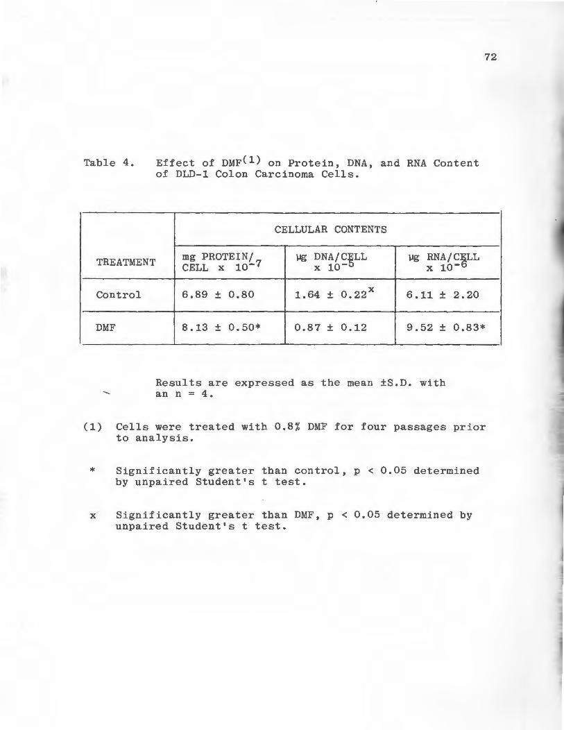
Table 4.
TREATMENT
Control
Effect of DMF(l) on Protein, DNA, and RNA Content of DLD-1 Colon Carcinoma Cells.
CELLULAR CONTENTS
mg PROTEIN/ lJg DNA/C~LL lJg RNA/CELL CELL x 10-7 x 10- x 10-6
6.89 ± 0.80 1. 64 ± o. 22 x 6.11 ± 2.20
72
DMF 8 .13 ± 0.50* o. 87 ± 0.12 9.52 ± 0.83*
Results are expressed as the mean ±S.D. with an n = 4.
(1) Cells were treated with 0.8% DMF for four passages prior to analysis.
* Significantly greater than control, p < 0.05 determined by unpaired Student's t test.
x Significantly greater than DMF, p < 0.05 determined by unpaired Student's t test.
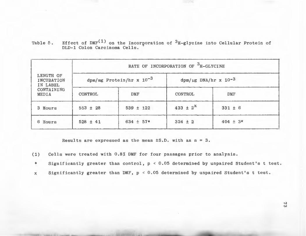
Table 5.
LENGTH OF
Effect of DMF(l) on the Incor~oration of 3H-glycine into Cellular Protein of DLD-1 Colon Carcinoma Cells.
RATE OF INCORPORATION OF 3H-GLYCINE
INCUBATION dpm/mg Protein/hr x 10-3 dpm/µg DNA/hr x 10-3 IN LABEL CONTAINING MEDIA CONTROL DMF CONTROL DMF
3 Hours 553 ± 28 539 ± 122 433 ± 2x 331 ± 6
6 Hours 528 ± 41 634 ± 57* 324 ± 2 404 ± 3*
Results are expressed as the mean ±S.D. with an n = 3.
(1) Cells were treated with 0.8/. DMF for four passages prior to analysis.
* Significantly greater than control, p < 0.05 determined by unpaired Student's t test.
x Significantly greater than DMF, p < 0.05 determined by unpaired Student's t test.
--l w

the media at a slower rate than controls at three hours
(Table 6). The rate of secretion of labeled proetin by
DMF treated cells was significantly slower than control
when expressed per unit of DNA but was not significantly
different per unit of protein (p < 0.05). At six hours,
74
the situation had reversed, DMF treated cells were secreting
labeled protein at a faster rate than controls. This in
crease was significant only when the data was expressed
per unit of DNA (p < 0.05).
Effect of Cycloheximide on the Synthesis of Connective
Tissue Degrading Enzymes by Control and DMF Treated
DLD-1 Cells
Control and DMF treated DLD-1 cells were treated with
cycloheximide to determine if the effects of DMF were
dependent on the synthesis of protein. Cells were exposed
to 1 µg cycloheximide/ml for the 24-hour period during
which enzyme production was quantified. Treatment of DMF
treated DLD-1 cells with cycloheximide resulted in a small
decrease in Type I collagenolytic activity expressed per
unit of protein and an insignificant increase in activity
per unit of DNA (p < 0.05) (Table 7). Cyclobeximide bad
the same effect on control cells; synthesis of Type I
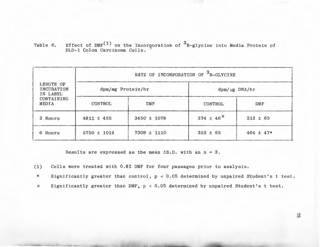
Table 6.
LENGTH OF
Effect of DMF(l) on the Incor~oration of 3H-glycine into Media Protein of DLD-1 Colon Carcinoma Cells.
RATE OF INCORPORATION OF 3H-GLYCINE
INCUBATION dpm/mg Protein/hr dpm/µg DNA/hr IN LABEL CONTAINING MEDIA CONTROL DMF CONTROL DMF
3 Hours 4811 ± 455 3450 ± 1078 374 ± 46 x 213 ± 65
6 Hours 5750 ± 1014 7309 ± 1110 353 ± 65 464 ± 47*
Results are expressed as the mean ±S.D. with an n = 3.
(1) Cells were treated with 0.8% DMF for four passages prior to analysis.
* Significantly greater than control, p < 0.05 determined by unpaired Student's t test.
x Significantly greater than DMF, p < 0.05 determined by unpaired Student's t test.
'I CJ1
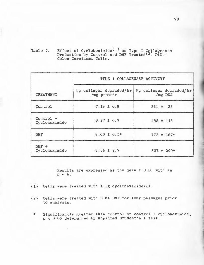
Table 7.
TREATMENT
Control
Control +
Effect of Cycloheximide(l) on Type I Collagenase Production by Control and DMF Treated(2) DLD-1 Colon Carcinoma Cells.
TYPE I COLLAGENASE ACTIVITY
76
µg collagen degraded/hr Ilg collagen degraded/hr /mg protein /mg DNA
7 .18 ± 0.8 311 ± 33
Cycloheximide 6.27 ± 0.7 458 ± 145
DMF 8.60 ± 0.5* 773 ± 167*
........
DMF + Cycloheximide 8.54 ± 2.7 867 ± 200*
Results are expressed as the mean ± S.D. with an n = 4.
(1) Cells were treated with 1 µg cycloheximide/ml.
(2) Cells were treated with 0.8% DMF for four passages prior to analysis.
* Significantly greater than control or control + cycloheximide, p < 0.05 determined by unpaired Student's t test.
....._
77
collagenolytic activity was diminished but not comnletely
abolished. Cycloheximide treatment could not inhibit the
increase in Tvpe I collagenolytic activity nroduced by
DMF treatment.
Treatment of DMF and control DLD-1 cells with
cycloheximide effected the synthesis of Type V collagen
olytic activity in a manner similar to that seen with
Type I collagenolytic activity. Increases in Type V
collagenolytic activity due to DMF were still present
when DMF and control cells were treated with cyclohexi
mide; however, the magnitude of the increase was less
(Table 8). DMF treated cells treated with cycloheximide
produced significantly more Type V collagenolytic activ-
ity than control cells treated with cycloheximide
(p < 0.05).
A similar pattern of reduction in the DMF effect
upon treatment with cycloheximide was seen with cellular
elastase activity (Table 9). DMF treated cells treated
with cycloheximide produced significantly more cellular
elastase activity than control cells treated with cyclo
heximide when the activity was expressed per unit of DNA
(p < 0.05). The increase was not significant when
expressed per unit of protein.
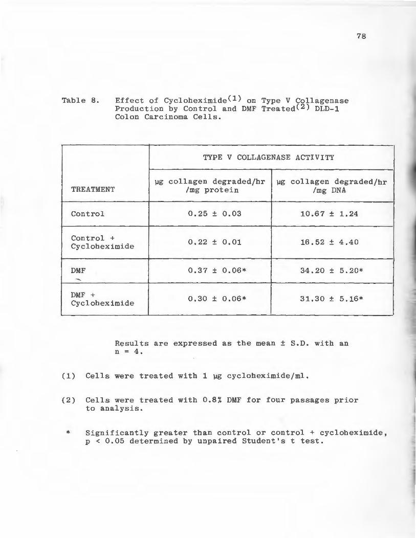
Table 8.
TREATMENT
Control
Control +
Effect of Cycloheximide(l) on Type V Collagenase Production by Control and DMF Treated(2) DLD-1 Colon Carcinoma Cells.
TYPE V COLLAGENASE ACTIVITY
78
µg collagen degraded/hr µg collagen degraded/hr /mg protein /mg DNA
0.25 ± 0.03 10. 6 7 ± 1.24
Cycloheximide 0.22 ± 0.01 16. 52 ± 4.40
DMF 0.37 ± 0.06* 34.20 ± 5.20*
---DMF + Cycloheximide 0.30 ± 0.06* 31.30 ± 5 .16*
Results are expressed as the mean ± S.D. with an n = 4.
(1) Cells were treated with 1 µg cycloheximide/ml.
(2) Cells were treated with 0.8% DMF for four passages prior to analysis.
* Significantly greater than control or control + cycloheximide, p < 0.05 determined by unpaired Student's t test.
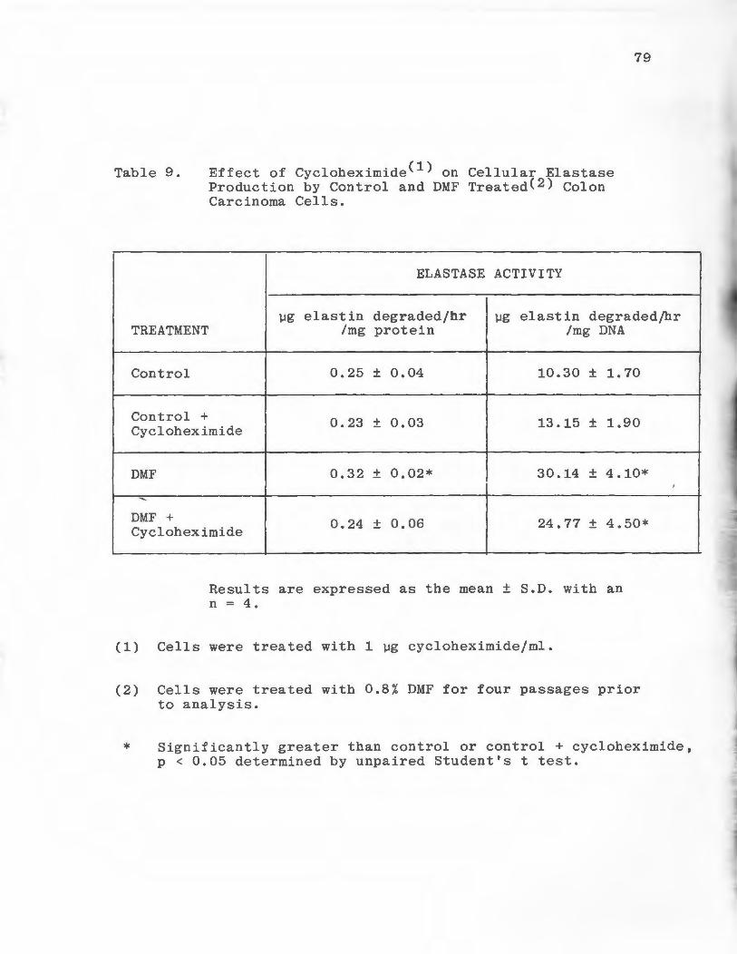
Table 9.
TREATMENT
Control
Control +
Effect of Cycloheximide(l) on Cellular Elastase Production by Control and DMF Treated(2) Colon Carcinoma Cells.
ELASTASE ACTIVITY
79
µg elastin degraded/hr µg elastin degraded/hr /mg protein /mg DNA
0.25 ± 0.04 10.30 ± 1. 70
Cycloheximide 0.23 ± 0.03 13.15 ± 1.90
DMF 0.32 ± 0.02* 30.14 ± 4.10*
......._
DMF + Cycloheximide
0.24 ± 0.06 24.77 ± 4.50*
Results are expressed as the mean ± S.D. with an n = 4.
(1) Cells were treated with 1 µg cycloheximide/ml.
(2) Cells were treated with 0.8% DMF for four passages prior to analysis.
* Significantly greater than control or control + cycloheximide, p < 0.05 determined by unpaired Student's t test.
A markedly different result due to cycloheximide
treatment was obtained when media elastase activity was
measured. Treatment of control cells with cycloheximide
resulted in a small decrease in media elastase activity
per unit of protein and a small but insignificant
80
increase in activity per unit of DNA (p < 0.05) (Table 10).
These results were analogous to those seen for Type I and
Type V collagenase and cellular elastase. However, when
DMF treated DLD-1 cells were treated with cycloheximide
media elastase activity increased above the levels seen
in cultures treated with DMF alone. These increases were
significant when the activity was expressed per unit of
protein and DNA (p < 0.05).

Table 10. Effect of Cycloheximide(l) on Media Elastase Production by Control and DMF Treated(2) DLD-1 Colon Carcinoma Cells.
ELASTASE ACTIVITY
81
µg elastin degraded/hr µg elastin degraded/hr TREATMENT /mg protein /mg DNA
Control 31.97 ± 2.8 1345 ± 108
Control + Cycloheximide 30.70 ± 11. 0 1959 ± 91 4
DMF 22.38 ± 11.9 2066 ± 1034
DMF + Cycloheximide 52.68 ± 20.6 5151 ± 814*
Results are expressed as the mean ± S.D. with an n = 4.
(1) Cells were treated with 1 µg cycloheximide/ml.
(2) Cells were treated with 0.8% DMF for four passages prior to analysis.
* Significantly greater than control + cycloheximide, p < 0.05 determined by unpaired Student's t test.
82
DISCUSSION
Type V Collagen Purification and Type V Collagenase Assay
In order to study the degradation of Type V collagen
by tumor proteases a fairly large quantity of the protein in
purified form was needed. Human placentas were a good
source of Type V collagen since they were easily obtain
able and contained fairly large quantities of collagenous
proteins. Initial attempts to purify Type V collagen
utilized the whole placenta. It was later found advan
tageous to use only the amnionic membrane as the starting
material since Type V collagen comprises a greater portion
~of the total collagen present in anmions than in whole
placentas (Sage and Bornstein, 1979). The exact molecular
configuration of Type V collagen is not established, many
preparations are composed of two different °'chains,
°' l(V) and °' 2(V) in a 2: 1 ratio (Rhodes and Miller, 1978;
Bentz et al, 1978; Burgeson et al, 1976). A third°' chain
d\3(V) has been isolated from whole placenta but is
absent from amnionic membranes (Sage and Bornstein, 1979).
The Type V collagen purified in this study was of the con
figuration cA l(V) 2 , <::\ 2(V). The molecular weights of the
~chains are in close agreement with those reported by
Rhodes and Miller (1978). The purified material was
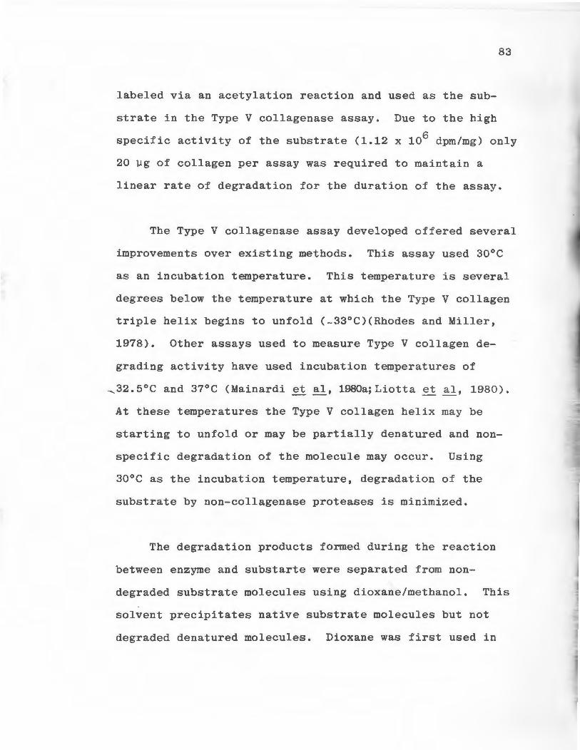
83
labeled via an acetylation reaction and used as the sub
strate in the Type V collagenase assay. Due to the high
specific activity of the substrate (1.12 x 10⁶ dpm/mg) only
20 µg of collagen per assay was required to maintain a
linear rate of degradation for the duration of the assay.
The Type V collagenase assay developed offered several
improvements over existing methods. This assay used 30°C
as an incubation temperature. This temperature is several
degrees below the temperature at which the Type V collagen
triple helix begins to unfold (-33°C)(Rhodes and Miller,
1978). Other assays used to measure Type V collagen de
grading activity have used incubation temperatures of
~32.5°C and 37°C (Mainardi et al, 1980a;Liotta et al, 1980).
At these temperatures the Type V collagen helix may be
starting to unfold or may be partially denatured and non
specific degradation of the molecule may occur. Using
30°C as the incubation temperature, degradation of the
substrate by non-collagenase proteases is minimized.
The degradation products formed during the reaction
between enzyme and substarte were separated from non
degraded substrate molecules using dioxane/methanol. This
solvent precipitates native substrate molecules but not
degraded denatured molecules. Dioxane was first used in
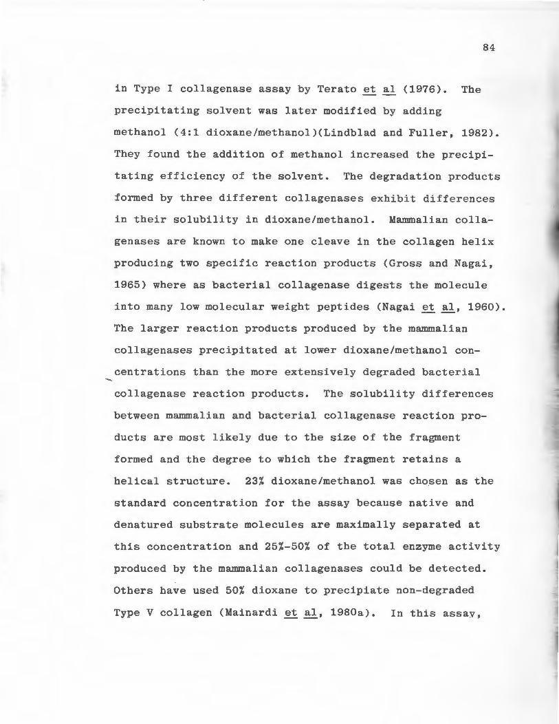
in Type I collagenase assay by Terato et al (1976). The
precipitating solvent was later modified by adding
84
methanol (4:1 dioxane/methanol)(Lindblad and Fuller, 1982).
They found the addition of methanol increased the precipi
tating efficiency of the solvent. The degradation products
formed by three different collagenases exhibit differences
in their solubility in dioxane/methanol. Mammalian colla
genases are known to make one cleave in the collagen helix
producing two specific reaction products (Gross and Nagai,
1965) where as bacterial collagenase digests the molecule
into many low molecular weight peptides (Nagai et al, 1960).
The larger reaction products produced by the mammalian
collagenases precipitated at lower dioxane/methanol con
centrations than the more extensively degraded bacterial
collagenase reaction products. The solubility differences
between mammalian and bacterial collagenase reaction pro
ducts are most likely due to the size of the fragment
formed and the degree to which the fragment retains a
helical structure. 23% dioxane/methanol was chosen as the
standard concentration for the assay because native and
denatured substrate molecules are maximally separated at
this concentration and 25%-50% of the total enzyme activity
produced by the mammalian collagenases could be detected.
Others have used 50% dioxane to precipiate non-degraded
Type V collagen (Mainardi et al, 1980a). In this assay,
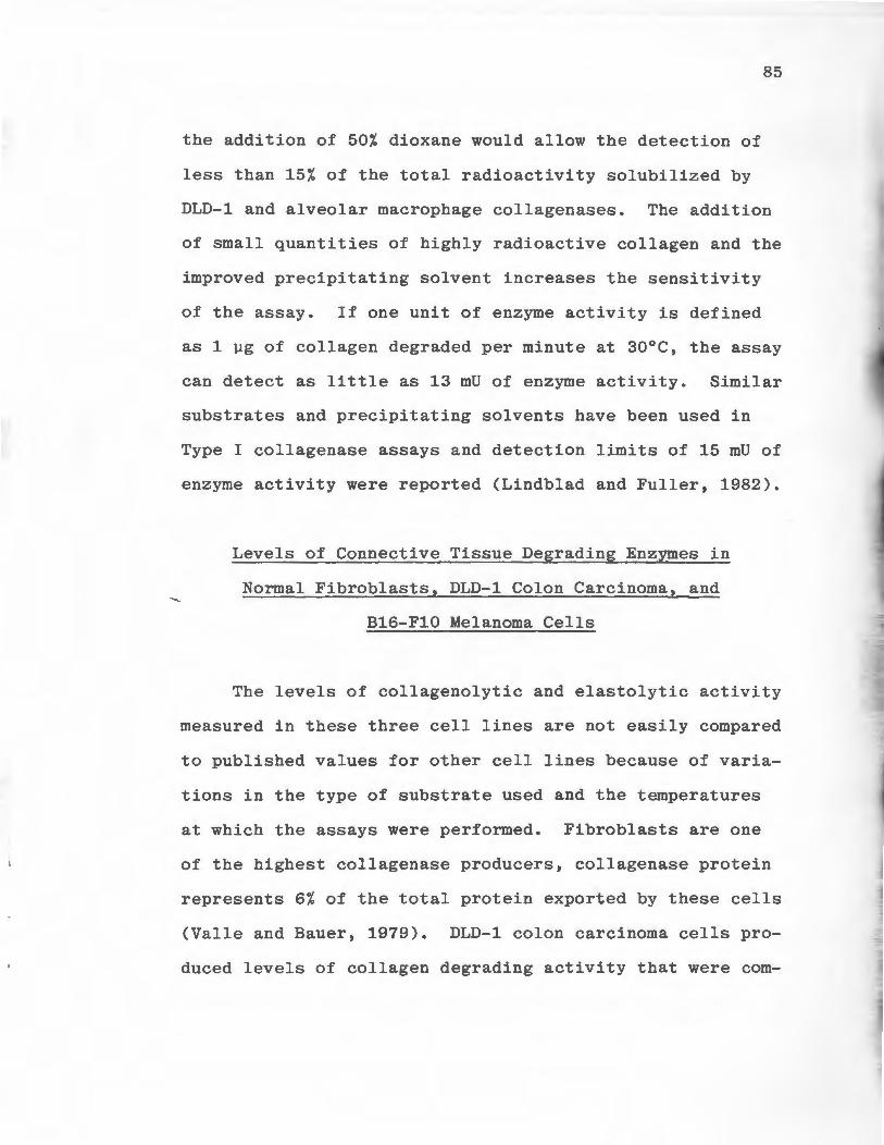
85
the addition of 50% dioxane would allow the detection of
less than 15% of the total radioactivity solubilized by
DLD-1 and alveolar macrophage collagenases. The addition
of small quantities of highly radioactive collagen and the
improved precipitating solvent increases the sensitivity
of the assay. If one unit of enzyme activity is defined
as 1 µg of collagen degraded per minute at 30°C, the assay
can detect as little as 13 mU of enzyme activity. Similar
substrates and precipitating solvents have been used in
Type I collagenase assays and detection limits of 15 mU of
enzyme activity were reported (Lindblad and Fuller, 1982).
Levels of Connective Tissue Degrading Enzymes in
Normal Fibroblasts, DLD-1 Colon Carcinoma, and
B16-F10 Melanoma Cells
The levels of collagenolytic and elastolytic activity
measured in these three cell lines are not easily compared
to published values for other cell lines because of varia
tions in the type of substrate used and the temperatures
at which the assays were performed. Fibroblasts are one
of the highest collagenase producers, collagenase protein
represents 6% of the total protein exported by these cells
(Valle and Bauer, 1979). DLD-1 colon carcinoma cells pro
duc ed levels of collagen degrading activity that were com-

86
parable to those produced by normal dermal fibroblasts.
DLD-1 cells produced considerably more collagenolytic
activity than highly metastatic B16-Fl0 murine melanoma
cells. Bl6-F10 cells are a subline of the B16 melanoma
parent line which have been selected in vivo and in vitro
for their ability to form experimental metastases (Fidler,
1973).
The location of elastase activity appears to vary
with different cell types (Werb et al, 1982). The elastin
degrading activity produced by DLD-1 cells was found almost
exclusively in the culture medium. Elastases from pan-
creatic cells and macrophages are also secreted into the
growth medium (Barrett et al, 1980; Banda and Werb, 1981).
Elastolytic activity has also been detected in the media of
cultured human breast carcinoma cells (Kao et al, 1982).
The elastin degrading activity produced by the normal fibro-
blasts and B16-F10 cells was found in both the cellular
compartment and the media, with a greater portion being
cell-associated. Hornebeck et al (1980) bas detected
elastase activity in the Triton X-100 extracts of fibro-
blasts and smooth muscle cells. Neutrophil elastase is
also known to be localized intracellularly (Werb et al,
1982).
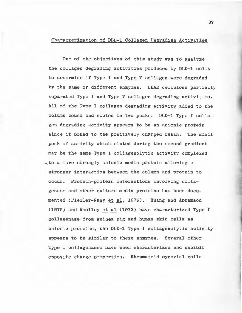
87
Characterization of DLD-1 Collagen Degrading Activities
One of the objectives of this study was to analyze
the collagen degrading activities produced by DLD-1 cells
to determine if Type I and Type V collagen were degraded
by the same or different enzymes. DEAE cellulose partially
separated Type I and Type V collagen degrading activities.
All of the Type I collagen degrading activity added to the
column bound and eluted in two peaks. DLD-1 Type I colla
gen degrading activity appears to be an anionic protein
since it bound to the positively charged resin. The small
peak of activity which eluted during the second gradient
may be the same Type I collagenolytic activity complexed
~ to a more strongly anionic media protein allowing a
stronger interaction between the column and protein to
occur. Protein-protein interactions involving colla
genase and other culture media proteins has been docu
mented (Fiedler-Nagy et al, 1976). Huang and Abramson
(1975) and Woolley et al (1973) have characterized Type I
collagenase from guinea pig and human skin cells as
anionic proteins, the DLD-1 Type I collagenolytic activity
appears to be similar to these enzymes. Several other
Type I collagenases have been characterized and exhibit
opposite charge properties. Rheumatoid synovial colla-
genase and rabbit v2 ascites tumor collagenase fail to
bind to anionic resins (Woolley et al, 1975a, McCroskery
et al, 1975). Type I collagenase from human skin fibro
blasts appears to have the capacity to bind to both
anionic and cationic resin under similar conditions
(Stricklin et al, 1977; Woolley et al, 1973).
The majority of the Type V collagen degrading
activity in DLD-1 media did not bind to the DEAE column
while a minor portion bound tightly. This protein
appears to be cationic in nature. The minor fraction of
the Type V collagenolytic activity which did bind to the
column may be a complex of enzyme and another media pro
tein which binds tightly to column. These results were
88
different from those obtained by Mainardi et al (1980a)
who separated macrophage Type I and Type V collagenolytic
activities. All the Type V collagen degrading activity
from macrophages bound to DEAE cellulose. The difference
in the physiochemical properties of these two Type V
collagenolytic activities may simply reflect a difference
in enzymes derived from macrophages and neoplastic
epithelial cells. Although the majority of Type V
collagenolytic activity from DLD-1 cells failed to bind
to the column, DEAE cellulose chromatography was an effec
tive method for partially separating Type I and Type V
89
collagen degrading activities.
The two partially separated collagenase activities
were further purified and characterized by gel filtration
chromatography on Ultrogel AcA44. The Type V collagen
degrading activity which did not bind to the DEAE column
was fractionated into two peaks of enzymatic activity by
gel filtration chromatography. The major peak of enzyme
activity from the Ultrogel chromatogram eluted in the
54,000 dalton M.W. range. A small peak of enzymatic
activity was present in the V , indicating a M.W. of 0
200,000 daltons or greater. Large M.W. peaks of enzyme
activity have been encountered during collagenase puri-
fications (Fidler-Nagy, 1977; Woolley et al, 1973; Huang
and Abramson, 1975; Brikedal-Hansen et al, 1976). These
peaks may be polymers of enzyme molecules or complexes of
enzyme molecules and fragments of the collagen substrate
(Woolley et al, 1973; Fidler-Nagy, 1977). Presently, only
three Type V collagen degrading activities have been
reported (Liotta et al, 1980; Mainardi et al, 1980a; Rich
et al, 1983), and only one study has reported a M.W.
Reticulium cell sarcoma Type V collagenolytic activity
has a M.W. of 80,000 daltons (Liotta et al, 1980). The
difference in M.W. between DLD-1 Type V collagen degrad-
ing activity and the reticulium cell sarcoma activity may
represent a difference in the enzyme produced by two
different cell types. Alternatively, the activity re-
ported by Liotta et al (1980) may be a complex of enzyme
and a collagen degradation fragment.
The major peak of Type I collagen degrading
activity from the DEAE chromatogram was also fraction-
ated into two peaks of enzymatic activity by Ultrogel
chromatography. One peak eluted in the V and may be a 0
complex of collagenase and collagen. Native collagens
have M.S.'s of approximately 300,000 daltons and would
elute from the Ultrogel column in the V • Collagenases 0
are known to bind tightly to their substrates (Stricklin
et al, 1978). The second peak of activity eluted in the
90
60,000 dalton M.W. range. This M.W. is in agreement with
M.W.'s reported for Type I collagenases isolated from two
other carcinoma cell lines. A M.W. of 63,000 daltons was
reported for a rat prostate carcinoma collagenase (Huang
et al, 1979) and 68,000 daltons has been cited as the M.W.
of Type I collagenase from a squamous cell carcinoma of
the parotid gland (Huang et al, 1982). These minor dif
ferences in M.W. may be due to the different types of gel
filtration media used in each study. A third Type I
collagenase isolated from an ascitic carcinoma cell line
91
has been characterized and a M.W. of 42,000 daltons was
reported (Wolf and Wirl, 1982). The differences in M.W.
between the ascites cell enzyme and the other Type I
collagenases may be due to the presence of substrate
fragments complexed with the enzyme or the same colla-
genases originating from different species or tissues may
simply be heterogeneous.
The third pool of collagen degrading activity gen-
erated by DEAE cellulose was fractionated into three peaks
of activity by gel filtration chromatography. A high M.W.
peak of activity was found in the V and contained both 0
Type I and Type V collagenolytic activities. As in the
two previous gel filtration chromatograms this peak was
probably an enzyme-substrate complex. A peak of Type V
collagenolytic activity eluted at a volume corresponding
to 80,000 daltons. Liotta et al (1980) has also reported
80,000 daltons as the M.W. of a reticulium cell sarcoma
Type V collagenolytic activity. The third peak of activity
generated by Ultrogel chromatography had a M.W. of 35,000
daltons and contained predominately Type I collagenolytic
activity. This activity has a M.W. similar to Type I
collagenase purified from rabbit v2 ascites tumor homo
genates (McCroskery et al, 1975).

......._
92
Type I collagenase from cultured human skin fibro
blasts has been extensively characterized and two species
of enzyme of different M.W. (45,000 and 50,000 daltons)
have been separated (Stricklin et al, 1977; Stricklin et
al, · 1978). The two enzyme were immunologically similar as
determined by immunodiffusion. They were also found to
have similar amino acid compositions, cyanogen bromide
peptide analysis revealed 9 peptides from each enzyme, 8
of which were common to both activities. Trypsinization
could not convert the higher M.W. enzyme into the smaller
one, the two proteins were considered to be different
species of the same enzyme. DLD-1 colon carcinoma cells
may also synthesize two different Type I and Type V colla
gen degrading activities. Since these activi t i e s are not
highly purified it is possible that the large M.W. forms
of each collagenase may be a complex of enzyme and sub
strate fragment. The two types of chromatography used in
this study were adequate methods for partially separating
DLD-1 Type I and Type V collagenolytic activities and
estimating their M.W. However, these methods were unable
to completely separate the enzymes from a large M.W. pro
tein, mostly likely their substrate. The complete separa
tion of the enzymes from their substrates may require more
extreme purification conditions. The addition of urea or
Triton X-100 to the buffer systems or heating the enzyme
93
containing sample to 60°C prior to subsequent purifica-
tion steps have all been used to completely dissociate
collagenases from their substrates (Harris and Vater,
1982).
Type I and Type V collagenolytic activities from
DLD-1 cells were found to have similar inhibition pro-
files. The Type V collagen degrading activities reported
by Mainardi et al (1980a) and Liotta et al (1980) were
characterized as metalloproteinases and had no serine pro-
tease activity. The DLD-1 Type V collagen degrading
activity can be similarly characterized since it was
inhibited by EDTA but not PMSF. All Type I collagenases
have been characterized as metalloproteinases and are
uneffected by serine protease inhibitors (Harris and
Vater, 1982). The DLD-1 Type I collagenolytic activity
is predominately a metalloproteinase. A non-collagenase
enzyme having serine protease activity may also be pre-
sent in the crude collagenase preparation. This activity
could act on the substrate following the initial cleavage
of the triple helix by the collagenolytic metalloprote-
inaseactivity. Collagenase from a rabbit v2 ascites cell
carcinoma was inhibited by DTT (McCroskery et al, 1975) as
were both Types I and V collagen degrading activities from
DLD-1 cells. These enzymes may require intact disulfide
bond for maximal activity since the reducing agent (DTT)
diminished enzymatic activity.
Effects of DMF on DLD-1 Colon Carcinoma Cells
94
The effects of DMF on the turmorigenicity, morphology
and in vitro growth properties of DLD-1 colon carcinoma
cells has been examined (Dexter et al, 1979; Dexter and
Hager, 1980). In vitro DMF reduces the growth rate of
DLD-1 cells and causes the cells to enlarge (Dexter et al,
1979). DMF treated cells appear less round and more
"leafy" than untreated cells. Concommitant with these
morphological changes phase optic microscopy has revealed
~that DMF treated cells have a higher cytoplasmic to nuclear
ratio (Dexter et al, 1979). In this study cells treated
with DMF contained more protein and RNA per cell and less
DNA per cell than control cells. This finding is in agree
ment with those obtained by phase optic microscopy.
Treatement of DLD-1 cells with DMF resulted in the
increased production of Type I and Type V collagenase and
cell-associated elastase activities. It is unlikely that
DMF had a selective effect stimulating the production of
these particular proteases. The increases in enzymatic
.......
95
activity probably resulted from a non-specific increase in
the rate of protein synthesis. This hypothesis, however,
cannot explain the decrease in media elastase activity seen
in DMF treated cultures. This decrease was not statisti-
cally significant and the value may be erroneous since the
corresponding standard deviation was very large.
To determine if the effect of DMF on protease produc
tion was the result of an increased rate of protein syn
thesis, the rate of incorporation of 3H-glycine into pro
tein was measured. After three hours of growth in label
containing media, the DMF cultures appeared to be incor
porating label slower than control cultures, although the
decrease was significant only when the data were expressed
per unit of DNA. After six hours of growth, the DMF
cultures were synthesizing protein faster than controls.
This increased rate of protein synthesis could explain the
increased levels of Type I and Type V collagenases and
cellular elastase activities present in DMF treated cul
tures. The decreased rate of incorporation of 3H-glycine
seen in DMF treated cells at the three hour point may not
be a true indicator of decreased macromolecule synthesis,
the decrease could have resulted from an initial lag in
the rate of glycine penetration into the cells. DMF treat
ment may have altered the cell membrane configuration
96
resulting in a decreased rate of amino acid uptake. After
longer growth periods in label-containing media sufficient
quantities of 3H-glycine may have entered the DMF treated
cells and allowed their rate of protein synthesis to exceed
controls.
If the DMF effect on enzyme production was due to
increased enzyme biosynthesis, blockade of protein syn
thesis with cycloheximide should inhibit the effect. Con
trol and DMF treated cultures were treated with 1 µg cyclo
heximide/ml, a dose which inhibited collagenase production
by endotoxin-activated macrophages (Wahl et al, 1974). The
effect of DMF on Type I and Type V collagenolytic activ-
~i ties was still apparent however, in the presence of cyclo
heximide. This result indicated that DMF could produce
its effect independent of protein synthesis. It has been
reported that collagenases are synthesized and stored
intracellularly as zymogens (Harper et al, 1971). The DMF
treated cells may have synthesized and stored intracellu
larly greater quantities of these enzymes prior to the
inhibition of protein synthesis. The increased levels of
these enzymes seen in DMF treated cells treated with cyclo
heximide could have been the result of the release of
greater quantities of stored enzyme.
97
When DMF treated cells were exposed to cycloheximide
for 24 hours, the cells secreted large amounts of elastase
into the media. Control cells did not respond to cyclo
heximide in the same manner. There was no significant
difference in media elastase levels in control and control
cells treated with cycloheximide, this was analogous to the
effects on collagenolytic activities. The elastase
activity secreated by DMF treated cells following cyclo
heximide treatement most likely originated from a pre
existing intracellular store of enzyme. Cycloheximide
has been shown to effect the turnover of tyrosine trans
aminase in cultured liver cells leading to increased
levels of transaminase activity (Kenney, 1967; Hershko and
Tomkins, 1971). It was postulated that the s ynthesis of
proteases responsible for the degradation of intracellular
proteins are more sensitive to cycloheximide treatment than
the synthesis of tyrosine transaminase (Kenney , 1967).
DLD-1 elastase levels may have been increased due to
decreased intracellular degradation of the enzyme.
CONCLUSIONS
1. Type V collagen was purified from human amnions,
labeled and used as a substrate in an improved
assay for measuring Type V collagen degrading
activity. Non-degraded substrate molecules were
separated from degraded substrate fragments by
precipitation with 23% dioxane/methanol.
2. DLD-1 colon carcinoma cells, B16-Fl0 murine
melanoma cells, and normal dermal fibroblasts
produced proteases which could degrade Type I
and Type V collagen and elastin.
3. Type I and Type V collagen degrading activities
secreted by DLD-1 cells were characterized as
metalloproteinases. These activities were
partially separated using DEAE cellulose chroma
tography.
4. Two forms of DLD-1 Type I collagenolytic activity
were partially purified and had molecular weights
of 60,000 and 35,000 daltons.
98
5. Two forms of DLD-1 Type V collagenolytic activity
were partially purified and had molecular weights
of 80,000 and 54,000 daltons.
6. DMF treatment increased the production of Type I
and Type V collagenolytic activities and cell
associated elastinolytic activity by DLD-1 cells.
7. DMF treatment increased the cellular protein and
RNA content and decreased the DNA content of DLD-1
cells.
99
REFERENCES
Aggeler, J., Enguall, E., and Werb, Z.: An Irreversible Tissue Inhibitor of Collagenase in Human Amniotic Fluid: Characterization and Separation from Fibronectin. Biochemical and Biophysical Research Communications 100: 1195-1201, 1981.
100
Anwar, R. A., Gerber, G. E., and Baie, K. M.: In Advances in Experimental Medicine and Biology: Protein Crosslinking. pp 709-727, ed. Mendel Friedman, Plenum Press, N. Y. 1977.
Banda, M. J., Clark, E. J., and Werb, Z.: Limited Proteolysis by Macrophage Elastase Inactivates Human°' 1-Proteinase Inhibitor. Journal of Experimental Medicine 152: 1563-1570, 1980.
Banda, M. J., and Werb, Z.: Mouse Macrophage Elastase. Biochemical Journal 193: 589-605, 1981.
Barrett, A. J., and McDonald, J. K.: Mammalian Protease, A Glossary and Bibliography, Vol. 1, Endopeptidase. New York, Academic Press, 1980.
Barsky, S. H., Rao, C. N., Grotendorst, G. R., and Liotta, ~L. A.: Increased Content of Type V Collagen in Desmo
plasia of Human Breast Carcinoma. American Journal of Pathology 108: 276-283, 1982.
Bauer, E. A., Stricklin, G. P., Jeffrey, J. J., and Eisen, A. Z.: Collagenase Production by Human Skin Fibroblasts. Biochemical and Biophysical Research Communications 64: 150-157, 1975
Bentz, H., Bachinger, H.P., Glanville, R., and Kuhn, K.: Physical Evidence for the Assembly of A and B Chains of Human Placental Collagen in a Single Triple Helix. European Journal of Biochemistry, 92: 563-567, 1978
Birbeck, M. S. C., and Wheatley, O. N.: An Electron Microscopic Study of the Invasion of Ascites Tumor Cells Into the Abdominal Wall. Cancer Research 25: 490-497, 1965.
Birkedal-Hensen, H., Cobb, C. M., Taylor, R. E., and Fullmer, H. M.: Activation of Fibroblast Procollagenase by Mast Cell Proteases. Biochemica et Biophysica Acta 438: 273-286, 1976.
Biswas, C.: Tumor Cell Stimulation of Collagenase Production by Fibroblasts. Biochemical and Biophysical Research Communications 109: 1026-1034, 1982.
101
Black, P.H.: Invasion and Metastasis. 156-161, 1980.
Advances in Cancer Research 32:
Bornstein, P., and Sage, H.: Structurally Distinct Collagen Types. Ann. Rev. Biochem. 49: 957-1003, 1980.
Boudillon, M. C., Brechemier, D., Blass, N., Deroutte, J.C., Hornbeck, W., and Robert, L.: Elastase-Like Enzymes in Skin Fibroblasts and Rat Smooth Muscle Cells, Cell Biology Int. Rep. !: 313-316, 1980.
Broth, W., Menzel, E. J., Salzer, M., and Steffen, C.: Human Serum Inhibitors of Collagenases as Revealed by Preparative Isoelectric Focusing. Clinica Chemica Acta 117: 219-225, 1981.
Brunson, K. W., Beattie, G., and Nicholson, G. L.: Selection and Altered Properties of Brain-Colonizing Metastatic Melanoma. Nature 272: 543-545, 1978.
Burgeson, R. E., Adki, D. W.: Fetal Membrane Collagen Alpha Chains.
~2579-2583, 1976.
F. A. Kaitila, I. I., and Hollister, Collagens: Identification of Two New
Proc. Natl. Acad. Sci. U.S.A. 73:
Burgeson, R. E., and Hollister, D. W.: Collagen Heterogeneity in Human Cartilage: Identification of Several New Collagen Chains. Biochemical and Biophysical Research Communications 87: 1124-1131, 1979.
Burgeson, R. E.: Genetic Heterogeneity of Collagens. Journal of Investigative Dermatology 79: 25s-30s, 1982.
Cawston, T. E., and Tyler, J. A.: Purification of Pig Synovial Collagenase to High Specific Activity. Biochemical Journal 183: 674-656, 1979.
Cawston, T. E., Galloway, W. A., Mercer, E., Murphy, G., and Reynolds, J. J.: Purification of Rabbit Bone Inhibitor of Collagenase. Biochemical Journal 195: 159-165, 1981.
Christner, P., Damato, D., Reinhart, M., and Abrams, W.: Purification of Human Neutrophil Collagenase and Production of a Monospecific Antiserum. Biochemistry 21: 6005-6011, 1982. ~
102
Chung, E., Keele, E. M., and Miller, E. J.: Isolation and Characterization of the Cyanogen Bromide Peptides from the o<1(III) Chain of Human Collagen. Biochemistry 13: 3459-3464, 1974. ~
Chung, E. R., Rhodes, K., and Miller, E. J.: Isolation of Three Collagenous Components of Probable Basement Membrane Origin from Several Tissues. Biochem. Biophys. Res. Com.~. 71: 1167-1174, 1976.
Cox, D. W., and Huber, D.: Rheumatoid Arthritis and Alpha-1-Antitrypsin, Lancet 1: 1216-1217, 1976.
Dayer, J. M., Breard, J., Chess, L., and Krane, S. M.: Participation of Monocyte-Macrophage and Lymphocytes in the Production of a Factor That Simulates Collagenase and Prostaglandin Release by Rheumatoid Synovial Cells. Journal Clinical Investigation 64: 1386-1392, 1979.
Dehm, P., and Kefalides, N. of Lens Basement Membrane. 253: 6680-6686, 1978.
A.: The Collagenous Component Journal of Biological Chemistry
Dexter, D. L.: N, N-Dimethylformamide-Induced Morphological Differentiation and Reduction of Tumorigenicity in Cultured Mouse Rhabdomyosarcoma Cells. Cancer Research 37: 3136-3140, 1977.
Dexter, D. L., Barbosa, J. A., and Calabresi, P.: N, NDimethylformamide-Induced Alteration of Cell Culture Characteristics and Loss of Tumorigenicity in Cultured Human Colon Carcinoma Cells. Cancer Research 39: 1020-1025, 1979.
Dexter, D. L., Hager, J. C.: Maturation-Induction of Tumor Cells Using a Human Colon Carcinoma Model. Cancer 45: 1178-1184, 1980.
Dobrossy, L., Pavelic, Z. P., and Bernacki, R. J.: A Correlation Between Cell Surface Sialyltransferase, Sialic Acid, and Glycosidase Activities and the Implantability of B16 Murine Melanoma. Cancer Research 41: 2262-2266, 1981.
Dresden, M. H., Heilman, S. A., and Schmidt, J. D.: Collagenolytic Enzymes in Human Neoplasms. Cancer Reserach 32: 993-996, 1972.
Eaves, G.: The Invasive Growth of Malignant Tumors as a Purely Mechanical Process. Journal of Pathology 109: 233-237, 1973.
103
Eeckhout, V., and Vaes, G.: Further Studies on the Activation of Procollagenase, The Latent Precursor of Bone Collagenase. Biochemical Journal 166: 21-31, 1977.
Eriksson, S.: Studies inoC1-Antitrypsin De f iciency. Medical Scandanavia 177: 432-435, 1979.
Acta
Evanson, M., Jeffrey, J. J., and Krane, S. M.: Studies on Collagenase from Rheumatoid Synovium in Tissue Culture. Journal of Clinical Investigation 47: 2639-2651, 1968.
Fahrney, D. E., and Gold, A.: Sulfonyl Fluorides as Inhibitors of Esterases. I. Rates of Reaction with Acetycholinestases, oi(-Chymotrypsin and Trypsin. Journal of the American Chemical Society 85: 997-1000, 1963.
Fessler, L. I., Morris, N. P., and Fessler, J. H.: Procollagen: Biological Scission of Amino and Carboxyl Extension Peptides. Proc. Natl. Acad. Sci. U.S.A. 72: 4905-4904, 1975.
Fessler, L. I., Shigaki, N., and Fessler, J.: Isolation of New Procollagen Type V From Chick Embryo Tendon. Federation Proceedings 42: 1886, 1983.
Fidler, I. J.: Selection of Successive Tumor Lines for ~ Matastasis. Nature New Biology 242: 148-1 49, 1973.
Fidler, I. J., and Nicoloson, G. L.: Organ Se l e ctivity for Implantation Survival and Growth of B16 Melanoma Variant Tumor Lines. Journal Natl. Cancer Inst. 57: 1199-1202, 1976.
Fidler, I. J., Gersten, D. M., and Hart, I. R.: The Biology of Cancer Invasion and Metastasis. Advances in Cancer Research 28: 149-150, 1978.
Fiedler-Nagy, C., Coffey, J. W., and Salvador, R. A.: Factors Influencing the Apparent Molecular Weight of Collagenase Produced by Human-Skin Explants. European Journal of Biochemistry 76: 291-297, 1977.
Foster, J. A., Bruenger, E., Gray, W.R., and Sandberg, L.B.: Isolation and Amino Acid Sequences of Tropoelastin Peptides. Journal of Biological Chemistry 248: 2876-2879, 1973.
Francis, G., John, R., and Thomas, J.: Biosynthetic Pathway of Desmosines in Elastin. Biochemical Journal 136: 45-55, 1973.
104
Friend, C. W., Scher, W., Holland, J. G., and Sato, T.: Hemoglobin Synthesis in Murine Virus Induced Leukemic Cell in vitro: Stimulation of Erythroid Differentiation by Dimethylsulfoxide. Proc. Natl. Acad. Sci. U.S.A. 68: 378-382, 1971.
Fujii, T., and Kuhn, K.: Isolation and Characterization of Pepsin Treated Type III Collagen from Calf Skin. Physiological Chemistry 356: 1793-1801, 1975.
Furuta, D. K., and Miller, E. J.: Isolation of a Unique Collagenous Fraction From Limited Pepsin Digests of Human Placental Tissue. Journal of Biological Chemistry 255: 290-295, 1980.
Gay, S., Rhodes, R. K., Gay, R. E., and Miller, E. J.: Collagen Molecules Comprised ofo(l(V) Chains (B-Chains): An Apparent Localization in the Exocytoskeleton. Collagen Research 1: 53-58, 1981.
Gillet, C., Eeckhout, Y., and Vaes, G.: Purification of Procollagenase and Collagenase by Affinity Chromatography on Sepharose-Collagen. FEBS Letters 74: 126-128, 1977.
Gisslow, M. T., and McBride, B. C.: A Rapid Sensitive Collagenase Assay. Analytical Biochemistry 68: 70-78, 1975.
Glanville, R. W., and Rauter, A.: Pepsin Fragments of Human Placental Basement-Membrane Collagens Showing Interrupted Triple-Helical Amino Acid Sequences. Hoppe-Seyler's Physiol. Chem. 362: 943-951, 1981.
Gross, J., and Nagai, Y.: Specific Degradation of the Collagen Molecule by Tadpole Collagenolytic Enzyme. Proc. Natl. Acad. Sci. U.S.A. 54: 1197-1204, 1965.
Gross, J., Harper, E., Harris, E. D., McCroskery, P.A., Highberger, J. H., Corbett, C., and Kang, A.H.: Animal Collagenases: Specificity of Action, and Structures of the Substrate Cleavage Site. Biochemical and Biophysical Research Communications 61: 605-612, 1974.
Hager, J. C., Gold, D. V., Barbosa, J. A., Fligiel, Z., Miller, F., and Dexter, D. L.: N~, N~DimethylforrnamideInduced Modulation of Organ- and Tumor-Associated Markers in Cultured Htiman Colon Carcinoma Cells. Journal of Natl. Cancer Inst. 64: 439-446, 1980.
Harper, E., Bloch, K. J., and Gross, J.: The Zymogen of Tadpole Collagenase. Biochemistry 10: 3035-3041, 1971.
105
Harper, E.: Collagenases. Ann. Rev. Biochem. 49: 1063-1078, 1980.
Harris, E. D., and Vater, C. A.: Vertebrate Collagenases. Methods in Enzymology 82: 443-452, 1982.
Hashimoto, K., Yamanishi, Y., Maeyens, E., Dabbous, M. K., and Kanzaki, T.: Collagenolytic Activities of Squamous Cell Carcinoma of the Skin. Cancer Research 33: 2790-2801, 1973.
Hershko, A., and Tomkins, G. M.: Studies on the Degradation of Tyrosine Aminotransferase in Hepatoma Cells in Culture. Journal of Biological Chemistry 246: 710-714, 197lb.
Hiatt, H. H., Watson, J. O., and Winsten, J. A.: (eds) Origins of Human Cancer Proliferation. Vol. 4, Cold Springs Harbor Laboratory, N. Y., 1977.
Hibbs, J.B., Taintor, R. R., Chapman, H. A., and Weinberg, J. B.: Macrophage Tumor Killing: Influence of the Local Environment. Science 197: 279-282, 1977.
~Hibbs, M. S., Mainardi, C. L., and Kang, A. H.: TypeSpecific Collagen Degradation by Eosinophils. Biochemical Journal 207: 621-624, 1982.
Hornebeck, W., Adnet, J. J., and Robert, L.: Age Dependent Variation of Elastin and Elastase in Aorta and Human Breast Cancerns. Experimental Gerontology 13: 293-298, 1978.
Hornebeck, W., Starkey, P. M., Gordon, J. L., Legrand, Y., Pighaud, G., Robert, L., Caen, J.P., Ehrlich, H.P., and Barrett, A. J.: The Elastase-Like Enzymes of Platelets. Thrombosis and Haemostasis 42: 1681-1685, 1980.
Hornebeck, W., Bellon, G., Brechemier, D., Godeau, G., and Robert, L.: Control of Elastic Tissue Destruction by Elastase Inhibitors, in Connective Tissue Research Chemistry, Biology, and Physiology, Deyl, A., and Adam, M. (eds): Alan R. Liss, Inc., New York, pp 233-246, 1981.
Horwitz, A. L., Kelman, J. A., and Crystal, R. G.: Activation of Alveolar Macrophage Collagenase by a Neutral Protienase Secreted by the Same Cell. Nature 264: 772-773, 1976.
106
Horwitz, A. L., Hance, A. J., and Crystal, R. G.: Granulocyte Collagenase: Selective Digestion of Type I Relative to Type III Collagen. Proc. Natl. Acad. Sci. U.S.A. 74: 897-901, 1977.
Huang, C. C., and Abramson, M.: Purification and Characterization of Collagenase from Guinea Pig Skin. Biochimica et Biophysica Acta 384: 484-492, 1975.
Huang, C. c., Wu, C.H., and Abramson, M.: Collagenase Activity in Cultures of Rat Prostate Carcinoma. Biochim. et Biophys. Acta 570: 149-156, 1979.
Huang, C. C., Blitzer, A., and Abramson, ~.: Collagenase Activity in Squamous Cell Carcinoma of the Human Parotid Gland. Ann. Oto. Rhin. Laryn. 91: 156-159, 1982.
Ishikawa, T., and Nimni, M. E.: A Modified Collagenase Assay Method Based on the Use of p-Dioxane. Analytical Biochemistry 92: 136-143, 1979.
Johnson-Muller, B., and Gross, J.: Regulation of Corneal Collagenase Production: Epithelial-Stromal Cell Interactions. Proc. Natl. Acad. Sci. U.S.A. 75: 4417-4421, 1978.
Jones, P. A., and DeClerck, Y. A.: Destruction of Extra~cellular Matrices Containing Glycoproteins, El astin, and
Collagen by Metastatic Human Tumor Cells. Can cer Reserach 40: 3222-3227, 1980.
Kao, R. T., Wong, M., and Stern, R.: Elastin Degradation by Proteases from Cultured Human Breast Cancer Cells. Biochemical and Biophysical Research Communciations 105: 383-389, 1982.
Keeley, F. W., and Partridge, S. M.: Amino Acid Composition and Calcification of Human Aortic Elastin. Atherosclerosis 19: 287-296, 1974.
Kenney, F. T.: Turnover of Rat Liver Tyrosine Transaminase: Stabilization After Inhibition of Protein Synthesis. Science 156: 525-528, 1967.
Kinsey, stasis.
R.: An Experimental Study of Preferential MetaCancer 13: 647-653, 1960.
Kivirikko, K. I., and Myllyla, L.: Collagen Gl ycosyltransferases: Int. Review of Connective Tissue. Research 8: 23-72, 1979.
107
Kreider, J. W., Rosethal, M., and Lengle, N.: Cyclic Adenosine 3~, 5~ Momophosphate in the Control of Melanoma Cell Replication and Differentiation. Journal of the National Cancer Institute 50: 555-558, 1973.
Kuettner, K. W., Hiti, J., Eisenstein, R., and Harper, E.: Collagenase Inhibition by Cationic Proteins Derived from Cartilage and Aorta. Biochemical and Biophysical Research Communications 72: 40-46, 1976.
Kumamoto, C., and Fessler, J. H.: Propeptides of Procollagen V (A, B) in Chick Embryo Crop. Journal of Biological Chemistry 256: 7053-7058, 1981.
Lapiere, C. M., Lenaers, A., and Kohn, L. D.: Procollagen Peptidase: An Enzyme Excising the Coordination Peptides of Procollagen. Proc. Natl. Acad. Sci., U.S.A. 68: 3054-3058, 1971.
Latner, A. L., Longstaff, E., and Pradham, K.: Inhibition of In Vitro Invasion by Protease Inhibitors. British J. Cancer 27: 460-464, 1973.
Lent, R. W., Smith, B. M., Salcedo, L. L., Faris, B., and Frazblau, C.: Studies on the Reduction of Elastin II. Evidence for the Presence of o-Amino-Adipic Semialdehyde
~and Its Aldol Condensation Product. Biochemistry 8: 2837-2845, 1969.
Lentner, M.: Introduction to Applied Statistics, 2nd Ed. Prindle, Weber and Schmidt, Inc., Boston, MA, 1976.
Lindblad, W. J., and Fuller, G. C.: An Improved Assay of Mammalian Collagenase Activity, and Its Use to Determine Hepatic Extracellular Matrix Susceptibility to Degradation. Clinical Chemistry 28: 2134-2138, 1982.
Liotta, L. A., Abe, S., Robey, P. G., and Martin, G. R.: Preferential Digestion of Basement Membrane Collagen by an Enzyme Derived from a Metastatic Murine Tumor. Proc. Natl. Acad. Sci. U.S.A. 76: 2268-2272, 1979.
Liotta, L.A., Trygguason, K., Garbisa, S., Robey, P. G., and Abe, S.: Partial Purification and Characterization of a Neutral Protease Which Cleaves Type IV Collagen. Biochemistry 20: 100-104, 1981a.
Liotta, L. A., Lanzer, W. L., and Garbisa, S.: Identification of a Type V Collagenolytic Enzyme. Biochemical and Biophysical Research Communications 98: 184-190, 1981b.
108
Lowry, O. H., Rosebrough, N. J., Farr, A. L., and Randall, R. J.: Protein Measurement with the Folin Phenol Reagent. Journal of Biological Chemistry 193: 265-275, 1951.
Lukens, L. N.: Time of Occurrence of Disulfide Linking Between Procollagen Chains. Journal of Biological Chemistry 251: 3530-3538, 1976.
Lundmark, C.: Breast Cancer and Elastosis. Cancer 30: 1195-1201, 1972.
Macartney, H. W., and Tschesche, H.: Latent Collagenase From Human Polymorphonuclear Leukocytes and Activation to Collagenase by Removal of an Inhibitor. FEBS Letters 119: 327-332, 1980.
Madri, J. A., and Furthmayr, H.: Collagen Polymorphism in the Lung. Human Pathology 11: 353-366, 1980.
Mainardi, C. L., Seyer, J.M., and Kang, A. H.: TypeSpecific Collagenolysis: A Type V Collagen-Degrading Enzyme from Macrophages. Biochemical and Biophysical Research Communciations 97: 1108-1115, 1980a.
Mainardi, C. L., Dixit, S. N., and Kang, A. H.: Degradation of Type IV (Basement Membrane) Collagen by a Proteinase Isolated From Human Polymorphonuclear Leukocyt e Granules.
~Journal of Biological Chemistry 255: 5435-5441, 1980b.
Market, C. L., Neoplasia: A Disease of Cell Differentiation. Cancer Research 28: 1908-1914, 1968.
Marshall, T. H., Whitaker, J. R., and Bender, M. L.: Procine Elastase I. The Presence of Tyrosinate Splitting Enzymes as Impurities in Elastase Preparations. Biochemistry 8: 4665-4677, 1969.
McCroskery, P. A., Richards, J. F., and Harris, E. D.: Purification and Characterization of a Collagenase Extracted from Rabbit Tumors. Biochemical Journal 152: 131-142, 1975.
Murphy, G., Cawston, T. E., and Reynolds, J. J.: An Inhibitor of Collagenase from Human Amniotic Fluid. Biochemical Journal 195: 167-170, 1981a.
Murphy, G., McGuire, M. B., Russell, R. G. G., and Reynolds, J. J.: Characterization of Collagenase, Other MetalloProteinases and an Inhibitor (TIMP) Produced by Human Synouium and Cartilage in Culture. Clinical Sciences 61: 711-716, 198lb.
Nagai, Y.: Vertebrate Collagenase: Further Characterization and the Significance of Its Latent Form In Vivo. Molecular Cellular Biochemistry != 137-145, 1973.~~
Nagai, Y., Sakakibara, S., Noda, H., and Akabori, S.: Hydrolysis of Synthetic Peptides by Collagenase. Biochemica et Biophysica Acta 37: 567-569, 1960.
109
Nagai, Y., Shinkai, H., and Ninomiya, Y.: The Release of Collagenase Inhibitors from Procollagen Additional Peptides by Pepsin Treatment. Proc. Japan Acad. 54: 140-144, 1978.
Neville, D. M.: Molecular Weight Determination of Protein Dodecyl Sulfate Complexes by Gel Electrophoresis in a Discontinuous Buffer System. Journal of Biological Chemistry 246: 6328-6334, 1971.
Nicolson, G. L., and Winkehake, J. L.: Organ Specificity of Blood-Borne Tumour Metastasis Determined by Cell Adhesion? Nature 255: 230-232, 1975.
O'Grady, R. L., Upfold, L. I., and Stephens, R. W.: Rat Mannary Carcinoma Cells Secrete Active Collagenase and Activate Latent Enzyme in the Stroma Via Plasminogen Activator. International Journal of Cancer 28: 509-515, 1981.
O'Grady, R. L., Harrop, P. J., and Cameron, O. A.: Collagenolytic Activity by Malignant Tumors. Pathology 14: 135-138, 1982.
Ohlsson, K., and Olsson, I.: The Nuetral Proteases of Human Granulocytes. European Journal of Biochemistry 42: 519-527, 1974.
Old. L. J., Benacerraf, B., and Stockert, E.: The Role System in the Host Reaction 21: 1281-1300, 1961.
Ossowski, L., and Reich, E.: tative Study of Metastasis. 1980.
Clarke, D. A., Carswell, E. A., of the Reticuloendothelial to Neoplasia. Cancer Research
Experimental Model for QuantiCancer Research 40: 2300-2309,
Parks, R. C.: Organ-Specific Metastasis of a Transplantable Reticulum Cell Sarcoma. Journal of the National Cancer Institute 52: 971-973, 1974.
Pettigrew, D. W., Wang, H. M., Sadek, J., and Brunette, D. M.: Synthesis of Collagenolytic Enzymes and Their Inhibitors by Gingival Tissue In Vitro. Journal of Peridontal Research 16: 637-645, 1981.
110
Pinell, S. R., and Martin, G. R.: The Cross-Linking of Collagen and Elastin: Enzymatic Conversion of Lysine in Peptide Linkage to o-Amino-Adipic-Semialdehyde (Allysine) by an Extract from Bone. Proc. Natl. Acad. Sci. U.S.A. 61: 708-716, 1968.
Poste, G., and Fidler, I.: The Pathogenesis of Cancer Metastasis. Nature 283: 139-146, 1980.
Prasad, A. S., DuMouchelle, E., Koniuch, D., and Oberlas, D.: A Simple Fluorometric Method for the Determination of RNA and DNA in Tissues. Journal of Lab. Clincal Med. 80: 598-602, 1972.
Prockop, D. J., and Juva, K.: Synthesis of Hydroxyproline In Vitro by the Hydroxylation of Proline in a Precursor of Collagen. Proc. Natl. Acad. Sci. U.S.A. 53: 661-668, 1965.
Prockop, D. J., Kivirikko, K. I., Tuderman, L., and Guzman, N. A.: The Biosynthesis of Collagen and Its Disorders. New England Journal of Medicine 301: 13-85, 1979.
Proctor, J. J.: Rat Sarcoma Model Supports Both "Soil Seed" and "Mechanical" Theories of Metastatic Spread. British Journal of Cancer 34: 651-654, 1976.
Puzas, J.E., and Brand, J. S.: Parathyroid Hormone Stimu~1ation of Collagenase Secretion by Isolated Bone Cells.
Endocrinology 104: 559-562, 1979.
Rabson, A. S., Stern, R., Tralka, T. S., Costa, J., and Wilczek, J.: Hexamethylene Bisacetamide Induces Morphologic Changes and Increased Synthesis of Procollagen in Cell Line from Glioblastoma Multiforme. Proc. Natl. Acad. Sci. U.S.A. 74: 5060-5064, 1977.
Rabinowitz, Z., and Sachs, L.: Properties in Transformed Cells.
Control of the Reversion of Nature 225: 136-139, 1970.
Raz, A., McLellan, W. L., Hart, I. R., Bucana, C. D., Hoyer, L. C., Sela, B. A., Dragsten, P., and Fidler, I. J.: Cell Surface Properties of B16 Melanoma Variants with Differing Metastatic Potential. Cancer Research 40: 1645-1651, 1980.
Reese, C. A., and Mayne, R.: Minor Collagens of Chicken Hyaline Cartilage. Biochemistry 20: 918-924, 1981.
Rhodes, R. K., and Miller, R. J.: Physiochemical Characterization and Molecular Organization of the Collagen A and B Chains. Biochemistry 17: 3442-3448, 1978.
111
Rich, E. A., Seyer, J.M., Kang, A. M., and Mainardi, C. L.: Identification of a Type V Collagen-Degrading Enzyme from Human Sputum. American Review of Respiratory Diseases 128: 166-169, 1983.
Risteli, J., Bachinger, H.P., Engel, J., Furthmayr, H., and Timpl, R.: 7-S Collagen: Characterization of an Unusual Basement Membrane Structure. European Journal of Biochemistry 108: 239-250, 1980.
Risteli, L., and Risteli, J.: Basement Membrane Research. Medical Biology 59: 185-189, 1981.
Roos, E., and Dingermans, K. P.: Mechanisms of Metastasis. Biochimica et Biophysica Acta 560: 135-166, 1979.
Sachs, L.: Induction of Normal Differentiation in Malignant Cells as an Approach to Cancer Therapy. In: Molecular Actions & Targets for Cancer Chemotherapeutic Agents; pp 579-589, Academic Press, 1981.
Sage, H., and Bornstein, P.: Characterization of a Novel Collagen Chain in Human Placenta and Its Relation to AB Collagen. Biochemistry 18: 3815-3822, 1979.
Sage, H., Pitzl, P., and Bornstein, P.: A Unique PepsinSensitive Collagen Synthesized by Aortic Endothe lial Cells
~ in Culture. Biochemistry 19: 5747-5755, 1980.
Sage, H., Pitzl, P., and Bornstein, P.: Susceptibility of Type V Collagen to Neutral Proteases: Evidence that the Major Molecular Species is a Thrombin-Sensitive Heteropolymer. Biochemistry 20: 3778-3784, 1981.
Sakai, T., and Gross, J.: Some Properties of the Products of Reaction of Tadpole Collagenase with Collagen. Biochemistry 6: 518-528, 1967.
Salo, T., Liotta, L. A., and Tryggvason, K.: Purification and Characterization of a Murine Basement Membrane Collagen-Degrading Enzyme Secreted by Metastatic Tumor Cells. Journal of Biological Chemistry 258: 3058-3063, 1983.
Sandberg, L.B., Gray, W.R., and Bruenger, E.: Structural Studies of Alanine and Lysine Rich Regions of Porcine Aortic Tropoelastin. Biochemica et Biophysica Acta 285: 453-458, 1972.
Sanberg, L. B., Soskel, N. T., and Leslie, J. Elastin Structure, Biosynthesis, and Relation States. New England Journal of Medicine 304: 1981.
G.: to Disease
566-579,
112
Schuppan, D., Timpl, R., and Glanville, R. W.: Discontinuities in the Triple Helical Sequence Gly-X-Y of Basement Membrane (Type IV) Collagen. FEBS Letters 115: 297-300, 1980.
Seifter, S., and Harper, E.: Collagenase: In Methods in Enzymology Vol. 19: 613-634, 1970.
Sellers, A., Cartwright, E., Murphy, G., and Reynolds, J. J.: Evidence that Latent Collagenases are Enzyme-Inhibitor Complexes. Biochemical Journal 163: 303-307, 1977.
Seltzer, J. L., Welgus, H. G., Jeffrey, J. J., and Eisen, A. Z.: The Function of a ca2+ in the Action of Mammalian Collagenase. Archives of Biochemistry and Biophysics 173: 355-361, 1976.
~Seltzer, J. L., Eisen, A. Z., Jeffrey, J. J., and Feder, J.: A Component of Normal Serum Which Enhances the Activity of Veretebrate Collagenases. Biochemical and Biophysical Research Communications 80: 637-645, 1978.
Siegel, R. C., and Martin, G. R.: Collagen Cross-Linking. Journal of Biological Chemistry 245: 1653-1658, 1970.
Spiro, M., and Spiro, R. G.: Studies on the Biosynthesis of Hydroxylysine-Linked Disaccharide Unit of Basement Membranes and Collagens. Journal of Biological Chemistry 246: 4910-4918, 1971. ~-
Stanley, J. R., Woodley, D. T., Katz, S. I., and Martin, G. R.: Structure and Function of Basement Membranes. Journal of Investigative Dermatology 79: 69s-72s, 1982.
Stenn, K. S., Madri, J. A., and Roll, F. J.: Migrating Epidermis Produces AB2 Collagen and Requires Continual Collagen Synthesis for Movement. Nature 277: 229-232, 1979.
113
Stone, P. J., Franzblau, C., and Kagan, H. M.: Proteolysis of Insoluble Elastin. In: Methods in Enzymology 82: 588-605, 1982.
Stricklin, G. P., Bauer, E. A., Jeffrey, J. J., and Eisen, A. Z.: Human Skin Collagenase: Isolation of Precursor and Active Forms from Both Fibroblast and Organ Cultures. Biochemistry 16: 1607-1615, 1977.
Stricklin, G. P., Eisen, A. Z., Bauer, E. A., and Jeffrey, J. J.: Human Skin Fibroblast Collagenase: Chemical Properties of Precursor and Active Forms. Biochemistry 17: 2331-2337, 1978.
Stricklin, G. P., Jeffrey, J. J., Roswit, W. T., and Eisen, A. Z.: Human Skin Fibroblast Procollagenases: Mechanisms of Activation by Organomercurials and Trypsin. Biochemistry 22: 61-68, 1983.
Terato, K., Nagai, Y., Kawanishi, K., and Yashimotoi S.: A Rapid Assay Method of Collagenase Activity Using 4c Labeled Soluble Collagen as Substrate. Biochemica et Biophysica Acta 445: 753-762, 1976.
Timpl, R., Wiedemann, H., vanDelden, V., Furthmayr, H., and Kuhn, K.: A Network Model for the Organization of Type IV Collagen Molecules in Basement Membranes. European Journal
~ of Biochemistry 120: 203-211, 1981.
Tryggvason, K., Robey, P. G., and Martin, G. R.: Biosynthesis of Type IV Procollagens. Biochemistry 19: 1284-1289, 1980.
Ditto, J., and Prockop, O. J.: Rate of Helix Intracellular Procollagen and Protocollagen. a Role for Disulfide Bonds. Biochemical and Research Communications 55: 904-910, 1973.
Formation by Evidence for
Biophysical
Ditto, V., Schwartz, D., and Veis. A.: Degradation of Basement Membrane Collagen by Neutral Proteases from Human Leukocytes. European Journal of Biochemistry 105: 409-417, 1980.
Vaes, G., and Eeckhout, Y.: In: Dynamics of Connective Tissue Macromolecule. Eds.: Burleigh, P. M. C., and Poole, A. R., pp 129-142, North-Holland, Amsterdam, 1975.
Vaes, G., Eeckhout, Y., Lenaers-Claeys, G., Francois-Gillet, C., and Druetz, J.E.: The Simultaneous Release by Bone Explants in Culture and the Parallel Activation of Procollagenase and a Latent Neutral Proteinase that Degrades Cartilage Proteoglycans and Denatured Collagen. Biochemical Journal 172: 261-274, 1978.
114
Valle, K., and Bauer, E. A.: Biosynthesis of Collagenase by Human Skin Fibroblasts in Monolayer Culture. Journal of Biological Chemistry 254: 10115-10122, 1979.
Vater, C. A., Harris, E. D., and Siegel, R. C.: Native Cross-Links in Collagen Fibrils Induce Resistance to Human Synovial Collagenase. Biochemical Journal 181: 634-645, 1979.
Vose, B. M.: Tumor Cells.
Specific T-Cell Killing of Autologous Lung Cellular Inununology 55: 12-16, 1980.
Wahl, L. M., Wahl, S. M., Mergehagen, S. E., and Martin, G. R.: Collagenase Production by Endotoxin-Activated Macrophages. Proc. Natl. Acad. Sci. USA 71: 3598-3601, 1974.
Weinstock, M., and Leblond, C. P.: Synthesis, Migration and Release of Precursor Colla§en by Odontoblasts as Visualized by Radioautography After [ H] Proline Administration. Journal of Cell Biology 60: 92-127, 1974.
Welgus, H. G., Stricklin, G. P., Eisen, A. Z., Bauer, E. A., Cooney, R. V., and Jeffrey, J. J.: A Specific Inhibitor of Vertebrate Collagenase Produced by Human Skin Fibroblasts. Journal of Biological Chemistry ·254: 1938-1943, 1979.
Welgus, H. G., Jeffrey, J. J., Stricklin, G. P., and Eisen, A. Z.: The Gelatinolytic Activity of Human Skin Fibroblast Collagenase. Journal of Biological Chemistry 257: 11534-11539, 1982.
Werb, Z., Banda, M. J., McKerrow, J. H., and Sandhaus, R. A.: Elastases and Elastin Degradation. Journal of Investigative Dermatology 79: 154s-159s, 1982.
White, R.R., Norby, D., Janoff, A., and Dearing, R.: Partial Purification and Characterization of Mouse Peritoneal Exudate Macrophage Elastase. Biochimica et Biophysica Acta 612: 233-244, 1980.
Winkelhake, J. L., and Nicholson, G. L.: Determination of Adhesive Properties of Variant Metastatic Melanoma Cells to BALB/3T3 Cells and Their Virus - Transformed Derivatives by a Monolayer Attachment Assay. Journal Natl. Cancer Inst. 56: 285-291, 1976.
Woessner, J. F.: A Latent Form of Collagenase in the Involuting Rat Uterus and Its Activation by a Serine Proteinase. Biochemical Journal 161: 535-542, 1977.
Wolf, W., and Wirl, G.: Collagenase in the Walker 256 Carcinoma. European Journal of Biochemistry 121: 623-629, 1982. ~-
Woolley, D. E., Glanville, R. W., and Evanson, J.M.: Differences in the Physical Properties of Collagenases Isolated from Rheumatoid Synovium and Human Skin. Biochemical and Biophysical Research Communications 51: 729-734, 1973. ~
115
Woolley, D. E., Glanville, R. W., Crossley, M. J., and Evanson, J. M.: Purification of Rheumatoid Synovial Collagenase and Its Action on Soluble and Insoluble Collagen. European Journal of Biochemistry 54: 611-622, 1975a.
Woolley, D. E., Roberts, 0. R., and Evanson, J.M.: Inhibition of Human Collagenase by a Small Molecular Weight Serum Protein. Biochemical and Biophysical Research Communications 66: 747-754, 1975b.
Woolley, D. E., Roberts, O. R., and Evanson,J. M.: Small Molecular Weight 81 Serum Protein Which Specifically
~ Inhibits Human Collagenases. Nature 261: 325-327, 1976.
Woolley, D. E., Harris, E. o., Mainardi, C. L., and Brinckerhoff, C. L.: Collagenase Immunolocalization in Cultures of Rheumatoid Synovial Cells. Science 200: 773-775, 1978.
Ziedman, I.: The Fate of Circulating Tumor Cells I. Passage of Cells Through Capillaries. Cancer Research 21: 38-39, 1961.