Embed Size (px)
Citation preview
316
Collateral Development After CarotidArtery Occlusion in Fischer 344 Rats
Peter Coyle, PhD, and Maret J. Panzenbeck, PhD
Mortality following permanent occlusion of both common carotid arteries decreases as the timebetween the first and second occlusions increases in Fischer 344 rats. Our goal was to examinethe possibility that collaterals develop after unilateral carotid artery occlusion. Duringtemporary occlusion of both carotid arteries in nine ketamine-anesthetized male rats,mean±SEM blood flow in both parietal cortices was 23 ±4% of the preocclusion (control) bloodflow (120±7 ml/min/100 g) measured by laser Doppler flowmetry (p<0.05). After permanentocclusion of one carotid artery for either 1-2 days (n=10) or 6 weeks (n=7), mean±SEM bloodflow was 16 ±2% and 30 ±3% of control, respectively, during a temporary test occlusion of theother carotid artery. During the test occlusion, blood flow in the cortex ipsilateral to the 6-weekocclusion was 170% that in the contralateral cortex (which was similar to the blood flowimmediately after temporary occlusion of both carotid arteries) and twice that after 1-2 daysof occlusion. Mean luminal diameter of the basilar-carotid anastomosis ipsilateral to the6-week occlusion was 186% that of the contralateral anastomosis, which showed only minimalchange, and 145% that after 1-2 days of occlusion. We conclude that during 6 weeks ofpermanent carotid artery occlusion the anastomosis enlarges and its blood flow or reserveincreases. Thus, collaterals developed ipsilateral to, but not contralateral to, the 6-week carotidartery occlusion, which suggests the possibility of greater collateral protection on thepermanently occluded side. (Stroke 1990;21316-321)
Simultaneous occlusion of the right and leftcommon carotid arteries produces 100% mor-tality in Fischer 344 rats.1 Survival rate rises to
100% as the time between the two occlusionsincreases to 1 month.1 During unilateral carotidartery occlusion, blood flow through the ipsilateralbasilar-carotid anastomosis2-4 may increase to pro-vide greater protection against mortality after thesecond occlusion. If blood flow or reserve increasesslowly after unilateral carotid artery occlusion, thenblood flow should be noticeably greater during tem-porary occlusion of the second carotid artery severalweeks later. Objectives of our inquiry were to exam-ine the anastomosis for a change in luminal diameterand to study blood flow 1-2 days and 6 weeks afterunilateral carotid artery occlusion in Fischer 344 rats.
From the Department of Anatomy and Cell Biology, Universityof Michigan, Ann Arbor, Michigan (P.C.) and the Pharmaceuticaland Research Development Division, Bristol-Myers Company,Wallingford, Connecticut (M.J.P.).
Supported by National Institutes of Health National Heart,Lung, and Blood Institute grant HL-18575 and by the Bristol-Myers Company.
Address for correspondence: Peter Coyle, PhD, 5714 MedicalScience II, Department of Anatomy and Cell Biology, Universityof Michigan, Ann Arbor, MI 48109-0616.
Received May 30, 1989; accepted September 26, 1989.
Materials and MethodsMale Fischer 344 rats weighing 200-250 g were
purchased from Charles River Laboratories, Inc.(Wilmington, Massachusetts). All rats were anesthe-tized with 130 mg/kg i.m. ketamine during surgeryand blood flow measurements. In nine rats (GroupI), snare ligatures were used to temporarily (15-45seconds) occlude the right or left (in random order)common carotid artery or both arteries by compres-sion against the sternohyoid muscle. Blood flowsample times were 1 second before (control) and 15seconds after initiation of the occlusion and 15seconds after occlusion release. In 17 additional rats,either the right (n=8) or the left (n=9) commoncarotid artery was permanently occluded by doubleligatures and vessel transection between the liga-tures. All 17 rats survived the first occlusion withoutan observable gross neurologic lesion or deficitexcept for ptosis in 14. In 10 rats (Group II) the othercommon carotid artery was temporarily occluded andblood flow was measured bilaterally 1-2 days afterthe first occlusion. In the remaining seven rats(Group III) the other artery was temporarilyoccluded and blood flow was measured 6 weeks(41-45 days) after the first occlusion. In both GroupsII and III, blood flow sample times were 1 secondbefore the second occlusion (control), 15 secondsafter its initiation, and 15 seconds after its release.
by guest on May 18, 2018
http://stroke.ahajournals.org/D
ownloaded from

Coyle and Panzenbeck Collateral Development 317
After dissection of the carotid arteries and place-ment of the ligatures, the rat's head was placed in astereotactic frame and cranial windows were made inthe parietal bone bilaterally to allow blood flowmeasurement. After a dorsal midline incision andskin reflection, the periosteum was removed by scrap-ing with a scalpel blade. A window 3-4 mm widex5-6 mm long was made by removing the outer table ofparietal bone and medullary substance with a num-ber 6 dental bur. Bone of the inner table was thinnedto 100-200 /tm, but the cranial cavity was not pene-trated. Physiological saline kept the bone moist andprevented its overheating during removal. The cra-nial window was covered with Aquasonic (ParkerLaboratories, Inc., Orange, New Jersey) to couplethe blood flow probe to the rat's head.
The rats were not paralyzed, no mechanical venti-lation was used, and no respiratory gases wereadministered. Blood pressure and blood gases inFischer 344 rats are within the physiologic range ofnormotensive rats.6 Blood flow was monitored bilat-erally using two Model BPM 403A Laserflo BloodPerfusion Monitors, each equipped with an 800-/tm-diameter probe and a time constant of 0.5 sec-onds (Thermo-System Inc., St. Paul, Minnesota).Parameters used to estimate blood flow by laserDoppler flowmetry have been presented else-where.7-8 Blood flow was recorded continuously atthree or four parietal locations. Instantaneous valueswere averaged to yield one blood flow value persample time per hemisphere.
Blood flow was expressed in absolute terms asmilliliters of blood per minute per 100 g tissuebecause the laser Doppler instrument was calibratedto such units by the manufacturer. We know of nopublished report that validates this calibration for ratbrain; thus, blood flow was also expressed in relativeterms as a percentage of blood flow 1 second beforethe temporary occlusion to control for possible cali-bration error.
Saturated papaverine hydrochloride in water wasinfused into a jugular vein after blood flow wasmeasured. Latex rubber was injected through a can-nula inserted into the ascending aorta to visualize theluminal diameter of the arteries (Number 563, Chi-cago Latex Products, Schaumburg, Illinois).5
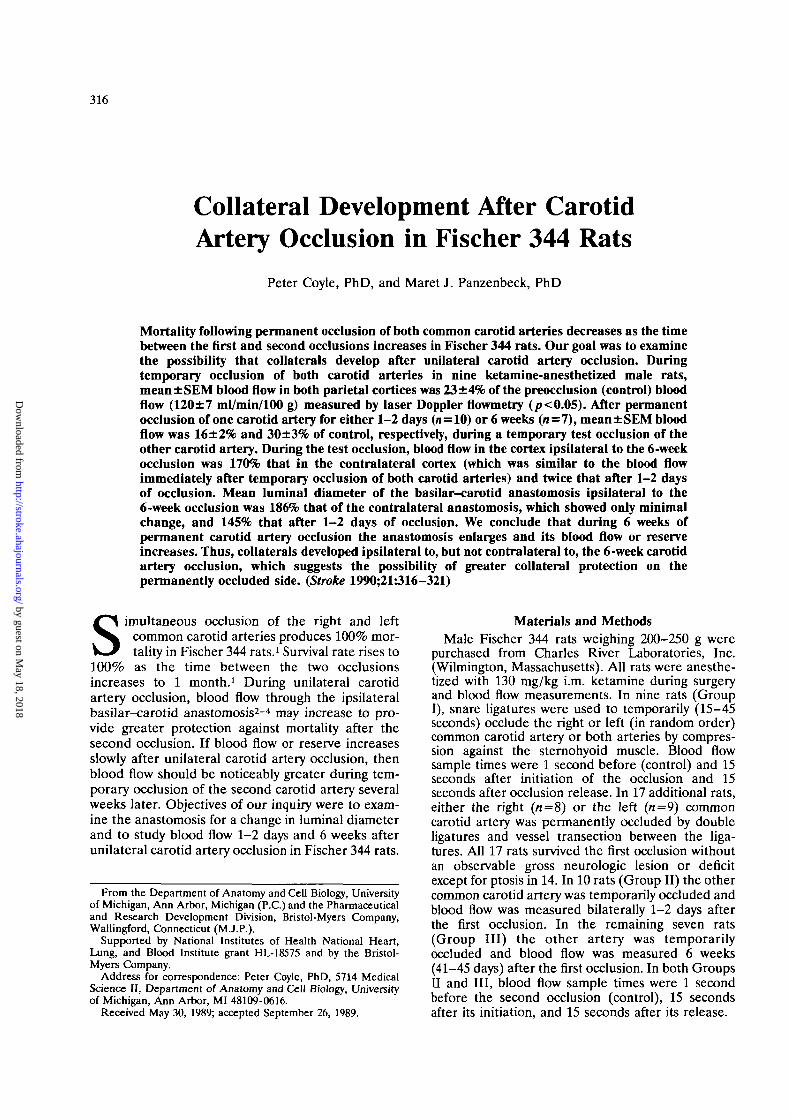
The internal carotid and basilar arteries supply thecircle of Willis, which is complete in Fischer 344 rats.To avoid confusing terminology of the posteriorcerebral artery and the posterior communicatingartery in rats,2"4 we designated the measured branchas the posterior communicating anastomosis (PC inFigure 1). A calibrated eyepiece micrometer in astereozoom dissecting microscope was used to mea-sure the luminal diameter at the arrow in Figure 1.
Blood flows and luminal diameters were expressedas mean±SEM. Analysis of variance was used tocompare blood flows and diameters in the threegroups. The paired t test was used to compare bloodflows and diameters within a group. An a error of<0.05 (i.e.,p<0.05) was considered significant.
ResultsThe posterior communicating anastomosis was
present bilaterally (Figure 1) in all 26 rats. In GroupI mean±SEM luminal diameters of the left and rightanastomoses were 153±10 and 151±12 yu,m, respec-tively. However, for each rat one anastomosis waslarger than the other. Luminal diameters for thelarge and small anastomoses were significantly dif-ferent (169±11 vs. 134±6 nm,p<0.Q5).
In Group II, the anastomosis ipsilateral to the firstocclusion was 74 jum larger than the contralateralanastomosis (/?<0.05, Table 1). In Group III, theanastomosis ipsilateral to the first occlusion was 158/*m larger than the contralateral anastomosis(p<0.05, Table 1). The anastomosis ipsilateral to thefirst occlusion was significantly larger at 6 weeks thanat 1-2 days (p<0.05, Table 1); diameter of thecontralateral anastomosis at 6 weeks and 1-2 daysdid not differ.
In Group I, control blood flow was 120±7 ml/min/100 g (Table 2). Within 5 seconds after the firstocclusion, ipsilateral blood flow fell to a minimumand then began to rise. After 15 seconds of unilateralocclusion, ipsilateral blood flow was significantly lessthan control (p<0.05, Table 2). During the firstocclusion, contralateral blood flow (116±5 ml/min/100 g) was essentially unchanged from thatbefore the occlusion (117±5 ml/min/100 g). Within 5seconds after the second occlusion blood flow ipsilat-eral and contralateral to the first occlusion fell to aminimum and remained there for the duration of theocclusion (Figure 2, A and B). After 15 seconds ofbilateral occlusion, mean blood flow was 23% ofcontrol (/?<0.05, Table 2), but there was no signifi-cant difference between the two sides (data notshown). By 15 seconds after the release of theocclusions, blood flow was restored to the controllevel (Table 2).
In Group II, after 1-2 days blood flow ipsilateral tothe first occlusion was not significantly different fromthe contralateral control (Table 2) nor from restingblood flow in rats without occlusion (control, GroupI). During the second occlusion, mean blood flow was16% of control (Table 2), not significantly differenton the two sides (Table 2, Figure 3). By 15 secondsafter the release of the second occlusion, blood flowwas restored to the control level (Table 2).
In Group III, after 6 weeks blood flow ipsilateralto the first occlusion was not significantly differentfrom the contralateral control (Table 2) nor fromresting blood flow in rats without occlusion (con-trol, Group I). During the second occlusion, meanabsolute blood flow was less than control (p<0.05,Table 2); both absolute and relative blood flowswere significantly greater ipsilateral to the firstocclusion than contralateral to it (p<0.05, Table 2;Figure 3). During the second occlusion, blood flowipsilateral to the 6 weeks' occlusion was twice thatipsilateral to the 1-2 days' occlusion (p<0.05,Table 2). Following release of the second occlusion,
by guest on May 18, 2018
http://stroke.ahajournals.org/D
ownloaded from

318 Stroke Vol 21, No 2, February 1990
FIGURE 1. Latex-filled arteries of circle of Willis in Fischer 344 rat 6 weeks after permanent occlusion of common carotid arteryipsilateral to labeled posterior communicating anastomosis (PC).
blood flow was restored to control levels by 15seconds (Table 2). Patterns of blood flow restora-tion included an initial hyperemia and a slow returnto control (Figure 2, C and D, respectively). Thus,blood flow restoration was variable, with no singleconsistent pattern.
During the second (bilateral) occlusion, the cortexwith higher blood flow was ipsilateral to the largeranastomosis in seven (78%) of the nine Group I rats,in eight (80%) of the 10 Group II rats, and in allseven (100%) Group III rats. The null hypothesisthat the higher collateral blood flow was related only
by guest on May 18, 2018
http://stroke.ahajournals.org/D
ownloaded from

Coyle and Panzenbeck Collateral Development 319
150%GROUP I
0
150%
B
150% GROUP III
6 weeks occl.
0150%
tI-
t15 sec.
FIGURE 2. Group I. Effect of temporary occlusion of both common carotid arteries in Fisher 344 rats on blood flow measuredbilaterally (A and B) in parietal cortex by laser Doppler flowmetry. Group III. Effect of temporary occlusion of one common carotidartery on blood flow measured bilaterally (C and D) 6 weeks after occclusion of other common carotid artery.
by chance to the cortex of either side was rejected(*2=12.5,<i/=l,p«).001).
DiscussionOur major findings of greater blood flow and a
larger communicating anastomosis at 6 weeks suggestcollateral development following permanent occlu-sion of a carotid artery. Blood flow and the anasto-mosis contralateral to the permanent occlusion wereessentially unchanged from their control levels,which suggests no or only minimal collateral devel-opment contralateral to the occlusion. Rather, thelarger anastomosis and increased blood flow werelateralized to the side of the permanently occludedartery.
Absolute blood flow values for resting brain mea-sured by laser Doppler flowmetry in our ketamine-anesthetized rats (120±7 ml/min/100 g) were similarto those measured with radioactive labeled micro-spheres in awake rats9 (117±13 ml/min/100 g) ormicrospheres in rats given nitrous oxide10 (128 ±10ml/min/100 g). Because of the high spatial and tem-
poral resolution of laser Doppler flowmetry and therelatively low resolution of the microsphere method,comparisons may be valid only if laser Dopplerreadings are made at multiple tissue sites11 in eachrat and then averaged, as we did.
Previous studies910'12-13 suggest that blood flow isrestored to normal resting levels during unilateralcarotid artery occlusion in some strains of rats. After15 seconds of unilateral carotid artery occlusion,blood flow returns to control levels in normotensiveWistar rats13 but not in Fischer 344 rats or hyperten-sive stroke-prone rats.13 The mechanism responsiblefor a blood flow less than control after 15 seconds ofunilateral carotid artery occlusion in Fischer 344 ratsis unknown. Because blood flow 15 seconds afterrelease of the second occlusion was at control levels,the mechanism appears to involve the collateralvessels, possibly at the anastomosis. Stroke-pronerats are known to have a defective collateral circula-tion, probably due to narrower anastomosing ves-sels.1415 An endothelium-dependent dilator responseappears to be impaired in hypertensive stroke-pronerats.16
by guest on May 18, 2018
http://stroke.ahajournals.org/D
ownloaded from

320 Stroke Vol 21, No 2, February 1990
TABLE 1. Luminal Diameter of Posterior Communicating Anasto-mosis in Fischer 344 Rats After Common Carotid Artery Occlusion
Group
IIIIII
Time afterfirst
occlusion
Same day1-2 days6 weeks
n
18107
Diameter
Firstocclusion
152+11238+9*
342±22*t
ipsilateral to
Secondocclusion
164±8184±3
Bonferroni correction used for multiple comparisons.Data are mean±SEM /im of n anastomoses (9 rats in Group I).*p<0.05 different from second occlusion by paired / test.t/7<0.05 different from Group II by analysis of variance.
Blood flow levels are similar in the two hemi-spheres later on the day of unilateral carotid arteryocclusion in Sprague-Dawley rats9 and Fischer rats.12
Blood flow was not evaluated for a bilateraldecrease,912 which is possible because the distalunpaired segment of the anterior cerebral arteryreceives blood from each internal carotid artery inrats. However, after 1-2 days of unilateral carotidartery occlusion, ipsilateral blood flow was similar tothat in rats without occlusion, thus suggesting restingblood flow was restored before 1-2 days.
During hypercapnia after 5 days of permanentunilateral carotid artery occlusion in Wistar rats,ipsilateral blood flow was 63% of that on the nonoc-cluded side, but the percentage difference dimin-ished over 1 month.10 Thus, after unilateral carotidartery occlusion, resting blood flow returns to normallevels sooner than blood flow during hypercapnia,which depends on a large dilator reserve.
We found that the posterior communicating anas-tomosis was wider following permanent occlusion ofa carotid artery. The luminal diameter may haveincreased by different mechanisms. Dilatation orcontraction, change in vascular wall compliance, orvascular wall remodeling could alter the diameter ofan anastomosis. We used papaverine to relax thesmooth muscle and prevent variability in luminaldiameter due to muscle contraction. After 1-2 daysof occlusion, the larger anastomosis was probablymore compliant because the time for structural rear-
TEMPORARILYOCCLUDED SIDE
GROUPS
PERMANENTLYOCCLUDED SIDE
GROUPS5 0 !
25-
"Ooj5CD
0
* p < 0.01vs group IIItemporarilyoccluded side
FIGURE 3. Bar graph. Effect of occlusion of both commoncarotid arteries on mean±SEM blood flow in Fischer 344 rats.Group I: n=9 (18 hemispheres), temporary occlusion; GroupII: n=10, 1-2 days' occlusion; Group III; n = 7, 6 weeks'occlusion. Probability determined using paired t test withoutBonferroni correction.
rangement was minimal. During 6 weeks of occlu-sion, structural remodeling of the vascular wall mostlikely produced an anastomosis having larger outerand inner diameters. Further study may define thesepossible mechanisms.
Despite a wider anastomosis after 1-2 days ofunilateral carotid artery occlusion, blood flow duringbilateral occlusion was not increased. One possibleexplanation of this discrepancy was that contractedsmooth muscle prevented widening of the anastomo-sis for increased blood flow and that the anastomosiswidened only following muscle relaxation caused bypapaverine or latex filling. Another possibility wasthat a steal phenomenon diverted blood from themonitored cortical site to elsewhere in the brain, tothe eye, or to skeletal muscle.
After 6 weeks of unilateral carotid artery occlu-sion, ipsilateral blood flow was increased twofoldduring bilateral occlusion, thus indicating either lesssteal or, more likely, greater blood flow through a
TABLE 2. Blood Flow in Parietal Cortex of Fischer 344 Rats
Time
Before occlusion (control)After first occlusion15 seconds after second occlusion
Ipsilateral to first occlusionContralateral to first occlusion
Mean of two sidesAfter release of second occlusion
I (15 seconds)
Mean±SEM(ml/min/100 g)
120±7
69±8*t
27±5t113±6
(71 = 18)
%
100
59±6*
23±4103±ll
II (1-2 days)
Mean±SEM(ml/min/100 g)
112±6124±5
22±315±319±2|
117±7
(II = 10)
%
100
113±6
19±314±316±2
109+10
III (6 weeks)
Mean+SEM(ml/min/100 g)
119+5124+3
46+5**27+4
36±4*f120+4
(/i = 7)
%
100
105±5
37±4*t23±330±3*
101±3
n, number of hemispheres (9 rats in Group I). Bonferroni correction used for multiple comparisons.*p<0.05 different from Group II by analysis of variance.tp<0.05 different from control by paired t test.^ 0 different from contralateral by paired t test.
by guest on May 18, 2018
http://stroke.ahajournals.org/D
ownloaded from

Coyle and Panzenbeck Collateral Development 321
wider anastomosis. That the side of the greater bloodflow and the side of the wider anastomosis wererelated, not by chance, suggests that the anastomosisdiameter was one factor, perhaps a major one, lim-iting blood flow during the second occlusion. Wepropose that during the days and weeks followingunilateral carotid artery occlusion the anastomosisundergoes structural enlargement, thus allowingincreased blood flow.
Others have found that blood flow to the cerebrumafter 2, 3, 4, and 5 hours of bilateral carotid arteryocclusion was appreciably less than flow after 5minutes of occlusion.17 After simultaneous occlusionof both carotid arteries in Fischer 344 rats for 4hours, cerebral edema is present,18 which may com-promise blood flow to the cerebrum.
After 6 weeks of unilateral carotid artery occlu-sion, the ipsilateral posterior communicating anasto-mosis is wider and more blood flows through it.When substantial time elapses between permanentocclusion of the first and second carotid arteries,mortality after the second occlusion is reducedappreciably,1 possibly due to protection fromedema.18 Our study suggests that protection may begreater on the side having the wider communicatinganastomosis and greater collateral blood flow inFischer 344 rats.
AcknowledgmentWe thank Dr. Donald Heistad for review and critical
comments on an early version of the manuscript.
References1. Payan HM, Levine S, Strebel R: Effects of cerebral ischemia in
various strains of rats. Proc Soc Exp Biol Med 1965;120:208-209
2. Greene EC: Anatomy of the rat. Trans Am Phil Soc NS1935;27:l-370
3. Brown JA: The morphology of circulus arteriosus cerebri inrats. Anat Rec 1966;156:99-106
4. Hodde KC: Cephalic Vascular Patterns in the Rat. Amsterdam,Rodopi, 1981, pp 1-94
5. Coyle P, Jokelainen PT: Dorsal cerebral arterial collaterals ofthe rat. Anat Rec 1982;203:397-404
6. Buchweitz-Milton E, Weiss HR: Cerebral oxygen consumptionand blood flow in Fischer-344 rats of different ages. NeurobiolAging 1987;8:55-60
7. Bonner R, Nossal R: Model for laser Doppler measurementsin tissue. Appl Optics 1981;20:2097-2107
8. Haberl RL, Heizer ML, Marmarou A, Ellis EF: Laser-Doppler assessment of brain microcirculation: Effect of sys-temic alterations. Am J Physiol 1989;256:H1247-H1254
9. Flaim SF, Nellis SH, Toggart EJ, Drexler H, Kanda K,Newman ED: Multiple simultaneous determinations of hemo-dynamics and flow distribution in conscious rat. J PharmacolMethods 1984;ll:l-39
10. De Ley G, Nishimyumuremyi J-B, Leusen I: Hemisphericblood flow in the rat after unilateral common carotid occlu-sion: Evolution with time. Stroke 1985;16:69-73
11. Shepherd AP, Riedel GL, Kiel JW, Haumschild DJ, MaxwellLC: Evaluation of an infrared laser-Doppler blood fiowmeter.Am J Physiol 1987;252:G832-G839
12. Hoffman WE, Miletich DJ, Albrecht RF: Repeated micro-sphere injections in rats. Life Sci 1981;28:2167-2172
13. Coyle P: Development of early collateral blood flow to cere-brum is compromised in chronic hypertension (abstract).Neurosci Abs 1988;14:47
14. Coyle P, Heistad DD: Blood flow through cerebral collateralvessels in hypertensive and normotensive rats. Hypertension1986;8(suppl II):II-67-11-71
15. Coyle P: Dorsal cerebral collaterals of stroke-prone hyperten-sive rats (SHRSP) and Wistar Kyoto rats (WKY). Anat Rec1987;218:40-44
16. Mayhan WG, Faraci FM, Heistad DD: Impairment ofendothelium-dependent responses of cerebral arterioles inchronic hypertension. Am J Physiol 1987;253:H1435-H1440
17. Fujishima M, Ishitsuka T, Nakatomi Y, Tamaki K, Omae T:Changes in local cerebral blood flow following bilateral carotidocclusion in spontaneously hypertensive and normotensiverats. Stroke 1981;12:874-876
18. Silvia RC, Slizgi GR, Ludens JH, Tang AH: Protection fromischemia-induced cerebral edema in the rat by U-50488H, akappa opioid receptor agonist. Brain Res 1987;403:52-57
KEY WORDS • carotid arteries • collateral circulationcerebral blood flow • rats
by guest on May 18, 2018
http://stroke.ahajournals.org/D
ownloaded from

P Coyle and M J PanzenbeckCollateral development after carotid artery occlusion in Fischer 344 rats.
Print ISSN: 0039-2499. Online ISSN: 1524-4628 Copyright © 1990 American Heart Association, Inc. All rights reserved.
is published by the American Heart Association, 7272 Greenville Avenue, Dallas, TX 75231Stroke doi: 10.1161/01.STR.21.2.316
1990;21:316-321Stroke.
http://stroke.ahajournals.org/content/21/2/316World Wide Web at:
The online version of this article, along with updated information and services, is located on the
http://stroke.ahajournals.org//subscriptions/
is online at: Stroke Information about subscribing to Subscriptions:
http://www.lww.com/reprints Information about reprints can be found online at: Reprints:
document. Permissions and Rights Question and Answer available in the
Permissions in the middle column of the Web page under Services. Further information about this process isOnce the online version of the published article for which permission is being requested is located, click Request
can be obtained via RightsLink, a service of the Copyright Clearance Center, not the Editorial Office.Stroke Requests for permissions to reproduce figures, tables, or portions of articles originally published inPermissions:
by guest on May 18, 2018
http://stroke.ahajournals.org/D
ownloaded from