Embed Size (px)
Citation preview
2014 www.kce.fgov.be
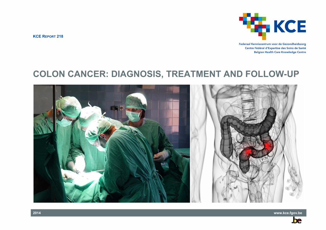
KCE REPORT 218
COLON CANCER: DIAGNOSIS, TREATMENT AND FOLLOW-UP

2011 www.kce.fgov.be
KCE REPORT 218 GOOD CLINICAL PRACTICE
COLON CANCER: DIAGNOSIS, TREATMENT AND FOLLOW-UP MARC PEETERS, ROOS LEROY, JO ROBAYS, GIGI VEEREMAN, DIDIER BIELEN, WIM CEELEN, ETIENNE DANSE, MARC DE MAN, PIETER DEMETTER, PATRICK FLAMEN, ALAIN HENDLISZ, ISABELLE SINAPI, DIRK VANBECKEVOORT, ERIC VAN CUTSEM, DIRK YSEBAERT, PAUL VAN GILS, LAETITIA VEERBEEK, YOLBA SMIT, LEEN VERLEYE
COLOPHON Title: Colon Cancer: Diagnosis, Treatment and Follow-Up Authors: Marc Peeters (UZA), Roos Leroy (KCE), Jo Robays (KCE), Gigi Veereman (KCE), Didier Bielen (UZ Leuven),
Wim Ceelen (UZ Gent), Etienne Danse (UCL), Marc De Man (OLV Ziekenhuis Aalst), Pieter Demetter (ULB), Patrick Flamen (Jules Bordet Instituut), Alain Hendlisz (Institut Jules Bordet), Isabelle Sinapi (Grand Hôpital de Charleroi), Dirk Vanbeckevoort (UZ Leuven), Eric Van Cutsem (UZ Leuven), Dirk Ysebaert (Universiteit Antwerpen), Paul van Gils (IKNL), Laetitia Veerbeek (IKNL), Yolba Smit (IKNL), Leen Verleye (KCE)
Project coordinator and Senior supervisor:
Sabine Stordeur (KCE)
Reviewers: Marijke Eyssen, Raf Mertens, Sabine Stordeur Stakeholders: Donald Claeys (Belgian Section of Colorectal Surgery), André D'Hoore (Royal Belgian Radiological Society),
Constant Jehaes (Belgian Section of Colorectal Surgery), Alex Kartheuser (Belgian Section of Colorectal Surgery), Daniel Léonard (Belgian Section of Colorectal Surgery), Ivo Nagels (Stichting Tegen Kanker), Bart Op De Beeck (Royal Belgian Radiological Society), Piet Pattyn (Belgian Section of Colorectal Surgery), Ward Rommel (Vlaamse Liga Tegen Kanker), Sabine Tejpar (Belgian Group of Digestive Oncology), Nancy Van Damme (Kankerregister), Vincent Vandecaveye (Royal Belgian Radiological Society), Didier Vander Steichel (Fondation Contre le Cancer)
External validators: Bert Aertgeerts (CEBAM, KU Leuven), Daniel Van Daele (CHU de Liège), Cornelis Van de Velde (Leids Universitair Medisch Centrum)
Other reported interests: Fees or other compensation for writing a publication or participating in its development: Patrick Flamen A grant, fees or funds for a member of staff or another form of compensation for the execution of research: Patrick Flamen (Sirtex, Bayer, Roche), Marc Peeters (Amgen, Roche), Dirk Ysebaert Consultancy or employment for a company, an association or an organisation that may gain or lose financially due to the results of this report: Patrick Flamen, Marc Peeters (Amgen, Merck Serono, Roche, Sanofi) Payments to speak, training remuneration, subsidised travel or payment for participation at a conference: Marc De Man (support participation in conferences (Merck Serono, Pfizer, Roche), Advisory Board (Merck Senoro)), Patrick Flamen, Marc Peeters (Amgen, Merck Serono, Roche, Sanofi) Presidency or accountable function within an institution, association, department or other entity on which the results of this report could have an impact: Donald Claeys (Chairman Collegium Chirurgicum), Constant Jehaes (Member of the board of the section of colorectal surgery of the Belgian Royal Society of Surgery), Didier Vander Steichel (Medical and Scientific Director Foundation against Cancer), Dirk Ysebaert (project Translational Research National Cancer Plan) Participation in scientific or experimental research as an initiator, principal investigator or researcher: Patrick
Flamen, Alain Hendlisz (Principal Investigator PePiTA trial), Marc Peeters (Amgen, Merck Serono), Cornelis Van de Velde (different colorectal trials) Further, it should be noted that all experts and stakeholders, as well as the validators consulted within this report were selected because of their expertise in the field of colon cancer. Therefore, by definition, all consulted experts, stakeholders and validators have a certain degree of conflict of interest to the main topic of this report.
Layout: Ine Verhulst Disclaimer: The stakeholders were consulted on a (pre-final) version of the scientific report. Their comments
were discussed during a meeting. They did not co-author the scientific report and did not necessarily agree with its content.
Subsequently, a final version was submitted to the validators. The validation of the report results from a consensus or a voting process between the validators. The validators did not co-author the scientific report and did not necessarily all agree with its content.
Finally, this report has been approved by common assent by the Executive Board. KCE is sole responsible for errors or omissions that might persist. The policy recommendations are
also the full responsibility of KCE. Publication date: 17 januari 2014 Domain: Good Clinical Practice (GCP) MeSH: Colonic Neoplasms, Practice guidelines NLM Classification: WI 529 Language: English Format: Adobe® PDF™ (A4) Legal depot: D/2014/10.273/15 Copyright: KCE reports are published under a “by/nc/nd” Creative Commons Licence
http://kce.fgov.be/content/about-copyrights-for-kce-reports.
How to refer to this document? Peeters M, Leroy R, Robays J, Veereman G, Bielen D, Ceelen W, Danse E, De Man M, Demetter P, Flamen P,
Hendlisz A, Sinapi I, Vanbeckevoort D, Van Cutsem E, Ysebaert D, van Gils P, Veerbeek L, Smit Y, Verleye L. Colon Cancer: Diagnosis, Treatment and Follow-Up. Good Clinical Practice (GCP) Brussels: Belgian Health Care Knowledge Centre (KCE). 2014. KCE Reports 218. D/2014/10.273/15.
This document is available on the website of the Belgian Health Care Knowledge Centre.

KCE Report 218 Colon cancer 1
TABLE OF CONTENTS
SCIENTIFIC REPORT ......................................................................................................................... 10 1 INTRODUCTION .................................................................................................................................. 10 1.1 BACKGROUND .................................................................................................................................... 10 1.2 THE NEED FOR A GUIDELINE ........................................................................................................... 11 1.3 INTERNATIONAL COLLABORATION ................................................................................................. 11 1.4 SCOPE ................................................................................................................................................. 11 1.5 REMIT OF THE GUIDELINE ................................................................................................................ 11
1.5.1 Overall objectives ................................................................................................................... 11 1.5.2 Target users of the guideline ................................................................................................. 11
1.6 STATEMENT OF INTENT .................................................................................................................... 12 1.7 FUNDING AND DECLARATION OF INTEREST ................................................................................. 12 2 METHODOLOGY ................................................................................................................................. 12 2.1 INTRODUCTION .................................................................................................................................. 12 2.2 GUIDELINE DEVELOPMENT GROUP ............................................................................................... 12 2.3 CLINICAL RESEARCH QUESTIONS .................................................................................................. 13 2.4 GENERAL APPROACH ....................................................................................................................... 14 2.5 LITERATURE SEARCH AND STUDY SELECTION ............................................................................ 14
2.5.1 Literature review ..................................................................................................................... 14 2.6 QUALITY APPRAISAL ......................................................................................................................... 15
2.6.1 Clinical practice guidelines ..................................................................................................... 16 2.6.2 Systematic reviews ................................................................................................................ 16 2.6.3 Primary articles ...................................................................................................................... 16
2.7 DATA EXTRACTION AND EVIDENCE SUMMARY ............................................................................ 16 2.8 STATISTICAL ANALYSIS .................................................................................................................... 16 2.9 GRADING EVIDENCE ......................................................................................................................... 16 2.10 FORMULATION OF RECOMMENDATIONS ....................................................................................... 19
2 Colon cancer KCE Report 218
2.11 EXTERNAL REVIEW ........................................................................................................................... 20 2.11.1 Healthcare professionals ....................................................................................................... 20 2.11.2 Patient representatives .......................................................................................................... 21
2.12 FINAL VALIDATION ............................................................................................................................. 21 3 RECOMMENDATIONS ........................................................................................................................ 21 3.1 DIAGNOSIS .......................................................................................................................................... 21 3.2 STAGING OF INVASIVE COLON CANCER ....................................................................................... 22
3.2.1 CT chest-abdomen................................................................................................................. 22 3.2.2 FDG PET-CT for staging ........................................................................................................ 23 3.2.3 MRI liver ................................................................................................................................. 25 3.2.4 Multidisciplinary team (MDT) meetings .................................................................................. 27
3.3 PATHOLOGY ....................................................................................................................................... 28 3.3.1 KRAS mutational analysis ...................................................................................................... 28 3.3.2 BRAF ...................................................................................................................................... 30 3.3.3 N-RAS as a predictor of treatment effectiveness ................................................................... 31 3.3.4 MSI testing as a predictor of treatment effectiveness ............................................................ 31 3.3.5 Standards for pathology report .............................................................................................. 33 3.3.6 Number of lymph nodes ......................................................................................................... 33
3.4 SURGICAL TREATMENT STAGE 0-III ............................................................................................... 34 3.4.1 Endoscopic treatment stage I: polypectomy .......................................................................... 34 3.4.2 Laparoscopic vs. open surgery .............................................................................................. 36 3.4.3 Surgical technique: complete mesocolic excision .................................................................. 41 3.4.4 Enhanced recovery after surgery (ERAS) ............................................................................. 42
3.5 TREATMENT OF ACUTE OBSTRUCTIONS ...................................................................................... 47 3.6 ADJUVANT CHEMOTHERAPY FOR STAGE II-III COLON CANCER ................................................ 52
3.6.1 (High risk) stage II colon cancer ............................................................................................ 52 3.6.2 Stage III colon cancer ............................................................................................................ 53 3.6.3 Adjuvant chemotherapy in elderly patients ............................................................................ 54
3.7 SURGICAL TREATMENT OF LIVER METASTASES ......................................................................... 57 3.7.1 Timing of surgical resection of primary tumour and synchronous liver metastasis ............... 57
KCE Report 218 Colon cancer 3
3.7.2 Neoadjuvant, perioperative and adjuvant chemotherapy ...................................................... 59 3.8 LOCAL TREATMENT MODALITIES FOR UNRESECTABLE LIVER METASTASES ........................ 61
3.8.1 Radio-frequency ablation (RFA) ............................................................................................ 61 3.8.2 Hepatic artery chemotherapy in unresectable CRC liver metastases ................................... 62 3.8.3 Chemo-embolisation of unresectable CRC liver metastases ................................................ 63 3.8.4 Radio-embolisation, Selective Internal Radiation Therapy (SIRT) for patients with
unresectable tumours ............................................................................................................ 64 3.8.5 Stereotactic Body Radiation Therapy (SBRT) for liver metastases ....................................... 66
3.9 LOCAL TREATMENT OF LUNG METASTASES ................................................................................ 67 3.9.1 Stereotactic Body Radiation Therapy (SBRT) for lung metastases ....................................... 67 3.9.2 Resection of lung metastases ................................................................................................ 67
3.10 TREATMENT OF PERITONEAL METASTASES: CYTOREDUCTIVE SURGERY AND HYPERTHERMIC INTRAPERITONEAL CHEMOTHERAPY (HIPEC) ................................................ 69
3.11 TREATMENT OF METASTATIC COLON CANCER: FIRST-LINE CHEMOTHERAPY +/- TARGETED THERAPY................................................................................................................... 71 3.11.1 Choice of chemotherapy agents ............................................................................................ 71 3.11.2 Sequential versus combined chemotherapy .......................................................................... 76 3.11.3 Targeted therapy .................................................................................................................... 77
3.12 SECOND LINE CHEMOTHERAPY FOR METASTATIC COLON CANCER ....................................... 84 3.13 FOLLOW-UP ........................................................................................................................................ 85
3.13.1 Summary of guidelines .......................................................................................................... 85 3.13.2 General recommendations ..................................................................................................... 85 3.13.3 CEA ........................................................................................................................................ 85 3.13.4 Colonoscopy .......................................................................................................................... 85 3.13.5 Imaging .................................................................................................................................. 85 3.13.6 Other tests .............................................................................................................................. 85 3.13.7 Update on the use of CT chest abdomen in follow-up ........................................................... 86
4 IMPLEMENTATION AND UPDATING OF THE GUIDELINE ............................................................. 87 4.1 IMPLEMENTATION ............................................................................................................................. 87
4.1.1 Multidisciplinary approach ...................................................................................................... 87
4 Colon cancer KCE Report 218
4.1.2 Patient-centred care ............................................................................................................... 87 4.1.3 Dissemination and implementation of this guideline .............................................................. 87
4.2 MONITORING THE QUALITY OF CARE ............................................................................................ 88 4.3 GUIDELINE UPDATE .......................................................................................................................... 88 REFERENCES ..................................................................................................................................... 89
KCE Report 218 Colon cancer 5
LIST OF FIGURES Figure 1 – Enhanced recovery programs vs. conventional recovery – outcome: mortality ................................ 44 Figure 2 – Enhanced recovery programs vs. conventional recovery – outcome: complications ....................... 45 Figure 3 – Enhanced recovery programs vs. conventional recovery – outcome: readmissions ........................ 46 Figure 4 – Enhanced recovery programs vs. conventional recovery – outcome: hospital stay ......................... 46 Figure 5 – Stent as a bridge to surgery vs. acute surgery – outcome: permanent stoma .................................. 50 Figure 6 – Stent as a bridge to surgery vs. acute surgery – outcome: peri-procedural mortality ....................... 51 Figure 7 – Chemotherapy combined with surgery vs. surgery alone - outcome: overall survival ...................... 60 Figure 8 – Oral versus IV fluoropyrimidines + oxaliplatin - outcome: progression-free survival ........................ 73 Figure 9 – Oral versus IV fluoropyrimidines + oxaliplatin - outcome: overall survival ........................................ 73 Figure 10 – Oral versus IV fluoropyrimidines + irinotecan - outcome: progression-free survival ....................... 74 Figure 11 – Sequential versus combined first-line chemotherapy for mCRC - outcome: overall survival ......... 77 Figure 12 – Adding bevacizumab to first-line chemotherapy for mCRC - outcome: progression-free survival.. 79 Figure 13 – Adding bevacizumab to first-line chemotherapy for mCRC - outcome: overall survival ................. 79 Figure 14 – Adding cetuximab or panitumumab to first-line chemotherapy +/- bevacizumab for
mCRC - outcome: progression-free survival .................................................................................... 82 Figure 15 – Adding cetuximab or panitumumab to first-line chemotherapy +/- bevacizumab for
mCRC - outcome: overall survival ................................................................................................... 83
LIST OF TABLES Table 1 – Searched websites of international guideline developers .................................................................. 15 Table 2 – Levels of evidence according to the GRADE system ......................................................................... 16 Table 3 – Downgrading the quality rating of evidence using GRADE ................................................................ 18 Table 4 – Strength of recommendations according to GRADE .......................................................................... 19 Table 5 – Factors that influence the strength of a recommendation .................................................................. 19 Table 6 – Interpretation of strong and conditional (weak) recommendations .................................................... 20 Table 7 – MRI results compared to standard (histopathology) ........................................................................... 26 Table 8 – PET- CT results compared to standard (histopathology) ................................................................... 27 Table 9 – Adjusted HR for colon cancer-specific mortality for surgery + adjuvant chemotherapy
versus surgery alone in different age strata ........................................................................................ 55 Table 10 – Evaluation of surgery and chemotherapy vs. surgery alone in different co-morbidity
strata in patients aged >67 years ..................................................................................................... 55 Table 11 – Evaluation of surgery and chemotherapy vs. surgery alone in different
co-morbidity/age strata in patients aged ≥65 years .......................................................................... 55
6 Colon cancer KCE Report 218
LIST OF ABBREVIATIONS
ABBREVIATION DEFINITION ACHBT AHRQ
Association of Hepatobiliary Surgery and Transplantation Agency for Healthcare Research and Quality
AJCC American Joint Committee on Cancer ASA ASCRS
American Society of Anaesthesiologists American Society of Colon and Rectal Surgeons
BSC CEBAM
Best supportive care Belgian Centre for Evidence-Based Medicine
CCO Cancer Care Ontario CEA Carcinoembryonic antigen CI CME
Confidence interval Complete mesocolic excision
CoI Conflict of interest CRC Colorectal cancer CRM Circumferential resection margin CRS Cytoreductive surgery CT Computed tomography CTC Computed tomographic colonography DFS Disease-free survival dMMR Defective Mismatch repair EGAPP Evaluation of Genomic Applications in Practice and Prevention EGFR EMR
Epidermal growth factor receptor Endoscopic mucosal resection
ERAS ESD
Enhanced recovery after surgery Endoscopic submucosal resection
EURECCA European registration of cancer care FDG FOD
Fluorodeoxyglucose Belgian Health Authorities
FOLFIRI Infusional 5-fluorouracil/leucovorin with irinotecan
KCE Report 218 Colon cancer 7
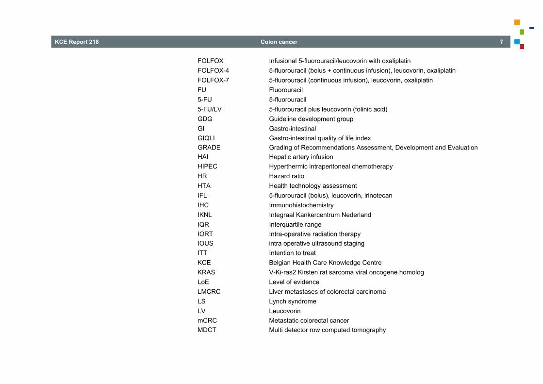
FOLFOX Infusional 5-fluorouracil/leucovorin with oxaliplatin FOLFOX-4 5-fluorouracil (bolus + continuous infusion), leucovorin, oxaliplatin FOLFOX-7 5-fluorouracil (continuous infusion), leucovorin, oxaliplatin FU Fluorouracil 5-FU 5-fluorouracil 5-FU/LV 5-fluorouracil plus leucovorin (folinic acid) GDG Guideline development group GI Gastro-intestinal GIQLI GRADE HAI
Gastro-intestinal quality of life index Grading of Recommendations Assessment, Development and Evaluation Hepatic artery infusion
HIPEC Hyperthermic intraperitoneal chemotherapy HR Hazard ratio HTA Health technology assessment IFL 5-fluorouracil (bolus), leucovorin, irinotecan IHC Immunohistochemistry IKNL Integraal Kankercentrum Nederland IQR Interquartile range IORT IOUS
Intra-operative radiation therapy intra operative ultrasound staging
ITT Intention to treat KCE Belgian Health Care Knowledge Centre KRAS V-Ki-ras2 Kirsten rat sarcoma viral oncogene homolog LoE Level of evidence LMCRC Liver metastases of colorectal carcinoma LS Lynch syndrome LV Leucovorin mCRC MDCT
Metastatic colorectal cancer Multi detector row computed tomography
8 Colon cancer KCE Report 218
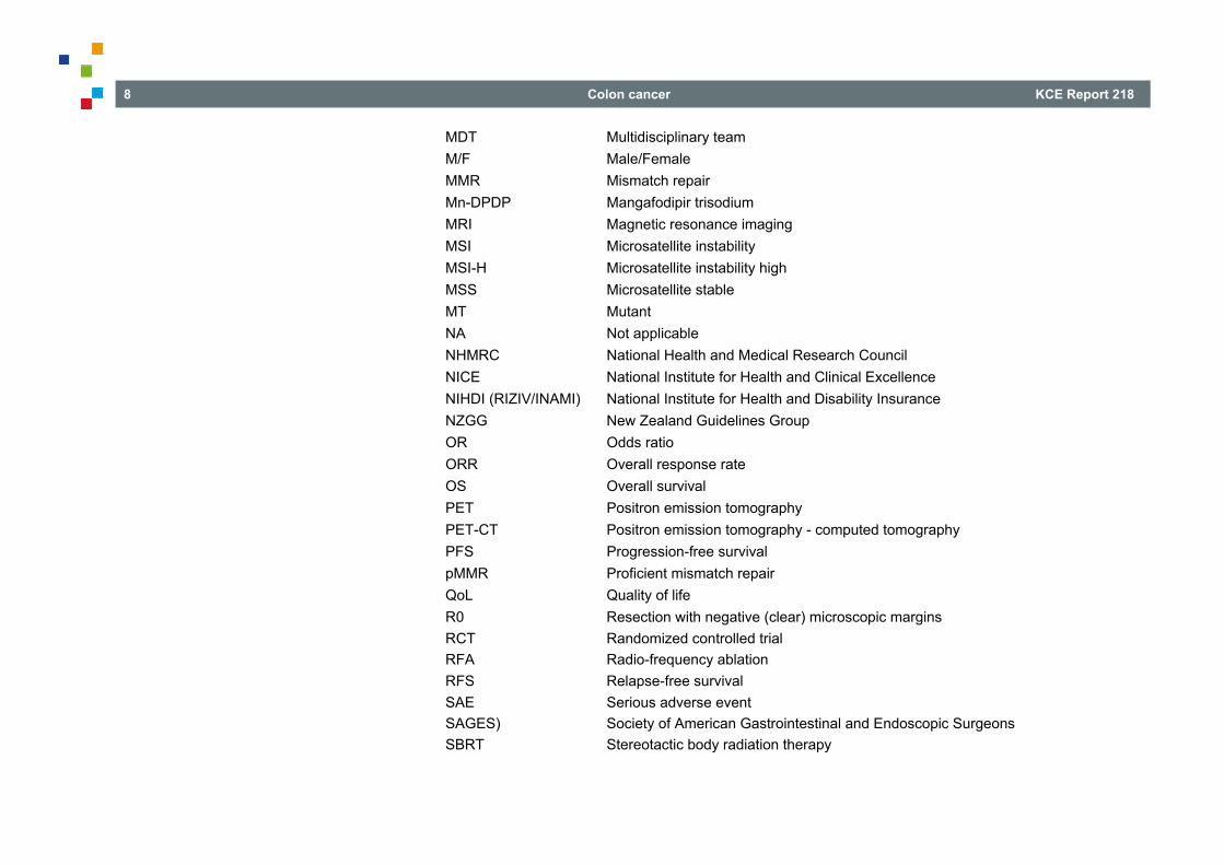
MDT Multidisciplinary team M/F Male/Female MMR Mismatch repair Mn-DPDP Mangafodipir trisodium MRI Magnetic resonance imaging MSI Microsatellite instability MSI-H Microsatellite instability high MSS Microsatellite stable MT Mutant NA Not applicable NHMRC National Health and Medical Research Council NICE National Institute for Health and Clinical Excellence NIHDI (RIZIV/INAMI) National Institute for Health and Disability Insurance NZGG New Zealand Guidelines Group OR Odds ratio ORR Overall response rate OS Overall survival PET Positron emission tomography PET-CT Positron emission tomography - computed tomography PFS Progression-free survival pMMR Proficient mismatch repair QoL Quality of life R0 Resection with negative (clear) microscopic margins RCT RFA
Randomized controlled trial Radio-frequency ablation
RFS Relapse-free survival SAE SAGES) SBRT
Serious adverse event Society of American Gastrointestinal and Endoscopic Surgeons Stereotactic body radiation therapy
KCE Report 218 Colon cancer 9
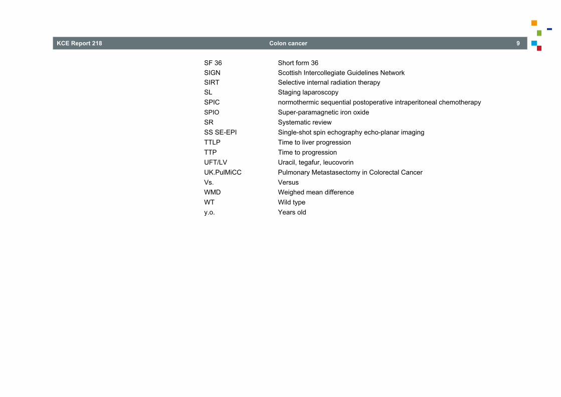
SF 36 Short form 36 SIGN SIRT
Scottish Intercollegiate Guidelines Network Selective internal radiation therapy
SL Staging laparoscopy SPIC normothermic sequential postoperative intraperitoneal chemotherapy SPIO Super-paramagnetic iron oxide SR Systematic review SS SE-EPI Single-shot spin echography echo-planar imaging TTLP Time to liver progression TTP Time to progression UFT/LV Uracil, tegafur, leucovorin UK.PulMiCC Pulmonary Metastasectomy in Colorectal Cancer Vs. Versus WMD Weighed mean difference WT Wild type y.o. Years old
10 Colon cancer KCE Report 218
SCIENTIFIC REPORT 1 INTRODUCTION The development of clinical care pathways is one of the main actions described in the Belgian National Cancer Plan 2008-2010 and at the same time one of the assignments of the College of Oncology. Since many years the Belgian Health Care Knowledge Centre (KCE) has collaborated with the College of Oncology. More precisely, it has provided scientific support for the development of clinical practice guidelines. This collaboration has resulted so far in the publication of clinical practice guidelines on breast cancer, colorectal cancer, testicular cancer, pancreatic cancer, upper gastrointestinal cancer, cervical cancer, prostate cancer and lung cancer.
1.1 Background According to the data collected by the Belgian Cancer Registry, colorectal cancer is the 3rd most frequent cancer in males and the 2nd in females.1 Colorectal cancer ranks as the 2nd most frequent cause of death by cancer in males and the 3rd in females (Belgium, 2008), affects males more often than females (male/female ratio: 1.56 in 2008) and primarily patients older than 64 years (69.5% in males and 72.9% in females in 2008). Due to ageing of the population, colorectal cancer will remain an important health problem for our society in the next decades. For colon cancers diagnosed in Belgium between 2004 and 2008, the 5-year relative survival rates were 62.3% in males and 64.6% in females, with few regional differences.1 Based on data collected in the Flanders between 1999 and 2008, it was apparent that from 3 to 4 years after diagnosis, females had a small survival advantage in comparison to males: 10-year relative survival rates were 58.5% in females and 55.6% in males. In both genders an age-dependent survival gradient was noted, with the best survival rates in patients between 15 and 49 years old (5-year relative survival: 71.0% in males and 74.7% in females) and the worst survival rates in patients over 64 years old (5-year relative survival: 59.8% in males and 62.7% in females).
KCE Report 218 Colon cancer 11
Stage at diagnosis is a very important prognostic factor for survival in colon cancer in men as well as in women.1 According to the clinical stage, the 5-year relative survival rates range from 91.8% to 91.3% in stage I and from 11.9% to 12.9% in stage IV for males and females respectively. According to the pathological stage, the 5-year relative survival estimates are 91.2% and 96.2% in stage I and 19.1% and 19.8% in stage IV, for males and females respectively. Pathological staging performs better in estimating survival results from stage III onwards because of the difficulty to distinguishing lymph-node positive from negative disease in the pre-operative setting.
1.2 The need for a guideline In 2006 a clinical practice guideline for colorectal cancer was published jointly by the College of Oncology and KCE.2 Since then, much has evolved in the diagnosis and treatment of colorectal cancer as well as in the methodology of developing clinical practice guidelines. As a consequence, an update of the recommendations with regard to the diagnosis, staging and treatment of colon cancer was indicated.
1.3 International collaboration The Dutch guideline developer ‘Integraal Kankercentrum Nederland’ (IKNL) decided to update the clinical guideline for the diagnosis and treatment of colorectal cancer and the guideline for the treatment of colorectal liver metastases. The update focused on eight research questions (see below) which were also of interest to KCE. An international collaboration was set up and the eight questions were divided equally between both IKNL and KCE. The mutual development process of a clinical practice guideline involved the search for evidence (search strategy + selection), quality appraisal, evidence tables, evaluation of the level of evidence using GRADE and the evidence report. The formulation of recommendations was the sole responsibility of each organisation.
1.4 Scope This guideline focuses on the diagnosis, staging, treatment and follow-up of patients with all stages of primary adenocarcinoma of the colon. Other (rare) histological types of colon cancer are not discussed in this guideline. The guideline does not cover population screening nor the surveillance of high-risk groups (e.g. patients with a family history or with inflammatory bowel disease). Cancer of the rectum is considered out of scope for this guideline, although many clinical trials include both patients with colon cancer and rectal cancer. Evidence from trials including both colon cancer and cancer of the rectum was taken into account. The specific clinical questions resulted from a scoping review of existing international guidelines and discussion with the stakeholders and the guideline development group (GDG) (see paragraph.2.3).
1.5 Remit of the guideline 1.5.1 Overall objectives This guideline provides recommendations based on current scientific evidence both for the diagnosis, treatment and follow-up of patients with colon cancer (CRC). Clinicians are encouraged to interpret these recommendations in the context of the individual patient situation, values and preferences. The guidelines are based on clinical evidence and may not always be in line with the current criteria for NIHDI (RIZIV/INAMI) reimbursement of diagnostic and therapeutic interventions. The NIHDI may consider adaptation of reimbursement or financing criteria based on these guidelines.
1.5.2 Target users of the guideline This guideline is intended to be used by all care providers involved in the management of patients with colon cancer, including gastroenterologists, surgeons, medical oncologists, radiologists and pathologists. It could also be of particular interest for patients and their families, for general practitioners, for hospital managers and policy makers.
12 Colon cancer KCE Report 218
1.6 Statement of intent Clinical guidelines are designed to improve the quality of health care and decrease the use of unnecessary or harmful interventions. This guideline has been developed by clinicians and researchers for use within the Belgian healthcare context. It provides advice regarding the care and management of patients with colon cancer. The recommendations are not intended to indicate an exclusive course of action or to serve as a standard of care. Standards of care are determined on the basis of all clinical data available for an individual case and are subject to change as scientific knowledge and technology advance and patterns of care evolve. Variations, which take into account individual circumstances, clinical judgement and patient choice may also be appropriate. The information in this guideline is not a substitute for proper diagnosis, treatment or the provision of advice by an appropriate health professional. It is advised, however, that significant deviations from the national guideline should be fully documented in the patient’s file at the time when a relevant decision is taken.
1.7 Funding and declaration of interest KCE is a federal institution which is financed for the largest part by INAMI/RIZIV, but also by the Federal Public Service of Health, food chain safety and environment, and Federal Public Service of social security. The development of clinical practice guidelines is part of the legal mission of KCE. Although the development of the guidelines is paid by KCE budget, the sole mission of KCE is providing scientifically valid information. KCE has no interest in companies (commercial or not, e.g. hospital, university), associations (e.g. professional association, syndicate), individuals or organisations (e.g. lobby group) on which the guidelines could have a positive or negative impact (financial or other). All clinicians involved in the GDG or the peer-review process completed a declaration of interest form. The information of possible conflicts of interest is published in the colophon of this report. All members of KCE Expert Team make yearly declarations of interest and further details of these are available on request.
2 METHODOLOGY 2.1 Introduction The KCE guideline is drawn up according to highly codified principles, based on scientific information regularly updated from the international literature. This guideline was developed using a standard methodology based on a systematic review of the evidence. Further details about KCE and the guideline development methodology are available at https://kce.fgov.be/content/kce-processes. Several steps were followed to elaborate this guideline. Firstly, clinical questions were developed in collaboration with the members of the GDG. Secondly a literature review was made (including search for recent, high quality guidelines). Thirdly, on the basis of the results of the literature review, recommendations were formulated and graded according to the GRADE approach.
2.2 Guideline development group The present guideline was developed by KCE in collaboration with a multidisciplinary group of experts assigned by the College of Oncology. Methodological expertise, support and facilitation were provided by the KCE Expert Team. The Guideline Development Group comprised the following experts: Medical Oncology & Gastroenterology Marc Peeters, Gastroenterology and Digestive Oncology, UZ Antwerp,
Antwerp - Coordinator of the GDG Eric Van Cutsem, Gastroenterology and Digestive Oncology,
University Hospitals Leuven, Leuven Isabelle Sinapi, Medical Oncology, Grand Hôpital de Charleroi,
Charleroi Alain Hendlisz, Digestive Oncology, Bordet Institute, Brussels Marc De Man, Gastro-enterology, Onze-Lieve-Vrouw Ziekenhuis,
Aalst
KCE Report 218 Colon cancer 13
Surgery Wim Ceelen, GI Surgery, UZ Gent, Ghent Dirk Ysebaert, Hepatobiliary Surgery, UZ Antwerp, Antwerp Nuclear medicine Patrick Flamen, Nuclear Medicine, Bordet Institute, Brussels Pathology Pieter Demetter, Pathology, Erasme University Hospital, Brussels Radiology Dirk Vanbeckevoort, Radiology, University Hospitals Leuven, Leuven Didier Bielen, Radiology, University Hospitals Leuven, Leuven Etienne Danse, Radiology, St Luc University Hospital, Brussels The roles assigned to the GDG were: The definition of the clinical questions, in close collaboration with the
KCE Expert Team and stakeholders; The identification of important outcomes; The feedback on the selection of papers and identification of papers
that were missed; The feedback on the content of the guideline; The judgement about indirectness of evidence; The feedback on the draft recommendations; The concerns that have to be reported under ‘other considerations’.
2.3 Clinical research questions Priority research questions to be included in this guideline were selected by the Dutch and the Belgian stakeholders. The following eight priority questions were selected by the Dutch stakeholders: Is PET-CT more sensitive and/or specific than CT to detect
metastases in patients with potentially resectable liver (or lung) metastases, resulting in a change of treatment plan?
What is the value of enhanced recovery programs after laparoscopic or open colectomy for colorectal cancer?
Is stenting or colostomy more beneficial than acute resection with or without primary anastomosis in acute obstruction due to left-sided colon carcinoma?
Does additional (segmental) colon resection yield better outcomes (PFS, OS, QoL) than watchful waiting in patients who are diagnosed with Tis/T1 colon carcinoma and who have undergone endoscopic polypectomy?
Which group of elderly patients with non-metastasized primary colorectal carcinoma does not benefit from surgery with or without preoperative radiotherapy or adjuvant chemotherapy?
What is the best therapeutic sequence for patients with o resectable metachronous liver metastases? o resectable synchronous liver metastases?
When to use local therapy for lung or unresectable liver metastases of colorectal cancer?
What is the current standard first line treatment for metastatic inoperable colorectal cancer?
14 Colon cancer KCE Report 218
The selection of research questions by the Belgian stakeholders was made during an initial expert meeting at KCE on May 3rd, 2012, based on a list of recommendations from international guidelines: Should MRI of the liver be performed in patients with potentially
resectable liver metastases on CT and PET-CT, to detect additional liver metastases and/or determine resectability?
What are the clinical indications for upfront testing of microsatellite instability (MSI) in a tumour?
Which factors should be determined to identify high-risk stage II colon cancer patients that are eligible for adjuvant chemotherapy?
Is laparoscopic colectomy beneficial compared to open surgery in terms of morbidity, recovery and oncological outcomes, with special attention to T4 tumours, tumours of the transverse colon, ‘single incision’ techniques and total mesocolic resection?
Is debulking surgery followed by hypertermic intraperitoneal chemotherapy (HIPEC) recommended for patients with resectable peritoneal metastases from colorectal cancer?
Should routine CT of the abdomen be performed on regular intervals during follow-up?
2.4 General approach The present clinical practice guideline was developed by adapting international guidelines to the Belgian context. For this procedure, the ADAPTE Collaboration, an international group of guideline developers and researchers, developed a formal methodology.3 It consists of three major phases: 1. Set-up Phase: outlines the necessary tasks to be completed prior to
beginning the adaptation process (e.g. identifying necessary skills and resources).
2. Adaptation Phase: assists guideline developers in moving from selection of a topic to identification of specific clinical questions; searching for and retrieving guidelines; assessing the consistency of the evidence therein, their quality, currency, content and applicability; decision making around adaptation and preparing the draft adapted guideline.
3. Finalization Phase: guides guideline developers through getting feedback on the document from stakeholders who will be impacted by the guideline, consulting with the source developers of guidelines used in the adaptation process, establishing a process for review and updating of the adapted guideline and the process of creating a final document.
For the selected priority research questions, the international guidelines were updated with more recently published evidence. For other topics, the recommendations formulated by international guidelines and the underlying evidence were reviewed by the GDG and adapted to the Belgian context.
2.5 Literature search and study selection 2.5.1 Literature review Evidence-based clinical practice guidelines on colon cancer were identified through searches on several databases and websites (see appendix). Search in OVID Medline for guidelines on colon cancer (2009-current date) resulted in more than 305 hits. Searching the website of the Guideline International network (GIN) (http://www.g-i-n.net) and the National Guideline Clearinghouse (http://www.guideline.gov/) revealed 8 and 58 hits respectively. All searches for guidelines were run in May/June, 2012. An overview of the search results can be found in the appendix. Additionally, the following websites of international guideline developers were searched:
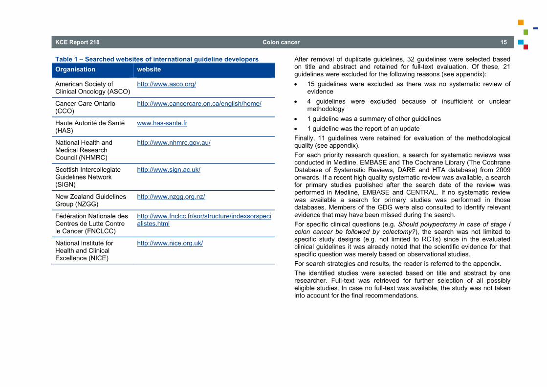
KCE Report 218 Colon cancer 15
Table 1 – Searched websites of international guideline developers Organisation website
American Society of Clinical Oncology (ASCO)
http://www.asco.org/
Cancer Care Ontario (CCO)
http://www.cancercare.on.ca/english/home/
Haute Autorité de Santé (HAS)
www.has-sante.fr
National Health and Medical Research Council (NHMRC)
http://www.nhmrc.gov.au/
Scottish Intercollegiate Guidelines Network (SIGN)
http://www.sign.ac.uk/
New Zealand Guidelines Group (NZGG)
http://www.nzgg.org.nz/
Fédération Nationale des Centres de Lutte Contre le Cancer (FNCLCC)
http://www.fnclcc.fr/sor/structure/indexsorspecialistes.html
National Institute for Health and Clinical Excellence (NICE)
http://www.nice.org.uk/
After removal of duplicate guidelines, 32 guidelines were selected based on title and abstract and retained for full-text evaluation. Of these, 21 guidelines were excluded for the following reasons (see appendix): 15 guidelines were excluded as there was no systematic review of
evidence 4 guidelines were excluded because of insufficient or unclear
methodology 1 guideline was a summary of other guidelines 1 guideline was the report of an update Finally, 11 guidelines were retained for evaluation of the methodological quality (see appendix). For each priority research question, a search for systematic reviews was conducted in Medline, EMBASE and The Cochrane Library (The Cochrane Database of Systematic Reviews, DARE and HTA database) from 2009 onwards. If a recent high quality systematic review was available, a search for primary studies published after the search date of the review was performed in Medline, EMBASE and CENTRAL. If no systematic review was available a search for primary studies was performed in those databases. Members of the GDG were also consulted to identify relevant evidence that may have been missed during the search. For specific clinical questions (e.g. Should polypectomy in case of stage I colon cancer be followed by colectomy?), the search was not limited to specific study designs (e.g. not limited to RCTs) since in the evaluated clinical guidelines it was already noted that the scientific evidence for that specific question was merely based on observational studies. For search strategies and results, the reader is referred to the appendix. The identified studies were selected based on title and abstract by one researcher. Full-text was retrieved for further selection of all possibly eligible studies. In case no full-text was available, the study was not taken into account for the final recommendations.
16 Colon cancer KCE Report 218
2.6 Quality appraisal 2.6.1 Clinical practice guidelines The AGREE II instrument was used to evaluate the methodological quality of the identified international guidelines (see appendix). Each guideline was scored by two independent researchers (RL and LV) and discussed in case of disagreement. Based on an overall assessment, 11 high quality guidelines were selected. 2.6.2 Systematic reviews Selected (systematic) reviews were critically appraised by a single KCE expert using the AMSTAR checklist (see appendix). In case of doubt, a second KCE expert was consulted. 2.6.3 Primary articles Diagnostic studies were assessed for risk of bias with the QUADAS-2 tool. For quality appraisal of RCTs for therapeutic interventions, the "Cochrane Collaboration’s tool for assessing risk of bias" was used (see appendix). Critical appraisal of each study was performed by a single KCE expert. In case of doubt, a second KCE expert was consulted. For the research questions elaborated by IKNL, the respective CocanCPG checklists were used. (http://www.cocancpg.eu/v1/fichiers/public/3_commontoolsandformats.pdf)
2.7 Data extraction and evidence summary For every clinical question, the recommendations and supporting evidence base were extracted from the selected guidelines. In addition the following data were extracted from the systematic reviews: the search date, publication year, included studies and main results. Similarly, the publication year, study population, study intervention, outcomes and results from the primary studies were summarized. Data extraction was done by one reviewer using the standard KCE template for evidence tables.
2.8 Statistical analysis We performed meta-analyses using Review Manager Version 5 (http://ims.cochrane.org/revman) whenever more recent RCTs were found in addition to a published meta-analysis or in case subgroup analysis was needed for certain items. For progression-free survival (PFS), disease-free survival (DFS) and overall survival (OS), a hazard ratio (HR) was extracted from the reported analyses. We used the extraction methods following Parmar et al.4 All meta-analyses were performed using a generic inverse variance method unless otherwise stated. Heterogeneity was statistically assessed using χ2 test and I² statistic. If heterogeneity was present, a random-effects model was used instead of a fixed-effect model. Possible reasons for heterogeneity were explored post-hoc. Sensitivity analysis was performed by removing outliers from the analysis.
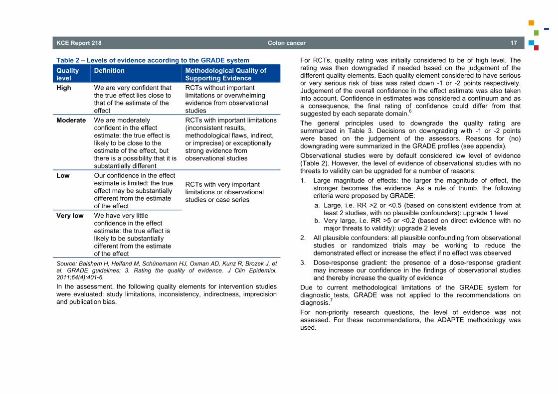
2.9 Grading evidence The results from selected systematic reviews and, where appropriate, the more recent RCTs were pooled and the quality of evidence was evaluated using GRADE methodology.5 More precisely, GRADE for guidelines was used, meaning that the evidence across all outcomes and across studies for a particular recommendation was assessed. This resulted in a level of evidence being assigned to each conclusion (Table 2).
KCE Report 218 Colon cancer 17
Table 2 – Levels of evidence according to the GRADE system Quality level
Definition Methodological Quality of Supporting Evidence
High We are very confident that the true effect lies close to that of the estimate of the effect
RCTs without important limitations or overwhelming evidence from observational studies
Moderate We are moderately confident in the effect estimate: the true effect is likely to be close to the estimate of the effect, but there is a possibility that it is substantially different
RCTs with important limitations (inconsistent results, methodological flaws, indirect, or imprecise) or exceptionally strong evidence from observational studies
Low Our confidence in the effect estimate is limited: the true effect may be substantially different from the estimate of the effect
RCTs with very important limitations or observational studies or case series
Very low We have very little confidence in the effect estimate: the true effect is likely to be substantially different from the estimate of the effect
Source: Balshem H, Helfand M, Schünemann HJ, Oxman AD, Kunz R, Brozek J, et al. GRADE guidelines: 3. Rating the quality of evidence. J Clin Epidemiol. 2011;64(4):401-6. In the assessment, the following quality elements for intervention studies were evaluated: study limitations, inconsistency, indirectness, imprecision and publication bias.
For RCTs, quality rating was initially considered to be of high level. The rating was then downgraded if needed based on the judgement of the different quality elements. Each quality element considered to have serious or very serious risk of bias was rated down -1 or -2 points respectively. Judgement of the overall confidence in the effect estimate was also taken into account. Confidence in estimates was considered a continuum and as a consequence, the final rating of confidence could differ from that suggested by each separate domain.6 The general principles used to downgrade the quality rating are summarized in Table 3. Decisions on downgrading with -1 or -2 points were based on the judgement of the assessors. Reasons for (no) downgrading were summarized in the GRADE profiles (see appendix). Observational studies were by default considered low level of evidence (Table 2). However, the level of evidence of observational studies with no threats to validity can be upgraded for a number of reasons: 1. Large magnitude of effects: the larger the magnitude of effect, the
stronger becomes the evidence. As a rule of thumb, the following criteria were proposed by GRADE: a. Large, i.e. RR >2 or <0.5 (based on consistent evidence from at
least 2 studies, with no plausible confounders): upgrade 1 level b. Very large, i.e. RR >5 or <0.2 (based on direct evidence with no
major threats to validity): upgrade 2 levels 2. All plausible confounders: all plausible confounding from observational
studies or randomized trials may be working to reduce the demonstrated effect or increase the effect if no effect was observed
3. Dose-response gradient: the presence of a dose-response gradient may increase our confidence in the findings of observational studies and thereby increase the quality of evidence
Due to current methodological limitations of the GRADE system for diagnostic tests, GRADE was not applied to the recommendations on diagnosis.7 For non-priority research questions, the level of evidence was not assessed. For these recommendations, the ADAPTE methodology was used.
18 Colon cancer KCE Report 218
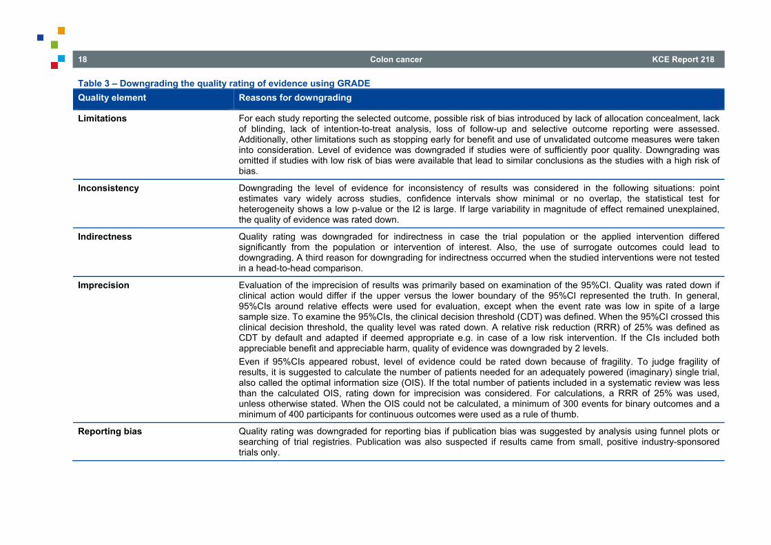
Table 3 – Downgrading the quality rating of evidence using GRADE Quality element Reasons for downgrading
Limitations For each study reporting the selected outcome, possible risk of bias introduced by lack of allocation concealment, lack of blinding, lack of intention-to-treat analysis, loss of follow-up and selective outcome reporting were assessed. Additionally, other limitations such as stopping early for benefit and use of unvalidated outcome measures were taken into consideration. Level of evidence was downgraded if studies were of sufficiently poor quality. Downgrading was omitted if studies with low risk of bias were available that lead to similar conclusions as the studies with a high risk of bias.
Inconsistency Downgrading the level of evidence for inconsistency of results was considered in the following situations: point estimates vary widely across studies, confidence intervals show minimal or no overlap, the statistical test for heterogeneity shows a low p-value or the I2 is large. If large variability in magnitude of effect remained unexplained, the quality of evidence was rated down.
Indirectness
Quality rating was downgraded for indirectness in case the trial population or the applied intervention differed significantly from the population or intervention of interest. Also, the use of surrogate outcomes could lead to downgrading. A third reason for downgrading for indirectness occurred when the studied interventions were not tested in a head-to-head comparison.
Imprecision Evaluation of the imprecision of results was primarily based on examination of the 95%CI. Quality was rated down if clinical action would differ if the upper versus the lower boundary of the 95%CI represented the truth. In general, 95%CIs around relative effects were used for evaluation, except when the event rate was low in spite of a large sample size. To examine the 95%CIs, the clinical decision threshold (CDT) was defined. When the 95%CI crossed this clinical decision threshold, the quality level was rated down. A relative risk reduction (RRR) of 25% was defined as CDT by default and adapted if deemed appropriate e.g. in case of a low risk intervention. If the CIs included both appreciable benefit and appreciable harm, quality of evidence was downgraded by 2 levels. Even if 95%CIs appeared robust, level of evidence could be rated down because of fragility. To judge fragility of results, it is suggested to calculate the number of patients needed for an adequately powered (imaginary) single trial, also called the optimal information size (OIS). If the total number of patients included in a systematic review was less than the calculated OIS, rating down for imprecision was considered. For calculations, a RRR of 25% was used, unless otherwise stated. When the OIS could not be calculated, a minimum of 300 events for binary outcomes and a minimum of 400 participants for continuous outcomes were used as a rule of thumb.
Reporting bias Quality rating was downgraded for reporting bias if publication bias was suggested by analysis using funnel plots or searching of trial registries. Publication was also suspected if results came from small, positive industry-sponsored trials only.
KCE Report 218 Colon cancer 19
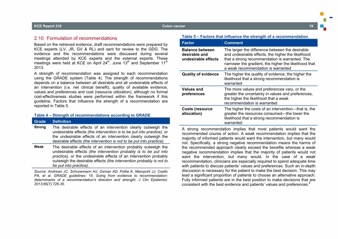
2.10 Formulation of recommendations Based on the retrieved evidence, draft recommendations were prepared by KCE experts (LV, JR, GV & RL) and sent for review to the GDG. The evidence and the recommendations were discussed during several meetings attended by KCE experts and the external experts. These meetings were held at KCE on April 24th, June 13th and September 11th 2013. A strength of recommendation was assigned to each recommendation using the GRADE system (Table 4). The strength of recommendations depends on a balance between all desirable and all undesirable effects of an intervention (i.e. net clinical benefit), quality of available evidence, values and preferences and cost (resource utilization), although no formal cost-effectiveness studies were performed within the framework if this guideline. Factors that influence the strength of a recommendation are reported in Table 5.
Table 4 – Strength of recommendations according to GRADE Grade Definition Strong The desirable effects of an intervention clearly outweigh the
undesirable effects (the intervention is to be put into practice), or the undesirable effects of an intervention clearly outweigh the desirable effects (the intervention is not to be put into practice).
Weak The desirable effects of an intervention probably outweigh the undesirable effects (the intervention probably is to be put into practice), or the undesirable effects of an intervention probably outweigh the desirable effects (the intervention probably is not to be put into practice).
Source: Andrews JC, Schunemann HJ, Oxman AD, Pottie K, Meerpohl JJ, Coello PA, et al. GRADE guidelines: 15. Going from evidence to recommendation-determinants of a recommendation's direction and strength. J Clin Epidemiol. 2013;66(7):726-35.
Table 5 – Factors that influence the strength of a recommendation Factor Comment
Balance between desirable and undesirable effects
The larger the difference between the desirable and undesirable effects, the higher the likelihood that a strong recommendation is warranted. The narrower the gradient, the higher the likelihood that a weak recommendation is warranted
Quality of evidence The higher the quality of evidence, the higher the likelihood that a strong recommendation is warranted
Values and preferences
The more values and preferences vary, or the greater the uncertainty in values and preferences, the higher the likelihood that a weak recommendation is warranted
Costs (resource allocation)
The higher the costs of an intervention—that is, the greater the resources consumed—the lower the likelihood that a strong recommendation is warranted
A strong recommendation implies that most patients would want the recommended course of action. A weak recommendation implies that the majority of informed patients would want the intervention, but many would not. Specifically, a strong negative recommendation means the harms of the recommended approach clearly exceed the benefits whereas a weak negative recommendation implies that the majority of patients would not want the intervention, but many would. In the case of a weak recommendation, clinicians are especially required to spend adequate time with patients to discuss patients’ values and preferences. Such an in-depth discussion is necessary for the patient to make the best decision. This may lead a significant proportion of patients to choose an alternative approach. Fully informed patients are in the best position to make decisions that are consistent with the best evidence and patients’ values and preferences.8
20 Colon cancer KCE Report 218
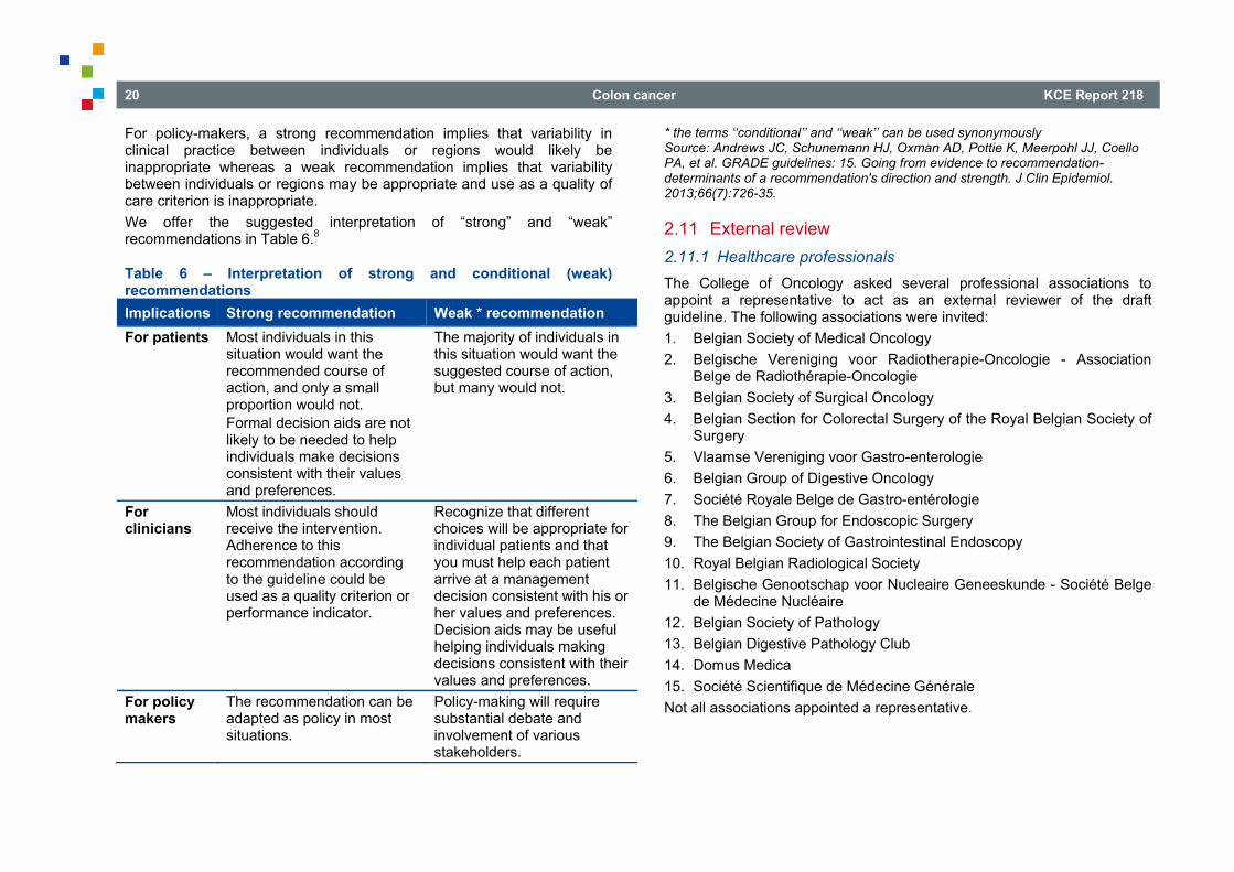
For policy-makers, a strong recommendation implies that variability in clinical practice between individuals or regions would likely be inappropriate whereas a weak recommendation implies that variability between individuals or regions may be appropriate and use as a quality of care criterion is inappropriate. We offer the suggested interpretation of “strong” and “weak” recommendations in Table 6.8
Table 6 – Interpretation of strong and conditional (weak) recommendations Implications Strong recommendation Weak * recommendation For patients Most individuals in this
situation would want the recommended course of action, and only a small proportion would not. Formal decision aids are not likely to be needed to help individuals make decisions consistent with their values and preferences.
The majority of individuals in this situation would want the suggested course of action, but many would not.
For clinicians
Most individuals should receive the intervention. Adherence to this recommendation according to the guideline could be used as a quality criterion or performance indicator.
Recognize that different choices will be appropriate for individual patients and that you must help each patient arrive at a management decision consistent with his or her values and preferences. Decision aids may be useful helping individuals making decisions consistent with their values and preferences.
For policy makers
The recommendation can be adapted as policy in most situations.
Policy-making will require substantial debate and involvement of various stakeholders.
* the terms ‘‘conditional’’ and ‘‘weak’’ can be used synonymously Source: Andrews JC, Schunemann HJ, Oxman AD, Pottie K, Meerpohl JJ, Coello PA, et al. GRADE guidelines: 15. Going from evidence to recommendation-determinants of a recommendation's direction and strength. J Clin Epidemiol. 2013;66(7):726-35.
2.11 External review 2.11.1 Healthcare professionals The College of Oncology asked several professional associations to appoint a representative to act as an external reviewer of the draft guideline. The following associations were invited: 1. Belgian Society of Medical Oncology 2. Belgische Vereniging voor Radiotherapie-Oncologie - Association
Belge de Radiothérapie-Oncologie 3. Belgian Society of Surgical Oncology 4. Belgian Section for Colorectal Surgery of the Royal Belgian Society of
Surgery 5. Vlaamse Vereniging voor Gastro-enterologie 6. Belgian Group of Digestive Oncology 7. Société Royale Belge de Gastro-entérologie 8. The Belgian Group for Endoscopic Surgery 9. The Belgian Society of Gastrointestinal Endoscopy 10. Royal Belgian Radiological Society 11. Belgische Genootschap voor Nucleaire Geneeskunde - Société Belge
de Médecine Nucléaire 12. Belgian Society of Pathology 13. Belgian Digestive Pathology Club 14. Domus Medica 15. Société Scientifique de Médecine Générale Not all associations appointed a representative.
KCE Report 218 Colon cancer 21
External experts received the recommendations two weeks before the stakeholder meeting and were asked to score each recommendation on a 5-point Likert-scale, with a score of ‘1’ indicating ‘completely disagree’, ‘2’ indicating ‘somewhat disagree’, ‘3’ indicating ‘not answered’, ‘4’ indicating ‘somewhat agree’, and ‘5’ indicating ‘completely agree’. In case they were not familiar with the underlying evidence, they had the option to answer ‘not applicable’. When an expert disagreed with the recommendation (score ‘1’ or ‘2’), (s)he was asked to provide appropriate evidence. The recommendations were discussed during a face-to-face meeting on October 16th, 2013. Based on this discussion a final draft of the recommendations was prepared. In the appendix, an account is provided on the external experts’ comments. 2.11.2 Patient representatives The ‘Vlaamse liga tegen kanker’ and the ‘Fondation contre le cancer’ were contacted to participate to the stakeholder meeting on October 16th, 2013. A key role for patient representatives is to ensure that patient views and experiences inform the group’s work. The patient representatives were asked the following questions: Are there any considerations from the patients’ perspective that we
missed in formulating our recommendations? Do we need to add information to clarify choices when doctors discuss
treatment options with patients? In the appendix, an overview is provided on how the comments of the patient representatives were taken into account.
2.12 Final validation As part of the standard KCE procedures, an external scientific validation of the report was conducted prior to its publication. The validation process was performed on November 18th, 2013. The current guideline was reviewed prior to its publication by three independent validators (see names in the colophon), making use of the AGREE II checklist. The validation process was chaired by CEBAM. The validation of the report resulted from a consensus or a voting process between the validators.
3 RECOMMENDATIONS 3.1 Diagnosis The diagnosis of colon cancer is based on history taking, a complete clinical examination and colonoscopy with biopsy.2 The diagnostic procedure is generally indicated for patients with the following symptoms:2 For all ages: rectal bleeding with alteration in bowel habits to looseness or increased frequency over a period of six weeks and/or palpable abdominal mass and/or iron-deficiency anaemia without overt cause. Over 60 years: rectal bleeding without any symptoms or alteration in bowel habits to looseness or increased frequency. A family history points to the high risk groups. Both the NICE and the SIGN guidelines consider colonoscopy with biopsy and/or polypectomy as the gold standard for making the diagnosis of colorectal cancer. These recommendations are based on systematic reviews of observational studies showing a pooled sensitivity of CT colonography of 69-70% and two poor quality RCTs showing that additional investigations are more frequently needed after air contrast barium enema compared to colonoscopy. Colonoscopy is preferred mainly because of the possibility for direct biopsy or polypectomy but is generally considered more invasive and has a higher morbidity than imaging procedures. Update The SIGGAR trials (see appendix), two parallel RCTs mentioned in the NICE guideline, have been published in 2013.9, 10 Results are summarized in the appendix. Detection rates of colorectal cancer were similar after computed tomographic colonography (CTC) and colonoscopy but more patients are referred for additional colonic investigations after CTC than after colonoscopy (RR 3.65; 95%CI 2.87-4.65). Colonography may be slightly more acceptable to patients (better satisfaction, less discomfort) but long term psychological effects are not significantly different. An advantage of colonoscopy is that results are more often communicated to the patient on the same day as the investigation and more often in a face-to-face conversation. Compared to barium enema, the detection rate of
22 Colon cancer KCE Report 218
colorectal cancer was higher after CTC (RR 1.31; 95%CI 1.01-1.68), at the cost of a higher rate of additional colonic investigations (23.5% vs. 18.6%; p=0.0003). In agreement with the GDG, no update of the literature search for RCTs or observational studies published since February 2011 (search date NICE) was performed.
Conclusions Computed tomographic colonography and colonoscopy have similar
detection rates for colorectal cancer. However, referral for additional colonic investigations is more frequent after computed tomographic colonography compared with colonoscopy (Atkin et al., 2013).
Patient acceptability of CT colonography is slightly higher compared to colonoscopy. However, psychological effects at three months are similar (von Wagner et al., 2013).
CT colonography results in a higher detection rate of colorectal cancer compared to barium enema at the cost of more additional colonic investigations (Halligan et al., 2013).
Other considerations Colonoscopy is generally considered more invasive than CT colonography as sedation is needed for most patients. Therefore, in patients less fit than the study population due to age or co-morbidities, CT colonography as first investigation can be considered. A CT colonography can be part of a full diagnostic CT scan of the abdomen. However, in case of strong suspicion of a cancerous lesion, colonoscopy with tailored sedation remains the preferred option.
Recommendations To confirm or rule out colon cancer, colonoscopy in conjunction
with histological confirmation is the technique of choice in fit patients (strong recommendation).
If colonoscopy is considered not feasible or contra-indicated, CT colonography is recommended (strong recommendation).
3.2 Staging of invasive colon cancer Staging procedures before the start of treatment should mainly answer the following three questions: Is the disease limited to the primary tumour? If yes, is the primary
tumour resectable? If no, are metastases limited to the liver (or lung)? If yes, are the
metastases resectable? If no, are metastases limited to the abdomen? If yes, are the
metastases eligible for debulking surgery followed by HIPEC? 3.2.1 CT chest-abdomen Both the SIGN11 and the NICE12 guideline recommend a contrast enhanced CT scan of the chest, abdomen and pelvis as the initial staging procedure for all patients diagnosed with CRC (see appendix). Supporting evidence reported by NICE12 consists of systematic reviews of cross-sectional studies. The included studies were of poor methodological quality and heterogeneous with regards to included patients and studied techniques. One systematic review specifically studied patients with colon cancer rather than colorectal cancer. A summary estimate for differentiating between T1/T2 and T3/T4 tumours was 86% (95%CI 78-92%) for sensitivity and 78% (95%CI for 71-84%) for specificity. The false positive rate was low in all included studies suggesting that CT reliably identifies T3/T4 tumours. Both guidelines also refer to diagnostic pathways most frequently used by clinicians.11-13 Further imaging is only recommended for patients showing possibly resectable metastatic disease on CT-scan.11-13 Update No update of the literature was performed.
KCE Report 218 Colon cancer 23
Other considerations In case of contra-indications for a contrast-enhanced CT-scan, such as contrast allergy or renal failure, other imaging modalities such as MRI should be considered.
Recommendations A CT scan including the chest and abdomen is recommended in
all patients diagnosed with colon cancer (strong recommendation).
3.2.2 FDG PET-CT for staging 3.2.2.1 Patients without distant metastases on CT-scan Routine PET-CT for patients with colorectal cancer is not recommended by the selected guidelines if metastases are not detected on CT scan. The CCO guideline14 on the use of PET-scan for diagnosis and staging of colon cancer recommends against the routine use of a PET-scan for staging of clinical stage I-III colorectal cancers. This recommendation is based on a small number of studies showing no obvious improvement in overall M-staging when PET or PET-CT is compared to CT alone. As solitary or oligo-metastasis is not a common presentation in the initial diagnosis of colorectal cancer, it would be unlikely for PET or PET-CT to detect such a situation in case of negative CT. However, in patients with suspected or confirmed metastases based on CT, it is quite possible that PET or PET-CT detects additional metastases in other sites/organs (see 3.2.2.2). Update No update of the literature was performed.
Recommendations PET-CT is not recommended as part of routine preoperative
assessment of non-metastatic colon cancer (strong recommendation).
3.2.2.2 Patients with potentially curable liver metastases on CT-scan or ultrasound
When CRC patients have potentially curable (resectable) liver (or lung) metastases, further imaging can be useful to exclude other distant metastases not detected by CT (such as peritoneal lesions) and to assess the technical operability of the detected liver metastases. The NICE guideline leaves the decision to perform additional imaging such as PET-CT to the specialist multidisciplinary team (MDT). The recommendation is based on evidence available from observational studies and case series. Although PET-CT shows a higher sensitivity for detection of liver metastases and lung metastases compared to CT, the limited availability in the UK and the high additional cost preclude PET-CT to be recommended for all patients. According to SIGN, a PET-CT should be considered in patients with apparently organ-restricted liver or lung metastases (either at primary presentation or during follow-up) who are being considered for resection, prior to the administration of cytoreductive chemotherapy. The identification of occult metastatic disease prior to resection or chemotherapy may render resection inappropriate or may alter patient’s management. Update Five systematic reviews about the accuracy of various imaging techniques to detect colorectal liver metastases were published since 2006, amongst which two meta-analyses, and one prospective study. One RCT included in the systematic reviews, investigated the clinical impact of PET-CT on patients with potentially resectable liver metastases. The review of Facey et al. is included in the study of Can et al. and is not discussed separately. The systematic review (SR) published by Niekel et al.15 included only prospective studies on patients suspected of having colorectal metastases and patients known to have colorectal liver metastases. Out of three studies, the authors calculated a patient based pooled sensitivity of 96.5% (95%CI 94.2%-97.9%) and specificity of 97.2% (95%CI 92.8%-99.0%) for FDG PET-CT for the detection of colorectal liver metastases. Out of nine studies, a patient based pooled sensitivity of 83.6% (95%CI 66.9%- 92.8%) and specificity of 94.9% (95%CI 92.9%-96.3%) was calculated for CT.
24 Colon cancer KCE Report 218
Out of seven prospective and retrospective studies (including 281 patients with known or suspected colorectal liver metastases) Brush et al.16 calculated a patient based pooled sensitivity of 91% (95%CI 87%-94%) and specificity of 76% (95%CI 58%-88%) for FDG PET-CT for the detection of colorectal liver metastases. For CT, no pooled sensitivity or specificity data were calculated. Four primary studies in this SR (362 patients) compared FDG PET-CT with CT. Two studies showed better accuracy data, one study showed comparable accuracy data and one study showed lower sensitivity but higher specificity of FDG PET-CT compared to CT (FDG PET-CT sensitivity range: 87%–100%, specificity range: 75%–100%, CT sensitivity range: 75%–98%, specificity range: 25%–100%). Patel et al.17 reviewed six prospective and retrospective studies (in total 440 patients with known liver metastases) and suggested that PET-CT has a higher patient based accuracy than CT for the detection of intra hepatic metastases and extra hepatic metastases. Based on five studies with 316 patients, PET-CT was more sensitive (range: 91%–100%) and more specific (range: 75%–100%) as compared to CT (sensitivity range: 78%–94%, specificity range: 25%–98%) for the detection of liver metastases. For extra hepatic metastases, based on three prospective studies with 178 patients PET-CT was more sensitive (range: 61%-97%) than CT (range: 64%-88%) but equally specific: PET-CT range: 95%-96% and CT range: 87%-97%. Overall, PET-CT affected clinical practice by changing the type of surgery or avoiding surgery in 8 to 20% of patients. Chan et al.18 compared the accuracy of PET, PET-CT and CT between seven prospective studies. The authors suggest an additional value of PET and PET-CT to CT, especially for patients suspected of having operable colorectal liver metastases. In those patients, PET and PET-CT may support the decision making by detecting additional metastases that CT would have missed. The authors based their conclusion on lesion based analysis. In the most recent prospective study19 on 34 patients with histologically proven CRC that used bimanual palpation at laparotomy and intra operative ultrasound staging (IOUS) as gold standard, PET-CT had a sensitivity of 100% and a specificity of 96% for the detection of liver metastases. Multi detector row computed tomography (MDCT) had a
sensitivity of 83% and a specificity of 96%. The differences between PET-CT and MDCT were not statistically significant. An RCT20 including 150 patients with three years follow-up calculated a significant decrease in the percentage of futile laparotomies in patients with liver metastases in the PET-CT arm (28%) as compared to the CT arm (45%). The relative risk reduction was 38% (95%CI 4%-60%, p=0.042). Overall survival (OS) and disease free survival (DFS) were comparable between the PET-CT arm and the CT arm (PET-CT arm: OS: 61.3%, DFS: 35.5%; CT arm: OS: 65.8%, DFS: 29.8%, p=0.378 en p=0.194 respectively).
Conclusions There are indications that PET-CT detects liver metastases more
accurately than CT in CRC patients with suspected or demonstrated liver metastases. However, the magnitude of the benefit remains uncertain (Patel et al., 2011; Niekel et al., 2010; Mainenti et al., 2010; Brush et al., 2009).
It is plausible that PET-CT is more sensitive than CT for the detection of extra hepatic metastases in colorectal cancer patients suspected of having liver metastases. It is plausible that the specificity of both imaging modalities for the detection of extra hepatic metastases is similar (Patel et al., 2011; Niekel et al., 2010; Mainenti et al., 2010; Brush et al., 2009).
It is plausible that PET-CT reduces the number of futile laparotomies in colorectal cancer patients with liver metastases compared to CT alone (Ruers et al., 2009; Patel et al., 2011).
KCE Report 218 Colon cancer 25
Other considerations Based on the findings for patients with potentially resectable liver metastases, PET-CT is also considered indicated for patients with potentially resectable lung or peritoneal metastases, although no evidence for these groups of patients could be identified in the literature.
Recommendation PET-CT is recommended to detect additional metastases in
colorectal cancer patients with potentially resectable metastases (strong recommendation).
3.2.3 MRI liver In current clinical practice a CT is performed early on during the course of the patient’s evaluation. Therefore, the information obtained by CT will be used for staging and determining whether a patient has potentially resectable liver metastases. Both PET-CT and MRI have been proposed for further evaluation. Since PET-CT becomes more widely used for the detection of possible other distant metastases, the question was raised as to whether MRI still has added value and should be performed in addition to PET-CT. In the NICE 201112 guideline, it appears that in a per-patient analysis PET-CT consistently had higher sensitivity for the detection of liver metastases compared to MRI and CT. Pooled analysis for PET-CT resulted in a superior summary sensitivity and accuracy (94% for both), compared with MRI (80% and 91% respectively) and CT (87% for both). On per-lesion analysis MRI appeared to be the modality showing higher sensitivities across individual studies compared to CT. Pooled data showed combined sensitivity and accuracy of 88% and 87% for MRI, 74% and 78% for CT and 79% and 97% respectively for PET-CT. NICE12 recommends that a specialist hepatobiliary MDT decides whether further imaging is needed to determine operability when CT-scan reveals metastatic disease in the liver only and the patient has no contraindications to further treatment. NICE also recommends further research on clinical and cost-effectiveness of the sequence MRI / PET/CT to determine resectability of the metastases. The other guidelines (SIGN 2011,11 SFCD et ACHBT,21 IKNL 2008,22 IKNL 200613) do not recommend the use of MRI over CT. PET-CT is not
considered first choice despite the mention that it could replace CT or MRI for detection of liver lesions (IKNL 2006).13 The recommendations made by the Belgian Health Authorities (SPF/FOD)23 state that ultrasound may be used as a first examination but has limited value in case of underlying liver disease. CT is the most used modality for diagnosing and following-up on liver lesions. MRI is considered a specialized examination, i.e. superior to CT, and should be used for preoperative assessment of liver metastases. PET-CT is reserved for specific cases to rule out extra hepatic involvement and discover the primary tumour location. Update Search for additional evidence was performed in May 2013, from February 2011 onwards for systematic reviews and from 2005 onwards for all other types of studies. After critical appraisal, three systematic reviews comparing different diagnostic modalities for the detection of liver metastases, including PET-CT and MRI, were included. One discussed imaging of the liver after neo-adjuvant chemotherapy and is summarized in the evidence table for additional information.24 All studies reviewed by Chan et al.18 were included in the other reviews or guidelines. The review of Niekel et al.15 was withheld but the reference list did not include any additional study when compared to the studies included in the NICE guideline. Overall, pooled sensitivity and specificity on a per-lesion basis and a per-patient basis were higher for MRI compared to PETCT. Due to methodological limitations of the pooled results in the NICE guideline (see below) and the lack of information on the additional diagnostic yield of MRI in patients who underwent PET-CT, studies in which the same patients underwent both PET-CT and MRI were extracted from the NICE guideline and summarized below in Table 7 and Table 8. For both imaging modalities the best available techniques to date were used. Note that patients having undergone chemotherapy at various time points were also allowed.
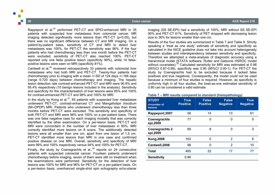
26 Colon cancer KCE Report 218
Rappeport et al.25 performed PET-CT and SPIO-enhanced MRI in 35 patients with suspected liver metastases from colorectal cancer. MR imaging detected significantly more lesions than PET-CT (p<0.05), but there was no significant difference between CT and MR imaging. On a patient-by-patient basis, sensitivity of CT and MRI to detect liver metastases was 100%, for PET-CT the sensitivity was 96%. If the four patients who had chemotherapy less than one month before the PET-CT were excluded, sensitivity of PET-CT also reached 100%. PET-CT reported only one false positive lesion (specificity 99%), while 14 false-positive lesions were seen on MRI (specificity 81%). Cantwell et al.26 reviewed retrospectively 33 patients with colorectal liver metastases who had both MRI and PET-CT. Twenty-four patients had chemotherapy prior to imaging with a mean +/-SD of 124 days +/-166 days (range 5-720 days) between chemotherapy and imaging. The hepatic lesion detection rate contrast-enhanced PET-CT and MRI were 90.9% and 95.4% respectively (10 benign lesions included in the analysis). Sensitivity and specificity for the characterisation of liver lesions were 85% and 100% for contrast-enhanced PET-CT and 98% and 100% for MRI. In the study by Kong et al.27, 65 patients with suspected liver metastases underwent PET-CT, contrast-enhanced CT and Mangafodipir trisodium (Mn-DPDP) MRI. Patients who underwent chemotherapy less than three months before PET-CT were excluded. The sensitivity and specificity of both PET-CT and MRI were 98% and 100% on a per-patient basis. There was one false negative case for each imaging modality that was correctly identified by the other examination. On a per-lesion basis, PET-CT and MRI were concordant in the number of liver metastases in 85%. MRI correctly identified more lesions on 8 scans. The additionally detected lesions were all smaller than one cm, apart from one lesion of 1.5 cm. PET-CT identified more lesions than MRI in one case and confirmed positive disease on one MRI. Overall, sensitivity and specificity of MRI were 99% and 100% respectively versus 94% and 100% for PET-CT. Finally, the study by Coenegrachts et al.,28 reports on 24 consecutive patients with suspected colorectal cancer. Fourteen patients underwent chemotherapy before imaging, seven of them were still on treatment when the examinations were performed. Sensitivity for the detection of liver lesions was 100% for MRI and 96% for PET-CT on a per-patient basis. On a per-lesion basis, unenhanced single-shot spin echography echo-planar
imaging (SS SE-EPI) had a sensitivity of 100%, MRI without SS SE-EPI 90% and PET-CT 61%. Sensitivity of PET dropped with decreasing lesion size to 36% for lesions smaller than one cm. Results of the four studies are summarized in Table 7 and Table 8. Strictly speaking a ’treat as one study’ estimate of sensitivity and specificity as calculated in the NICE guideline does not take into account heterogeneity between studies and interdependency between sensitivity and specificity. Therefore we attempted a meta-analysis of diagnostic accuracy using a hierarchical model (STATA software, Rutter and Gatsonis HSROC model without covariates).29 Calculated sensitivity for MRI was estimated at 0.98 (95%CI 0.87-0.99), specificity was 0.95 (95%CI 0.00-1). For PET-CT the study by Coenegrachts had to be excluded because it lacked false positives and true negatives. Consequently, the model could not be used because a minimum of four studies is required. However, as specificity is uniformly high in all four studies, the treat-as-one estimated sensitivity of 0.80 can be considered a valid estimate.
Table 7 – MRI results compared to standard (histopathology) STUDY n=number of lesions
True Positive
False Positive
False Negative
True Negative
Rappeport,2007 58 14 13 61
Coenegrachts epi,2009
77 0 0 0
Coenegrachts 2 spi,2009
69 8 0 0
Kong,2008 163 0 2 6
Cantwell,2008 98 0 2 10
Total 465 22 17 77
Sensitivity 0.96
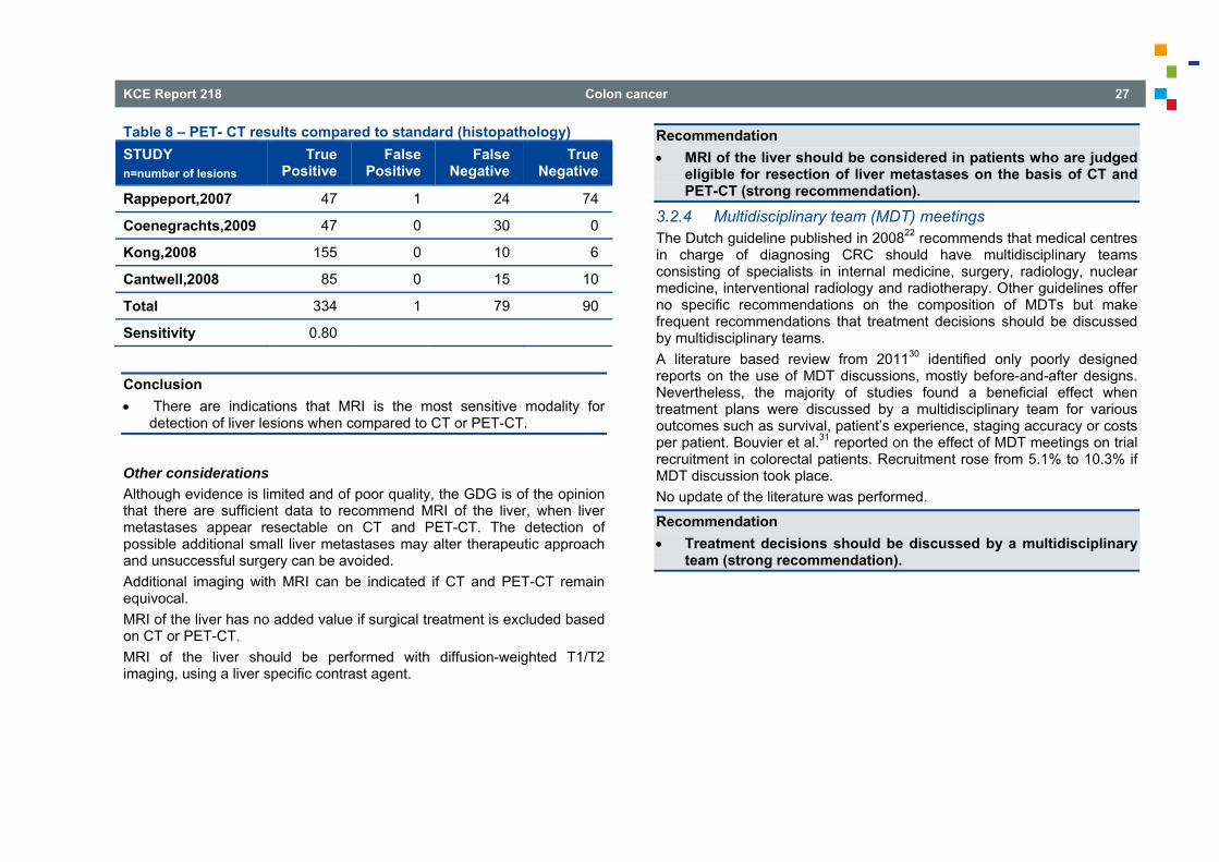
KCE Report 218 Colon cancer 27
Table 8 – PET- CT results compared to standard (histopathology) STUDY n=number of lesions
True Positive
False Positive
False Negative
True Negative
Rappeport,2007 47 1 24 74
Coenegrachts,2009 47 0 30 0
Kong,2008 155 0 10 6
Cantwell,2008 85 0 15 10
Total 334 1 79 90
Sensitivity 0.80
Conclusion There are indications that MRI is the most sensitive modality for
detection of liver lesions when compared to CT or PET-CT.
Other considerations Although evidence is limited and of poor quality, the GDG is of the opinion that there are sufficient data to recommend MRI of the liver, when liver metastases appear resectable on CT and PET-CT. The detection of possible additional small liver metastases may alter therapeutic approach and unsuccessful surgery can be avoided. Additional imaging with MRI can be indicated if CT and PET-CT remain equivocal. MRI of the liver has no added value if surgical treatment is excluded based on CT or PET-CT. MRI of the liver should be performed with diffusion-weighted T1/T2 imaging, using a liver specific contrast agent.
Recommendation MRI of the liver should be considered in patients who are judged
eligible for resection of liver metastases on the basis of CT and PET-CT (strong recommendation).
3.2.4 Multidisciplinary team (MDT) meetings The Dutch guideline published in 200822 recommends that medical centres in charge of diagnosing CRC should have multidisciplinary teams consisting of specialists in internal medicine, surgery, radiology, nuclear medicine, interventional radiology and radiotherapy. Other guidelines offer no specific recommendations on the composition of MDTs but make frequent recommendations that treatment decisions should be discussed by multidisciplinary teams. A literature based review from 201130 identified only poorly designed reports on the use of MDT discussions, mostly before-and-after designs. Nevertheless, the majority of studies found a beneficial effect when treatment plans were discussed by a multidisciplinary team for various outcomes such as survival, patient’s experience, staging accuracy or costs per patient. Bouvier et al.31 reported on the effect of MDT meetings on trial recruitment in colorectal patients. Recruitment rose from 5.1% to 10.3% if MDT discussion took place. No update of the literature was performed.
Recommendation Treatment decisions should be discussed by a multidisciplinary
team (strong recommendation).
28 Colon cancer KCE Report 218
3.3 Pathology 3.3.1 KRAS mutational analysis This section focuses on the ability of KRAS mutational analysis to predict the effect of epidermal growth factor receptor (EGFR) treatment. We did not focus on KRAS as a prognostic marker. A high quality SR performed in the framework of the Ontario Health Technology Assessment Series was updated.32 In the update we focused on systematic reviews and subgroup analysis of RCTs comparing the effect of EGFR treatment in KRAS mutant and KRAS wild type patients. Three systematic reviews and twelve primary studies were identified, most studies were however already included in the systematic reviews. Fourteen observational studies were identified, four for cetuximab monotherapy, seven for the cetuximab-irinotecan combination therapy and three for panitumumab monotherapy. The findings of the HTA report supplemented with studies published since 2010 (search date of the HTA) are presented.
3.3.1.1 KRAS predicting efficacy of cetuximab monotherapy (third line)
Four observational studies were included in the Ontario Health Technology Assessment review. Karapetis et al. (2008) is a retrospective analysis (stratified by KRAS status) of an RCT which compared the effectiveness of cetuximab and best-supportive care (BSC) compared to BSC alone in the treatment of patients with advanced colorectal cancer refractory to chemotherapy. The Karapetis data included 394 patients out of 572 patients in the original trial (68.8%) for whom KRAS status was available. There was no evidence of a prognostic effect as survival times were similar in the BSC arm for OS (4.6 and 4.8 months) and PFS (1.8 and 1.9 months) for KRAS mutated and KRAS wild type (WT) patients, respectively. However, Karapetis et al. showed a significant KRAS-treatment interaction (p<0.05) for both OS and PFS. This significant interaction indicates that the effect of cetuximab differs by KRAS status, with improved OS and PFS favouring the KRAS WT compared to the mutated patients treated with cetuximab. Three more observational (single arm) treatment studies
reached similar conclusions but due to the lack of a control arm these studies can only evaluate the prognostic value. The systematic reviews by Vale et al.33 and Adelstein et al.34 confirmed this result. No additional primary studies on the role of KRAS predicting efficacy of cetuximab monotherapy were identified.
3.3.1.2 KRAS predicting efficacy of Panitumumab Monotherapy (third line)
Similar to the analysis of Karapetis et al.32 for cetuximab monotherapy, Amado et al. published a retrospective analysis in 2008 (stratified by KRAS status) of a previously conducted RCT of panitumumab compared to BSC by Van Cutsem et al.; 427 of the 463 patients in the Van Cutsem trial with known KRAS status were included. Testing of the KRAS-treatment interaction term was significant, although not for the outcome of OS. These results suggest a benefit with respect to PFS for KRAS WT patients treated with panitumumab. The lack of a significant effect for OS, however is likely attributable to the fact that cross-over was allowed in this RCT, with approximately 76% of the BSC patients receiving treatment. Two additional observational studies without control arm were identified by the systematic review. The systematic reviews by Vale et al.33 and Adelstein et al.34 confirmed this result. No additional primary studies were identified. Adelstein et al.34 also pooled the effect of cetuximab and panitumumab monotherapy coming to the same conclusion.
3.3.1.3 KRAS predicting efficacy of Cetuximab-Irinotecan Combination Therapy
The Ontario review32 identified seven observational studies. Pooling five of the seven studies for which relevant data were available, they found a mean difference in PFS of 3.32 months (6 months against 3) (95%CI 1.78-4.86) in favour of the KRAS WT patients (p<0.00001), however with considerable heterogeneity. The median OS was also highest for patients without the mutation (approximately 14 months) compared to those with the mutation (8 months), with a pooled mean difference of 4.11 months (95%CI 2.62-5.60) in favour of the WT patients. Similar to the pooled estimates of the PFS data, there was significant heterogeneity in the pooling of the OS data (p<0.00001, I²=95%).
KCE Report 218 Colon cancer 29
In contrast with the data on cetuximab, there is only evidence that KRAS has a prognostic value but the evidence does conclude on treatment effect modification by KRAS.
3.3.1.4 KRAS predicting efficacy of Cetuximab- FOLFIRI Combination Therapy (first line)
Significant interaction for treatment effect between KRAS mutant and KRAS WT patients was demonstrated for PFS (p=0.0028), overall survival (p=0.0463), and best overall response (p=0.0005) by Van Cutsem et al.35 in an updated secondary subgroup analysis of the Crystal randomized trial, where patients were randomly assigned to receive FOLFIRI with or without cetuximab. Bokemeyer et al.36 pooled the CRYSTAL and OPUS randomised clinical trials, both demonstrating that adding cetuximab to first-line chemotherapy in patients with KRAS WT metastatic colorectal cancer (mCRC) significantly improved treatment outcome compared with chemotherapy alone. Pooled individual patient data from each study were analysed for overall survival (OS), progression-free survival (PFS) and best overall response rate (ORR) in patients evaluable for KRAS and BRAF mutation status. In 845 patients with KRAS WT tumours adding cetuximab to chemotherapy led to a significant improvement in OS (hazard ratio [HR] 0.81; p=0.0062), PFS (HR 0.66; p<0.001) and ORR (odds ratio 2.16; p<0.0001). BRAF mutations were detected in 70/800 evaluable tumours. No significant differences were found in outcome between the treatment groups for these patients. Prognosis was worse in each treatment arm for patients with BRAF tumour mutations compared with those with BRAF WT tumours. In conclusion, analysis of pooled data from the CRYSTAL and OPUS studies confirms the consistent benefit obtained across all efficacy end-points by adding cetuximab to first-line chemotherapy in patients with KRAS wild-type mCRC.
3.3.1.5 KRAS predicting efficacy of Panitumumab - FOLFIRI Combination Therapy in second line therapy
A trial reported by Peeters et al.37 evaluated the efficacy and safety of panitumumab plus fluorouracil, leucovorin, and irinotecan (FOLFIRI) compared with FOLFIRI alone after failure of initial treatment for mCRC patients by tumour KRAS status. KRAS status was available for 91% of patients: 597 (55%) with WT KRAS tumours, and 486 (45%) with mutant (MT) KRAS tumours. In the WT KRAS subpopulation, PFS was improved (HR 0.73; 95%CI 0.59 - 0.90; p=0.004); median PFS was 5.9 months for panitumumab-FOLFIRI versus 3.9 months for FOLFIRI. A non-significant trend toward increased OS was observed. In patients with MT KRAS no difference in efficacy was demonstrated. There was no statistically significant difference in PFS (HR 0.85; 95%CI 0.68 - 1.06; p=0.14, stratified log-rank). Note however that the statistical test for interaction between KRAS result and effect was not significant.
3.3.1.6 KRAS predicting efficacy of Panitumumab or Cetuximab - with oxaliplatin based Combination Therapy in first line therapy
Adelstein et al.34 pooled four studies (Bokemeyer et al. 2009, Maughan et al. 2010, Douillard et al. 2010, Tveit et al. 2010) comparing the addition of Panitumumab or Cetuximab to an oxaliplatin based combination therapy with combination therapy alone. Whereas a significant effect was demonstrated in the pooled effect in KRAS WT patients for PFS, HR 0.86 (95%CI 0.70-1.05) and not in the KRAS mutated HR 1.13 (95%CI 0.86-1.47), the overall assessment of interaction was not significant (HR for interaction 0.75 (95%CI 0.47-1.18)). However, there was considerable heterogeneity, Bokemeyer et al., 2009 and Douillard et al., 2010 showing an effect in KRAS WT and a clear interaction as opposed to Tveit et al., 2010 and Maughan et al., 2010 who show no effect in any KRAS subgroup.
30 Colon cancer KCE Report 218
3.3.1.7 KRAS predicting efficacy of Panitumumab or Cetuximab added to a bevacizumab based therapy in first line therapy
Adelstein et al.34 pooled the studies by Hecht et al., 2009 and Tol et al., 2009, both studies showed a shortened PFS and no difference in effect between KRAS mutated and wild type. However these regimens are not recommended anyway for this indication.
3.3.1.8 Studies reporting an overall pooling of treatment effects and interactions
Adelstein et al.34 provided a statistically significant pooled estimate for the interaction between KRAS mutated and non mutated overall treatments. Dahabreth et al.38 reported an overall sensitivity and specificity in predicting response. The clinical relevance of those pooled data is questionable.
3.3.1.9 Mutant KRAS codon 12 and 13 alleles as predictive biomarkers.
Tejpar et al.39 assessed the associations between tumour KRAS mutation status (WT, G13D, G12V or other mutations) and progression-free survival (PFS), survival and response in pooled data from 1 378 evaluable patients from the CRYSTAL and OPUS studies. In patients with G13D-mutant tumours, cetuximab plus chemotherapy versus chemotherapy alone significantly improved PFS (median 7.4 vs. 6.0 months; HR 0.47; p=0.039) and tumour response (40.5% vs. 22.0%; OR 3.38; p=0.042) but not survival (median 15.4 vs. 14.7 months; HR 0.89; p=0.68). Patients with G12V and other mutations did not benefit from this treatment combination. Peeters et al.40 did a retrospective analysis of three randomized phase III studies assessing the prognostic and predictive impact of individual mutant KRAS codon 12 and 13 alleles. Patients were randomly assigned 1:1 to FOLFOX4 (infusional fluorouracil, leucovorin, and oxaliplatin) in study 20050203, FOLFIRI (fluorouracil, leucovorin, and irinotecan) in study 20050181, or best supportive care in study 20020408 with or without panitumumab 6.0 mg/kg once every 2 weeks. In all, 441 (study 20050203), 486 (study 20050181), and 126 (study 20020408) patients with mutant KRAS codon 12 or 13 alleles were included in the analysis. They found
that no mutant KRAS allele in patients treated in the control arm emerged as a consistent prognostic factor for PFS or OS. In addition, no mutant KRAS allele was consistently identified as a predictive factor for PFS or OS in patients receiving panitumumab treatment. Significant interactions for individual mutant KRAS alleles were observed only in study 20050203 with G13D negatively and G12V positively associated with OS in the panitumumab-containing arm. Pooled analysis indicated that only G12A was associated with a negative predictive effect on OS. They conclude that mutant KRAS codon 12 or 13 mCRC tumours are unlikely to benefit from panitumumab therapy and that panitumumab therapy should be limited to patients with WT KRAS mCRC.
3.3.2 BRAF BRAF mutations are associated with poor prognosis41 but here the focus is on the potential role of BRAF mutations in predicting treatment effects. We identified a HTA report by Lea et al.42 that assessed the clinical validity of testing for the BRAF p.Val600Glu sequence variant and found 7 mainly retrospective cohort studies investigating the ability to predict response to treatment with cetuximab or panitumumab. Across these studies, the incidence of BRAF sequence variants ranged from 4.3% to 14.9% of patients with CRC. Across these 7 studies, only 3 of 78 patients with a BRAF p.Val600Glu sequence variant responded to treatment with cetuximab or panitumumab. In the update we focused on systematic review and subgroup analysis of RCTs comparing the effect of EGFR treatment in BRAF mutant and BRAF WT patients. We found one pooled analysis and one retrospective analysis of RCTs. Bokemeyer et al.36 pooled the CRYSTAL and OPUS randomised clinical trials, both demonstrating that adding cetuximab to first-line chemotherapy in patients with KRAS wild-type metastatic mCRC significantly improved treatment outcome compared with chemotherapy alone. Pooled individual patient data from each study were analysed for overall survival (OS), progression-free survival (PFS) and best overall response rate (ORR) in patients evaluable for KRAS and BRAF mutation status. BRAF mutations were detected in 70/800 evaluable (wild type) tumours. No significant differences were found in outcome between the treatment groups in these patients. Prognosis was worse in each treatment arm for
KCE Report 218 Colon cancer 31
patients with BRAF tumour mutations compared with those with BRAF WT tumours. Therefore, BRAF mutation does not appear to be a predictive biomarker in this setting, but is a marker of poor prognosis. Ogino et al.43 assessed status of BRAF c.1799T>A (p.V600E) mutation and MSI in 506 stage III colon cancer patients enrolled in a randomized adjuvant chemotherapy trial [5-fluorouracil and leucovorin (FU/LV) vs. irinotecan (CPT11), FU and LV (IFL); (study CALGB 89803). They found that BRAF mutation status was a prognostic marker but could not ascertain the role as a predictor of treatment efficacy.
3.3.3 N-RAS as a predictor of treatment effectiveness In a study identified by the GDG group but published after the search date, Douillard et al.44 assessed the efficacy and safety of panitumumab plus oxaliplatin, fluorouracil, and leucovorin (FOLFOX4) as compared with FOLFOX4 alone, according to RAS (KRAS or NRAS) or BRAF mutation status. A total of 639 patients who had metastatic colorectal cancer without KRAS mutations in exon 2 had results for at least one of the following: KRAS exon 3 or 4; NRAS exon 2, 3, or 4; or BRAF exon 15. The overall rate of ascertainment of RAS status was 90%. A total of 108 patients (17%) with non-mutated KRAS exon 2 had other RAS mutations. These mutations were associated with inferior progression-free survival and overall survival with panitumumab-FOLFOX4 treatment. Non KRAS exon 2 mutations were pooled (mutations in KRAS exon 3 (at codon 61) and exon 4 (at codons 117 and 146); NRAS exon 2 (at codons 12 and 13), exon 3 (at codon 61), and exon 4 (at codons 117 and 146). In the primary analysis, interaction testing between the subgroups that did not have RAS mutations and the subgroups that did not have KRAS mutations in exon 2 but did have other RAS mutations was significant for progression-free survival (p=0.04) but not for overall survival (p=0.07). In the updated analysis of overall survival, which was based on a larger number of deaths from any cause, the results of interaction testing were significant (p=0.01). Although these results indicate that treatment effects differed between the subgroups of patients without RAS mutations and those without KRAS mutations in exon 2 but with other RAS mutations - suggesting that RAS mutations in addition to KRAS mutations in exon 2, were negative predictive factors – it is based on a pooled analysis of different non KRAS
exon 2 KRAS and NRAS mutations, hence the individual contribution NRAS testing needs further assessment.
3.3.4 MSI testing as a predictor of treatment effectiveness
3.3.4.1 Predicting the effect of adjuvant therapy According to Sargent et al.,45 approximately 15% of CRCs have defective DNA-mismatch repair (MMR). Defective MMR (dMMR) has frequently been measured by either the presence of microsatellite instability (MSI) or by testing for loss of the protein products for genes involved in DNA mismatch repair, most commonly MLH1, MSH2, MSH6, and PMS2. CRCs with dMMR have distinctive features that include proximal colon predominance, poor differentiation and/or mucinous histology, intra- and peritumoural lymphocytic infiltration and diploid DNA content. 45 Des Guetz et al.46 reviewed the role of microsatellite instability status in predicting the efficacy of adjuvant chemotherapy and performed a meta-analysis on seven studies representing 3 690 patients; mean age 65.5 years; 810 stage II and 2 444 stage III (75%). MSI-high (MSI-H) was found in 454 patients (14% of the global population), and microsatellite stable (MSS) in 2 871. A total of 1 444 patients received 5-fluorouracil (5FU)-based chemotherapy, whereas 1 518 patients did not. For MSI-H patients, there was no statistically significant difference for relapse-free survival (RFS) whether or not they received chemotherapy (5 studies; HR RFS: 0.96, 95%CI 0.62–1.49; HR OS (6 studies): 0.70, 95%CI 0.44-1.09; p=0.12). They found a significant interaction between MSI status (MSI-H or MSS) and therapeutic status suggesting a smaller benefit for MSI-H than for MSS patients (HR interaction RFS 0.77; 95%CI 0.67-0.87). Sargent et al.45 distinguished between stage II and III (Des Guetz et al. stated that they were not able to obtain data sufficiently detailed to do this) and found no benefit from 5-FU based treatment in a pooled data set (of which part of the data were included in Des Guetz et al.), including data for patients with either stage II (HR 2.30; 95%CI 0.85-6.24; p=0.09) or stage III (HR 1.01; 95%CI 0.41-2.51; p=0.98) disease with dMMR. No treatment benefit was present in patients with pMMR and stage II disease (HR 0.84; 95%CI 0.57-1.24; p=0.38). In patients with stage III disease and pMMR tumours, a benefit from treatment was observed (HR 0.64; p=0.001). The interaction test between MMR status and treatment efficacy for DFS was
32 Colon cancer KCE Report 218
significant (p=0.04), which indicated that the effect of treatment differs by MMR status. All findings were consistent for the OS end point, with one exception. For the OS end point, there was a statistically significant decreased OS in patients with stage II disease and dMMR tumours who were treated compared with patients in the surgery-alone control (HR 2.95; 95%CI 1.02-8.54; p=0.04). Hutchins et al.47 found no evidence for less sensitivity of 5-FU based chemotherapy in dMMR patients; however, the confidence interval around the estimation was large (0.81; 95%CI 0.29-2.22), and the study was underpowered. A pooled analysis by Sinicrope et al.48 was excluded as it overlapped with Sargent et al.;45 it focused (inconclusively, due to lack of power) on the difference between germline vs. sporadic CRC. Observational studies examined the relation between oxaliplatin based treatments (FOLFOX) and MSI instability but were inconclusive and not included in the review.
3.3.4.2 Metastatic colon cancer Des Guetz et al.49 pooled six studies representing 964 patients (mean age 63 years; 91 MSI-H and 873 microsatellite stable (MSS) tumours). A total of 287 patients received 5-fluorouracil based chemotherapy, whereas 678 patients received combinations of 5FU or capecitabine with oxaliplatin and/or irinotecan. They found no benefit of metastatic chemotherapy in terms of RR for MSI-H patients compared with MSS patients. The global hazard ratio (HR) for RR was 0.82 (95%CI 0.65-1.03; p=0.09). Different treatment schedules containing FU were pooled; the appropriateness could be questioned but separate analysis would reach the same conclusion, i.e. there is no proof that MSI instability has the power to predict the effectiveness of FU containing regimens. Two observational studies concerning FOLFOX and FOLFIRI were found in the update but not included as they only assessed the prognostic value and not the predictive value of MSI instability.
Conclusions KRAS mutation status is an effect modifier for anti-EGFR antibodies,
with the efficacy of anti-EGFR antibodies limited to KRAS wild type patients.
There are indications that G13D-mutant tumours may benefit from cetuximab treatment but this needs confirmation since a large study with panitumumab could not confirm this finding.
Recent data suggest that in addition to KRAS mutations in exon 2, other RAS mutations are negative predictive factors. However, this conclusion is based on a pooled analysis of different (non KRAS exon 2) KRAS and NRAS mutations, implying that the individual contribution NRAS testing needs further assessment.
BRAF mutation is a strong indicator of poor prognosis but its ability to predict treatment response remains unclear.
MSI predicts treatment effectiveness of adjuvant treatment with 5-FU alone in stage (I and) II colorectal cancer. However, the predictive value of MSI concerning combination therapies with oxaliplatin (FOLFOX) is uncertain.
There is no proof that MSI instability predicts treatment effectiveness in metastatic colorectal cancer.
Recommendations RAS mutation status should be assessed in all patients when
anti-EGFR treatment is considered (strong recommendation). If a patient is considered for adjuvant 5FU-monotherapy, MSI
testing should be performed. If the tumour is MSI-high, no 5FU-monotherapy should be given (strong recommendation).
KCE Report 218 Colon cancer 33
3.3.5 Standards for pathology report Pathology reports of resected colon cancer should contain all information necessary to determine stage and prognosis and to decide on further treatment. The Dutch guideline22 lists the following items that need to be reported in a pathology report: Histological type of the tumour Differentiation grade of the tumour Depth of invasion Distance of the tumour to the resection margin and completeness of
the resection Number of resected lymph nodes and number of lymph nodes
containing metastases Size of the tumour Optionally, also a macroscopic description of the tumour, perineural invasion, vascular invasion and lymphatic invasion can also be reported. The guideline recommends the use of standardized pathology reports. Similarly, the SIGN guideline11 recommends that all reporting of colorectal cancer specimens should be done according to, or supplemented by, the Royal College of pathologists’ minimum data set. Reports should include the following information: Tumour differentiation Staging (Dukes and TNM systems) Margins (peritoneal and CRM) Extramural vascular invasion The SIGN guideline refers to two randomized studies showing that the use of template proformas significantly increases the rate of inclusion of data items in pathology reports.
The need for synoptic reporting and inclusion of all information for complete staging of the tumour is confirmed in the guideline of NZGG.50 The minimal dataset developed by the British Royal College of Pathologists can be found on their website (http://www.rcpath.org/Resources/RCPath/Migrated%20Resources/Documents/G/G049-ColorectalDataset-Sep07.pdf) and in the appendix. Update No update of the literature was performed. We refer to the Protocol for the Examination of Specimens From Patients With Primary Carcinoma of the Colon and Rectum of the College of American Pathologists as additional information (http://www.cap.org/apps/docs/committees/cancer/cancer_protocols/2012/Colon_12protocol_3200.pdf)
Recommendation Pathology reports should at least contain the minimal datasets as
defined by international professional organizations; it should always include the pathological TNM classification (strong recommendation).
3.3.6 Number of lymph nodes The presence or absence of tumour involvement of lymph nodes differentiates between stage II and III colon cancer. This differentiation is crucial to decide on the use of adjuvant chemotherapy. The Dutch guideline22 reviewed the literature on the number of lymph nodes needed for examination. Observational studies have shown that determination of stage and prognosis improves with an increasing number of examined lymph nodes. However, it is not possible to define necessary minimum number of lymph nodes from the literature. The recommended number varies from nine to as many as possible. According to the seventh edition of the TNM Classification of Malignant Tumours of the International Union Against Cancer (UICC), a minimum of 12 lymph nodes should be investigated.51 The Dutch guideline concluded that as many lymph nodes as possible should be examined but at least 10 as that is the threshold for decisions on the use of adjuvant chemotherapy. They further mention that current staging is based on routine hematoxylin and eosin stained samples
34 Colon cancer KCE Report 218
and that therefore no specialised preparation techniques or immunohistochemistry should be used. SIGN11 does not make a specific recommendation on the number of lymph nodes needed to be resected. It is advised that the fat tissue is carefully dissected to retrieve all lymph nodes. Other considerations At least 12 lymph nodes should be examined to comply with the current TNM classification of Malignant Tumours. However, the tumour can be staged pN0 regardless of the number of lymph nodes examined. In vivo and ex vivo detection of sentinel lymph nodes have been investigated in colon cancer. Serial sectioning and immunohistochemistry results in a mean upstaging of 18.9% (range 0-50%). However, as it is still unclear how results should be implemented in clinical practice, sentinel node procedures are not further discussed in this guideline.52, 53
Recommendation For the pathological examination of resection specimens of
colorectal cancer, as many lymph nodes as possible should be assessed for the presence of tumour cells. Only routine hematoxylin and eosin stained samples should be used (strong recommendation).
3.4 Surgical treatment stage 0-III 3.4.1 Endoscopic treatment stage I: polypectomy The NHMRC guideline54 advocates polypectomy alone in the management of malignant polyps as standard and safe practice, providing that there is adherence to a strict policy of case selection and histopathological assessment recognising four key features needed to identify a very low risk of lymph node metastasis: (1) a clear margin of excision (1 to 2 mm) (2) cancer which is well - or moderately-differentiated (3) absence of lymphatic and venous invasion (4) complete removal as assessed endoscopically Their recommendations stem from one case-control study, five case series and one narrative review.
The Australian guideline further suggests that malignant polyps with unfavourable features may require further treatment but this decision should be made on the basis of the age, site, health and wishes of the patient. Further excision can be achieved successfully by laparotomy with colonic resection or laparoscopically assisted colectomy.55 The advice is based on one SR (Hassan et al. 2005,56 which is solely based on retrospective case series), one RCT (Liang et al. 2002,57 comparing laparoscopy-assisted colectomy versus laparotomy), three narrative reviews and five case series. The SR concluded that a positive resection margin is largely predictive of residual local disease, the presence of poorly differentiated carcinoma is mainly associated with a higher cancer-related mortality and vascular invasion with a higher risk of lymph node metastasis.56 SIGN11 recommends further surgery for pedunculated polyp cancers that have been removed endoscopically if at least one of the following conditions is present: (1) there is histological evidence of tumour at, or within 1 mm of, the resection margin (2) there is lymphovascular invasion (3) the invasive tumour is poorly differentiated This recommendation is based on two cohort studies with a low risk of confounding or bias. Furthermore, it is stressed that the results of a case series illustrated that although T1 tumours (those with the smallest local spread) are often deemed suitable for local excision, extensive involvement of the submucosa is associated with a 17% rate of lymph node involvement.3 Minimal involvement of the submucosa (T1 sm1 tumours) appears to be associated with minimal risk of lymph node involvement. NICE 201112 states that the colorectal MDT should consider further treatment for patients with locally excised, pathologically confirmed stage I cancer taking into account pathological characteristics of the lesion, imaging results and any previous treatments. It suggests further treatment to patients whose tumour had involved resection margins (less than 1 mm); a recommendation that is based on retrospective case series. Furthermore, the risks and benefits of all treatment options should be discussed with the patient after discussion in the MDT.
KCE Report 218 Colon cancer 35
The authors of the NICE guideline concluded that there was no evidence to answer the question "What are the prognostic factors for determining the most effective curative treatment for patients diagnosed with stage I colorectal cancer, including/or polyp cancer?", since much of the literature concentrates on identifying the unfavourable prognostic features rather than focusing on the long term outcomes related to such features or on assessing which type of treatment is best for patients with specific unfavourable characteristics. The Dutch guideline does not cover this topic.22 Update The additional search did not yield any (randomized or non-randomized) comparative study that reported the primary outcomes of interest (Overall survival, PFS, QoL) for polypectomy followed by surveillance versus polypectomy followed by surgery.
Conclusion There is no evidence to compare the effect of polypectomy followed by
surveillance with polypectomy followed by (segmental) colon resection in patients who were diagnosed with Tis/T1 colorectal cancer after endoscopic polypectomy, in terms of overall survival, progression-free survival or quality of life.
Observational studies There are some observational studies that suggest that polypectomy followed by surveillance may be safe for low-risk Tis/T1 colorectal cancer but not in high risk cancer. The results of one review of observational studies and five observational studies (all with methodological limitations) are listed below. All studies are considered of very low quality as no appropriate eligibility criteria for the different treatment groups were applied, confounding was not appropriately controlled and there were no data on the completeness of follow-up. In 2012, Di Gregorio et al.58 performed a review on the available literature on the outcome of low- and high-risk malignant colorectal polyps. No quality assessment of the included observational studies was performed. High risk polyps were defined by the presence of at least one of the following histological features: positive resection margin, poorly
differentiated adenocarcinoma, lymphatic/vascular invasion or tumour budding. If none of those features were present, polyps were classified as low risk. Overall, there were 345 patients with a low risk polyp reported of whom 53 underwent surgery after polypectomy. In one of the 53 surgical specimens, residual disease was reported. One of the 345 low risk cancer patients died due to cancer. There were in total 471 patients with a high risk polyp included, 335 of them underwent surgery. In 49 of the 335 (14.6%) surgical specimens, residual cancer was seen; 23/471 (4.9%) patients died due to cancer. Results for the separate risk factors (present vs. absent) are summarized in the appendix. These results should be interpreted with great caution as it is not clear which patients underwent surgery and there is no correction for the other risk factors. Benizri et al.59 summarized a retrospective case series of 64 patients with T1 CRC in whom resection (either by laparotomy or laparoscopy) and regional lymphadenectomy was performed after analysis of the polypectomy specimen had revealed at least one of the following adverse criteria: inadequate excision with cancer free distance of the resection margin ≤1 mm, lymphovascular invasion, poorly differentiated carcinoma (grade III), submucosal SM 2-3 involvement, tumour budding, sessile morphology or piecemeal resection (see appendix). The rate of residual adenocarcinoma and/or lymph node metastasis was 7/64 (11%). Post-operative complications were observed in 16/64 (25%) patients. Butte et al.60 reported on a retrospective case series of 143 consecutive patients with T1 CRC undergoing polypectomy followed by colectomy (see appendix). At colectomy, invasive residual disease was observed in sixteen (11%) patients, non-invasive in three (2.1%) and lymph node metastasis in ten (7%). Collectively, in 13% of patients residual disease was diagnosed at the moment of surgery. In case of positive or unknown resection margin, the rate of residual invasive disease in the colonic wall was 16% vs. 0% in case of a negative resection margin. After a median follow-up period of 63 months, no recurrences were identified; 122 patients were still alive, 15 died of unknown causes and 6 died of other causes. Kim et al.61 followed retrospectively a case series of 64 patients with intramucosal CRC and 65 patients with submucosal CRC who all had either EMR (Endoscopic mucosal resection) or ESD (Endoscopic submucosal resection) performed (see appendix). After a mean follow-up period of 19 months, 62 patients with intramucosal CRC were still alive;
36 Colon cancer KCE Report 218
two died of unrelated diseases. The survival rate for patients who had submucosal CRC was not reported. Seven patients with submucosal cancer had colectomy performed during the follow-up period, five because of positive resection margin or lymphovascular involvement, one because of bowel perforation and one patient requested surgery (see appendix). The recurrence rate (i.e. local recurrence and/or distant recurrence) was 0/64 in the intramucosal group and 7/65 in the submucosal group (3/7 underwent colectomy and 4/7 only had polypectomy). Of the seven patients who suffered from recurrence, five had a high risk polyp and two a low risk polyp. The total number of high risk and low risk polyps included in the study is unclear. Meining et al.62 documented on 390 patients with T1 CRC: 141 patients had polypectomy and surgical removal of T1 CRC (group A) and 249 only had polypectomy (group B) (see appendix). Decision in favour or against surgery was based on risk patterns, patients’ personal wishes and patients’ fitness. Both low-risk and high-risk polyps were included in both groups. An unfavourable outcome was defined as locoregional cancer relapse, distant metastasis, lymph node metastasis or death related to CRC. In the polypectomy only group, an unfavourable outcome was observed in 17/249 (6.8%) patients. In this polypectomy only group, the rate of unfavourable outcome was 20% in case of incomplete resection versus 4% in case of complete resection; poorly differentiated tumours had an unfavourable outcome in 43% of cases versus 6% in other tumours and 44% of tumours with lymphovascular infiltration had an unfavourable outcome versus 5% in other cases. Oka et al.63 reported on retrospective case series of 792 patients with submucosal CRC who only had surveillance after endoscopic resection (see appendix). The data were collected from 15 centres in Japan. The recurrence rate was 18/792 (2.3%) (local recurrence in 11 cases and metastatic recurrence in 13 cases). The association between histopathological characteristics at polypectomy and recurrence was evaluated by means of a multivariate logistic regression analysis: lymphatic invasion was significantly associated with recurrence after ER in patients with submucosal CRC (OR: 6.36, 95% C.I. 1.46-27.79). It has to be mentioned though that this analysis was only based on 387 cases as the histopathological data were missing for 49% of the sample. The mean interval between ER and recurrence was 19.7 (+/- 9.2) months.
Other considerations At present, there are insufficient data from randomized trials that polypectomy is non-inferior to surgical resection. For low risk in situ tumours, the risk for metastases and recurrence is considered sufficiently low to accept polypectomy as definitive treatment. For T1 tumours, the evidence is considered insufficient to accept polypectomy as a safe option, unless the invasion is limited to SM1 level or if surgery is not feasible. Morbidity and impact on quality of life associated with segmental colectomy is considered to be low (as opposed to rectal surgery).
Recommendations In patients in whom Tis is diagnosed after polypectomy, no
additional treatment is indicated on the condition that (strong recommendation):
(1) there is a clear margin of excision (1 to 2 mm), (2) the tumour is well or moderately differentiated (3) there is no lymphatic or venous invasion. In patients in whom T1 is diagnosed after polypectomy, surgical
resection should be considered (strong recommendation). 3.4.2 Laparoscopic vs. open surgery SIGN11 suggests that both laparoscopic and open surgery can be offered for resection of colorectal cancer. The recommendation is based on a Cochrane Systematic Review covering studies up to 2008 that concluded that laparoscopic resection of carcinoma of the colon is associated with a long term outcome no different from that of open colectomy.64 The authors also mentioned that further studies are required to determine whether the method of approach has an impact on the incidence of incisional hernias and adhesions. NICE65 comes to the same conclusion: laparoscopic (including laparoscopically assisted) resection is recommended as an alternative to open resection for individuals with colorectal cancer in whom both laparoscopic and open surgery are considered suitable.12 They copied their recommendations from an earlier NICE document, in which the
KCE Report 218 Colon cancer 37
recommendations were formulated by the technology appraisal and not by the guideline developers. The NZGG50 as well as the IKNL guideline22 are in line with the previously cited: in experienced hands, laparoscopic surgery for colon cancer has equivalent outcomes to conventional surgery. NICE12 further states that the decision about which procedures (open or laparoscopic) is undertaken should be made after informed discussion between the patient and the surgeon. In particular, they should consider: the suitability of the lesion for laparoscopic resection, the risks and benefits of the two procedures and the experience of the surgeon in both procedures. In all selected guidelines it is unanimously stated that elective surgery for colon cancer should be performed by a surgeon with specific training and experience in colorectal surgery, and with sufficient caseload to maintain surgical skills.12,11,22,50 In addition, NICE12 recommends that the relevant national professional bodies should determine the exact criteria to be used. The American Society of Colon and Rectal Surgeons (ASCRS) and the Society of American Gastrointestinal and Endoscopic Surgeons (SAGES) have developed minimum requirements that surgeons must meet before they can perform laparoscopic surgery with curative intent in patients with cancer. The IKNL guideline states that surgeons must perform at least 20 laparoscopic colon operations for benign or incurable diseases before starting laparoscopic colon surgery with curative intent.22 Furthermore, the first 20 procedures are performed preferably under the supervision of an expert surgeon. Under these conditions, both benign and curative laparoscopic colorectal resections can be performed. Update The search for recent systematic reviews/meta-analyses yielded 1 550 references (after exclusion of doubles); since 2011 (i.e. final search date of the NICE guideline). Fifteen systematic reviews and meta-analyses examined laparotomy versus laparoscopy in colorectal patients. After evaluation of title and abstract six systematic reviews/meta-analyses were excluded (four narrative reviews, one cost-effectiveness study and one critical review of SRs). Full text assessment eliminated another four manuscripts (two narrative reviews, one article with a search until 2008 and one with the same studies included as a more recent SR published by
the same group (Ohtani et al. 201166). The critical appraisal, according to the AMSTAR criteria, of the five remaining publications is summarized in the appendix. The most recent (general) review (Ohtani et al. 2012)67 included 12 RCTs published between 2000 and 2011 that compared laparoscopy (2 444 patients) to laparotomy (2 170 patients) for colon cancer. The follow-up ranged between 30 and 95 months. No statistically significant differences were observed between intervention and control group with respect to long-term oncologic outcomes, i.e. overall mortality, cancer-related mortality, peri-operative mortality, overall recurrence, local recurrence, wound-site recurrence and distant metastases. In the short-term, laparoscopy was associated with significantly better outcomes with regard to intra-operative blood loss (in ml, WMD -103.90; 95%CI -180.88 to -26.91), peri-operative overall complications (OR 0.73, 95%CI 0.56 to 0.95), ileus (OR 0.40, 95%CI 0.25 to 0.66), time to oral resumption (in days, WMD -0.81, 95%CI -1.03 to -0.60) and hospital stay (in days, WMD -2.28, 95%CI -4.05 to -0.52). On the other hand, the operative time was significantly shorter for laparotomies (in minutes, WMD 42.08, 95%CI 29.87 to 54.30). The conversion rate in the laparoscopy group ranged between 3 and 46.4%. The review by Ma et al.68 included 15 RCTs that compared laparoscopy (2 126 patients) with laparotomy (2 081 patients) for colorectal cancer and that were published between 1997 and 2008; six of these RCTs were also included in the Ohtani et al. review. The authors come to identical conclusions: after a follow-up period that ranged between 1 and 59 months, both surgery types yielded comparable long-term oncologic outcomes (i.e. overall mortality, cancer-related mortality, overall recurrence, local recurrence, wound-site recurrence and distant metastases). Also in line with Ohtani et al., the overall complication rate was significantly lower in the laparoscopy group (OR: 0.71, 95% CI: 0.58-0.87). The authors comment that patients lost to follow-up were not included in the meta-analyses, which may have biased the results. In addition, it must be noted that the critical appraisal of individual studies was not reported.
38 Colon cancer KCE Report 218
The review by Sammour et al.69 concentrated on intra-operative complications. It is based on ten RCTs, that included patients with any indication for colonic or rectal surgery (among which one study on rectal cancer, one on Crohn's disease and one on diverticulitis). Their meta-analyses demonstrated a statistically significantly higher intra-operative complication rate (OR 1.37; 95%CI 1.06-1.76) and intra-operative bowel injury rate (OR 1.88; 95%CI 1.10-3.21) in the laparoscopy group. No statistically significant differences with respect to intra-operative haemorrhage or solid organ injury between the two treatment groups were observed. The authors comment that patients who presented acutely, with synchronous or invading neoplastic lesion or a cancer in the transverse colon were excluded from studies and hence not adopted in the meta-analyses. In addition, 20 trials were excluded because intra-operative complications were not reported separately. The review published by Grailey et al.70 focused on older patients. They evaluated ten comparative studies and one RCT that included 1 280 patients who were aged 70 or older. There was no statistically significant difference in operative mortality (i.e. 30-day mortality and in-hospital mortality taken together; nine trials), anastomotic leak rate or postoperative pneumonia between the laparoscopy and the open surgery group. Laparoscopic surgery resulted in a significantly shorter hospital stay (WMD -1.23, 95%CI -1.78 to -0.67), earlier return to normal bowel function (WMD: -1.23, 95%CI -1.84 to -0.61), shorter operative time (in minutes, WMD: 3.46, 95% CI 1.55-5.37) and reduced intra-operative blood loss (in ml, WMD -2.79; 95%CI -4.18 to -1.39). As most included studies were not randomized, selection bias cannot be excluded. In addition, the critical appraisal of individual studies was not reported. The review by Ding et al.71 focused on right hemicolectomies; one RCT and eleven retrospective non-randomized trials that included 1 057 patients, were assessed. The meta-analyses indicated no statistically significant difference in mortality rate or recurrence rate between the two treatment groups. Laparoscopic surgery was associated with significantly longer operative times (in minutes, WMD: 33.37; 95%CI 14.23-52.51). On the other hand, laparoscopic surgery resulted in reduced blood loss (in ml, WMD, -128.97, 95%CI -232.01 to -25.94), postoperative hospital stay (in days, WMD: -1.62, 95%CI -2.98 to -0.26) and reduced time to flatus (in days, WMD -0.96, 95% CI: -1.25 to -0.66). Conversions were
recorded in 0-21.4% of laparoscopy cases; the main reasons were: tumour invasion to adjacent structures, bulky tumours, extensive adhesions, obscure bleeding, cecum injury and hypercapnia. As most included studies were not randomized, selection bias cannot be excluded. The search for recent RCTs yielded 1 067 references after exclusion of duplicates. Since 2011 (i.e. final search date of the SR by Ohtani et al.) eight RCTs were published on the topic. After evaluation of title and abstract three studies were excluded (all were conference abstracts72-74). Full text assessment eliminated another manuscript (description of a study protocol, Kennedy et al. 201275). The evidence extracted from the four remaining RCTs is summarized in the appendix. The most recent publication describes the long-term results of the CLASICC trial; earlier published results of the trial were also adopted in the meta-analyses described in the previous paragraphs.76 In this non-blinded multi-centre study, 794 patients with colorectal cancer were randomized to either laparoscopically assisted colorectal resection (n=526) or open colorectal resection (n=268). After a median follow-up of 62.9 months, there were no statistically significant differences in median overall survival or median disease free survival between both groups. On the other hand, median disease free survival was significantly better for left-sided and sigmoid colonic resections than right-sided resections. The intra-operative conversion rate was 27% and the authors noticed that median overall survival as well as disease-free survival were significantly worse in these patients, even after adjustment for age, sex and TNM stage. Additional sensitivity analysis revealed that surgical experience was unlikely to have influenced that outcome. In addition, no significant differences in local or distant recurrence was observed. Again, 10-year local recurrence rate was significantly higher for right colonic cancers (14.7%) vs. left colonic cancers (5.2; p=0.019).
KCE Report 218 Colon cancer 39
Bagshaw et al.77 described a non-blinded multi-centre RCT with a median follow-up of 5.2 years in which 587 patients with colonic cancer participated. In spite of some significant differences in tumour pathology (distal resection margin, perineural invasion of the tumour, positive harvested lymph nodes and difference in N-stage) between both study-arms, there were no significant differences with respect to overall survival, disease free survival or freedom from recurrence. Again, disease free survival was significantly worse in patients with colonic cancer whose operation was converted. Kaltoft et al.78 described short-term results of a small (n=18 patients with sigmoid cancer) double blinded RCT, which was prematurely closed (due to the re-organisation of the hospital structure). Patients assigned to the laparoscopy arm performed better with respect to median hospital stay, returning to normal activity after 30 days, fatigue and sleep during the day after 30 days. It should be noted however that patients in whom surgery was converted from laparoscopic to open surgery were excluded from the analysis. In addition, no correction for multiple testing was performed. Li et al.79 described a non-blinded single-centre RCT in which 145 patients with right-sided colon cancer were included. After 5 years, there was no significant difference in probability of survival or probability of being disease-free between both groups. In addition, no significant differences between groups were observed with regard to total morbidity rate, mean number of lymph nodes removed, median time to flatus, median time to first bowel motion, postoperative pain, analgesic requirement or median time to ambulation. The laparoscopic procedure was associated with a longer operation time, but on the other hand also with a shorter hospital stay and shorter median time to resumption to normal diet. The authors noted that the sample size was insufficient for survival comparison. Since the outcome measures of the retrieved meta-analyses were odds ratios and (weighted) mean differences and the outcomes for the recent RCTs were hazard ratios (derived from survival analyses), it was not possible to update the published meta-analyses with the data derived from the more recent RCTs. The conclusions of the recent RCTS were in line with the systematic reviews/meta-analyses. GRADE profiles are summarized in the appendix.
Conclusions There is no proof that laparotomy results in better survival or lower
mortality than laparoscopy (Ohtani et al. 201267; Moderate level of evidence).
There is no proof that the surgical technique (laparotomy or laparoscopy) affects recurrence rate or the occurrence of distant metastasis (Ohtani et al. 201267; Low level of evidence).
Hospital stay is shorter after laparoscopically assisted colonic resection compared to open surgery (Ohtani et al. 201267; Moderate level of evidence).
Operation time is longer for laparoscopically assisted colonic resection compared to open surgery (Ohtani et al. 201267; High level of evidence).
There are indications that overall per-operative complication rate is lower for laparoscopically assisted colonic resection compared to open surgery (Ohtani et al. 201267; Low level of evidence).
Other considerations In general, laparoscopy is preferred if the surgeon has sufficient expertise. However, in certain situations (e.g. patient with pulmonary disease, previous abdominal surgery, advanced tumours), laparoscopy may not be feasible and open surgery is a valid alternative.
Recommendation In the absence of contra-indications, laparoscopic surgery is a
valid option in patients with resectable stage I-III colon cancer (weak recommendation).

40 Colon cancer KCE Report 218
3.4.2.1 Single-incision vs. traditional multiport laparoscopic colorectal surgery
The search for systematic reviews and meta-analyses yielded eight references on single-incision vs. multiport laparoscopic colorectal surgery; one additional systematic review was obtained through Pubmed.80 After evaluation of title and abstract four studies were excluded (three narrative reviews, one report on case series). Full text assessment eliminated two additional studies (inclusion of non-comparative studies). The critical appraisal according to the AMSTAR criteria of the three remaining publications is summarized in the appendix. The review by Lv et al.80 included two RCTs and 18 comparative studies, with 670 patients who underwent single-incision laparoscopic colorectal surgery and 838 who had conventional multiport laparoscopic colorectal surgery. No statistically significant differences were observed between intervention and control group with respect to overall conversion rate, overall complication rate or operative time. On the other hand, single-incision laparoscopic colorectal surgery was associated with significantly better outcomes with regard to intra-operative blood loss (MD -18.61; 95%CI -31.33 to -5.90, but statistically significant heterogeneity) and post-operative hospital stay (MD -0.54, 95%CI -0.95 to -0.12, but statistically significant heterogeneity). These results should be interpreted with caution: the sample size of most included studies was small and 18 included studies were non-randomized, hence selection bias is very plausible since single-incision laparoscopic colorectal surgery tended to be performed in "selected patients" by "experienced laparoscopic surgeons". In addition, the authors did not perform a critical appraisal of the included studies. There was no evaluation of long-term (oncologic) outcomes. All 14 studies included in the Zhou et al.81 review were also included in the Lv et al.80 SR, but were subjected to a critical appraisal. Zhou et al.81 concluded to comparable results as the Lv systematic review. In addition, they evaluated some more outcomes: no statistically significant differences were observed with regard to postoperative mortality, 1-year disease-free survival (1 trial), port-site recurrence (1 trial) or number of harvested lymph nodes. Single-incision laparoscopic surgery performed better with respect to need for blood transfusion (3 trials, OR 0.42; 95%CI 0.19-0.94), time to
first flatus (2 trials, WMD -0.58; 95%CI -0.85 to -0.30) and length of largest incision (5 trials, WMD -0.84; 95%CI: -1.54 to -0.14). All but one (Gandhi et al. 201082) primary studies included in the meta-analysis reported by Maggiori et al.83 were also included in the Lv et al.80 meta-analysis. Maggiori et al. confirmed that no statistically significant differences were observed between intervention and control group with respect to overall conversion rate, overall complication rate or operative time. The search for recent RCTs yielded 1 067 references (after exclusion of doubles, see supra); since 2012 (i.e. final search date of the SR by Lv et al.) two RCTs were published on the topic.84, 85 Both had been adopted in the systematic review of Lv et al. and hence are not separately described.80 GRADE profiles are summarized in the appendix.
Conclusions There is no proof that the surgical technique (single incision vs.
multiple incision laparoscopy) has an effect on overall complication rate or conversion rate (Lv et al. 2013; Very low level of evidence).
There is no proof that the operation time is shorter after single incision laparoscopic colorectal surgery (Lv et al. 2013; Very low level of evidence).
Hospital stay is marginally shorter after single incision laparoscopic colorectal surgery (Lv et al. 2013; Very low level of evidence).
Other considerations There are almost no data on short-term oncological outcomes and no data on long-term oncological outcomes. The studies available at present were performed in a selected group of patients who were treated by very skilled surgeons.
Recommendation Single incision laparoscopy can be considered an alternative to
multiple incision laparoscopy (weak recommendation).
KCE Report 218 Colon cancer 41
3.4.2.2 Robotic vs. traditional laparoscopic colorectal surgery The search for systematic reviews and meta-analyses yielded four references on robot assisted laparoscopic colorectal surgery. After evaluation of title and abstract three studies were excluded (two narrative reviews, one conference abstract with too few data). The critical appraisal according to the AMSTAR criteria of the remaining publication is summarized in the appendix. The review by Mirnezami et al.86 included one RCT (on robotic tumour-specific mesorectal excision of rectal cancer), seven comparative studies and nine case series, with 288 patients who underwent robot assisted laparoscopic colorectal surgery. Due to the heterogeneity of the data, no meta-analysis of the results could be performed. Overall, the conversion rate for robotic surgery was 6% and the complication rate 11% (no data for the control groups were presented). No instances of 30-day mortality were reported. The evaluated short-term oncological outcomes (i.e. number of lymph nodes harvested and resection margin clearance) yielded comparable results. Robotic colorectal surgery was associated with statistically significantly longer operative times (three trials, no pooled data presented) but also with a significant reduction in hospital stay (one RCT, 6.9 vs. 8.7 days, p<0.001). With regard to intra-operative blood loss, the results of the comparative studies were inconsistent and the RCT yielded no statistically significant differences. The results of the review have to be interpreted with caution, since only one RCT was included (hence selection bias is very plausible for the other included studies) and most studies only had very small sample sizes (n=2-53). In addition, the data were not reported in a consistent way, patient characteristics of included studies were not reported, publication bias was not assessed and there was no evaluation of long-term (oncological) outcomes. The search for recent RCTs yielded 1 067 references (after exclusion of doubles, cfr supra); since 2009 (i.e. final search date of the systematic review by Mirnezami et al.86) one RCT was published on the topic.87 The extracted evidence is summarized in the appendix. Park et al. 87 randomly assigned 70 patients with right-sided colon cancer to either robot-assisted or laparoscopically assisted colectomy. Robot-assisted colectomy was associated with a significantly longer operation time and significantly higher costs. In addition, no significant differences
were observed with respect to mean hospital stay, pain, mean estimated blood loss, mean time to first flatus, morbidity rate or number of retrieved lymph nodes. It must be noted that the results were derived from a small sized, non-blinded study where it was noted by the authors that the surgeon had more experience with laparoscopy than with robot-assisted surgery. Since the RCT that was adopted in the Mirnezami SR86 (Baik et al. 2008)88 focused on rectal cancer, we opted not to pool these data with the results described in the recent RCT by Park et al.,87 which focused on colonic cancer. GRADE profiles are summarized in the appendix.
Conclusions There is no proof that the surgical technique (robot-assisted vs.
laparoscopically assisted colectomy) has an effect on total morbidity rate, conversion rate or post-operative hospital stay (Park et al. 2013; Very low level of evidence).
There is limited evidence that the operation time for robot-assisted colectomy is longer (Park et al. 2013; Very low level of evidence).
Other considerations No data on long-term oncological outcomes are available. Robot-assisted colectomy is associated with a significantly higher cost than laparoscopically assisted colectomy.
Recommendation Given its high cost, robot-assisted colectomy is not
recommended in colon cancer patients (strong recommendation).
42 Colon cancer KCE Report 218
3.4.3 Surgical technique: complete mesocolic excision The technique of complete mesocolic excision (CME) for colonic cancer aims at the separation of the mesocolon from the parietal plane and true central ligation of the supplying arteries and draining veins right at their roots.89 The NHMRC guideline54 states that high ligation of the lymphovascular pedicle does not confer any oncological benefit. Resection where feasible should extend to the origin of segmental vessels. Their recommendation is based on a literature review performed by Sugarbaker and Corless who concluded that high ligation of the mesenteric pedicle did not produce substantial improvement in survival.90 The NZGG 201150 guideline adopted the Australian guideline. The SIGN guideline11 recommends the treatment of colon cancer with radical surgery involving complete mesocolic excision and flush ligation of the colonic vessels. This advice is merely based on three observational studies.89, 91, 92 The cohort study by Hohenberger et al.87 compared the use of complete mesocolic excision and flush ligation of the colonic vessels in the treatment of patients with colon cancer with historical controls; they reported reduced risk of local recurrence (from 6.5% in the period 1978-1984 to 3.6% in 1995-2002) and improved 5-year survival rates (from 82.1% to 89.1% in the same time spans).89 A comparison of this technique used in a German hospital with a conventional technique used in Leeds concentrated on surrogate end-points: the authors reported a significantly larger harvest of lymph nodes and more mesocolic tissue.92 A retrospective observational study reported that complete mesocolic excision may be associated with an overall survival advantage, especially in Stage III colon cancer patients.91 Update The search for recent systematic reviews/meta-analyses yielded 124 references (after exclusion of duplicates) but none were relevant for the research question. The search from 2011 (i.e. final search date of the SIGN guideline) to May 2013 for recent RCTs yielded 116 references (after exclusion of duplicates), among which 1 RCT on the topic.93 The publication of the RCT was limited to a conference abstract. Thirty-nine patients were randomly allocated to either laparoscopic complete mesocolic excision (n=20) or to
D3-laparoscopic colectomy (L-D3; n=19) for colon cancer. No significant differences were observed in terms of the median operation time, median time of hospitalization and complications between the two groups. The results should be interpreted with caution since they are based on a very small sample size and no critical appraisal of the study could be performed in the absence of a full manuscript.
Conclusion There is insufficient evidence on the use of complete mesocolic
excision in colon cancer.
Recommendation There is insufficient evidence to formulate any recommendation
regarding the use of complete mesocolic excision in colon cancer.
3.4.4 Enhanced recovery after surgery (ERAS) Fast-track or enhanced recovery programs consist of a number of peri-operative measures that aim at maintaining physiological function and facilitate postoperative recovery. Interventions recommended by the Enhanced Recovery After Surgery (ERAS) Group based on at least two good-quality RCTs or a meta-analysis, include the following:94 Patients undergoing elective colonic resection above the peritoneal
reflection should not receive routine oral bowel preparation. The duration of preoperative fasting should be 2 hours for liquids and
6 hours for solids. Patients should receive carbohydrate loading preoperatively.
The preferred methods for prophylaxis against thromboembolism in patients undergoing elective colorectal surgery are subcutaneous low-dose unfractionated heparin or subcutaneous low-molecular weight heparin.
KCE Report 218 Colon cancer 43
Long-acting opioids should be avoided in patients undergoing anaesthesia. Patients should receive a mid-thoracic epidural commenced preoperatively and containing local anaesthetic in combination with a low-dose opioid.
Nasogastric tubes should not be used routinely in the postoperative period. They should be inserted if ileus develops.
Intra-operative maintenance of normothermia with an upper-body forced-air heating cover should be used routinely.
Intra-operative and postoperative fluid restriction in major colonic surgery with avoidance of hypovolemia is safe. When compared with excessive fluid regimens, normovolemic regimens in major colonic surgery lead to more favourable outcomes. Intra-operative goal-directed therapy (e.g. with transesophageal Doppler monitoring) is superior to a non-protocol-based standard with respect to outcome and should be considered on an individual basis.
Drains are not indicated following routine colonic resection above the peritoneal reflection.
Mid-thoracic epidural analgesia and avoidance of fluid overload are recommended to prevent postoperative ileus. A laparoscopic approach is recommended if validated locally.
Patients should receive continuous epidural mid-thoracic low-dose local anaesthetic and opioid combinations for approximately 48 hours following elective colonic surgery.
Patients should be encouraged to commence an oral diet at will after surgery.
None of the selected clinical practice guidelines comments on fast-track recovery programs. Update A Cochrane review on fast track surgery vs. conventional recovery strategies for colorectal surgery95 was identified and updated with more recently published RCTs. After selection based on title and abstract, 11 reviews and nine RCTs were found. Reference lists of the reviews did not lead to additional RCTs. Three RCTs were further excluded based on full text evaluation. The critical appraisal and characteristics of the six included studies are summarized in the appendix.96-102
Four RCTs were included in the Cochrane review.95 Based on these four studies a reduction in minor complications was seen but there was no reduction in the number of major complications. Duration of hospital stay was significantly shorter for ERAS patients by almost three days. No differences in mortality or re-admission were found. Four of the six more recently published studies had mortality as an outcome measure but none of these studies found any differences. Six studies had complications as an outcome measure. Five of the six found no statistically significant differences, although one study did.99 None of the four studies with re-admission as an outcome measure found any difference between ERAS and standard care. Finally, all six of these studies took reduction of the number of postoperative hospital stay days as an outcome and all six of these studies found a statistically significant reduction in length of hospital stay. One study included data on ‘quality of life’.98 General quality of life was measured by the Short Form 36 (SF 36), and bowel problem-related quality of life was measured using the Gastro-Intestinal Quality of Life Index (GIQLI). Generally, physical and social functioning and level of physical pain were significantly reduced during the first two postoperative weeks. Four weeks after the operation, pain and social functioning had returned to baseline level. All other functions remained significantly lower. Neither of the scales had any single point of measurement that showed a significant difference between the four groups. On the basis of this study, ERAS does not appear to make any difference to quality of life. The meta-analyses of the Cochrane review were updated for mortality, complications and readmissions. A reduction of complications was seen with the use of fast-track programs compared with conventional recovery after colorectal surgery with a RR of 0.67 (95%CI 0.52-0.86) (Figure 2). For mortality, no difference was found. For the number of readmissions, the results remained inconclusive. For mortality, due to the low rate of events, we pooled risk differences instead of risk ratio’s (Figure 1 and Figure 3).
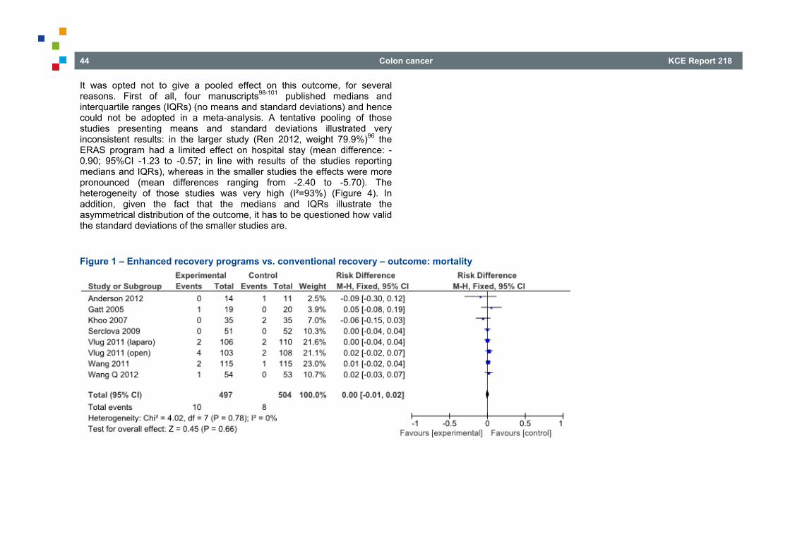
44 Colon cancer KCE Report 218
It was opted not to give a pooled effect on this outcome, for several reasons. First of all, four manuscripts98-101 published medians and interquartile ranges (IQRs) (no means and standard deviations) and hence could not be adopted in a meta-analysis. A tentative pooling of those studies presenting means and standard deviations illustrated very inconsistent results: in the larger study (Ren 2012, weight 79.9%)96 the ERAS program had a limited effect on hospital stay (mean difference: -0.90; 95%CI -1.23 to -0.57; in line with results of the studies reporting medians and IQRs), whereas in the smaller studies the effects were more pronounced (mean differences ranging from -2.40 to -5.70). The heterogeneity of those studies was very high (I²=93%) (Figure 4). In addition, given the fact that the medians and IQRs illustrate the asymmetrical distribution of the outcome, it has to be questioned how valid the standard deviations of the smaller studies are.
Figure 1 – Enhanced recovery programs vs. conventional recovery – outcome: mortality
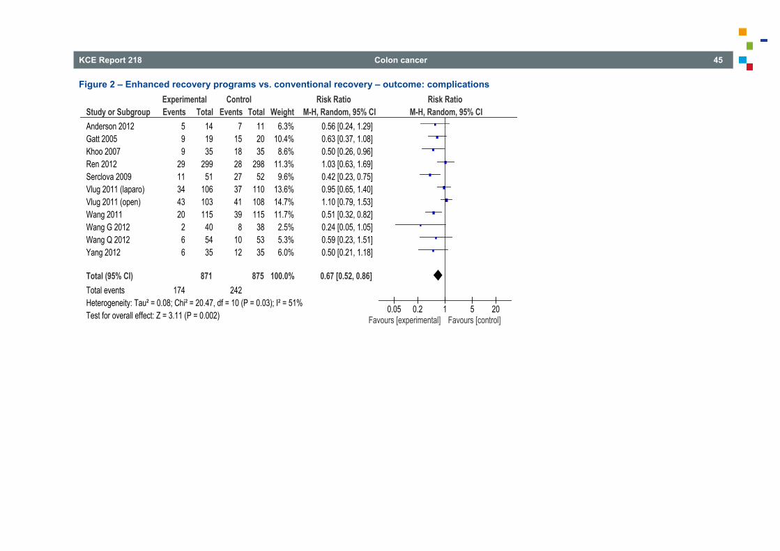
KCE Report 218 Colon cancer 45
Figure 2 – Enhanced recovery programs vs. conventional recovery – outcome: complications
Study or SubgroupAnderson 2012Gatt 2005Khoo 2007Ren 2012Serclova 2009Vlug 2011 (laparo)Vlug 2011 (open)Wang 2011Wang G 2012Wang Q 2012Yang 2012
Total (95% CI)Total eventsHeterogeneity: Tau² = 0.08; Chi² = 20.47, df = 10 (P = 0.03); I² = 51%Test for overall effect: Z = 3.11 (P = 0.002)
Events599
2911344320
266
174
Total141935
29951
106103115405435
871
Events7
151828273741398
1012
242
Total112035
29852
110108115385335
875
Weight6.3%
10.4%8.6%
11.3%9.6%
13.6%14.7%11.7%2.5%5.3%6.0%
100.0%
M-H, Random, 95% CI0.56 [0.24, 1.29]0.63 [0.37, 1.08]0.50 [0.26, 0.96]1.03 [0.63, 1.69]0.42 [0.23, 0.75]0.95 [0.65, 1.40]1.10 [0.79, 1.53]0.51 [0.32, 0.82]0.24 [0.05, 1.05]0.59 [0.23, 1.51]0.50 [0.21, 1.18]
0.67 [0.52, 0.86]
Experimental Control Risk Ratio Risk RatioM-H, Random, 95% CI
0.05 0.2 1 5 20Favours [experimental] Favours [control]
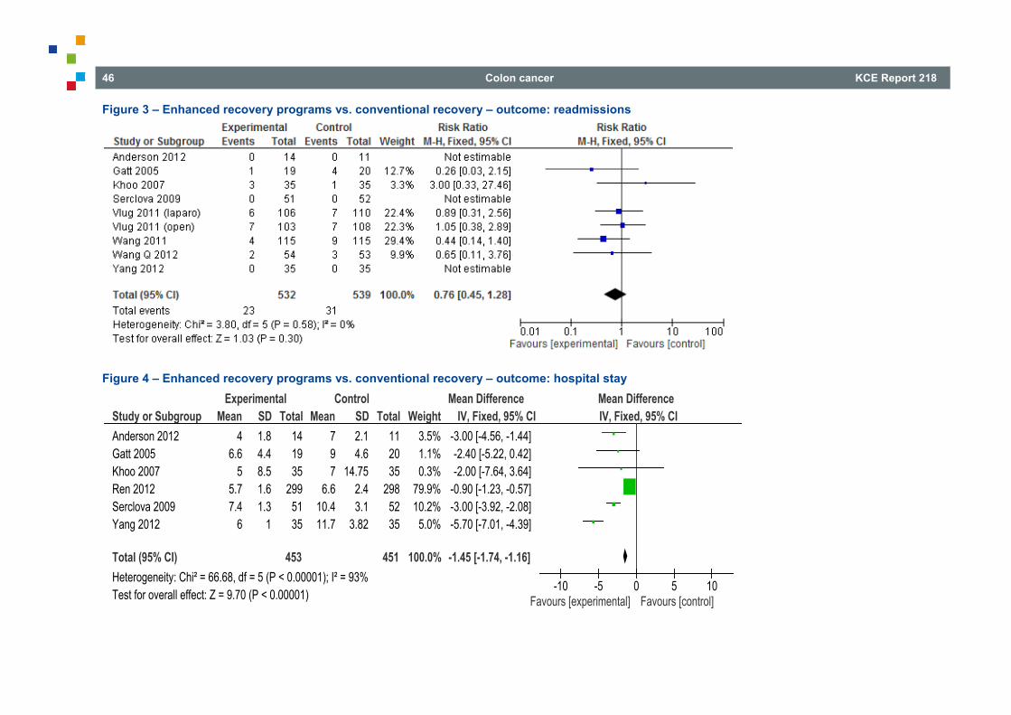
46 Colon cancer KCE Report 218
Figure 3 – Enhanced recovery programs vs. conventional recovery – outcome: readmissions
Figure 4 – Enhanced recovery programs vs. conventional recovery – outcome: hospital stay
Study or SubgroupAnderson 2012Gatt 2005Khoo 2007Ren 2012Serclova 2009Yang 2012
Total (95% CI)Heterogeneity: Chi² = 66.68, df = 5 (P < 0.00001); I² = 93%Test for overall effect: Z = 9.70 (P < 0.00001)
Mean4
6.65
5.77.4
6
SD1.84.48.51.61.3
1
Total141935
2995135
453
Mean797
6.610.411.7
SD2.14.6
14.752.43.1
3.82
Total112035
2985235
451
Weight3.5%1.1%0.3%
79.9%10.2%
5.0%
100.0%
IV, Fixed, 95% CI-3.00 [-4.56, -1.44]-2.40 [-5.22, 0.42]-2.00 [-7.64, 3.64]
-0.90 [-1.23, -0.57]-3.00 [-3.92, -2.08]-5.70 [-7.01, -4.39]
-1.45 [-1.74, -1.16]
Experimental Control Mean Difference Mean DifferenceIV, Fixed, 95% CI
-10 -5 0 5 10Favours [experimental] Favours [control]
KCE Report 218 Colon cancer 47
Conclusions ERAS has no clinically important impact on mortality (Anderson 2012;
Gatt 2005; Khoo 2007; Serclova 2009; Vlug 2011, Wang 2011; Wang 2012; moderate level of evidence)
There are less complications after ERAS (Anderson 2012; Gatt 2005; Khoo 2007; Ren 2012; Serclova 2009; Vlug 2011, Wang 2011; Wang 2012; Wang 2012; Yang 2012; moderate level of evidence)
There is no proof that ERAS results in less readmissions (Anderson 2012; Gatt 2005; Khoo 2007; Serclova 2009; Vlug 2011, Wang 2011; Wang 2012; Yang 2012; very low level of evidence)
There is limited evidence that ERAS has a positive impact on hospital stay (Anderson 2012; Gatt 2005; Khoo 2007; Ren 2012; Serclova 2009; Yang 2012; very low level of evidence)
Other considerations Further development and confirmation in larger studies is needed. Although evidence is limited and the impact on hospital stay may be limited (one day), all evidence points at a beneficial effect for fast track recovery with no indication of any disadvantage. Auditing compliance and outcomes is an essential part of an ERAS program.103
Recommendation An enhanced recovery after surgery (ERAS) program is
recommended after colon cancer surgery (strong recommendation).
3.5 Treatment of acute obstructions SIGN is rather hesitant to recommend colonic stenting in case of obstruction: “Where facilities and expertise are available, colonic stenting can be considered for the palliation of patients with obstructing colon cancer, i.e. in those who are not fit for immediate resection or in those with advanced disease.” Their advise is based on a 2007 SR by Watt and et al. who concluded -based on little high-level evidence- that placement of self expanding metallic stents is both a safe and effective technique for relieving left-sided malignant colonic obstruction.104 But SIGN also refers to two RCTs that were closed prematurely because of colonic perforations caused by stenting.105, 106
One of these trials was in patients with stage IV disease and it was hypothesised that stenting may not be safe in patients undergoing chemotherapy. The other trial was closed because two colonic perforations directly related to stent placement occurred among 30 randomised patients. Hence, SIGN warns that the risk of colonic perforation should be taken into account. SIGN advocates that patients with malignant obstruction of the large bowel should be considered for immediate resection and that segmental resection is preferred for left-sided lesions if immediate reconstruction after resection is deemed feasible.11 The first recommendation is based on one SR and the latter on one RCT. They further suggest that in patients fit for resection, stenting as a bridge to surgery should only be performed as part of a RCT. NZGG states that primary resection of obstructing carcinoma is recommended unless the patient is moribund.50 They adopt fully the NHMRC guideline since new high level evidence is lacking. In addition, NZGG recommends colonic stenting in patients with left-sided bowel obstruction due to colorectal cancer for palliation or as a bridge to surgery, if endoscopic expertise can be readily accessed.50 The NZGG advice stems from one SR based on (historical) case-control studies. IKNL and NZGG encourage enrolment of patients with acute colon obstruction in clinical trials to compare surgical treatment with stent placement with or without subsequent surgery.22, 50 IKNL further states that if participation in a study is not possible, primary stent placement followed within a few weeks by resection with primary anastomosis is preferred over immediate surgical treatment, provided that sufficient expertise is available.22 Their advice is based on two SRs, one RCT, one prospective study, three cohort studies and one decision-theory analysis.
48 Colon cancer KCE Report 218
IKNL as well as NZGG remark that large RCTs comparing prospectively and clearly defined treatment strategies (surgery with or without stents) are lacking.22,50 In the NICE guideline it is recommended that if the use of a colonic stent is considered in patients presenting with acute large bowel obstruction, a CT of the chest, abdomen and pelvis should be offered to confirm the diagnosis of mechanical obstruction and to determine whether the patient has metastatic disease or colonic perforation. This recommendation is based on two case series.12 In addition, NICE suggests that contrast enema studies should not be used as the only imaging modality in patients presenting with acute large bowel obstruction. On the other hand, SIGN suggests that when a mechanical large bowel obstruction is suspected, confirmation should be obtained with a water-soluble contrast enema in order to avoid operative intervention for pseudo-obstruction.11 Their advice is based on one article published in 1985, which received a 2+ level of evidence (no full text available). SIGN further suggests that abdominal CT may also be used in the context. IKNL advocates that the gastroenterologist and the surgeon should be consulted before treating patients with acute obstruction due to colon cancer.22 Stent placement should be discussed, particularly if there is evident colon dilatation proximal to the obstruction and if acute stent placement is feasible. NICE suggests that in patients presenting with acute large bowel obstruction, a consultant colorectal surgeon should consider inserting a colonic stent. He/she should do this together with an endoscopist or a radiologist (or both) who is experienced in using colonic stents.12 The NICE guideline further recommends to resuscitate patients with acute large bowel obstruction and to consider placing a self-expanding metallic stent to initially manage a left-sided complete or near-complete colonic obstruction.12 Based on evidence from case series, NICE advocates not to place self-expanding metallic stents for low rectal lesions, in case of clinical or radiological evidence of perforation or peritonitis nor to relieve right-sided colonic obstruction.12 NICE also warns not to dilate the tumour in order to inserting the self-expanding metallic stent.12
NICE further recommends that only experienced healthcare professionals who have access to fluoroscopic equipment and trained support staff should insert colonic stents.12 NICE suggests that whenever a self-expanding metallic stent is suitable, the insertion should be attempted urgently, i.e. within 24 hours of presentation.12 IKNL advises resecting the tumour during the first operation in patients undergoing primary surgical correction of an acute colon obstruction (based on one SR, which was based on one low-level comparative study).22 With respect to the clinical question: when should primary anastomosis be considered, the New Zealand guideline (NZGG, 2011) adopted the Australian guideline (NHMRC, 2005).50 They state that colectomy with primary anastomosis (ileocolic or ileorectal) could be considered for left-sided obstruction and may need to be preceded by on table colonic lavage. Their advice stems from 1 SR which was based on (historical) case-control studies. For patients with an albumin level < 20 mmol/l, ascites, or poor nutritional status (> 5% weight loss in 1 month or > 10% over the last 6 months), IKNL warns that a very conservative approach should be used with regard to primary anastomosis following resection (based on 1 RCT, 1 comparative study and 1 case series).22 Update In a search for systematic reviews published from 2005 onwards, six systematic reviews were identified, one additional review was found through reference tracking.104, 107-112 One SR compared primary resection with staged resection.110 The other six reviews compared stenting with acute resection.104, 107-109, 111,
112 These six reviews included a total of nine RCTs.105, 106, 113-119 However, inclusion varied from one trial to eight trials and the reasons for exclusion were often not reported. For this reason we decided not to describe the systematic reviews themselves, but the nine trials included in these reviews (see appendix). In a search RCTs from 2005 onwards, no new studies were found. In addition there is the CReST trial which is still running and which is intending to randomize 400 patients between stenting and acute resection
KCE Report 218 Colon cancer 49
(http://www.birmingham.ac.uk/research/activity/mds/trials/bctu/trials/coloproctology/crest/index.aspx). One trial, published by Alacantara et al., compared stenting followed by resection with emergency surgery consisting of intra-operative colonic lavage with primary colo-colic anastomosis.113 This single centre trial was prematurely ended after randomization of 28 patients due to high peri-operative morbidity in the acute resection group. In addition, another five trials compared stenting followed by elective surgery with immediate surgery.105, 106, 114, 116-118 Cheung et al. compared stenting followed by laparoscopic resection
with acute open resection in their centre.114 The type of resection during emergency surgery and the need for defunctioning stoma were to be decided by the surgeon. One quarter of the 48 patients (12 patients) had a stage IV tumour, nine of these patients were in the open surgery group and three in the stenting group (p=0.02). Four patients had failed endoluminal stenting but no complications due to stent placement were noted. Stent placement followed by laparoscopic resection resulted in more 1-stage operations and less patients with a permanent stoma. Also anastomotic leakage (0/24 versus 2/24; p=0.45) and wound infection (2/24 versus 8/24; p=0.4) were less frequent in the stent followed by laparoscopy group.
Ho et al. compared stenting followed by elective laparoscopic or open resection with acute resection in a single centre RCT.116 Type of surgery were to be decided by the surgeon. One quarter of the patients had a stage IV carcinoma; three were in the stenting group and seven in the acute resection group (p=0.227). A higher proportion of patients in the stent group underwent segmental resection, the number of defunctioning stomas was lower in that group (2/20 vs. 6/19; p=0.127). The overall complication rate and postoperative mortality were higher in the emergency group, although differences were not statistically significant.
The French multicentre trial of Pirlet et al. was prematurely discontinued after 60 patients were randomized due to two perforations during stent placement and an unexpected high technical failure rate (53%).105 Ten out of seventy randomized patients were excluded from the analysis because of protocol violations. Overall, no
significant differences were seen for stoma, in-hospital morbidity and mortality.
The trial of Sankararajah et al. was an interim analysis after 19 patients were randomized, and was described only in an abstract.117 78% of stent placement procedures were successful, 57% of stented patients underwent elective surgery. Postoperative morbidity occurred in 66% of patients after emergency surgery versus 24% after stent placement and 14% after elective surgery.
In 2011 van Hooft et al. randomized 98 patients between stenting followed by elective surgery or acute surgery.118 This multicentre trial was prematurely discontinued, after 98 of the 120 planned patients were randomized, as the data safety monitoring board considered the morbidity in the stenting group too high compared to the emergency surgery group (RR 1.62; 95%CI 0.94-2.78). Final analysis shows no significant difference in 30-day mortality or morbidity. More patients in the immediate surgery group needed a stoma (RR 1.46; 95%CI 1.06-2.01), but the number of permanent stomas during follow-up was not significantly different.
The last three of the nine trials evaluated palliative stenting compared with the elective creation of a colostomy or palliative resection in patients with threatening obstruction.115, 119, 120 Originally Fiori et al. intended to include 30 patients, but after two
years there had been no complications associated with stenting so it was decided to discontinue recruitment as stenting apparently involved no more complications than the elective creation of a colostomy.115, 120 Twenty-two pateints with incurable metastatic cancer of the rectum or sigmoid colon were randomized between endoscopic stenting or a proximal diverting colostomy. There was no significant difference in peri-procedural morbidity. Patients with a colostomy and their family reported interference with their lifestyles;
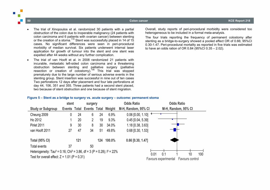
50 Colon cancer KCE Report 218
The trial of Xinopoulos et al. randomized 30 patients with a partial obstruction of the colon due to inoperable malignancy (24 patients with colon carcinoma and 6 patients with ovarian cancer) between stenting or the creation of a stoma.119 Stent was successfully placed in 14 of 15 cases. No significant differences were seen in peri-procedural morbidity of median survival. Six patients underwent internal laser application for growth of tumour into the stent and one stent was expelled after 44 weeks without any further complication.
The trial of van Hooft et al. in 2008 randomized 21 patients with incurable, metastatic left-sided colon carcinoma and a threatening obstruction between stenting and palliative surgery (palliative resection or creation of colostomy).106 This trial was stopped prematurely due to the large number of serious adverse events in the stenting group. Stent insertion was successful in nine out of ten cases Two perforations 12 days after placement and four late perforations at day 44, 106, 351 and 355. Three patients had a second stent placed, two because of stent obstruction and one because of stent migration.
Overall, study reports of peri-procedural morbidity were considered too heterogeneous to be included in a formal meta-analysis. The four trials reporting the frequency of permanent colostomy after stenting as a bridge-to-surgery showed a pooled effect OR of 0.66; 95%CI 0.30-1.47. Peri-procedural mortality as reported in five trials was estimated to have an odds ration of OR 0.84 (95%CI 0.35 – 2.02).
Figure 5 – Stent as a bridge to surgery vs. acute surgery – outcome: permanent stoma
Study or SubgroupCheung 2009Ho 2012Pirlet 2011van Hooft 2011
Total (95% CI)Total eventsHeterogeneity: Tau² = 0.16; Chi² = 3.86, df = 3 (P = 0.28); I² = 22%Test for overall effect: Z = 1.01 (P = 0.31)
Events019
27
37
Total24203047
121
Events628
34
50
Total24193051
124
Weight6.9%9.3%
34.0%49.8%
100.0%
M-H, Random, 95% CI0.06 [0.00, 1.10]0.45 [0.04, 5.39]1.18 [0.38, 3.63]0.68 [0.30, 1.53]
0.66 [0.30, 1.47]
stent surgery Odds Ratio Odds RatioM-H, Random, 95% CI
0.01 0.1 1 10 100Favours experimental Favours control
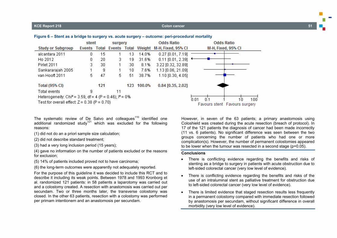
KCE Report 218 Colon cancer 51
Figure 6 – Stent as a bridge to surgery vs. acute surgery – outcome: peri-procedural mortality
The systematic review of De Salvo and colleagues110 identified one additional randomized study121 which was excluded for the following reasons: (1) did not do an a priori sample size calculation; (2) did not describe standard treatment; (3) had a very long inclusion period (15 years); (4) gave no information on the number of patients excluded or the reasons for exclusion; (5) 14% of patients included proved not to have carcinoma; (6) the long-term outcomes were apparently not adequately reported. For the purpose of this guideline it was decided to include this RCT and to describe it including its weak points. Between 1978 and 1993 Kronborg et al. randomized 121 patients: in 58 patients a laparotomy was carried out and a colostomy created. A resection with anastomosis was carried out per secundam. Two or three months later, the transverse colostomy was closed. In the other 63 patients, resection with a colostomy was performed per primam intentionem and an anastomosis per secundam.
However, in seven of the 63 patients; a primary anastomosis using Coloshield was created during the acute resection (breach of protocol). In 17 of the 121 patients the diagnosis of cancer had been made incorrectly (11 vs. 6 patients). No significant difference was seen between the two groups concerning the number of patients who had one or more complication(s). However, the number of permanent colostomies appeared to be lower when the tumour was resected in a second stage (p=0.05).
Conclusions There is conflicting evidence regarding the benefits and risks of
stenting as a bridge to surgery in patients with acute obstruction due to left-sided colorectal cancer (very low level of evidence).
There is conflicting evidence regarding the benefits and risks of the use of an intraluminal stent as palliative treatment for obstruction due to left-sided colorectal cancer (very low level of evidence).
There is limited evidence that staged resection results less frequently in a permanent colostomy compared with immediate resection followed by anastomosis per secundam, without significant difference in overall morbidity (very low level of evidence).
52 Colon cancer KCE Report 218
Other considerations There are two clinical situations for which the use of an intraluminal stent has been studied: 1. Stent as a bridge to surgery in a curative setting 2. Stent as symptomatic treatment in a palliative setting For patients with a curable, resectable left-sided colorectal cancer who present with signs of acute obstruction, it is hypothesised that relieving the obstruction using an intraluminal stent followed by planned surgery would reduce the high morbidity and mortality associated with emergency surgery with resection. Conflicting evidence from randomized trials shows no proof of a beneficial effect of stenting. Two large international trials were closed early because of technical failures and an unexpectedly high complication rate: early and late perforations after stent placement. Furthermore, observational studies have raised doubt about the long term oncological safety of stenting.122 For the symptomatic treatment of patients who are in the palliative stage of their disease, a intraluminal stent can be considered if sufficient local expertise is available. However, since patients with incurable disease have a longer survival period with contemporary systemic treatment and stenting is a contraindication for some treatment options (anti-VEGF therapy), surgery remains the first choice. If a stent is considered, it should be integrated within the overall treatment plan.
Recommendations The use of an intraluminal stent as a bridge to surgery in patients
with acute obstruction due to curable colorectal cancer is not recommended (strong recommendation).
For the treatment of patients with acute obstruction due to incurable colorectal cancer, intraluminal stenting can be considered in selected patients (weak recommendation).
3.6 Adjuvant chemotherapy for stage II-III colon cancer 3.6.1 (High risk) stage II colon cancer NICE, the New-Zealand NGG and CCO state that adjuvant chemotherapy may be offered to patients with completely resected stage II colon cancer with high-risk features such as T4 lesions, perforation and poorly differentiated histology. As the underlying evidence is low, all guidelines comment on the need for discussions of the uncertain benefits and potential side effects with the patients. SIGN considers the identified evidence of too insufficient quality to determine the use of any novel prognostic or predictive marker to aid decision making. The NZGG recommendation is based on three reviews, of which only one shows a benefit for stage II patients. Two reviews show no difference in disease-free survival. Four additional RCTs show mixed results: three show little or no difference, one shows a marginal benefit. Overall, it is concluded that adjuvant chemotherapy appears to offer limited, if any, survival benefit to stage II colon cancer patients. The evidence review of the NICE guideline identified three pooled analyses (non-systematic pooling of specific trial data, a single RCT, one systematic review on MSI and two case-series studies). None of the included pooled analyses found a proven beneficial effect on survival for patients with Dukes B or B2 colorectal cancer. Data were taken from subgroup-analyses from RCTs that included patients with colon or rectal cancer of different stages. Also used chemotherapy agents were very heterogeneous. The RCT randomized patients with high-risk stage II or III colon cancer. Results show a survival benefit for patients who received chemotherapy (95%CI of 1.12-2.45 for RR of 5-year survival after covariate adjustment) without significant interactions between treatment and any of the prognostic variables such as number of lymph nodes containing disease.
KCE Report 218 Colon cancer 53
Update The evidence review of the NICE guideline was used as a starting point to update the literature search. Meta-analyses and RCTs containing separate date for stage II colon cancer comparing adjuvant chemotherapy with observation in stage II colon cancer were included. After removal of duplicates, 869 citations of possible interest were identified. After a first selection based on title and abstract, 50 were evaluated based on the full text. Two systematic reviews were included. One abstract of an updated Cochrane review was identified. The original review and meta-analysis was used for critical appraisal (see appendix). Search for the updated review was performed in September 2012. RCTs containing data on stage II colon cancer patients undergoing adjuvant 5-fluorouracil containing chemotherapy (with levamisole or folinic acid) were included. Seven studies were identified that all could be included in the meta-analysis for overall survival, showing an improvement with adjuvant 5-FU (HR 0.87; 95%CI 0.78-0.97). Six studies reported data for disease-free survival, pooled result shows a RR of 0.84 (95%CI 0.75-0.94). Wu et al.123 performed a systematic review and meta-analysis on RCTs published between 1985 and 2010. Twelve studies were identified of which nine were included in the analysis for stage II colon cancer. In all but one studies, adjuvant therapy contained 5-fluorouracil. For the majority of the studies, HR was not reported in the text but had to be estimated from available data or graphical representations. Overall survival and disease-free survival were improved with the use of adjuvant chemotherapy (HR 0.81; 95%CI 0.71-0.91 and HR 0.86; 95%CI 0.75-0.98 respectively).
Conclusions There are indications that 5-Fluorouracil containing adjuvant
chemotherapy improves DFS and OS in (high-risk) stage II colon cancer, however the effect may be not clinically important (Meyers et al.,2013; Wu et al., 2012; low level of evidence).
Other considerations Although the meta-analyses for DFS and OS survival showed a statistically significant beneficial effect for 5-FU containing adjuvant chemotherapy, the effect appears to be of limited clinical importance and must be weighed against the side effects. Furthermore, included studies did not separate patients with low and high risk stage II features which should also be taken into account when deciding on the use of adjuvant chemotherapy. Patient preferences towards expected benefits and risks may differ depending on different factors such as co-morbidities, age and risk-profile of the tumour. As 5FU-monotherapy in the adjuvant setting appears not to be beneficial in MSI-high tumours (see 3.3.4), MSI testing should be performed if adjuvant 5FU-monotherapy is considered.
Recommendations Adjuvant chemotherapy can be considered for stage II colon
cancer taking into account the presence of high risk features in the tumour, co-morbidities and patient preferences (weak recommendation).
If a patient is considered for 5FU-monotherapy, MSI testing should be performed. If the tumour is MSI-high, no 5FU-monotherapy should be given (strong recommendation).
3.6.2 Stage III colon cancer NICE, the Scottish SIGN, the New Zealand NZGG, the Canadian CCO and the Dutch IKNL all recommend adjuvant chemotherapy for stage III colon cancer patients. NICE recommends capecitabine or oxaliplatin in combination with 5-fluorouracil and folinic acid as the preferred regimens in the adjuvant setting. IKNL, CCO and NZGG consider the combination with oxaliplatin to be the first choice except for patients with contra-indications for oxaliplatin e.g. increased risk for neuropathy or infections. For patients receiving monotherapy, both capecitabine or 5-FU and leucovorin are proposed by IKNL and CCO. NZGG recommends not to use irinotecan in the adjuvant setting. The CCO guideline also stresses that adjuvant therapy should be initiated within eight weeks after surgery.
54 Colon cancer KCE Report 218
The NZGG guideline identified one systematic review including over 3 000 patients and three more recent RCTs reporting survival outcomes for stage III colon cancer. The meta-analysis shows an overall survival benefit for adjuvant chemotherapy compared to observation in patients with stage II or III colorectal cancer (HR 0.74; range 0.66-0.83). The effect is more pronounced in patients with node-positive disease. A joint analysis of Scandinavian trials published in 2005 also found a 7% difference in overall survival after 5-year follow-up for stage III colon cancer, but that difference did not reach statistical significance (p=0.15).124 Another Nordic trial did not find a difference in overall survival for stage II-III colorectal cancer but for the subgroup of stage III colon cancer, 5-year cancer-specific survival improved from 47% to 65% with the use of adjuvant chemotherapy (p=0.032).125 The CCO guideline is based on the same meta-analysis comparing adjuvant chemotherapy with observation alone mentioned in the NZGG guideline.126 Update No update of the literature was performed. Other considerations Although findings on the role of MSI testing when considering 5FU monotherapy may mainly apply in stage II patients, the GDG considered that the findings are also valid for stage III, considering that 5FU-monotherapy is sometimes used and that it is plausible that MSI status also predicts non responsiveness in this case.
Recommendations Adjuvant chemotherapy is recommended for stage III colon
cancer. In fit patients, fluoropyrimidine and oxaliplatin is the combination of choice (strong recommendation).
If a patient is considered for 5FU-monotherapy, MSI testing should be performed. If the tumour is MSI-high, no 5FU-monotherapy should be given (strong recommendation).
3.6.3 Adjuvant chemotherapy in elderly patients Only one guideline issues a specific recommendation on adjuvant chemotherapy in elderly patients (see appendix). The SIGN guideline11 states that decisions concerning adjuvant chemotherapy for patients over the age of 75 with stage III colorectal cancer should be based on a balance between the risks and the potential benefits of treatment. Biological age may be more relevant than chronological age in making these decisions. Update A comprehensive search for studies investigating the use of adjuvant chemotherapy in patients older than 65 years was run in April 2013 (see appendix). Ten studies were selected, one individual patient meta-analysis127 and nine observational studies.128-136 The individual patient meta-analysis combined three Japanese randomised controlled trials that included patients with colorectal carcinoma Dukes A, B or C. Only studies that used central randomisation and were initiated before 1990 were included. Patients were randomised between surgery followed by oral fluoropyrimidines or surgery alone.127 Overall survival for all age groups was slightly better with the use of adjuvant chemotherapy although the difference was small (HR 0.86; 95%CI 0.73-1.00) but no difference was seen for the age-group 65-69 years or the age group 70-75 years old. The nine observational studies used data from cancer registries or hospital registries and analysed data on over 45 000 patients aged 65 years or older who received either surgery and adjuvant chemotherapy or surgery alone.128-136 Seven observational studies included patients with stage III colon cancer;128-132, 134, 136 one observational study included stage III colorectal cancer patients135 and one observational study included stage II colorectal patients.133 One of the observational studies described a progressive decline in completion of chemotherapy with aging, with 74% of patients aged 65-69 years completing chemotherapy vs. 65% in patients aged 70-74 years, 59% in patients aged 75-79 years, 49% in patients aged 80-84 years and 46% of patients aged 85 years or older.130
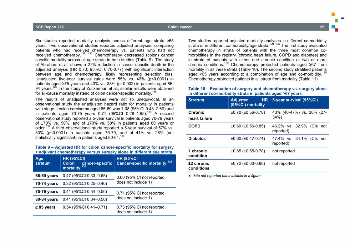
KCE Report 218 Colon cancer 55
Six studies reported mortality analysis across different age strata ≥65 years. Two observational studies reported adjusted analyses, comparing patients who had received chemotherapy vs. patients who had not received chemotherapy.128, 136 Chemotherapy decreased (colon) cancer specific mortality across all age strata in both studies (Table 9). The study of Abraham et al. shows a 27% reduction in cancer-specific death in the adjusted analysis (HR 0.73; 95%CI 0.70-0.77) with significant interaction between age and chemotherapy, likely representing selection bias. Unadjusted five-year survival rates were 55% vs. 43% (p<0.0001) in patients aged ≥75 years and 43% vs. 38% (p=0.002) in patients aged 85-94 years.128 In the study of Zuckerman et al., similar results were obtained for all-cause mortality instead of colon cancer-specific mortality.136 The results of unadjusted analyses were not so unequivocal. In an observational study the unadjusted hazard ratio for mortality in patients with stage II colon carcinoma aged 65-69 was 1.08 (95%CI 0.43–2.69) and in patients aged 70-75 years 0.71 (95%CI 0.26–1.95).133 A second observational study reported a 5-year survival in patients aged 70-79 years of ±70% vs. 50%, and of ±70% vs. 50% in patients aged 80 years or older.131 A third observational study reported a 5-year survival of 57% vs. 33% (p<0.0001) in patients aged 75-79, and of 41% vs. 28% (not statistically significant) in patients aged 80-84.134
Table 9 – Adjusted HR for colon cancer-specific mortality for surgery + adjuvant chemotherapy versus surgery alone in different age strata Age stratum
HR (95%CI) Colon cancer-specific mortality 136
HR (95%CI Cancer-specific mortality 128
66-69 years 0.47 (95%CI 0.33–0.65) 0.80 (95% CI not reported, does not include 1) 70-74 years 0.32 (95%CI 0.25–0.40)
75-79 years 0.41 (95%CI 0.34–0.50) 0.71 (95% CI not reported, does not include 1) 80-84 years 0.41 (95%CI 0.34–0.50)
≥ 85 years 0.54 (95%CI 0.41–0.71) 0.73 (95% CI not reported, does not include 1)
Two studies reported adjusted mortality analyses in different co-morbidity strata or in different co-morbidity/age strata.129, 135 The first study evaluated chemotherapy in strata of patients with the three most common co-morbidities in the registry (chronic heart failure, COPD and diabetes) and in strata of patients with either one chronic condition or two or more chronic conditions.129 Chemotherapy protected patients aged ≥67 from mortality in all these strata (Table 10). The second study stratified patients aged ≥65 years according to a combination of age and co-morbidity.135 Chemotherapy protected patients in all strata from mortality (Table 11).
Table 10 – Evaluation of surgery and chemotherapy vs. surgery alone in different co-morbidity strata in patients aged >67 years Stratum Adjusted HR
(95%CI) mortality 5-year survival (95%CI)
Chronic heart failure
±0.70 (±0.56-0.78) 43% (40-47%) vs. 30% (27-34%)
COPD ±0.69 (±0.56-0.85) 46.2% vs. 32.9% (CIs not reported)
Diabetes ±0.60 (±0.47-0.74) 47.4% vs. 34.1% (CIs not reported)
1 chronic condition
±0.65 (±0.55-0.78) not reported
≥2 chronic conditions
±0.72 (±0.60-0.88) not reported
±: data not reported but available in a figure
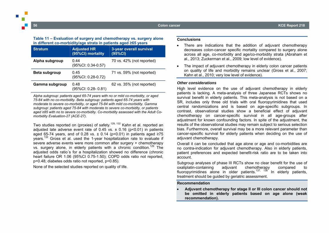
56 Colon cancer KCE Report 218
Table 11 – Evaluation of surgery and chemotherapy vs. surgery alone in different co-morbidity/age strata in patients aged ≥65 years Stratum Adjusted HR
(95%CI) mortality 3-year overall survival (95%CI)
Alpha subgroup 0.44 (95%CI: 0.34-0.57)
70 vs. 42% (not reported)
Beta subgroup 0.45 (95%CI: 0.28-0.72)
71 vs. 59% (not reported)
Gamma subgroup 0.48 (95%CI: 0.28- 0.81)
62 vs. 35% (not reported)
Alpha subgroup: patients aged 65-74 years with no or mild co-morbidity, or aged 75-84 with no co-morbidity. Beta subgroup: patients aged 65-74 years with moderate to severe co-morbidity, or aged 75-84 with mild co-morbidity. Gamma subgroup: patients aged 75-84 with moderate to severe co-morbidity, or patients aged ≥85 with no to severe co-morbidity. Co-morbidity assessed with the Adult Co-morbidity Evaluation-27 (ACE-27).
Two studies reported on (proxies) of safety.129, 132 Kahn et al. reported an adjusted late adverse event rate of 0.45 vs. ± 0.16 (p<0.01) in patients aged 65-74 years, and of 0.28 vs. ± 0.14 (p<0.01) in patients aged ≥75 years.132 Gross et al. used the 1-year hospitalization rate to evaluate if severe adverse events were more common after surgery + chemotherapy vs. surgery alone, in elderly patients with a chronic condition.129 The adjusted odds ratio´s for a hospitalization showed no difference (chronic heart failure OR 1.06 (95%CI 0.75-1.50); COPD odds ratio not reported, p=0.48; diabetes odds ratio not reported, p=0.85). None of the selected studies reported on quality of life.
Conclusions There are indications that the addition of adjuvant chemotherapy
decreases colon-cancer specific mortality compared to surgery alone across all age, co-morbidity and age/co-morbidity strata (Abraham et al., 2013; Zuckerman et al., 2009; low level of evidence).
The impact of adjuvant chemotherapy in elderly colon cancer patients on quality of life and morbidity remains unclear (Gross et al., 2007; Kahn et al., 2010; very low level of evidence).
Other considerations High level evidence on the use of adjuvant chemotherapy in elderly patients is lacking. A meta-analysis of three Japanese RCTs shows no survival benefit in elderly patients. This meta-analysis is not based on a SR, includes only three old trials with oral fluoropyrimidines that used central randomizations and is based on age-specific subgroups. In contrast, observational studies show a beneficial effect of adjuvant chemotherapy on cancer-specific survival in all age-groups after adjustment for known confounding factors. In spite of the adjustment, the results of the observational studies may remain subject to serious selection bias. Furthermore, overall survival may be a more relevant parameter than cancer-specific survival for elderly patients when deciding on the use of adjuvant chemotherapy. Overall it can be concluded that age alone or age and co-morbidities are no contra-indication for adjuvant chemotherapy. Also in elderly patients, patient preferences and expected benefit-risk ratio are to be taken into account. Subgroup analyses of phase III RCTs show no clear benefit for the use of oxaliplatin-containing adjuvant chemotherapy compared to fluoropyrimidines alone in older patients.137, 138 In elderly patients, treatment should be guided by geriatric assessment.
Recommendation Adjuvant chemotherapy for stage II or III colon cancer should not
be omitted in elderly patients based on age alone (weak recommendation).
KCE Report 218 Colon cancer 57
3.7 Surgical treatment of liver metastases Case series have shown that patients with liver metastases can achieve long-term survival if the metastases can be completely resected. Based on these observations, all recent guidelines recommend to attempt curative resection of CRC liver metastases, in some cases in combination with other local treatment modalities such as radiofrequency ablation (RFA) in spite of the lack of evidence from randomized trials.11, 12, 139 KCE performed no systematic search on this subject. A recent review reports that 15 to 30 % of patients with liver metastases are apt for curative resection. Five year survival varies between 30 and 60%. 140 The criteria for resectability are discussed in the 2006 IKNL guideline.13 Important criteria are the estimated residual liver volume, the number and localisation of lesions and the resection margins. Co-existing medical conditions need to be taken into account. Age per se is not a limiting factor. Vena porta embolisation has been suggested to optimize residual volume in the contra-lateral side. A recent review141 reports a 37.9% gain in liver volume following embolisation after a two to four weeks period in patients with preserved liver function and in a six to eight weeks period in patients with cirrhosis or diabetes. A SR on resection criteria and vena porta embolisation are out of the scope of this guideline. When patients are considered for resection of metastases, questions arise about the best timing of the liver surgery (sequential or simultaneous with surgery of the primary tumour) and the timing of chemotherapy. Current guidelines recommend MDT discussion11 and staged surgery.13, 21 The rules of oncologic surgery must always be followed. The importance of a centre's expertise is stressed.13 Expert opinion was the sole basis to recommend peri-operative chemotherapy with a combination of oxaliplatin and 5-FU/leucovorin for a total period of six months11 and similar modalities of neoadjuvant chemotherapy, whether or not combined with biological therapy, both for synchronous and metachronous liver metastases.21
Update Literature was updated from 2006 to answer the following question: What is the best therapeutic sequence for patients with resectable synchronous or metachronous metastases to the liver? Seven systematic reviews and one retrospective study were selected. In the literature addressing the use of chemotherapy before (neoadjuvant), before and after (perioperative) or after (adjuvant) surgery, the distinction is rarely made between synchronous metastases and metachronous metastases. The question of using and timing chemotherapy for resectable liver metastases has therefore been considered as one entity. However, overall prognosis is better for metachronous than for synchronous liver metastases 142. The use of chemotherapy to enhance resectability in case of initially unresectable liver metastases was excluded from this chapter. The sequence of surgical resection of the primary tumour and synchronous liver metastases has been addressed separately. The term ‘simultaneous’ resection is commonly used but the term ‘concomitant’ may be more appropriate. Staged resection involves the classical approach with initial resection of the primary tumour. Recently a ‘liver first’ or reverse strategy has been advocated. In this approach, the liver surgery is performed first, usually after a period of down-staging chemotherapy. Possible advantages are that progression of the liver metastases is avoided during the treatment of the primary tumour, the chances of potentially curative liver resection are optimized and that the response to neoadjuvant chemotherapy helps selecting patients for this aggressive approach. Currently, there are no randomized data comparing the ‘liver first’ strategy with the standard approach. It is mostly considered in patients whose liver disease is not easily resectable at diagnosis. The ‘liver first’ strategy is not further elaborated in this guideline.143
58 Colon cancer KCE Report 218
3.7.1 Timing of surgical resection of primary tumour and synchronous liver metastasis
Chen144 performed a meta-analysis on 14 studies that retrospectively compared concomitant resection to staged resection in patients with resectable synchronous hepatic metastases. The analysis was performed on a total of 2 204 patients of whom 1 384 (ages 56-64.9 yrs) had received simultaneous resection and 817 (ages 58-61 yrs) a staged resection. The median follow up was 2.5 yrs, maximal follow-up 5 yrs. The outcomes of interest are PFS, OS and QoL. There are no data on PFS. Overall survival did not significantly differ at 1, 3 or 5 yrs (1yr: OR 0.77; 95%CI 0.51–1.16, p=0.21; 3 yrs: OR 1.12; 95%CI 0.85–1.47, p=0.43, 5 yrs: OR 1.14; 95%CI 0.86–1.50, p=0.37). The following variables can be considered as indirect QoL indicators. Operative time (weighted mean difference [WMD] −34.19; 95%CI −81.32–12.95, p=0.16) and intra-operative blood loss (WMD −161.33; 95%CI −351.45–28.79, p=0.10) were similar in both groups. Hospital stay (WMD, −4.77; 95%CI −7.26–2.28, p<0.01) and postoperative morbidity rate (odds ratio [OR] 0.71; 95%CI 0.57–0.88, p=0.002) were significantly lower in patients undergoing simultaneous resection of the primary tumour and the synchronous liver metastases. The authors caution the reader because of heterogeneity of the high quality studies. A 2012 single-centre retrospective study, reports higher morbidity rates in patients who were referred for a (staged) liver resection (n=32) as compared to patients diagnosed and treated in their own centre (n=47).145 Simultaneous resection was performed in 53% of the non-referred patients. The median follow-up was 43 months. Treatment with chemotherapy or not was variable, as was the number of surgical interventions. Overall PFS and OS were not significantly different between the two groups but postoperative morbidity was significantly higher in the referred group (75% vs. 47%, P=0.023). Simultaneous resection was one of the many variables that may play a role in this improved outcome. This study was not included in the GRADE profile. After completing this review, an e-publication came under our attention.146 The conclusions of the meta-analysis of 2 880 patients are in line with ours: OS (HR 0.96; 95%CI 0.81-1.14; p = 0.64; I2 = 0) and DFS survival (HR 1.04; 95%CI 0.76-1.43; p= 0.79; I2 = 53%) are similar for both
simultaneous and delayed resections and the simultaneous group has a lower incidence of postoperative complications (modified RR = 0.77; 95%CI: 0.67-0.89; p=0.0002; I2 = 10%).
Conclusions It is plausible that there is no difference in OS after simultaneous
resection compared to staged resection of the primary tumour and resectable synchronous liver metastases. (Chen 2011, moderate level of evidence).
It is plausible that simultaneous resection of the primary tumour and synchronous liver metastases results in lower postoperative morbidity compared to staged resection (Chen 2011, Goyer 2012, moderate level of evidence).
Other considerations Estimation of the (functional) residual liver volume remains challenging. The assessment and treatment of liver metastases requires expert skill, experience, high level technical support and MDT discussion. Although simultaneous resection may be advantageous if the patient is fit (good performance status, low ASA-score), it may not be feasible in case of significant co-morbidity. Technical factors associated with the extent of the liver resection may preclude simultaneous resection.
Recommendations Liver metastases should be resected if imaging techniques
indicate that surgery is an option (strong recommendation). Radiofrequency ablation (RFA) should be considered in addition
to surgery in patients with liver metastases in order to achieve complete response and sufficient residual liver function (strong recommendation).
Simultaneous resection of the primary colon tumour and liver metastases can be considered if the patient is sufficiently fit and a simultaneous operation is judged technically feasible (weak recommendation).
KCE Report 218 Colon cancer 59
3.7.2 Neoadjuvant, perioperative and adjuvant chemotherapy Update Since RCTs comparing neoadjuvant to adjuvant chemotherapy combined with resection of CRC liver metastases are lacking, the PICO can only be addressed indirectly. Outcomes are overall survival (OS) and depending on the study, progression free survival (PFS), recurrence free survival (RFS) or disease free survival (DFS). QoL could be appreciated from surrogate outcomes such as morbidity, adverse events, hospital stay etc. RCTs have compared adjuvant chemotherapy to none (n=7) and peri-operative chemotherapy to none (n=1). Adjuvant chemotherapy can be systemic or loco regional i.e. administered by hepatic artery infusion (HAI). Wieser et al. performed a meta-analysis on 8 RCTs,147 including different regimens (systemic and loco regional) of peri-operative and adjuvant chemotherapy. Ciliberto et al. published a meta-analysis focusing on the three RCTs involving systemic adjuvant chemotherapy148 whereas Nelson’s meta-analysis reports on adjuvant hepatic artery infusion.149 These same RCTs were included in the systematic review by Quan et al.150 The effect of neoadjuvant chemotherapy has been solely reported in observational studies. The single available RCT on peri-operative chemotherapy151 was included in the systematic reviews of neoadjuvant as well as adjuvant chemotherapy. A recent analysis of this RCT is available in an abstract form.152 Chua et al. performed a SR including this one RCT, three phase 2 and 19 observational studies.153 A later review by Lehman et al. details the different systemic regimens for 14 studies, of which 11 were included by Chua. This report adds no new information related to the PICO.154 The last SR discusses the three RCTs on systemic peri-operative and adjuvant chemotherapy and 28 retrospective and prospective studies on neoadjuvant and adjuvant chemotherapy.150 The authors state that heterogeneity and poor quality of the evidence were incompatible with a meta-analysis comparing different sequences of chemotherapy. The Cancer Care Ontario guidelines are based on this last review.139
In conclusion, direct evidence needed to answer the PICO directly is currently lacking. The evidence on related aspects of the question has been reviewed hereunder. 3.7.2.1 Adjuvant chemotherapy Systemic adjuvant chemotherapy Wieser’s meta-analysis includes the largest number of patients: a total of 1 058 patients from which 525 were randomized to surgery with peri-operative or adjuvant chemotherapy and 533 to surgery alone.147 Their median mean age was under 65 yrs and the median follow-up 81 months. The intervention included both HAI and systemic chemotherapy. OS did not differ in the intervention group (HR 0.94; 95%CI 0.8-1.10; p = 0.43) but RFS was clearly improved by the intervention (HR 0.77; 95%CI 0.67-0.88; p=0.0001). A QoL measure in this study are the adverse events which were rated as mild and acceptable toxicities: grade 3-4 leucopoenia in 4.9%, grade 3-4 neutropenia in 13%, grade 3 nausea and vomiting in 13.9%, diarrhoea in 7.3% and hepatic toxicity in 6.4%. Noteworthy are 12% treatment related deaths in the HAI group. For the subgroup treated with systemic chemotherapy the benefit in OS approached statistical significance (HR 0.74; 95%CI 0.53-1.04; p=0.08; I2=0%) and RFS was clearly improved (HR 0.75; 95%CI 0.62-0.91; p=0.003). Ciliberto et al.148 published a separate meta-analysis of the three available RCTs (one in abstract form) comparing peri and adjuvant systemic CT combined with surgery to surgery alone. They demonstrate improved DFS (HR 0.71; 95%CI 0.58-0.88; p=0.001) and PFS (HR 0.75, 95%CI 0.62-0.91, p=0.003) in 666 patients. OS based on two studies was unaffected by combined systemic CT (HR 0.74, 95%CI 0.53-1.04, p=0.088).148 These numbers are identical to the subgroup with systemic CT reported by Wieser et al.147 We updated the meta-analysis for overall survival with the recently presented results of the EORTC study.152 A total of 642 patients were included in this analysis. OS was not improved by the intervention (HR 0.83, 95%CI 0.67-1.02, p=0.07) (Figure 7). .
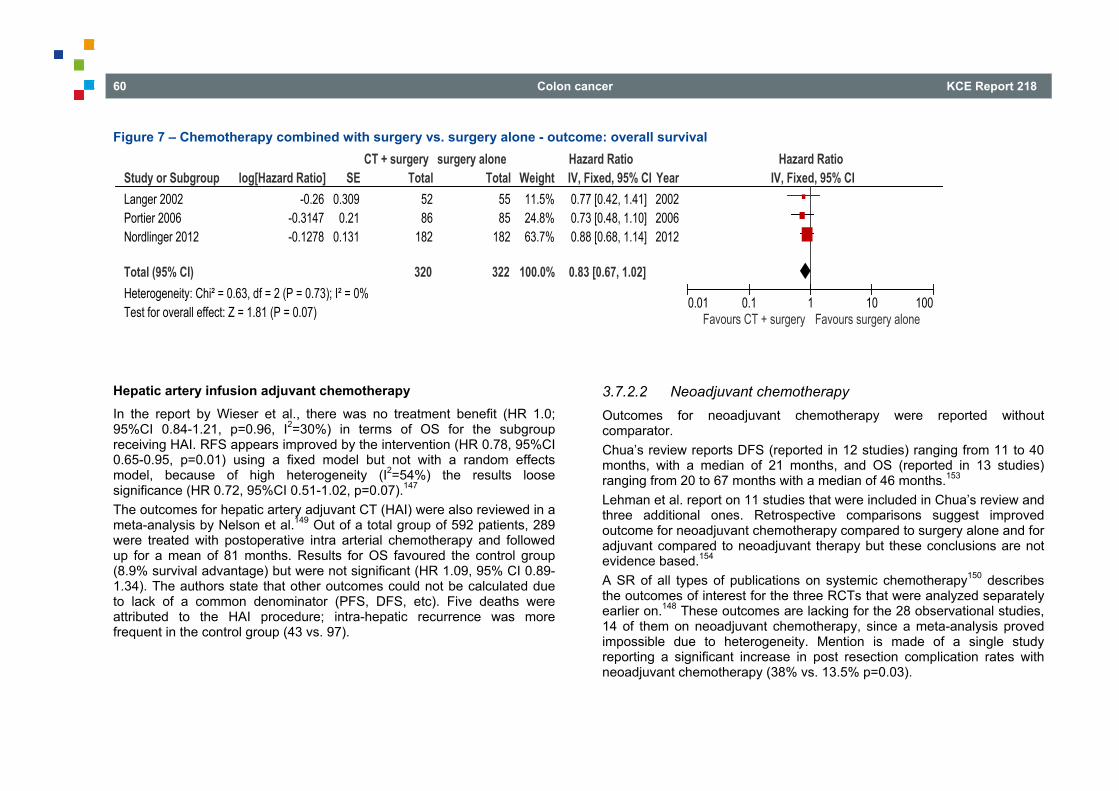
60 Colon cancer KCE Report 218
Figure 7 – Chemotherapy combined with surgery vs. surgery alone - outcome: overall survival
Hepatic artery infusion adjuvant chemotherapy In the report by Wieser et al., there was no treatment benefit (HR 1.0; 95%CI 0.84-1.21, p=0.96, I2=30%) in terms of OS for the subgroup receiving HAI. RFS appears improved by the intervention (HR 0.78, 95%CI 0.65-0.95, p=0.01) using a fixed model but not with a random effects model, because of high heterogeneity (I2=54%) the results loose significance (HR 0.72, 95%CI 0.51-1.02, p=0.07).147 The outcomes for hepatic artery adjuvant CT (HAI) were also reviewed in a meta-analysis by Nelson et al.149 Out of a total group of 592 patients, 289 were treated with postoperative intra arterial chemotherapy and followed up for a mean of 81 months. Results for OS favoured the control group (8.9% survival advantage) but were not significant (HR 1.09, 95% CI 0.89-1.34). The authors state that other outcomes could not be calculated due to lack of a common denominator (PFS, DFS, etc). Five deaths were attributed to the HAI procedure; intra-hepatic recurrence was more frequent in the control group (43 vs. 97).
3.7.2.2 Neoadjuvant chemotherapy Outcomes for neoadjuvant chemotherapy were reported without comparator. Chua’s review reports DFS (reported in 12 studies) ranging from 11 to 40 months, with a median of 21 months, and OS (reported in 13 studies) ranging from 20 to 67 months with a median of 46 months.153 Lehman et al. report on 11 studies that were included in Chua’s review and three additional ones. Retrospective comparisons suggest improved outcome for neoadjuvant chemotherapy compared to surgery alone and for adjuvant compared to neoadjuvant therapy but these conclusions are not evidence based.154 A SR of all types of publications on systemic chemotherapy150 describes the outcomes of interest for the three RCTs that were analyzed separately earlier on.148 These outcomes are lacking for the 28 observational studies, 14 of them on neoadjuvant chemotherapy, since a meta-analysis proved impossible due to heterogeneity. Mention is made of a single study reporting a significant increase in post resection complication rates with neoadjuvant chemotherapy (38% vs. 13.5% p=0.03).
Study or SubgroupLanger 2002Portier 2006Nordlinger 2012
Total (95% CI)Heterogeneity: Chi² = 0.63, df = 2 (P = 0.73); I² = 0%Test for overall effect: Z = 1.81 (P = 0.07)
log[Hazard Ratio]-0.26
-0.3147-0.1278
SE0.3090.21
0.131
Total5286
182
320
Total5585
182
322
Weight11.5%24.8%63.7%
100.0%
IV, Fixed, 95% CI0.77 [0.42, 1.41]0.73 [0.48, 1.10]0.88 [0.68, 1.14]
0.83 [0.67, 1.02]
Year200220062012
CT + surgery surgery alone Hazard Ratio Hazard RatioIV, Fixed, 95% CI
0.01 0.1 1 10 100Favours CT + surgery Favours surgery alone
KCE Report 218 Colon cancer 61
Since evidence is lacking for the outcomes on neoadjuvant chemotherapy, GRADE profiles could not be provided.
Conclusions It is plausible that systemic peri-operative or adjuvant chemotherapy
improves PFS compared to surgery alone in patients with resectable CRC liver metastases. (Wieser et al. 2010; Ciliberto et al. 2012; Quan et al. 2012; moderate level of evidence).
There is no proof that systemic peri-operative or adjuvant chemotherapy has a clinically significant effect on OS compared to surgery alone in patients with resectable CRC liver metastases. (Wieser et al. 2010; Ciliberto et al. 2012; Quan et al. 2012; Norlinger et al. 2012; moderate level of evidence).
There are indications that adjuvant HAI CT does not provide a survival advantage compared to surgery alone in patients with resectable CRC liver metastases. (Wieser et al. 2010; Nelson et al. 2009; very low level of evidence).
An effect of adjuvant HAI CT on PFS could neither be demonstrated nor refuted compared to surgery alone in patients with resectable CRC liver metastases. (Wieser et al. 2010; Nelson et al. 2009; very low level of evidence).
There is no evidence to inform on the effect of neoadjuvant chemotherapy compared to surgery alone on PFS or OS in patients with resectable CRC liver metastases (Chua et al. 2010; Lehman et al. 2012; Quan et al. 2012).
Other considerations The experts expressed that in the clinical setting the choice of chemotherapy (perioperative or adjuvant) often depends on perceived resectability of the tumour and is therefore tailored to the patient. In borderline cases neoadjuvant chemotherapy improves resectability but this aspect was beyond the scope of this review.
Recommendations Systemic peri-operative or adjuvant chemotherapy can be
considered in patients with resectable colorectal liver metastasis (weak recommendation).
(Neo)adjuvant hepatic arterial infusion chemotherapy is not recommended in patients with resectable colorectal liver metastasis (strong recommendation).
3.8 Local treatment modalities for unresectable liver metastases
For unresectable liver metastases, other local treatment modalities have been studied, aiming at achieving favourable long-term outcomes as seen after resection. Radiofrequency ablation (RFA) and stereotactic radiotherapy are the best known local therapies for liver and lung metastases, but also hepatic artery (HAI) chemotherapy, chemo-embolisation, radio-embolisation and selective internal radiotherapy (SIRT) have been proposed. Local treatment of metastases has been considered as part of first-line treatment of metastatic disease and in later stages when the disease has become resistant to systemic chemotherapy. It should be stressed that, as is the case for surgery, local treatment of metastatic disease should only be attempted when complete eradication of the metastases is expected since it is a necessary requirement for good tumour control and prolonged survival. 3.8.1 Radio-frequency ablation (RFA) Radiofrequency ablation of liver metastasis has been used in addition to surgery in order to achieve complete removal of all metastatic disease. However, no data from controlled trials are available. A randomized controlled trial was initiated but closed early due to slow accrual. RFA in addition to surgery is therefore not discussed in this guideline. RFA has been studied in unresectable metastatic disease as addition to first-line chemotherapy and in a palliative setting compared to chemotherapy.
62 Colon cancer KCE Report 218
Update Cirocchi et al.155 identified in a Cochrane review 18 studies comparing radio-frequency ablation (RFA) with other treatment modalities among patients with resectable and unresectable liver metastases. Seventeen studies were not randomised with an increased risk for selection bias and an imbalance in the baseline characteristics of the participants included in all studies. All studies were classified as having an elevated risk of bias. Survival and local recurrence vary widely between studies; main results are reported in the appendix. The heterogeneity regarding interventions, comparisons and outcomes rendered the data not suitable for pooling, and the general conclusion of the review was that there is insufficient evidence regarding the use of RFA. Weng et al.156 attempted to pool the same studies but considered this pooling as inappropriate. A single RCT was included (Ruers 2010) from an abstract of 2010 ASCO Annual Meeting. The final results of the study were published in 2012.157 It compared 60 patients receiving RFA plus (first-line) chemotherapy versus 59 patients receiving chemotherapy alone. Prior adjuvant chemotherapy was allowed. It showed that PFS at 3 years was significantly higher in the group that received RFA (HR 0.63; 95%CI 0.42-0.95), but no effect on overall mortality could be demonstrated. Thirty-month OS was high in both groups, 61.7% (95%CI 48.2-73.9%) in the RFA group and 57.6% (95%CI 44.1-70.4%) in the systemic therapy only group. We updated the SR of Cirocchi et al. using the same strategy from the search date of the review. No additional RCTs were found, three observational case series were excluded since only the abstracts were available and insufficient information was available to assess quality and methodology. GRADE profile is given in the appendix.
Conclusions There is limited evidence that radiofrequency ablation added to first-
line systemic chemotherapy improves PFS at 3 years in CRC patients with unresectable liver metastases (Ruers et al. 2012; low level of evidence).
In CRC patients with unresectable liver metastases, an effect on overall survival when adding radiofrequency ablation to first-line systemic chemotherapy could neither be demonstrated nor refuted (Ruers et al. 2012; very low level of evidence).
Other considerations Radiofrequency ablation can be considered as an adjunct to surgery in order to achieve complete removal of all liver metastases. As previously mentioned, good long-term outcomes have been observed but no high level of evidence is available. The results of the RCT that investigated the use of RFA in combination with first-line chemotherapy for metastatic disease shows a benefit in PFS at 3-years but no benefit of overall survival. Taking the morbidity of the intervention into account leads to a weak recommendation. 3.8.2 Hepatic artery chemotherapy in unresectable CRC liver
metastases Update Mocellin et al.158 included ten RCTs that compared hepatic artery infusion (HAI) with systemic chemotherapy in CRC patients with unresectable liver metastases. HAI regimens were based on fluoxuridine (FUDR), 5-fluorouracil or either one of these two fluoropyrimidines in eight and one RCT, respectively. Systemic chemotherapy (SCT) consisted of FUDR or 5-fluorouracil in three and seven RCT, respectively. Only two out of ten studies were considered to be of high quality. Crossover to HAI was reported in 4/10 trials and the proportion of patients who received allocated treatment was often low. By pooling the summary data, tumour response rate resulted in 42.9% and 18.4% for HAI and SCT, respectively (RR=2.26; 95%CI 1.80-2.84, p<0.0001). Mean weighted median OS times were 15.9 and 12.4 months for HAI and SCT, respectively: the meta-risk of death was not statistically different between the two treatment groups (HR=0.90; 95%CI 0.76-1.07, p=0.24). Subgroup analysis taking into account quality of
KCE Report 218 Colon cancer 63
the studies confirmed this result. No additional RCTs were identified, starting from the search date (January 2011).
Conclusion In CRC patients with unresectable liver metastases, HAI has an effect
on tumour response but an effect of HAI compared to systemic chemotherapy on overall survival could neither be demonstrated nor refuted (Mocellin et al. 2011; very low level of evidence).
Other considerations Although ten RCTs investigating the use of HAI, the studies have several methodological limitations and a beneficial effect on overall survival remains unproven. Furthermore, the chemotherapy regimens used in the trials are outdated. Modern systemic chemotherapy regimens achieve better outcomes compared to the control arms in the HAI trials. Overall, HAI cannot be recommended based on currently available evidence. 3.8.3 Chemo-embolisation of unresectable CRC liver metastases Carter et al.159 did a SR on chemo-embolisation and identified two abstracts on two small case series. An update starting from the search date (2008) was performed. Four case series and one phase III trial for which at least a full text report existed were published since the Carter review (see appendix). Fiorentini et al.160 randomised 75 patients to either chemo-embolisation with irinotecan eluting beads (DEBIRI) or FOLFIRI in an RCT with unclear risk of bias (unclear randomization, no or unclear allocation concealment, no blinding of outcome assessment, albeit only important for progression free survival). The primary end-point was survival; secondary end points were response, recurrence, toxicity, quality of life, cost and influence of molecular markers. At 50 months, overall survival was significantly longer for patients treated with DEBIRI than for those treated with FOLFIRI (p=0.031, log-rank, HR 0.60; 95%CI 0.37-0.97). Median survival was 22 months (95%CI 21-23 months) for DEBIRI and 15 months (95%CI 12-18 months) for FOLFIRI. Progression-free survival was 7 months (95%CI 3-11 months) in the DEBIRI group compared to 4 months (95%CI 3-5 months) in the FOLFIRI group and the difference between groups was statistically significant (p=0.006, log-rank). Extra-hepatic progression had occurred in
all patients by the end of the study, at a median time of 13 (95%CI 10-16) months in the DEBIRI group compared to 9 (95% CI 5-13) months in the FOLFIRI group. A statistically significant difference between groups was not observed (p=0.064, log-rank). The median time for duration of improvement to QoL was 8 (95%CI 3-13) months in the DEBIRI group and 3 (95%CI 2-4) months in the FOLFIRI group. The difference in duration of improvement was statistically significant (p=0.00002, log-rank). Martin et al.161 reported on 55 cases of unresectable colorectal hepatic metastasis patients who had failed standard therapy who received repeated embolisations with irinotecan loaded beads (max. 100 mg per embolisation) per treating physician’s discretion. The median disease free and overall survival from the time of first treatment was 247 days and 343 days. Vogl et al.162 treated 463 patients (mean age, 62.5 years; range, 34.7-88.1 years) with unresectable liver metastases of colorectal cancer that did not respond to systemic chemotherapy repeatedly treated chemo-embolisation in 4-week intervals. In total, 2 441 chemo-embolisation procedures were performed (mean, 5.3 sessions per patient). The local chemotherapy protocol consisted of mitomycin C alone (n = 243), mitomycin C with gemcitabine (n = 153), or mitomycin C with irinotecan (n = 67). Embolisation was performed with lipiodol and starch microspheres for vessel occlusion. Tumour response was evaluated with magnetic resonance imaging. Evaluation of local tumour control resulted in partial response in 68 patients [14.7%], stable disease in 223 patients [48.2%] and progressive disease in 172 patients [37.1%]. The 1-year survival rate after chemo-embolisation was 62%, and the 2-year survival rate was 28%. Median survival from date of diagnosis of liver metastases was 38 months and from the start of chemo-embolisation treatment was 14 months. There was no statistically significant difference between the three treatment protocols.
64 Colon cancer KCE Report 218
Albert et al.163 reported on 121 patients undergoing chemo-embolisation with cisplatin, doxorubicin, mitomycin C, ethiodized oil and polyvinyl alcohol particles, performed at monthly intervals for one to four sessions. Two patients (2%) had partial response, 39 (41%) stable disease and 54 (57%) progression. Median time to disease progression (TTP) in the treated liver was five months, and median TTP anywhere was three months. Median survival was 33 months from diagnosis of the primary colon cancer, 27 months from development of liver metastases and 9 months from chemo-embolisation. Aliberti et al.164 reported on 82 patients presenting with metastatic colorectal carcinoma to the liver after failing chemotherapy undergoing chemo-embolisations with drug eluding beads with irinotecan. The primary endpoints were tumour shrinkage, safety, feasibility, compliance, and overall survival. RECIST was used to assess response. Observed adverse effects were: right upper quadrant pain (40%), fever (80%), nausea (27%) and increased transaminases (70%). The median follow-up was 29 months. After the first treatment, 75 out 82 patients declared an improvement of their well being lasting more than 18 weeks. The median duration of response was 6 (range 3-10) months; the median follow up time was 29 (range 7-48) months. The median survival was 25 (range 6-34) months, with progression free survival at 8 (range 4-16) months.
Conclusion There are indications that chemo-embolisation for the treatment of liver
metastases from colorectal cancer may improve progression-free and overall survival compared to systemic chemotherapy in patients with liver metastases from colorectal cancer (Very low level of evidence).
Other considerations Comparative evidence for chemo-embolisation and systemic chemotherapy is derived from one single RCT with methodological limitations. Survival outcomes in the control arm of that trial are lower than currently seen in practice. As such, the GDG considers available evidence insufficient to recommend the use of chemo-embolisation outside the framework of clinical research.
3.8.4 Radio-embolisation, Selective Internal Radiation Therapy (SIRT) for patients with unresectable tumours
Rizell et al.165 identified eight studies on patients with unresectable liver metastases from CRC. Three studies were controlled studies, two of which being randomised (RCT). The two RCTs were also reported by the Cochrane review of Townsend et al.166 which only focused on RCTs. Main results of those RCTs are reported in the appendix. The other five studies were case series. One of the RCTs was of moderate and the other of low quality. The non-randomised, controlled study was also of low quality. The frequency of patients with a complete or partial tumour response varied between 34–75 %. Most patients experienced nausea, abdominal pain and extreme fatigue. A serious adverse effect occurred in 2–4 % with regard to liver toxicity, in 1% with regard to bilirubin toxicity and in 5–8 % with regard to gastrointestinal toxicity. The review was updated from the search date on (January 2010) (see appendix). Hendlisz et al.167 reported on a prospective, multicenter, randomized phase III trial in patients with unresectable, chemotherapy-refractory liver-limited metastatic CRC (mCRC) comparing arm A (fluorouracil [FU] protracted intravenous infusion 300 mg/m2 days 1 through 14 every 3 weeks) and arm B (radio-embolisation plus intravenous FU 225 mg/m2 days 1 through 14 then 300 mg/m2 days 1 through 14 every 3 weeks) until hepatic progression. The primary end point was time to liver progression (TTLP). Cross-over to radio-embolisation was permitted after progression in arm A. Forty-six patients were randomly assigned and 44 were eligible for analysis (arm A, n=23; arm B, n=21). Median follow-up was 24.8 months. Median TTLP was 2.1 and 5.5 months in arms A and B, respectively (hazard ratio [HR]=0.38; 95%CI 0.20-0.72, p=0.003). Median time to tumour progression (TTP) was 2.1 and 4.5 months, respectively (HR=0.51, 95%CI 0.28-0.94, p=0.03). Grade 3 or 4 toxicities were recorded in six patients after FU monotherapy and in one patient after radio-embolisation plus FU treatment (p=0.10). Twenty-five of 44 patients received further treatment after progression, including 10 patients in arm A who received radio-embolisation. Median overall survival was 7.3 and 10.0 months in arms A and B, respectively (HR=0.92, 95%CI 0.47-1.78, p=0.80). The study had following limitations: trial was prematurely closed (with the number of enrolled patients lower than required (based on power analysis);
KCE Report 218 Colon cancer 65
open label design; liver progression not documented in three patients of arm B; in four patients from arm B there was an unjustified change in the treatment allocated by randomization. Chua et al.168 reported on a prospective database of a major yttrium-90 microsphere radio-embolisation treatment centre in Sydney, Australia, that included 140 patients with unresectable colorectal liver metastases. One hundred and thirty-three patients (95%) had a single treatment, and seven patients (5%) had repeated treatments. Response following treatment was complete in two patients (1%), partial in 43 patients (31%), stable in 44 patients (31%), and 51 patients (37%) developed progressive disease. Combining chemotherapy with radio-embolisation was associated with a favourable treatment response (p=0.007). The median overall survival was 9 (95%CI 6.4-11.3) months with a 1-, 2-, and 3-year survival rate of 42, 22, and 20%, respectively. Primary tumour site (p=0.019), presence of extra-hepatic disease (p 0.033), and a favourable treatment response (p< 0.001) were identified as independent predictors for survival. Kosmider et al.169 reported on 19 patients who underwent radio-embolisation (RE) plus systemic chemotherapy as a first-line treatment for unresectable liver metastases from CRC. Overall response rate according to RECIST was 84% (two complete responses and 14 partial responses). Median progression-free survival (PFS) time was 10.4 months and median overall survival (OS) time was 29.4 months. For patients with disease confined to the liver, PFS improved (10.7 mo versus 3.6 mo, p=0.09), with significant prolongation of OS (median, 37.8 mo versus 13.4 mo, p=0.03) compared with those who had extra-hepatic disease. Serious treatment-related toxicities included febrile neutropenia with concurrent FOLFOX treatment, a perforated duodenal ulcer and one death from hepatic toxicity. Bester et al.170 reported on a retrospective study including 224 patients with chemotherapy-refractory liver metastases treated with yttrium-90 (90Y) resin microspheres, The median OS embolisation group was 11.9 months (95%CI 10.1-14.9 months). A comparison was made to a control group of 29 patients who underwent standard care but we did not consider this comparison as valid.
Martin et al.171 reported on twenty-four patients with unresectable mCRC with liver metastases treated with yttrium-90 microsphere radio-embolisation. Among them, 54% had extra-hepatic disease; 67% had bilobar involvement. The patients had received a median of three prior therapies. No objective responses were observed. Five patients had a CEA response. Median PFS and OS were 3.9 months (95%CI 2.4-4.8 months) and 8.9 months (95%CI 4.2-16.7 months), respectively. Patients older than 65 years had improved PFS (4.6 vs. 2.4 months) and OS (14 vs. 5.5 months) vs. younger patients, likely due to receipt of 90Y treatment earlier in their disease course. The presence of extrahepatic disease and the absence of CEA response appeared negatively predictive of efficacy. Seidensticker et al.172 reported on a matched-pair comparison of patients who received radio-embolisation plus BSC or BSC alone for extensive liver disease. The study included 29 patients who received radio-embolisation, retrospectively matched with patients for prior treatments and tumour burden and then 29 patients were consecutively identified with two or more of four matching criteria: synchronous/metachronous metastases, tumour burden, increased ALP, and/or CEA >200 U/ml. Of 29 patients in each study arm, 16 pairs (55.2%) matched for all four criteria and 11 pairs (37.9%) matched three criteria. Compared with BSC alone, radio-embolisation prolonged survival (median, 8.3 vs. 3.5 months; p< 0.001) with a hazard ratio of 0.3 (95%CI 0.16-0.55, p< 0.001) in a multivariate Cox proportional hazard model. Treatment-related adverse events following radio-embolisation included: grade 1-2 fatigue (n=20, 69%), grade 1 abdominal pain/nausea (n=14, 48.3%) and grade 2 gastrointestinal ulceration (n=3, 10.3%). Three cases of grade 3 radiation-induced liver disease were symptomatically managed. This small observational study attempted to control for confounding by matching on a number of criteria but nevertheless there remains a high risk of residual confounding.
66 Colon cancer KCE Report 218
Conclusions There is limited evidence that SIRT may improve PFS and OS if added
to systemic chemotherapy in CRC patient with unresectable liver metastases (very low level of evidence).
In patients with unresectable liver metastases, an effect of SIRT on PFS or OS when added to hepatic artery infusion could neither be demonstrated nor refuted (very low level of evidence).
There is moderate level of evidence that SIRT improves PFS if added to systemic chemotherapy in CRC patient with unresectable liver metastases refractory to chemotherapy (moderate level of evidence).
There is no direct evidence that SIRT improves OS if added to systemic chemotherapy in CRC patient with unresectable liver metastases refractory to chemotherapy, mainly due to cross over but there is moderate level of evidence that there is effect on PFS (Hendlisz et al. 2010; moderate level of evidence).
Other considerations The studies that compared SIRT added to chemotherapy with chemotherapy alone for unresectable liver metastases (not refractory to chemotherapy) cannot be applied to current practice since improved outcomes are now achieved with more contemporary systemic chemotherapy compared to the control arm in the studies. The GDG does not recommend SIRT for the treatment of non-refractory disease. For patients with metastatic disease limited to the liver that is refractory to standard chemotherapy, SIRT and 5-FU can be considered taking into account the limited absolute effect on PFS, the side effects and the uncertain effect on overall survival.
3.8.5 Stereotactic Body Radiation Therapy (SBRT) for liver metastases
One SR of Tipton et al.173 was excluded because no primary results were reported, however the same systematic review was reported in a more detailed way in a HTA report by Agency for Healthcare Research and Quality (AHRQ).174 Two HTA reports were identified. The National Radiotherapy Implementation Group Report from the NHS identified nine case series on liver metastases, with overall survival ranging from 16 to 92 months and local control ranging from 71 to 92 %.175 However, in some of those studies colorectal and other metastases are mixed. The AHRQ, in the report mentioned above, identified the same studies and added two small case series with results within the same range. Neither comparative studies nor RCTs were found after the search date of the AHRQ review (December 2010). One large case series of Chang et al.176 was identified, reporting 12-month, 18-month, and 24-month OS rates of 72%, 55%, and 38%, respectively and 12-month, 18-month and 24-month local control rates of 67%, 65%, and 55%, respectively.
Conclusion In CRC patients with unresectable liver metastases, an effect of
stereotactic body radiotherapy on overall survival could neither be demonstrated nor refuted (very low level of evidence).
Other considerations Since only case series are reported and no comparative data are available, the GDG considers that SBRT is not recommended for CRC liver metastases outside the framework of clinical research. In a recent KCE report, the evidence available for the treatment of liver metastases with stereotactic radiotherapy was considered of sufficiently high level to receive research financing by the RIZIV-INAMI (for a 4 year period) without the requirement of a clinical trial setting but with obligatory registration at the Belgian Cancer Registry. Such financing of “a model to introduce innovative techniques in health insurance based on evaluation of outcomes and costs” is covered by RIZIV–INAMI art 56 §1.
KCE Report 218 Colon cancer 67
Conclusions are mainly based on The National Radiotherapy Implementation Group Report from the NHS mentioned above.175 General comments Although there is evidence that different local therapies may be beneficial compared to standard care there is no evidence that one is superior to another. Effect on time to progression or progression free survival could be demonstrated but due to the cross over designs effect on overall survival is lacking. It is unclear whether this evidence will ever be provided as cross over designs are difficult to avoid for ethical reasons and acceptability by care givers and patients. Justification of the GRADE and evidence profiles are given in the appendix.
Recommendations Radiofrequency ablation is not recommended in patients with
unresectable liver metastases (strong recommendation). Hepatic artery chemotherapy is not recommended as a treatment
of liver metastases from colorectal cancer (strong recommendation).
Chemoembolisation of CRC liver metastases is not recommended outside the framework of clinical research (weak recommendation).
Adding radioembolisation to systemic chemotherapy in patients with unresectable liver metastases is not recommended (weak recommendation).
Radioembolisation can be considered in patients with unresectable liver metastases refractory to systemic chemotherapy (weak recommendation).
The use of stereotactic body radiation therapy in the treatment of CRC liver metastases is not recommended outside the framework of clinical research (strong recommendation).
3.9 Local treatment of lung metastases 3.9.1 Stereotactic Body Radiation Therapy (SBRT) for lung
metastases The NHS National Radiotherapy Implementation Group Report summarized eight case series of patients treated with SBRT for lung metastases from different tumours.175 The number of patients with a colorectal tumour included is unclear. A review of the literature reported grade 3 to 5 toxicity in up to 15% of patients, with a mortality rate of 0.3%. Stated 2 year survival ranged from approximately 40% to 90%. Survival appears to depend on prognostic factors such as the number of lung metastases, extra-thoracic disease and length of prior disease-free interval. The HTA report by Agency for Healthcare Research and Quality (AHRQ)174 identified 68 studies, none of them with a comparison group, on lung tumours and lung metastases reporting similar results. Reported side effects were the following grade 1–4 toxicities, rash, pneumonitis, cough, rib fracture, pneumothorax, (fiducial placements), chest wall pain, fatigue, nausea, interstitial lung tissue changes, shortness of breath, dermatitis, pleural effusion, fibrosis. Search for recent publications could not identify any reports on treatment of lung metastases from colorectal cancer with stereotactic radiotherapy. Update for lung metastases in general is considered out of the scope of this guideline.
Conclusion In CRC patients with limited lung metastases, an effect of stereotactic
body radiotherapy on overall survival could neither be demonstrated nor refuted.
68 Colon cancer KCE Report 218
Other considerations In the previously mentioned KCE report the available evidence for treatment of lung metastases with stereotactic radiotherapy was considered of sufficiently high level to be reimbursed by the RIZIV-INAMI without the requirement of a clinical trial setting but with obligatory registration at the Belgian Cancer Registry. Conclusions are mainly based on The National Radiotherapy Implementation Group Report from the NHS mentioned above. 3.9.2 Resection of lung metastases Gonzalez et al.177 did a meta-analysis of 25 series that included more than 40 patients each, with a total number of 2 925 patients. Overall 5-year survival after complete resection of lung metastases ranged from 27 to 68%, median survival ranged from 18 to 72 months. Factors associated with poor survival were: (1) a short disease-free interval between primary tumour resection and development of lung metastases (HR 1.59, 95%CI 1.27–1.98) (2) multiple lung metastases (HR 2.04, 95%CI 1.72–2.41) (3) positive hilar and/or mediastinal lymph nodes (HR 1.65, 95%CI 1.35-2.02) (4) elevated pre-thoracotomy carcinoembryonic antigen (HR 1.91, 95%CI 1.57–2.32) Schule et al.178 reported on 65 patients who underwent surgery for liver and lung metastases. Five- and 10-year survival rates for all patients are 57% and 15 % from diagnosis of the primary tumour, 37% and 14 % from resection of the first metastasis and 20% and 15% from resection of the second metastasis. After complete resection, 5- and 10-year survival rates increased to 61% and 18%, 43% and 17% as well as 25% and 19%, respectively. Long-term survivors (≥ 10 years) were seen only after complete resection of both metastases. Complete resection was achieved in 51 patients (79%) and was less likely in patients with synchronous disease (p=0.017). Negative margins (p=0.002), the absence of pulmonary involvement in synchronous metastases (p=0.0003) and single metastases in both organs (p=0.036) were associated with a better prognosis. Hirosawa et al.179 reported on 266 CRC patients undergoing complete pulmonary resection collected from 19 institutions. The cumulative 2- and
5-year survival rates of the patients who underwent pulmonary resection were 76.6% and 46.7%, respectively. The independent unfavourable prognostic factors after pulmonary resection included stage T4 (p=0.0004) and N2 (p=0.0082) as primary cancer-related factors and more than three metastases (p=0.0342), bilateral distribution (p=0.0450), metastatic disease-free interval (DFI) of less than 2 years (p=0.0257) and a preoperative carcinoembryonic antigen (CEA) level greater than 5.0 ng/mL (p=0.0209) as pulmonary metastases-related factors. Gonzalez et al.180 reported on a retrospective analysis of 27 consecutive patients (median age 62 years; range: 33-75 years) who underwent resection of pulmonary metastases after previous hepatic metastasectomy from CRC in two institutions from 1996 to 2009. All patients underwent complete resection (R0) for both colorectal and hepatic metastases. Median follow-up was 32 months (range: 3-69 months) after resection of lung metastases and 65 months (range: 19-146 months) after resection of primary CRC. Three- and 5-year overall survival rates after lung surgery were 56% and 39%, respectively, and median survival was 46 months (95%CI 35-57). Median disease-free survival after pulmonary metastasectomy was 13 months (95%CI 5-21). At the time of last follow-up, seven patients (26%) had no evidence of recurrent disease and 6 of these 7 patients presented initially with a single lung metastasis. Tampellini et al.181 reported on a retrospective cohort comprising of 155 patients with pulmonary and extra-pulmonary metastases; 104 patients with lung metastases only and no surgery and 50 patients with lung metastases only and submitted to surgery. Median progression-free survival (PFS) times were: 10.3 months, 10.5 months, and 26.2 months for the 3 respective groups. Median overall survival times were 24.2 months, 31.5 months and 72.4 months, respectively. Survival times were longer in resected patients: 17 survived >5 years and three survived >10 years. In patients with lung metastases only and no surgery, four survived for 5 years and none survived >10 years. In a Cox regression model, adjusting for some confounders, lung surgery was associated with longer progression free survival (HR 0.46, 95%CI 0.31-0.57) and overall survival (HR 0.26, 95% CI.0.06-0.47). Although the authors attempted an adjustment for confounders, the characteristics of patients undergoing lung surgery or not were very different and it is unclear how much residual confounding persists.
KCE Report 218 Colon cancer 69
Marin et al.182 reported on 44 patients who were strictly selected for pulmonary resection. There was no postoperative mortality and the morbidity rate after pulmonary resection was 1.8%. No patient was lost to follow-up. Overall survival was 93% at 1 year, 81% at 3 years and 64% at 5 years. Factors related to poor prognosis in the univariate analysis were presence of more than 1 pulmonary metastasis (p=0.04), invasion of the surgical margin (p=0.006) and administration of neoadjuvant chemotherapy (p=0.01 for hepatic metastases and p=0.02 for pulmonary metastases). Lida et al.183 retrospectively analyzed 1 030 patients who underwent pulmonary metastasectomy for colorectal cancer from 1990 to 2008. Overall 5-year survival was 53.5%. Median survival time was 69.5 months. Univariate analysis showed tumour number (p<0.0001), tumour size (p<0.0001), pre-thoracotomy serum CEA level (p<0.0001), lymph node involvement (p<0.0001) and completeness of resection (p<0.0001) to significantly influence survival. In multivariate analysis all remained independent predictors of outcome. A RCT funded by Cancer Research UK.PulMiCC (Pulmonary Metastasectomy in Colorectal Cancer) with random allocation to 'active monitoring' or 'active monitoring with pulmonary metastasectomy' is ongoing, no results are available to date.184
Conclusion There is insufficient evidence in favour or against the resection of lung
metastases from colorectal cancer.
Other considerations As is the case for the resection of isolated liver metastases comparative data are lacking. However, since observational studies report long-term tumour control the GDG is of the opinion that resection of lung metastases can be considered if the patients accepts the uncertain benefit-risk balance.
Recommendations Resection of lung metastases should be considered if complete
resection can be achieved (strong recommendation). The use of stereotactic body radiation therapy can be considered
for unresectable or inoperable limited CRC lung metastases (weak recommendation).
3.10 Treatment of peritoneal metastases: cytoreductive surgery and hyperthermic intraperitoneal chemotherapy (HIPEC)
In the NHMRC guideline (2005) it is recommended that cytoreductive surgery with or without chemotherapy should be performed in an appropriate RCT.54 The recommendation is based on the single RCT published so far on this topic. It compared cytoreductive surgery with hyperthermic intraperitoneal chemotherapy (HIPEC, using mitomycin C), followed by systemic 5FU/lv versus systemic chemotherapy (5 FU/leucovorin) and palliative surgery when indicated.185,186 With 50 patients in each arm, the median survival was 12.6 months in the systemic chemotherapy arm and 22.3 months in the surgery with HIPEC arm (p=0.032). The median progression-free survival was 7.7 months in the control arm and 22.2 months in the HIPEC arm (p=0.028).186 However, this trial was criticised for using older generation systemic chemotherapy protocols. In addition, the number of patients was small and the method of randomisation not clearly described. Moreover, the question remains unanswered whether extensive cytoreduction itself (i.e. without HIPEC) would yield comparable results. Another piece of evidence comes from a multi-centre study with 506 patients, from 28 institutions, who were treated with cytoreductive surgery and HIPEC.187 Overall, the median survival was 19.2 months, with morbidity and mortality rates of 23% and 4% respectively. The Dutch guideline states that treatment with HIPEC may be considered for patients with metastases limited to the abdominal cavity, provided that the number of metastatic sites is limited and the metastases can be removed radically by surgery.22 They also warn that this approach is associated with more severe toxicity. They also refer to the RCT by Verwaal and colleagues.185,186
70 Colon cancer KCE Report 218
In 2010 NICE issued an Interventional Procedure Guidance on cytoreductive surgery followed by hyperthermic intraoperative peritoneal chemotherapy for peritoneal carcinomatosis (from various types of cancers).188 The document states that the procedure should only be used with special arrangements for clinical governance, consent and audit or research, since it carries significant risks of morbidity and mortality which needs to be balanced against the perceived benefit (i.e. improvement in survival for patients with colorectal cancer). The evidence for CRC is mainly based on one RCT and three non-randomized comparative studies.185-187, 189, 190 Update A search from 2009 (i.e. final search date of the NICE guidance) for recent systematic reviews/meta-analyses yielded 33 references (after exclusion of doubles), with ten pertinent to the research question. Four of them were excluded as they were narrative. After the critical appraisal of the remaining six reviews, it was decided not to rely on these reviews because none of them had performed a systematic search (i.e. at least two databases and a supplementary strategy, indicating the period of the search), none had performed a critical appraisal of the literature and few reported on the characteristics of the individual studies. As a consequence, a search for primary studies (RCTs as well as observational studies) from 2009 was performed. The search from 2009 to June 2013 for recent primary studies yielded 361 references (after exclusion of duplicates); 81 references describing cytoreductive surgery and HIPEC in mixed cases (i.e. primary tumour from different origins) were excluded. In 54 studies, the primary tumour was the colon. After evaluation of title and abstract, 22 case series and 30 conference abstracts were excluded. The evidence extracted from the three remaining comparative studies is summarized in the appendix. The most recent publication describes a matched cohort study (which the authors erroneously called a case-controlled study).191 The intervention comprised cytoreductive surgery (CRS) and peroperative hyperthermic intraperitoneal chemotherapy (HIPEC) and systemic chemotherapy whereas the control group received CRS and normothermic sequential postoperative intraperitoneal chemotherapy (SPIC). After a median follow-up of 38 months (HIPEC) and 66 months (SPIC), HIPEC resulted in significantly better median overall (36.5 vs. 23.9 months) and disease-free
(22.8 vs. 13.0 months) survival; there were no significant differences between both groups with respect to morbidity (37% vs. 19%) or mortality (6% vs. 6%). It should be noted however that the study had several methodological limitations, e.g. a non-randomised design, retrospective set-up, small sample size (n=32) and a very heterogeneous chemotherapy regimen in and between both groups (see appendix). The non-randomized study performed by Franko et al.192 compared CRS and HIPEC and systemic chemotherapy (n=67) with a control group receiving only systemic chemotherapy (n=38). The primary tumours were located either in the colon or in the rectum. Median overall survival was significantly better for the HIPEC group (34.7 (HIPEC) vs. 16.8 months (SPIC)). This study as well had several limitations, e.g. no randomisation; retrospective analysis of control (and intervention?) data; the control group was statistically older (mean age: 59 y. vs. 51 y., p<0.001), had a higher proportion of patients diagnosed with carcinomatosis at initial presentation (76% vs. 42%, p< 0.001) and a higher proportion of patients with liver lesions (35% vs. 15%, p=0.014) and there was a large heterogeneity in chemotherapy regimens in and between both groups (see appendix). The comparative study by Elias et al. 190 was also adopted in the NICE Guidance188 on CRS and HIPEC; it compared CRS and HIPEC in the intervention group (n=48) with standard (palliative) chemotherapy with or without palliative surgery in the control group (n=48).188, 190 The primary tumours were either located in the colon or in the rectum (HIPEC: 8/48 vs. control 7/48). After a median follow-up of 63 months (HIPEC) and 96 months (control), HIPEC resulted in a significantly better median overall survival (62.7 vs. 23.9 months). This study had several limitations, e.g. no randomisation, HIPEC patients were significantly younger (46 vs. 51 y., p=0.01), significantly more HIPEC patients had well differentiated tumours (37/48 vs. 29/48, p=0.02), prospective inclusion of cases vs. retrospective inclusion of controls based on diagnosis and there was a great diversity in chemotherapy used in both groups (see appendix).
KCE Report 218 Colon cancer 71
Conclusions HIPEC may result in a significantly longer overall survival (Very low
level of evidence).
HIPEC may result in a significantly longer disease-free survival (Very low level of evidence).
When compared with sequential postoperative intraperitoneal chemotherapy, HIPEC does not result in significantly higher rates of postoperative mortality or morbidity (Very low level of evidence).
Other considerations The three studies are considered to be too heterogeneous to pool results. Cytoreductive surgery (CRS) requires a highly skilled surgeon; it should only be performed in expert centres in highly selected patients. It is very unlikely that new RCTs on this treatment modality will be performed.
Recommendations Cytoreductive surgery and HIPEC should be offered to highly
selected, fit patients with metastases limited to the abdominal cavity, provided that the number of metastatic sites is limited and the metastases can be removed radically by surgery (strong recommendation).
HIPEC should only be used with special arrangements for consent and either appropriate clinical governance, including audit or it should be used in the framework of clinical research, since it carries significant risks of morbidity and mortality which needs to be balanced against the perceived benefit (i.e. improvement in survival for patients with colorectal cancer).
3.11 Treatment of metastatic colon cancer: first-line chemotherapy +/- targeted therapy
3.11.1 Choice of chemotherapy agents 3.11.1.1 Oral versus IV fluoropyrimidines Six meta-analyses193-198 were published since 2009 on the comparison between oral fluoropyrimidines (capecitabine) and IV 5-fluorouracil, administered as a continued infusion or as a bolus (see appendix). The meta-analyses of Montagnani195 and Petrelli196 were excluded based on AMSTAR criteria because critical appraisal of included studies was lacking. However, since both publications performed a comprehensive search strategy, we checked reference lists and included two additional RCTs199, 200 in the meta-analyses by KCE. The review by Cao et al.193, with search date September 2008, included all RCTs comparing oxaliplatin in combination with capecitabine or fluorouracil as first-line treatment of metastatic colorectal cancer. No significant difference was detected for progression-free or overall survival (HR 1.08; 95%CI 0.98-1.18 and HR 1.04; 95%CI 0.95-1.14 respectively). Grade 3-4 thrombocytopenia and grade 3-4 hand-foot syndrome were significantly more frequent in the capecitabine arm, whilst grade 3-4 neutropenia was significantly more frequent in the 5FU arm. All first-line studies included in the review by Zhao et al.198 had been included in the review by Cao et al. except the study by Hochster et al. That study reported insufficient information on PFS and OS to be included in the meta-analysis. The study is summarized in the appendix. The review by Ling et al.194 with search date March 2010, included all studies comparing capecitabine with IV 5FU as monotherapy or in combination with oxaliplatin or irinotecan. Studies in the first-line, second-line and neo-adjuvant setting were included. Overall, PFS was in favour of capecitabine (WMD 1.24 months, p=0.04). No significant difference was seen for OS (WMD 0.29 months, p=0.75). The risk for severe adverse events was significantly lower in patients treated with capecitabine (OR 0.73, 95%CI 0.59-0.92).
72 Colon cancer KCE Report 218
The review by Zhang et al.201 included all studies comparing capecitabine-oxaliplatin with 5FU-oxaliplatin. Search date of the review was April 2011. One study202 was added to the updated meta-analysis by Cao et al. (see below). A search for RCTs published since 2011 retrieved three publications, 203-205 comparing oral or IV fluoropyrimidines in combination with oxaliplatin, including one with updated survival results of the study by Cassidy et al. published in 2008. Two RCTs published in 2012 compared capecitabine and 5FU in combination with irinotecan and bevacizumab.204, 205 Oxaliplatin-based chemotherapy The meta-analysis by Cao et al. including all first-line studies comparing oral and IV fluoropyridoxines and oxaliplatin was updated with the study of Ducreux et al.202 and the updated survival results of Cassidy et al.203 Cassidy et al. enrolled 2 034 patients who received XELOX or FOLFOX with or without bevacizumab. The trial was designed as a 2X2 design and was considered to have a low risk of bias. Overall, no significant difference was noted for overall survival (HR 0.95; 97.5%CI 0.85-1.06). Exclusion of patients who received bevacizumab led to the same conclusion (HR 0.95; 97.5%CI 0.83-1.09). Ducreux et al. randomized 306 patients between XELOX and FOLFOX6. The trial was considered to have a low risk of bias. No significant difference between the two treatment arms was seen for overall (HR 1.02; 90%CI 0.81-1.30) or progression-free survival (HR 1.00; 90%CI 0.82-1.22). A meta-analysis performed by KCE shows that there is no significant difference in progression-free or overall survival between capecitabine and IV fluoro-uracil in combination with oxaliplatin for first-line treatment of unresectable metastatic colorectal cancer. Hazard ratios are 1.07 (95%CI 0.98-1.16) and 1.01 (95%CI 0.93-1.11) respectively. (Figure 8 and Figure 9).
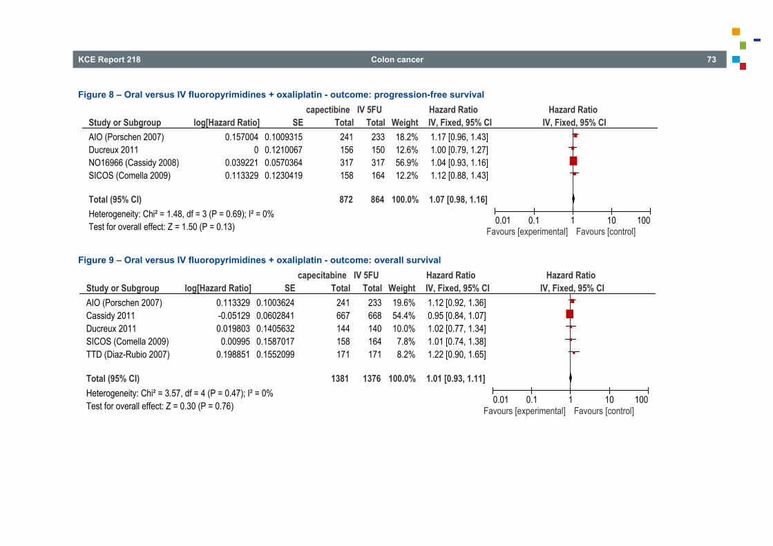
KCE Report 218 Colon cancer 73
Figure 8 – Oral versus IV fluoropyrimidines + oxaliplatin - outcome: progression-free survival
Figure 9 – Oral versus IV fluoropyrimidines + oxaliplatin - outcome: overall survival
Study or SubgroupAIO (Porschen 2007)Ducreux 2011NO16966 (Cassidy 2008)SICOS (Comella 2009)
Total (95% CI)Heterogeneity: Chi² = 1.48, df = 3 (P = 0.69); I² = 0%Test for overall effect: Z = 1.50 (P = 0.13)
log[Hazard Ratio]0.157004
00.0392210.113329
SE0.10093150.12100670.05703640.1230419
Total241156317158
872
Total233150317164
864
Weight18.2%12.6%56.9%12.2%
100.0%
IV, Fixed, 95% CI1.17 [0.96, 1.43]1.00 [0.79, 1.27]1.04 [0.93, 1.16]1.12 [0.88, 1.43]
1.07 [0.98, 1.16]
capectibine IV 5FU Hazard Ratio Hazard RatioIV, Fixed, 95% CI
0.01 0.1 1 10 100Favours [experimental] Favours [control]
Study or SubgroupAIO (Porschen 2007)Cassidy 2011Ducreux 2011SICOS (Comella 2009)TTD (Diaz-Rubio 2007)
Total (95% CI)Heterogeneity: Chi² = 3.57, df = 4 (P = 0.47); I² = 0%Test for overall effect: Z = 0.30 (P = 0.76)
log[Hazard Ratio]0.113329-0.051290.019803
0.009950.198851
SE0.10036240.06028410.14056320.15870170.1552099
Total241667144158171
1381
Total233668140164171
1376
Weight19.6%54.4%10.0%7.8%8.2%
100.0%
IV, Fixed, 95% CI1.12 [0.92, 1.36]0.95 [0.84, 1.07]1.02 [0.77, 1.34]1.01 [0.74, 1.38]1.22 [0.90, 1.65]
1.01 [0.93, 1.11]
capecitabine IV 5FU Hazard Ratio Hazard RatioIV, Fixed, 95% CI
0.01 0.1 1 10 100Favours [experimental] Favours [control]
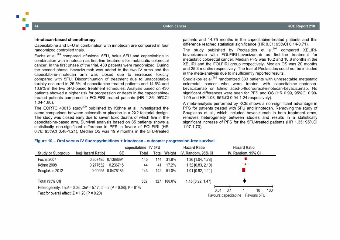
74 Colon cancer KCE Report 218
Irinotecan-based chemotherapy Capecitabine and 5FU in combination with irinotecan are compared in four randomized controlled trials. Fuchs et al.199 compared infusional 5FU, bolus 5FU and capecitabine in combination with irinotecan as first-line treatment for metastatic colorectal cancer. In the first phase of the trial, 430 patients were randomized. During the second phase, bevacizumab was added to the two IV arms and the capecitabine-irinotecan arm was closed due to increased toxicity compared with 5FU. Discontinuation of treatment due to unacceptable toxicity occurred in 25.5% of capecitabine treated patients and 14.6% and 13.9% in the two 5FU-based treatment schedules. Analysis based on 430 patients showed a higher risk for progression or death in the capecitabine-treated patients compared to FOLFIRI-treated patients (HR 1.36; 95%CI 1.04-1.80). The EORTC 40015 study200 published by Köhne et al. investigated the same comparison between celecoxib or placebo in a 2X2 factorial design. The study was closed early due to seven toxic deaths of which five in the capecitabine-based arm. Survival analysis based on 85 patients shows a statistically non-significant difference in PFS in favour of FOLFIRI (HR 0.76; 95%CI 0.48-1.21). Median OS was 19.9 months in the 5FU-treated
patients and 14.75 months in the capecitabine-treated patients and this difference reached statistical significance (HR 0.31; 95%CI 0.14-0.71). The study published by Pectasides et al.204 compared XELIRI-bevacizumab with FOLFIRI-bevacizumab as first-line treatment for metastatic colorectal cancer. Median PFS was 10.2 and 10.8 months in the XELIRI and the FOLFIRI group respectively. Median OS was 20 months and 25.3 months respectively. The trial of Pectasides could not be included in the meta-analysis due to insufficiently reported results. Souglakos et al.205 randomized 333 patients with unresectable metastatic colorectal cancer who were treated with capecitabine-irinotecan-bevacizumab or folinic aced-5-fluorouracil-irinotecan-bevacizumab. No significant differences were seen for PFS and OS (HR 0.99, 95%CI 0.90-1.09 and HR 1.08, 95%CI 0.94-1.24 respectively). A meta-analysis performed by KCE shows a non-significant advantage in PFS for patients treated with 5FU and irinotecan. Removing the study of Souglakos et al., which included bevacizumab in both treatment arms, removes heterogeneity between studies and results in a statistically significant increase of PFS for the 5FU-treated patients (HR 1.35; 95%CI 1.07-1.70).
Figure 10 – Oral versus IV fluoropyrimidines + irinotecan - outcome: progression-free survival
Study or SubgroupFuchs 2007Köhne 2008Souglakos 2012
Total (95% CI)Heterogeneity: Tau² = 0.03; Chi² = 5.17, df = 2 (P = 0.08); I² = 61%Test for overall effect: Z = 1.28 (P = 0.20)
log[Hazard Ratio]0.3074850.277632
0.00995
SE0.1368694
0.2367150.0476183
Total14544
143
332
Total144
41142
327
Weight31.8%17.2%51.0%
100.0%
IV, Random, 95% CI1.36 [1.04, 1.78]1.32 [0.83, 2.10]1.01 [0.92, 1.11]
1.16 [0.92, 1.47]
capecitabine IV 5FU Hazard Ratio Hazard RatioIV, Random, 95% CI
0.01 0.1 1 10 100Favours capecitabine Favours 5FU
KCE Report 218 Colon cancer 75
The study by Fuchs et al. contained insufficient data to be included in a meta-analysis for OS. Combining the studies of Köhne et al. and Souglakos et al. was not considered meaningful given the heterogeneity.
Conclusions There are indications that there is no significant difference in
progression-free survival if fluoropyrimidines are administered orally or intravenously in combination with oxaliplatin as first-line treatment for metastatic colorectal cancer (Cao et al. 2010; Ducreux et al. 2011; Cassidy et al. 2011; Low level of evidence).
It is plausible that there is no significant difference in overall survival if fluoropyrimidines are administered orally or intravenously in combination with oxaliplatin as first-line treatment for metastatic colorectal cancer (Cao et al. 2010; Ducreux et al. 2011; Cassidy et al. 2011; Moderate level of evidence).
There are indications that oral administration of fluoropyrimidines shortens progression-free survival compared to intravenous fluoropyrimidines if combined with irinotecan as first-line treatment for metastatic colorectal cancer (Fuchs et al. 2007; Köhne et al. 2008; Very low level of evidence).
3.11.1.2 Oxaliplatin versus irinotecan As recognized in the IKNL 2008, SIGN 2011 and NICE 2011 guidelines11,
12, fluoropyrimidines in combination with oxaliplatin or irinotecan are valuable options for the first-line treatment of unresectable metastatic colorectal cancer. Comparisons between oxaliplatin and a fluoropyrimidine with irinotecan combinations do not result in significant differences in the majority of trials but toxicities differ.12 However, a meta-analysis of seven RCTs published in 2010 by Liang et al.206 (see appendix) shows a survival benefit of approximately two months in favour of the oxaliplatin-fluorouracil combination (WMD -2.04; 95%CI -3.54 to -0.54). The quality of included studies was judged to be poor as allocation concealment was unclear in all studies and blinding procedures were not used. Search date of the systematic review was January 2010.
The meta-analysis published by Zhuang et al.197 was based on a systematic review of the literature performed in May 2008; all studies were included in the paper of Liang et al. No additional RCTs comparing oxaliplatin-based with irinotecan-based chemotherapy in a first-line setting were identified in the NICE guideline or in the literature published since 2011.
Conclusion There are indications that first-line treatment of metastatic colorectal
cancer with the combination oxaliplatin-fluoropyrimidines results in longer overall survival compared to first-line treatment with the combination irinotecan-fluoropyrimidines (Liang et al. 2010; Very low level of evidence).
Other considerations In the NICE 2011 guideline12, ten different combinations of first- and second-line therapy including FOLFOX, XELOX, FOLFIRI, XELIRI and irinotecan monotherapy were compared using mixed and indirect treatment comparison techniques as no head-to-head comparisons are available. Efficacy data, quality of life and cost-effectiveness considerations are taken into account. The following three sequences are recommended: FOLFOX as first-line treatment then single agent irinotecan as
second-line treatment FOLFOX as first-line treatment then FOLFIRI as second-line treatment XELOX as first-line treatment then FOLFIRI as second-line treatment Patient-specific factors, such as prior oxaliplatin-containing adjuvant chemotherapy, are not considered in the comparison. Overall, there is no clinically meaningful difference in activity between oxaliplatin and irinotecan based chemotherapy, but both regimens have a different toxicity profile.207, 208 The long term neurotoxicity often associated with oxaliplatin can be a limiting factor for its use.
76 Colon cancer KCE Report 218
Recommendations Combination chemotherapy containing oral or intravenous
fluoropyramidines and oxaliplatin or irinotecan is considered the first choice regimen for first–line treatment of metastatic colorectal cancer (strong recommendation).
If combination chemotherapy contains fluoropyrimidines and irinotecan, fluoropyrimidines should be administered intravenously (weak recommendation).
3.11.2 Sequential versus combined chemotherapy The IKNL 2008 guideline and the NICE 2011 guideline identified two RCTs comparing sequential versus combination chemotherapy for first-line treatment of metastatic colorectal cancer. In 2011, one additional RCT on the same comparison was published. The three trials are summarized in the appendix. All trials are considered to have a low risk of bias. None of the trials showed a significant difference in overall survival between sequential and combination therapy. Koopman et al.209 randomized 820 patients with metastatic colorectal cancer between sequential treatment (first-line treatment with capecitabine, second-line with irinotecan and third line capecitabine and oxaliplatin) or combination treatment (first-line treatment with capecitabine plus irinotecan and second-line with capectiabine with oxaliplatin). Treatment in both arms was continued for at least 6 months or until disease progression or unacceptable toxicity, whichever came first. Before a patient entered the next line of treatment, initial eligibility criteria had to be met. No significant difference in overall survival was seen (HR 0.92; 95%CI 0.79-1.08).
Seymour et al.210 randomized 2 135 patients with advanced colorectal cancer starting treatment with non-curative intent. The trial contained multiple comparisons. Patients in the control arm (arm A) were treated with single-agent 5FU until treatment failure, then single agent irinotecan. In treatment arm B, the deferred combination arm, patients were treated with single agent 5FU first, then with 5FU-irinotecan or 5-FU-oxaliplatin as determined by a second randomization. In treatment arm C, the first-line combination arm, patients also underwent a second randomization and were treated immediately with combined 5FU-irinotecan or 5-FU-oxaliplatin. Treatments were continued until treatment failure. Breaks were not allowed during the first three months and were restricted to four weeks during the second three months. Median survival was slightly longer for all groups of treatment arm B and C compared to treatment arm A but the difference was only significant for the group treated with first-line 5FU-irinotecan. A non-inferiority analysis for group B versus group C (considered standard treatment at the end of the trial period) was added post-hoc. HR for overall survival was 1.06; 90%CI 0.97-1.17, which was within the predetermined non-inferiority boundary of HR=1.18. In the trial of Ducreux et al.211 410 patients were randomly assigned to either sequential therapy consisting of monotherapy 5FU followed by FOLFOX6 and then FOLFIRI or combination therapy consisting of FOLFOX6 followed by FOLFIRI. Further lines of therapy were at the investigator’s discretion. There was no significant difference between the two treatment arms in terms of progression-free survival after two lines of therapy (HR 0.95, 95%CI 0.77-1.16) or overall survival (HR 1.02, 95%CI 0.82-1.27). Meta-analysis for overall survival performed by KCE shows a hazard ratio of 1.01, 95%CI 0.93-1.10 (see appendix)).
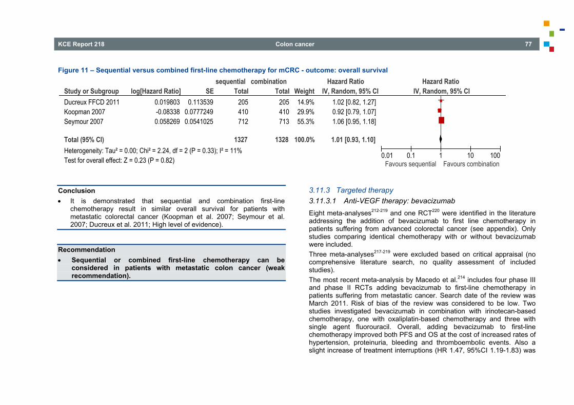
KCE Report 218 Colon cancer 77
Figure 11 – Sequential versus combined first-line chemotherapy for mCRC - outcome: overall survival
Conclusion It is demonstrated that sequential and combination first-line
chemotherapy result in similar overall survival for patients with metastatic colorectal cancer (Koopman et al. 2007; Seymour et al. 2007; Ducreux et al. 2011; High level of evidence).
Recommendation Sequential or combined first-line chemotherapy can be
considered in patients with metastatic colon cancer (weak recommendation).
3.11.3 Targeted therapy 3.11.3.1 Anti-VEGF therapy: bevacizumab Eight meta-analyses212-219 and one RCT220 were identified in the literature addressing the addition of bevacizumab to first line chemotherapy in patients suffering from advanced colorectal cancer (see appendix). Only studies comparing identical chemotherapy with or without bevacizumab were included. Three meta-analyses217-219 were excluded based on critical appraisal (no comprehensive literature search, no quality assessment of included studies). The most recent meta-analysis by Macedo et al.214 includes four phase III and phase II RCTs adding bevacizumab to first-line chemotherapy in patients suffering from metastatic cancer. Search date of the review was March 2011. Risk of bias of the review was considered to be low. Two studies investigated bevacizumab in combination with irinotecan-based chemotherapy, one with oxaliplatin-based chemotherapy and three with single agent fluorouracil. Overall, adding bevacizumab to first-line chemotherapy improved both PFS and OS at the cost of increased rates of hypertension, proteinuria, bleeding and thromboembolic events. Also a slight increase of treatment interruptions (HR 1.47, 95%CI 1.19-1.83) was
Study or SubgroupDucreux FFCD 2011Koopman 2007Seymour 2007
Total (95% CI)Heterogeneity: Tau² = 0.00; Chi² = 2.24, df = 2 (P = 0.33); I² = 11%Test for overall effect: Z = 0.23 (P = 0.82)
log[Hazard Ratio]0.019803-0.083380.058269
SE0.113539
0.07772490.0541025
Total205410712
1327
Total205410713
1328
Weight14.9%29.9%55.3%
100.0%
IV, Random, 95% CI1.02 [0.82, 1.27]0.92 [0.79, 1.07]1.06 [0.95, 1.18]
1.01 [0.93, 1.10]
sequential combination Hazard Ratio Hazard RatioIV, Random, 95% CI
0.01 0.1 1 10 100Favours sequential Favours combination
78 Colon cancer KCE Report 218
seen. Other meta-analyses reported first-line studies that were included in the publication of Macedo et.al.214 One more recent RCT was found: the study by Guan et al.220 including 214 Chinese patients who were randomized to receive irinotecan, leucovorin bolus and 5FU intravenous infusion with or without bevacizumab. The trial was considered to have a low risk of bias. Treatment was continued until documented progressive disease, death or unacceptable toxicity. Hazard ratios for progression-free and overall survival were 0.44 (95%CI 0.31-0.63) and 0.62 (95%CI 0.41-0.95) respectively, in favour of bevacizumab. The meta-analysis of Macedo et al. was updated with the data of the Guan trial. Overall, adding bevacizumab to first-line chemotherapy improves progression-free and overall survival (HR 0.59, 95%CI 0.46-0.74 and 0.82, 95%CI 0.71-0.94 respectively) as summarized in Figure 12 and Figure 13. There was substantial in and between study heterogeneity for the PFS outcome, which disappeared when the only study using oxaliplatin-based chemotherapy (Saltz et al. 2008) was removed from the analysis but results were not significantly altered (PFS HR 0.55; 95%CI 0.48-0.62). The study by Saltz et al. shows a more modest effect of bevacizumab when added to oxaliplatin-based chemotherapy.
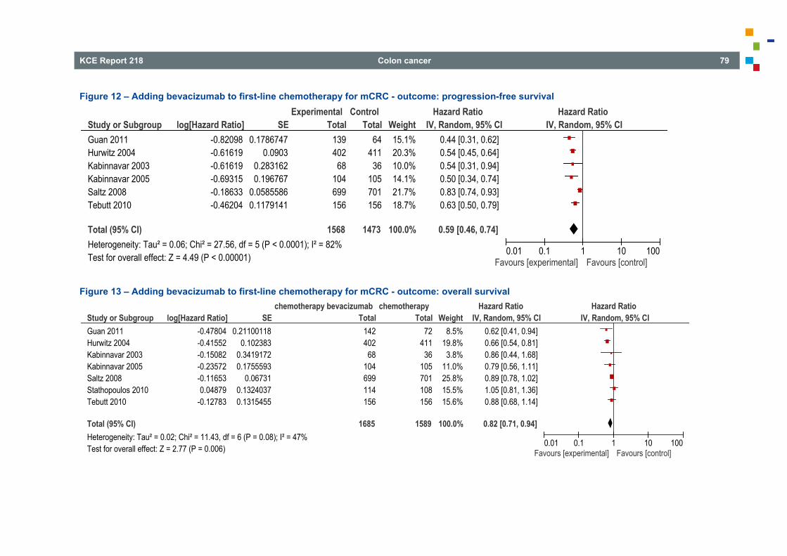
KCE Report 218 Colon cancer 79
Figure 12 – Adding bevacizumab to first-line chemotherapy for mCRC - outcome: progression-free survival
Figure 13 – Adding bevacizumab to first-line chemotherapy for mCRC - outcome: overall survival
Study or SubgroupGuan 2011Hurwitz 2004Kabinnavar 2003Kabinnavar 2005Saltz 2008Tebutt 2010
Total (95% CI)Heterogeneity: Tau² = 0.06; Chi² = 27.56, df = 5 (P < 0.0001); I² = 82%Test for overall effect: Z = 4.49 (P < 0.00001)
log[Hazard Ratio]-0.82098-0.61619-0.61619-0.69315-0.18633-0.46204
SE0.1786747
0.09030.2831620.196767
0.05855860.1179141
Total139402
68104699156
1568
Total64
41136
105701156
1473
Weight15.1%20.3%10.0%14.1%21.7%18.7%
100.0%
IV, Random, 95% CI0.44 [0.31, 0.62]0.54 [0.45, 0.64]0.54 [0.31, 0.94]0.50 [0.34, 0.74]0.83 [0.74, 0.93]0.63 [0.50, 0.79]
0.59 [0.46, 0.74]
Experimental Control Hazard Ratio Hazard RatioIV, Random, 95% CI
0.01 0.1 1 10 100Favours [experimental] Favours [control]
Study or SubgroupGuan 2011Hurwitz 2004Kabinnavar 2003Kabinnavar 2005Saltz 2008Stathopoulos 2010Tebutt 2010
Total (95% CI)Heterogeneity: Tau² = 0.02; Chi² = 11.43, df = 6 (P = 0.08); I² = 47%Test for overall effect: Z = 2.77 (P = 0.006)
log[Hazard Ratio]-0.47804-0.41552-0.15082-0.23572-0.116530.04879
-0.12783
SE0.21100118
0.1023830.34191720.1755593
0.067310.13240370.1315455
Total142402
68104699114156
1685
Total72
41136
105701108156
1589
Weight8.5%
19.8%3.8%
11.0%25.8%15.5%15.6%
100.0%
IV, Random, 95% CI0.62 [0.41, 0.94]0.66 [0.54, 0.81]0.86 [0.44, 1.68]0.79 [0.56, 1.11]0.89 [0.78, 1.02]1.05 [0.81, 1.36]0.88 [0.68, 1.14]
0.82 [0.71, 0.94]
chemotherapy bevacizumab chemotherapy Hazard Ratio Hazard RatioIV, Random, 95% CI
0.01 0.1 1 10 100Favours [experimental] Favours [control]
80 Colon cancer KCE Report 218
Conclusions It is plausible that the addition of bevacizumab to first-line
chemotherapy for metastatic colorectal cancer improves progression-free survival (Macedo et al. 2012; Guan et al. 2011; Moderate level of evidence).
It is plausible that the addition of bevacizumab to first-line chemotherapy for metastatic colorectal cancer improves overall survival (Macedo et al. 2012; Guan et al. 2011; Moderate level of evidence).
3.11.3.2 Anti-EGFR therapy: cetuximab and panitumumab NB: two monoclonal antibodies (MAbs) directed at the epidermal growth factor receptor (EGFR) are registered with the European Medicines Agency (EMA) for the treatment of metastatic colorectal cancer: cetuximab and panitumumab.221, 222 Both agents are authorized for use in tumours containing wild-type (WT) KRAS genes. Therefore, only results from patients with wild-type KRAS tumour are reported. For panitumumab, authorization changed to wild-type RAS genes (including KRAS an NRAS mutated tumours) after the meta-analysis below was performed. Fourteen systematic reviews on the efficacy of anti-EGFR therapy in the treatment of metastatic colorectal cancer, of which eight were excluded based on critical appraisal34, 223-229 (no search in at least two databases or absence of quality appraisal of included studies). The most recent systematic review with meta-analysis published in 2012 by Vale et al.230 included all RCTs comparing chemotherapy with or without cetuximab or panitumumab in patients with advanced colorectal cancer (see appendix). The review was considered to have a low risk of bias. Fourteen eligible trails were included, of which seven in the first line setting. Results were primarily reported for wild type (WT) KRAS tumours. Three trials did not report KRAS status and were included only in an additional sensitivity analysis. Also the trials including bevacizumab in the two treatment arms were considered in a separate analysis.
There was benefit of anti-EGFR MAbs in patients with WT KRAS tumours for both PFS and OS (HR 0.83, 95%CI 0.76-0.90 for PFS, HR 0.89, 95%CI 0.82-0.97 for OS) in trials of first or second line treatment. Where reported, baseline characteristics for the subset of patients in whom KRAS status was assessed were similar to those for all randomised patients suggesting a low risk of patient selection bias in the KRAS tested population. There was significant heterogeneity between trials (p=0.02, I2=60%) assessing progression-free survival, most probably explained by the choice of fluoropyrimidines. Analysis confined to trials using 5FU based chemotherapy yielded HR for PFS of 0.77 (95%CI 0.70-0.85). For OS, the benefit appeared to be confined to trials using 5FU (HR 0.86, 95%CI 0.78-0.95). Adding the results of all randomised patients from three trials without KRAS subgroup data did not change the conclusions for PFS. Meta-analysis of the WT KRAS patients treated in two trials adding bevacizumab to both arms shows improved PFS and OS for patients treated with chemotherapy and bevacizumab (HR 1.27, 95%CI 1.06-1.51 and HR 1.51, 95%CI 0.74-3.08 respectively). All first-line studies included in the other five systematic reviews were also included in the analysis of Vale et al. Two more recent RCTs35, 231 were retrieved from the literature, one reports updated results of the CRYSTAL trial,35 but they were already included in the meta-analysis of Vale et al. The CRYSTAL trial compared FOLFIRI with or without cetuximab in 1 217 patients with previously untreated metastatic colorectal cancer. In the updated report, median follow-up time was 46 months. KRAS status was known for 88% of participants (45% in the original publication). Baseline characteristics and survival data for patients with known KRAS status were similar to the overall population. A benefit in progression-free survival and overall survival was confirmed for treatment with cetuximab and chemotherapy compared to chemotherapy alone in patients with wild-type KRAS tumours (HR 0.69, 95%CI 0.56-0.87 and HR 0.79, 95%CI 0.67-0.95 respectively).
KCE Report 218 Colon cancer 81
Tveit et al.231 published the results of the NORDIC-VII study in 2012. Patients were randomized to receive either standard Nordic FLOX (bolus 5FU + folinic acid + oxaliplatin) or cetuximab and FLOX or cetuximab with intermittent FLOX. In the first two arms, treatment was continued until disease progression or intolerable toxicity. In the third arm, FLOX was stopped after 16 weeks of treatment, cetuximab was continued in case of objective response. When progressive disease was reported, FLOX was reintroduced. Comparing the first two arms with identical chemotherapy, no significant advantage was seen for cetuximab in terms of progression-free or overall survival (HR 1.07; 95%CI 0.79-1.45 and HR 1.14; 95%CI 0.80-1.61). A meta-analysis was performed including all first-line trials included in the MA of Vale et al. and the two recent publications. Only data for wild-type KRAS tumours were included. Oxaliplatin-based chemotherapy was used in the majority of studies. Only in the CRYSTAL study and part of the PACCE study the chemotherapy was irinotecan-based. Overall, a statistically significant benefit was seen when cetuximab was added to chemotherapy in terms of progression-free survival (HR 0.82; 95%CI 0.69-0.96) and overall survival (HR 0.89; 95%CI 0.80-0.99). For progression-free survival, there was evidence of heterogeneity between studies which disappeared if the two studies using oral (COIN Xelox) or bolus IV (Nordic VII) fluoropyrimidines were removed. Analysis limited to studies using continuous IV administration of 5FU only shows a HR of 0.74; 95%CI 0.66-0.84 (data not shown). Adding cetuximab to combined chemotherapy and bevacizumab results in a shorter PFS for patients treated with cetuximab (HR 1.27; 95%CI 1.06-1.51). Differences were not statistically significant for overall survival (HR 1.51; 95%CI 0.74-3.08).
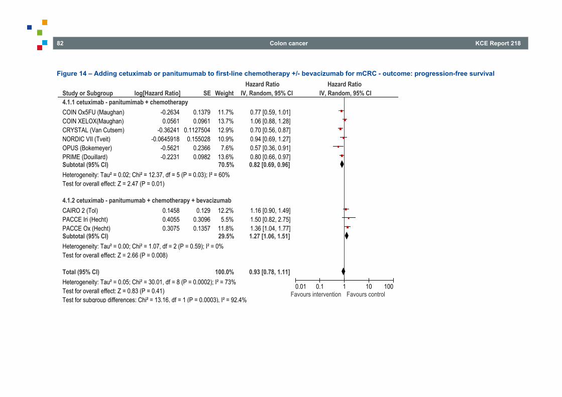
82 Colon cancer KCE Report 218
Figure 14 – Adding cetuximab or panitumumab to first-line chemotherapy +/- bevacizumab for mCRC - outcome: progression-free survival
Study or Subgroup4.1.1 cetuximab - panitumimab + chemotherapyCOIN Ox5FU (Maughan)COIN XELOX(Maughan)CRYSTAL (Van Cutsem)NORDIC VII (Tveit)OPUS (Bokemeyer)PRIME (Douillard)Subtotal (95% CI)Heterogeneity: Tau² = 0.02; Chi² = 12.37, df = 5 (P = 0.03); I² = 60%Test for overall effect: Z = 2.47 (P = 0.01)
4.1.2 cetuximab - panitumumab + chemotherapy + bevacizumabCAIRO 2 (Tol)PACCE Iri (Hecht)PACCE Ox (Hecht)Subtotal (95% CI)Heterogeneity: Tau² = 0.00; Chi² = 1.07, df = 2 (P = 0.59); I² = 0%Test for overall effect: Z = 2.66 (P = 0.008)
Total (95% CI)Heterogeneity: Tau² = 0.05; Chi² = 30.01, df = 8 (P = 0.0002); I² = 73%Test for overall effect: Z = 0.83 (P = 0.41)Test for subgroup differences: Chi² = 13.16, df = 1 (P = 0.0003), I² = 92.4%
log[Hazard Ratio]
-0.26340.0561
-0.36241-0.0645918
-0.5621-0.2231
0.14580.40550.3075
SE
0.13790.0961
0.11275040.155028
0.23660.0982
0.1290.30960.1357
Weight
11.7%13.7%12.9%10.9%
7.6%13.6%70.5%
12.2%5.5%
11.8%29.5%
100.0%
IV, Random, 95% CI
0.77 [0.59, 1.01]1.06 [0.88, 1.28]0.70 [0.56, 0.87]0.94 [0.69, 1.27]0.57 [0.36, 0.91]0.80 [0.66, 0.97]0.82 [0.69, 0.96]
1.16 [0.90, 1.49]1.50 [0.82, 2.75]1.36 [1.04, 1.77]1.27 [1.06, 1.51]
0.93 [0.78, 1.11]
Hazard Ratio Hazard RatioIV, Random, 95% CI
0.01 0.1 1 10 100Favours intervention Favours control
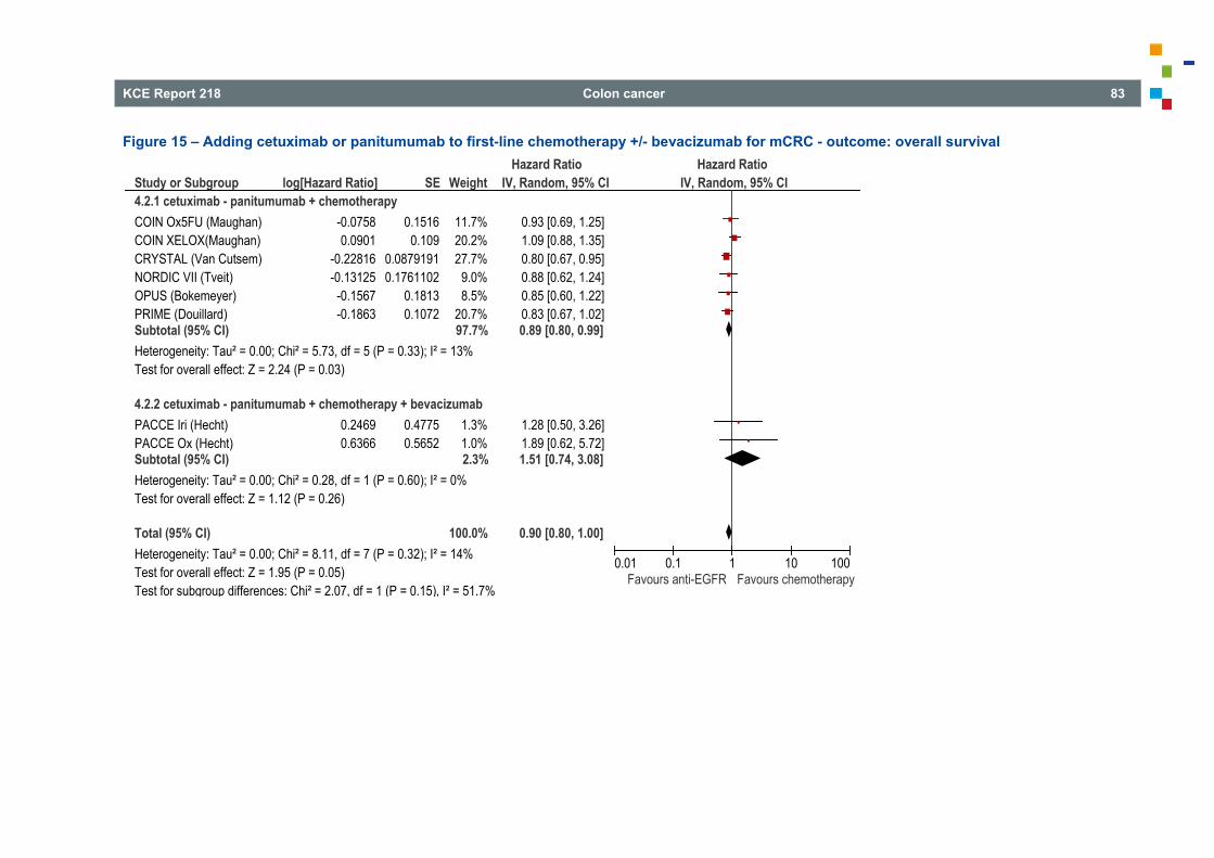
KCE Report 218 Colon cancer 83
Figure 15 – Adding cetuximab or panitumumab to first-line chemotherapy +/- bevacizumab for mCRC - outcome: overall survival
Study or Subgroup4.2.1 cetuximab - panitumumab + chemotherapyCOIN Ox5FU (Maughan)COIN XELOX(Maughan)CRYSTAL (Van Cutsem)NORDIC VII (Tveit)OPUS (Bokemeyer)PRIME (Douillard)Subtotal (95% CI)Heterogeneity: Tau² = 0.00; Chi² = 5.73, df = 5 (P = 0.33); I² = 13%Test for overall effect: Z = 2.24 (P = 0.03)
4.2.2 cetuximab - panitumumab + chemotherapy + bevacizumabPACCE Iri (Hecht)PACCE Ox (Hecht)Subtotal (95% CI)Heterogeneity: Tau² = 0.00; Chi² = 0.28, df = 1 (P = 0.60); I² = 0%Test for overall effect: Z = 1.12 (P = 0.26)
Total (95% CI)Heterogeneity: Tau² = 0.00; Chi² = 8.11, df = 7 (P = 0.32); I² = 14%Test for overall effect: Z = 1.95 (P = 0.05)Test for subgroup differences: Chi² = 2.07, df = 1 (P = 0.15), I² = 51.7%
log[Hazard Ratio]
-0.07580.0901
-0.22816-0.13125
-0.1567-0.1863
0.24690.6366
SE
0.15160.109
0.08791910.1761102
0.18130.1072
0.47750.5652
Weight
11.7%20.2%27.7%
9.0%8.5%
20.7%97.7%
1.3%1.0%2.3%
100.0%
IV, Random, 95% CI
0.93 [0.69, 1.25]1.09 [0.88, 1.35]0.80 [0.67, 0.95]0.88 [0.62, 1.24]0.85 [0.60, 1.22]0.83 [0.67, 1.02]0.89 [0.80, 0.99]
1.28 [0.50, 3.26]1.89 [0.62, 5.72]1.51 [0.74, 3.08]
0.90 [0.80, 1.00]
Hazard Ratio Hazard RatioIV, Random, 95% CI
0.01 0.1 1 10 100Favours anti-EGFR Favours chemotherapy

84 Colon cancer KCE Report 218
Conclusions There are indications that the addition of cetuximab or panitumumab to
first-line chemotherapy for wild-type KRAS metastatic colorectal cancer improves progression-free survival (Vale et al. 2012; Van Cutsem et al. 2011; Tveit et al. 2012; Low level of evidence).
There are indications that the addition of cetuximab or panitumumab to first-line chemotherapy for wild-type KRAS metastatic colorectal cancer improves overall survival (Vale et al. 2012; Van Cutsem et al. 2011; Tveit et al. 2012; Low level of evidence).
There are indications that the addition of cetuximab or panitumumab to chemotherapy and bevacizumab as first-line treatment for wild-type KRAS metastatic colorectal cancer shortens progression-free survival (Vale et al. 2012; Low level of evidence).
The addition of cetuximab or panitumumab to chemotherapy and bevacizumab as first-line treatment for wild-type KRAS metastatic colorectal cancer did not result in significant harm or significant benefit in terms of overall survival. (Vale et al. 2012; Very low level of evidence).
Other considerations Although adding bevacizumab or anti-EGFR treatment (in wild-type RAS patients) to first-line chemotherapy results in a proven beneficial effect on PFS and OS, the benefit may be of limited clinical importance and must be balanced against the additional side effects associated with the targeted therapy. Furthermore, information on the optimal sequence of therapeutic agents is limited.
Recommendations In RAS wild type patients, the addition of anti-EGFR therapy
(cetuximab or panitumumab) or bevacizumab to first-line chemotherapy should be considered (strong recommendation).
In RAS mutated patients, the addition of bevacizumab to first-line chemotherapy should be considered (strong recommendation).
3.12 Second line chemotherapy for metastatic colon cancer SIGN11 identified two reviews on the use of second-line chemotherapy for advanced colorectal cancer. In a Cochrane review published in 2009, Roqué i Figuls et al. 232 identified seven RCTs assessing the efficacy of (single or combined) second-line chemotherapy. Improved progression-free survival and overall survival is seen with second-line irinotecan compared to BSC or 5FU chemotherapy. Combined 5FU and oxaliplatin appeared to be superior in terms of response, time to progression and control of tumour-related symptoms compared to single agent treatment with one of them. A HTA published in 2008 233 concludes that staged combination therapy (combination oxaliplatin followed by combination irinotecan or vice versa) provided the best OS and PFS. In the only trial comparing the use of three active chemotherapies in any staged combination, median OS was over 20 months. Other considerations Clinical trials have assessed the use of several targeted therapies in the second-line treatment (and later stages) of metastatic colorectal cancer, e.g. bevacizumab, aflibercept, cetuximab and panitumumab.234, 235 A systematic review of targeted treatment in second-line and further lines of treatment is not within the scope of this guideline. Update No update of the literature was performed.
Recommendations Second-line chemotherapy should be considered for patients
with metastatic colorectal cancer with good performance status and adequate organ function (strong recommendation).
In fit patients who have progressive disease after first line therapy with oxaliplatin or irinotecan containing chemotherapy, a change in the cytotoxic regimen from oxaliplatin to irinotecan or from irinotecan to oxaliplatin should be considered (strong recommendation).
KCE Report 218 Colon cancer 85
3.13 Follow-up 3.13.1 Summary of guidelines The objective was to identify the recommendations for follow-up in order to detect recurrences of the primary tumour or new metastases after curative therapy. Previously selected guidelines11-13, 22, 50 were reviewed for recommendations on schedule and type of examination (see appendix). The report from NICE is the most recent (2011) and comprehensive12, including reports that generated previous recommendations (2006).22 Overall, there is very little evidence and recommendations are based on expert opinion.
3.13.2 General recommendations It is generally recommended that a coordinator and a plan are communicated to the patient and that regular follow-up is initiated within four to six weeks after completing treatment. Proposed schedules are expert based and vary around every six months for two to three years, thereafter yearly for up to five years or whenever symptoms occur. Follow-up should be discontinued when the benefits do not outweigh the risks of diagnostic tests or when the patient cannot tolerate further treatment.
3.13.3 CEA CEA testing is widely available and performed during office visits. NICE proposes to offer regular serum CEA tests, i.e. every six months during the first three years. However, in the guidelines there is no clear demonstration of the usefulness of CEA and no consensus on diagnostic yield. The possible role of CEA screening is discussed in the update of the literature on the use of CT scan.
3.13.4 Colonoscopy There is consensus that full colonoscopy should be performed whenever the full examination was impossible prior to tumour resection in order to exclude residual disease. However, the proposed timing of the colonoscopy is expert based and varies between three11, 22 and twelve50 months after surgery.
Nor is there consensus on timing of surveillance colonoscopy: it is recommended one year after initial treatment and five years thereafter by NICE,12 after five years by SIGN,11 every three to five years by NZGG,50 every six years for local tumours or according to the consensus on follow-up of colon polyps (six years for 0-2 polyps, three years for 3 or more polyps) by IKNL.22
3.13.5 Imaging The diagnostic yield of imaging may be low for local (T1N0) tumours.22
3.13.5.1 Hepatic ultrasound Hepatic ultrasound is often practised but the relevance and diagnostic yield of this examination in follow-up is unknown. The subject was not covered by recent guidelines. IKNL22 mentions that ultrasound may be sufficient to screen for liver metastases if the liver can be well visualised.
3.13.5.2 CT chest abdomen In Belgium, physicians may base their practice on the 2007 KCE guidelines that recommend a CT thorax and abdomen at 3 months and every year during 3 years in patients at higher risk of recurrence or on the 2005 ASCO guidelines that recommend a yearly CT scan. According to the more recent NICE guidelines,12 patients should be offered a minimum of 2 CTs of the chest, abdomen and pelvis in the first 3 years. The evidence for use of CT scan during follow-up after treatment with curative intent was updated.
3.13.5.3 FDG PET-CT The use of FDG PET-CT was only addressed by SIGN11 reflecting the lack of consensus on the use of this examination during follow-up. SIGN suggests that FDG PET-CT be used in the evaluation of patients with raised tumour marker CEA with negative or equivocal conventional imaging or in the assessment of possible pelvic or pre-sacral recurrence.
3.13.6 Other tests Occult blood testing is not mentioned as part of follow-up.
86 Colon cancer KCE Report 218
3.13.7 Update on the use of CT chest abdomen in follow-up A systematic search on the use of CT scan as a tool for surveillance of CRC patients after curative treatment was performed in Medline, Premedline, Embase and Cochrane database between the dates January 1st 2011 and July 16th 2013. The search strategy is documented in the appendix. Following selection and appraisal, two relevant studies were identified and summarized in the evidence tables (see appendix). The first, a retrospective case series of 177 patients followed during 60 months indicates that CT was the first sign of recurrence in 68% of patients.236 The second is a long awaited RCT on the effect of scheduled CEA and CT follow-up.237 This study is currently submitted for full publication. The authors kindly allowed us to review the full text confidentially, allowing appraisal with the Cochrane Collaboration’s Risk of Bias Tool (see appendix). This study demonstrates that a more intensive follow-up schedule with either CEA measurement 3 monthly for 2 years, then 6 monthly for 3 years, with a single chest, abdominal and pelvic CT scan at 12-18 months or CT chest, abdominal and pelvis 6 monthly for 2 years, then annually for 3 years, detects about three times more recurrences compared to a minimum follow-up with a CT of chest, abdomen and pelvis at 12-18 months (p=0.019). The adjusted odds were 2.7 for CEA only (p=0.035) and 3.4 for CT only (p=0.007); the absolute differences in detection rate in the more intensive arms compared to minimum follow-up were 4.3-5.7% (5.8-8.0% per-protocol analysis). Combining CEA and CT provided no additional benefit (adjusted odds for CT+CEA arm = 2.9). The absolute difference in the proportion of participants with recurrence treated surgically with curative intent in the factorial comparison was 1.4% for CEA (p=0.28) and 2.8% for CT (p=0.04). There was no statistical difference in colorectal cancer deaths (DFS) nor in overall deaths (OS) amongst the different groups for 3.7 years follow-up (3-5 years).
Conclusions A follow-up plan should be communicated to the patient after curative
resection. Every effort should be made to exclude residual disease after surgery:
full colonoscopy should be performed within a year whenever the examination could not be performed prior to tumour resection.
After curative treatment of CRC, an intensive follow-up plan with CEA measurement 3 monthly for 2 years, then 6 monthly for 3 years, with a single chest, abdominal and pelvic CT scan at 12-18 months or CT chest, abdominal and pelvis every 6 months for 2 years, then annually for 3 years increases the detection of potentially treatable recurrences compared with a minimal follow-up with CT at 12 to 18 months.
In case of positive CEA, a CT is performed.
Adding CEA in case of planned CT follow-up is probably not meaningful.
Other considerations Cost has not been considered in these comparisons. Although widely used, the relevance of US is questionable.
KCE Report 218 Colon cancer 87
Recommendations Identify a coordinator who communicates a follow-up plan to the
patient after curative resection. A full colonoscopy should be performed as soon as possible and
no later than 6 months after curative surgery in cases where complete colonoscopy was impossible preoperatively.
Surveillance colonoscopy is recommended one and five years after curative treatment.
After curative treatment, propose: o a first clinic visit (including baseline CT and CEA) 4-6 weeks
after treatment; these data will serve as baseline for further follow-up
o during the first 2 years 3-monthly clinical exams and CEA and 6-monthly CT
o during follow-up years 3-5: 6-monthly clinical exams and CEA and annual CT
Occult blood testing has no role in CRC follow-up.
4 IMPLEMENTATION AND UPDATING OF THE GUIDELINE
4.1 Implementation 4.1.1 Multidisciplinary approach In this report we focused on the effectiveness of specific (medical) interventions, without taking into account the organization of health services. In clinical practice, a multidisciplinary approach by different health care professionals should be encouraged. This approach should not only cover the medical needs of the patient but should also consider their psychosocial needs. 4.1.2 Patient-centred care The choice of a treatment should not only consider medical aspects but should also take into account patient preferences. Patients should be well and timely informed about all treatment options and the advantages and disadvantages related to these treatments. Indeed, patients and patient representatives involved in the development of this report emphasized the need for patient information. This information should be clear and ideally be repeated over time. More emphasis should also be put on potential adverse events related to each treatment.
4.1.3 Dissemination and implementation of this guideline Clinical guidelines provide a tool for physicians to consult at different stages of the patient management pathway: screening, diagnosis, treatment and follow-up. They are developed according to highly codified principles, based on scientific information regularly updated from the international literature. KCE formulates recommendations addressed to specific audiences (clinicians, decision-makers, sickness funds, NIHDI, professional organizations, hospital managers,...). KCE is not involved in the decision making process itself, or in the execution of the decisions. The implementation of this guideline will be facilitated by the College of Oncology. An online implementation tool similar to the tools accompanying previous guidelines will be developed (www.collegeoncologie.be).
88 Colon cancer KCE Report 218
Barriers and facilitators Additionally, the members of the guideline development group and consulted professional organisations agreed to facilitate the dissemination and implementation of this guideline e.g. during future scientific congresses and medical education programs. At the time of the external review, representatives of the professional organisations were asked for factors that, in their view, could facilitate or hinder the implementation of the guideline. Also during the stakeholder meeting, the potential barriers and facilitators related to the use of this guideline were discussed. A possible barrier for implementation could be that the guideline is not sufficiently known by the health care professionals involved in colon cancer care. Stakeholders stressed the importance of a wide dissemination of the guideline through several websites and the professional societies. No other barriers were suggested. More information on the identification of barriers and facilitators in guidelines can be found in a KCE-report 212 (see KCE website).238
4.2 Monitoring the quality of care Ultimately, the pursue of quality in oncologic care should be conceived in the framework of an integrative quality system, covering the development and implementation of clinical practice guidelines, the monitoring of the quality of care by means of quality indicators, feedback to health care providers and organizations, and targeted actions to improve the quality if needed (see KCE report 152).239 Accordingly, supplementing this guideline with an appropriate set of quality indicators would provide an opportunity to systematically assess the quality of colon cancer care delivered in Belgium. However, while quality indicator sets covering the diagnostic and therapeutic options have been developed for other cancer types, this is as yet not the case for colon cancer.240-242 Several other countries e.g. Norway and the Netherlands have shown that auditing and feedback can improve the quality of colon cancer care and its outcomes. Results and a proposal for a harmonised data set can be found on the website of the European registration of cancer care (EURECCA) project: www.canceraudit.eu.
Molecular tests used to guide therapy deserve specific attention in terms of the quality of the sample and of the test itself. Centralisation of tests may be required to guarantee robust and accurate test results, ensuring that the very expensive targeted treatments reach the right patients. Mandatory ISO accreditation for the test and participation of the laboratory to external quality assurance have been recommended in a previous KCE report on molecular diagnosis. Reimbursement decisions of targeted therapy at RIZIV-INAMI level should include a joint and coordinated evaluation of both the drug and the test.243
4.3 Guideline update In view of the rapidly evolving evidence, this guideline should be updated every 5 years. If, in the meantime, important new evidence would become available, this should be taken into consideration. Pending a full update of the guideline, important new evidence should be posted on the website of the College of Oncology (http://www.collegeoncologie.be). The KCE processes foresee that the relevance of an update would be yearly assessed for each published guideline by the authors. Decisions are made on the basis of new scientific publications on a specific topic (e.g. Cochrane reviews, RCTs on medications or interventions). Potential interest for groups of health practitioners is also considered in this process. This appraisal leads to a decision on whether to update or not a guideline or specific parts of it to ensure the recommendations stay in line with the latest scientific developments.
KCE Report 218 Colon cancer 89
REFERENCES
1. Belgian Cancer Registry. Cancer Survival in Belgium. 2012. 2. Peeters M, Zlotta A, Roucoux F, De Greve J, Van Belle S,
Haelterman M, et al. National Clinical Practice Guidelines of the College of Oncology : A - General framework for a Multidisciplinary Handbook for Oncology. B - Scientific base for clinical pathways for the diagnosis and treatment of colorectal cancer and testicular cancer. 2006;KCE Reports 29.
3. ADAPTE collaboration. Available from: http://www.adapte.org/www/
4. Parmar MK, Torri V, Stewart L. Extracting summary statistics to perform meta-analyses of the published literature for survival endpoints. Stat Med. 1998;17(24):2815-34.
5. Balshem H, Helfand M, Schunemann HJ, Oxman AD, Kunz R, Brozek J, et al. GRADE guidelines: 3. Rating the quality of evidence. J Clin Epidemiol. 2011;64(4):401-6.
6. Guyatt G, Oxman AD, Sultan S, Brozek J, Glasziou P, Alonso-Coello P, et al. GRADE guidelines: 11. Making an overall rating of confidence in effect estimates for a single outcome and for all outcomes. J Clin Epidemiol. 2013;66(2):151-7.
7. Schunemann HJ, Oxman AD, Brozek J, Glasziou P, Bossuyt P, Chang S, et al. GRADE: assessing the quality of evidence for diagnostic recommendations. Evid Based Med. 2008;13(6):162-3.
8. Andrews JC, Schunemann HJ, Oxman AD, Pottie K, Meerpohl JJ, Coello PA, et al. GRADE guidelines: 15. Going from evidence to recommendation-determinants of a recommendation's direction and strength. J Clin Epidemiol. 2013;66(7):726-35.
9. Atkin W, Dadswell E, Wooldrage K, Kralj-Hans I, von Wagner C, Edwards R, et al. Computed tomographic colonography versus colonoscopy for investigation of patients with symptoms suggestive of colorectal cancer (SIGGAR): a multicentre randomised trial. Lancet. 2013.
10. Halligan S, Wooldrage K, Dadswell E, Kralj-Hans I, von Wagner C, Edwards R, et al. Computed tomographic colonography versus barium enema for diagnosis of colorectal cancer or large polyps in
90 Colon cancer KCE Report 218
symptomatic patients (SIGGAR): a multicentre randomised trial. Lancet. 2013.
11. SIGN. SIGN 126: Diagnosis and management of colorectal cancer. 2011.
12. NICE. Clinical Guideline Colorectal cancer: the diagnosis and management of colorectal cancer. 2011.
13. IKNL. Colorectale levermetastasen. 2006. 14. Chan K, Welch S, Walker-Dilks C, Raifu AO. PET Imaging in
Colorectal Cancer. Cancer Care Ontario program in evidence-based care. 2010.
15. Niekel MC, Bipat S, Stoker J. Diagnostic imaging of colorectal liver metastases with CT, MR imaging, FDG PET, and/or FDG PET/CT: a meta-analysis of prospective studies including patients who have not previously undergone treatment. Radiology. 2010;257(3):674-84.
16. Brush J, Boyd K, Chappell F, Crawford F, Dozier M, Fenwick E, et al. The value of FDG positron emission tomography/computerised tomography (PET/CT) in pre-operative staging of colorectal cancer: a systematic review and economic evaluation. Health Technol Assess. 2011;15(35):1-192, iii-iv.
17. Patel S, McCall M, Ohinmaa A, Bigam D, Dryden DM. Positron emission tomography/computed tomographic scans compared to computed tomographic scans for detecting colorectal liver metastases: a systematic review. Ann Surg. 2011;253(4):666-71.
18. Chan K, Welch S, Walker-Dilks C, Raifu A. Evidence-based guideline recommendations on the use of positron emission tomography imaging in colorectal cancer. Clin Oncol (R Coll Radiol). 2012;24(4):232-49.
19. Mainenti PP, Mancini M, Mainolfi C, Camera L, Maurea S, Manchia A, et al. Detection of colo-rectal liver metastases: prospective comparison of contrast enhanced US, multidetector CT, PET/CT, and 1.5 Tesla MR with extracellular and reticulo-endothelial cell specific contrast agents. Abdom Imaging. 2010;35(5):511-21.
20. Ruers TJ, Wiering B, van der Sijp JR, Roumen RM, de Jong KP, Comans EF, et al. Improved selection of patients for hepatic surgery of colorectal liver metastases with (18)F-FDG PET: a randomized study. J Nucl Med. 2009;50(7):1036-41.
21. Zalinski S, Mariette C, Farges O, Sfcd-Achbt evaluation committee, French Society of Gastrointestinal Surgery, Association of Hepatobiliary Surgery, et al. Management of patients with synchronous liver metastases of colorectal cancer. Clinical practice guidelines. Guidelines of the French society of gastrointestinal surgery (SFCD) and of the association of hepatobiliary surgery and liver transplantation (ACHBT). Short version. J Visc Surg. 2011;148(3):e171-82.
22. IKNL. Coloncarcinoom. Landelijke richtlijn met regionale toevoegingen. 2008.
23. Federale overheidsdienst Volksgezondheid. Richtlijnen medische beeldvorming [Web page].2012. Available from: http://www.health.belgium.be/eportal/Healthcare/Consultativebodies/Doctorscolleges/Medicalimagingandnuclearmedici/recommandationmedicalimaging/index.htm
24. van Kessel CS, Buckens CF, van den Bosch MA, van Leeuwen MS, van Hillegersberg R, Verkooijen HM. Preoperative imaging of colorectal liver metastases after neoadjuvant chemotherapy: a meta-analysis. Ann Surg Oncol. 2012;19(9):2805-13.
25. Rappeport ED, Loft A, Berthelsen AK, von der Recke P, Larsen PN, Mogensen AM, et al. Contrast-enhanced FDG-PET/CT vs. SPIO-enhanced MRI vs. FDG-PET vs. CT in patients with liver metastases from colorectal cancer: a prospective study with intraoperative confirmation. Acta Radiol. 2007;48(4):369-78.
26. Cantwell CP, Setty BN, Holalkere N, Sahani DV, Fischman AJ, Blake MA. Liver lesion detection and characterization in patients with colorectal cancer: a comparison of low radiation dose non-enhanced PET/CT, contrast-enhanced PET/CT, and liver MRI. J Comput Assist Tomogr. 2008;32(5):738-44.
27. Kong G, Jackson C, Koh DM, Lewington V, Sharma B, Brown G, et al. The use of 18F-FDG PET/CT in colorectal liver metastases--
KCE Report 218 Colon cancer 91
comparison with CT and liver MRI. Eur J Nucl Med Mol Imaging. 2008;35(7):1323-9.
28. Coenegrachts K, De Geeter F, ter Beek L, Walgraeve N, Bipat S, Stoker J, et al. Comparison of MRI (including SS SE-EPI and SPIO-enhanced MRI) and FDG-PET/CT for the detection of colorectal liver metastases. Eur Radiol. 2009;19(2):370-9.
29. Leeflang MM, Deeks JJ, Gatsonis C, Bossuyt PM. Systematic reviews of diagnostic test accuracy. Ann Intern Med. 2008;149(12):889-97.
30. Patkar V, Acosta D, Davidson T, Jones A, Fox J, Keshtgar M. Cancer multidisciplinary team meetings: evidence, challenges, and the role of clinical decision support technology. Int J Breast Cancer. 2011;2011:831605.
31. Bouvier AM, Bauvin E, Danzon A, Grosclaude P, Delafosse P, Buemi A, et al. Place of multidisciplinary consulting meetings and clinical trials in the management of colorectal cancer in France in 2000. Gastroenterol Clin Biol. 2007;31(3):286-91.
32. Secretariat MA. KRAS testing for anti-EGFR therapy in advanced colorectal cancer: an evidence-based and economic analysis. 2010. Ontario Health Technology Assessment Series
33. Vale CL, Tierney JF, Fisher D, Adams RA, Kaplan R, Maughan TS, et al. Does anti-EGFR therapy improve outcome in advanced colorectal cancer? A systematic review and meta-analysis. Cancer Treat Rev. 2012;38(6):618-25.
34. Adelstein BA, Dobbins TA, Harris CA, Marschner IC, Ward RL. A systematic review and meta-analysis of KRAS status as the determinant of response to anti-EGFR antibodies and the impact of partner chemotherapy in metastatic colorectal cancer (Provisional abstract). European Journal of Cancer. 2011;47(9):1343-54.
35. Van Cutsem E, Kohne CH, Lang I, Folprecht G, Nowacki MP, Cascinu S, et al. Cetuximab plus irinotecan, fluorouracil, and leucovorin as first-line treatment for metastatic colorectal cancer: Updated analysis of overall survival according to tumor KRAS and BRAF mutation status. J. Clin. Oncol. 2011;29(15):2011-9.
36. Bokemeyer C, Cutsem EV, Rougier P, Ciardiello F, Heeger S, Schlichting M, et al. Addition of cetuximab to chemotherapy as first-line treatment for KRAS wild-type metastatic colorectal cancer: pooled analysis of the CRYSTAL and OPUS randomised clinical trials. Eur J Cancer. 2012;48(10):1466-75.
37. Peeters M, Price TJ, Cervantes A, Sobrero AF, Ducreux M, Hotko Y, et al. Randomized phase III study of panitumumab with fluorouracil, leucovorin, and irinotecan (FOLFIRI) compared with FOLFIRI alone as second-line treatment in patients with metastatic colorectal cancer. J Clin Oncol. 2010;28(31):4706-13.
38. Dahabreth IJ, Terasawa T, Castaldi PJ, Trikalinos TA. Systematic review: anti-epidermal growth factor receptor treatment effect modification by KRAS mutations in advanced colorectal cancer (Provisional abstract). Ann Intern Med. 2011;154(1):37-49.
39. Tejpar S, Celik I, Schlichting M, Sartorius U, Bokemeyer C, Van Cutsem E. Association of KRAS G13D tumor mutations with outcome in patients with metastatic colorectal cancer treated with first-line chemotherapy with or without cetuximab. J Clin Oncol. 2012;30(29):3570-7.
40. Peeters M, Douillard JY, Van Cutsem E, Siena S, Zhang K, Williams R, et al. Mutant KRAS codon 12 and 13 alleles in patients with metastatic colorectal cancer: assessment as prognostic and predictive biomarkers of response to panitumumab. J Clin Oncol. 2013;31(6):759-65.
41. Safaee Ardekani G, Jafarnejad SM, Tan L, Saeedi A, Li G. The prognostic value of BRAF mutation in colorectal cancer and melanoma: a systematic review and meta-analysis. PLoS ONE. 2012;7(10):e47054.
42. Lea A, Allingham-Hawkins D, Levine S. BRAF p.Val600Glu (V600E) Testing for Assessment of Treatment Options in Metastatic Colorectal Cancer. PLoS Curr. 2010;2:RRN1187.
43. Ogino S, Shima K, Meyerhardt JA, McCleary NJ, Ng K, Hollis D, et al. Predictive and prognostic roles of BRAF mutation in stage III colon cancer: results from intergroup trial CALGB 89803. Clinical cancer research : an official journal of the American Association for Cancer Research. 2012;18(3):890-900.
92 Colon cancer KCE Report 218
44. Douillard J-Y, Oliner KS, Siena S, Tabernero J, Burkes R, Barugel M, et al. Panitumumab-FOLFOX4 treatment and RAS mutations in colorectal cancer. N Engl J Med. 2013;369(11):1023-34.
45. Sargent DJ, Marsoni S, Monges G, Thibodeau SN, Labianca R, Hamilton SR, et al. Defective mismatch repair as a predictive marker for lack of efficacy of fluorouracil-based adjuvant therapy in colon cancer. J Clin Oncol. 2010;28(20):3219-26.
46. Des Guetz G, Schischmanoff O, Nicolas P, Perret G-Y, Morere J-F, Uzzan B. Does microsatellite instability predict the efficacy of adjuvant chemotherapy in colorectal cancer? A systematic review with meta-analysis. Eur J Cancer. 2009;45(10):1890-6.
47. Hutchins G, Southward K, Handley K, Magill L, Beaumont C, Stahlschmidt J, et al. Value of mismatch repair, KRAS, and BRAF mutations in predicting recurrence and benefits from chemotherapy in colorectal cancer.[Erratum appears in J Clin Oncol. 2011 Jul 20;29(21):2949]. J Clin Oncol. 2011;29(10):1261-70.
48. Sinicrope FA, Foster NR, Thibodeau SN, Marsoni S, Monges G, Labianca R, et al. DNA mismatch repair status and colon cancer recurrence and survival in clinical trials of 5-fluorouracil-based adjuvant therapy.[Erratum appears in J Natl Cancer Inst. 2011 Nov 2;103(21):1639 Note: Kim, George P [added]]. J Natl Cancer Inst. 2011;103(11):863-75.
49. Des Guetz G, Uzzan B, Nicolas P, Schischmanoff O, Perret G-Y, Morere J-F. Microsatellite instability does not predict the efficacy of chemotherapy in metastatic colorectal cancer. A systematic review and meta-analysis. Anticancer Res. 2009;29(5):1615-20.
50. New Zealand guidelines group. Management of Early Colorectal Cancer. Evidence-based Best Practice Guideline. 2011.
51. Sobin L, Gospodarowicz M, Wittekind C, editors. UICC International Union Against Cancer - TNM Classification of Malignant Tumours, Seventh edition. Chichester, West Sussex, UK: Wiley-Blackwell; 2009.
52. van der Zaag ES, Bouma WH, Tanis PJ, Ubbink DT, Bemelman WA, Buskens CJ. Systematic review of sentinel lymph node mapping procedure in colorectal cancer. Ann Surg Oncol. 2012;19(11):3449-59.
53. Nordgard O, Oltedal S, Aasprong OG, Soreide JA, Soreide K, Tjensvoll K, et al. Prognostic relevance of occult metastases detected by cytokeratin 20 and mucin 2 mRNA levels in sentinel lymph nodes from colon cancer patients. Ann Surg Oncol. 2012;19(12):3719-26.
54. Australian Government - National Health and Medical Research Council. Clinical Practice Guideline for the prevention, early detection and management of colorectal cancer. 2005.
55. Australian Government - National Health and Medical Research Council. Clinical Practice Guidelines for Surveillance Colonoscopy - in adenoma follow-up, following curative resection of colorectal cancer, and for cancer surveillance in inflammatory bowel disease. 2011.
56. Hassan C, Zullo A, Risio M, Rossini FP, Morini S. Histologic risk factors and clinical outcome in colorectal malignant polyp: a pooled-data analysis. Diseases of the Colon & Rectum. 2005;48(8):1588-96.
57. Liang J-T, Shieh M-J, Chen C-N, Cheng Y-M, Chang K-J, Wang S-M. Prospective evaluation of laparoscopy-assisted colectomy versus laparotomy with resection for management of complex polyps of the sigmoid colon. World Journal of Surgery. 2002;26(3):377-83.
58. Di Gregorio C Fau - Bonetti LR, Bonetti Lr Fau - de Gaetani C, de Gaetani C Fau - Pedroni M, Pedroni M Fau - Kaleci S, Kaleci S Fau - Ponz de Leon M, Ponz de Leon M. Clinical outcome of low- and high-risk malignant colorectal polyps: results of a population-based study and meta-analysis of the available literature. Intern Emerg Med. 2012;27:27.
59. Benizri EI, Bereder JM, Rahili A, Bernard JL, Vanbiervliet G, Filippi J, et al. Additional colectomy after colonoscopic polypectomy for T1 colon cancer: A fine balance between oncologic benefit and operative risk. Int. J. Colorectal Dis. 2012;27(11):1473-8.
KCE Report 218 Colon cancer 93
60. Butte JM, Tang P, Gonen M, Shia J, Schattner M, Nash GM, et al. Rate of residual disease after complete endoscopic resection of malignant colonic polyp.[Erratum appears in Dis Colon Rectum. 2012 Apr;55(4):498 Note: Nash, Garret M [corrected to Nash, Garrett M]]. Dis Colon Rectum. 2012;55(2):122-7.
61. Kim MN, Kang JM, Yang JI, Kim BK, Im JP, Kim SG, et al. Clinical features and prognosis of early colorectal cancer treated by endoscopic mucosal resection. J Gastroenterol Hepatol. 2011;26(11):1619-25.
62. Meining A, von Delius S, Eames TM, Popp B, Seib HJ, Schmitt W. Risk factors for unfavorable outcomes after endoscopic removal of submucosal invasive colorectal tumors. Clin Gastroenterol Hepatol. 2011;9(7):590-4.
63. Oka S, Tanaka S, Kanao H, Ishikawa H, Watanabe T, Igarashi M, et al. Mid-term prognosis after endoscopic resection for submucosal colorectal carcinoma: summary of a multicenter questionnaire survey conducted by the colorectal endoscopic resection standardization implementation working group in Japanese Society for Cancer of the Colon and Rectum. DIG. ENDOSC. 2011;23(2):190-4.
64. Kuhry E, Schwenk W, Gaupset R, Romild U, Bonjer HJ. Long-term results of laparoscopic colorectal cancer resection. Cochrane Database of Systematic Reviews. 2012(2).
65. NICE. Laparoscopic surgery for colorectal cancer. Review of NICE technology appraisal 17. 2006.
66. Ohtani H, Tamamori Y, Arimoto Y, Nishiguchi Y, Maeda K, Hirakawa K. A meta-analysis of the short- and long-term results of randomized controlled trials that compared laparoscopy-assisted and conventional open surgery for colorectal cancer. Journal of Cancer. 2011;2:425-34.
67. Ohtani H, Tamamori Y, Arimoto Y, Nishiguchi Y, Maeda K, Hirakawa K. and long-term results of randomized con-trolled trials that compared laparoscopy-assisted and open colectomy for colon cancer. J. Cancer. 2012;3(1):49-57.
68. Ma Y, Yang Z, Qin H, Wang Y. A meta-analysis of laparoscopy compared with open colorectal resection for colorectal cancer. Med Oncol. 2011;28(4):925-33.
69. Sammour T, Kahokehr A, Srinivasa S, Bissett IP, Hill AG. Laparoscopic colorectal surgery is associated with a higher intraoperative complication rate than open surgery. Ann. Surg. 2011;253(1):35-43.
70. Grailey K, Markar SR, Karthikesalingam A, Aboud R, Ziprin P, Faiz O. Laparoscopic versus open colorectal resection in the elderly population. Surg. Endosc. Interv. Tech. 2013;27(1):19-30.
71. Ding J, Liao GQ, Xia Y, Zhang ZM, Liu S, Yan ZS. Laparoscopic versus open right hemicolectomy for colon cancer: A meta-analysis. J. Laparoendosc. Adv. Surg. Techn. 2013;23(1):8-16.
72. Yamamoto S, Inomata M, Kitano S, Katayama H, Mizusawa J, Konishi F, et al. Short-term clinical outcomes from a randomized controlled trial to evaluate laparoscopic and open surgery for stage II-III colorectal cancer: Japan Clinical Oncology Group study JCOG 0404 (NCT00147134). J. Clin. Oncol. 2012;30(4).
73. Nishizawa Y, Saito N, Inomata M, Etoh T, Kitano S, Katayama H, et al. Short-term clinical outcomes from a randomized controlled trial to evaluate laparoscopic versus open complete mesocolic excision for stage II,III colorectal cancer (CRC): Japan Clinical Oncology Group study JCOG0404 (NCT00147134). J. Clin. Oncol. 2012;30(15).
74. Allardyce R, Bagshaw PF, Frampton CM, Frizelle FA, Hewett PJ, McMurrick PJ, et al. Long-term outcomes of the Australasian laparoscopic colon cancer trial comparing laparoscopic and open surgery. Eur. J. Surg. Oncol. 2012;38(9):790.
75. Kennedy RH, Francis A, Dutton S, Love S, Pearson S, Blazeby JM, et al. EnROL: a multicentre randomised trial of conventional versus laparoscopic surgery for colorectal cancer within an enhanced recovery programme. BMC Cancer. 2012;12(181).
76. Green BL, Marshall HC, Collinson F, Quirke P, Guillou P, Jayne DG, et al. Long-term follow-up of the Medical Research Council CLASICC trial of conventional versus laparoscopically assisted resection in colorectal cancer. Br J Surg. 2013;100(1):75-82.
94 Colon cancer KCE Report 218
77. Bagshaw PF, Allardyce RA, Frampton CM, Frizelle FA, Hewett PJ, McMurrick PJ, et al. Long-term outcomes of the australasian randomized clinical trial comparing laparoscopic and conventional open surgical treatments for colon cancer: the Australasian Laparoscopic Colon Cancer Study trial. Ann Surg. 2012;256(6):915-9.
78. Kaltoft B, Gogenur I, Rosenberg J. Reduced length of stay and convalescence in laparoscopic vs open sigmoid resection with traditional care: a double blinded randomized clinical trial. Colorectal Dis. 2011;13(6):e123-30.
79. Li JC, Leung KL, Ng SS, Liu SY, Lee JF, Hon SS. Laparoscopic-assisted versus open resection of right-sided colonic cancer--a prospective randomized controlled trial. Int J Colorectal Dis. 2012;27(1):95-102.
80. Lv C, Wu S, Wu Y, Shi J, Su Y, Fan Y, et al. Single-incision laparoscopic versus traditional multiport laparoscopic colorectal surgery-a cumulative meta-analysis and systematic review. Int. J. Colorectal Dis. 2013.
81. Zhou YM, Wu LP, Zhao YF, Xu DH, Li B. Single-incision versus conventional laparoscopy for colorectal disease: A meta-analysis. Dig. Dis. Sci. 2012;57(8):2103-12.
82. Gandhi DP, Ragupathi M, Patel CB, Ramos-Valadez DI, Pickron TB, Haas EM. Single-incision versus hand-assisted laparoscopic colectomy: a case-matched series. J Gastrointest Surg. 2010;14:6.
83. Maggiori L, Gaujoux S, Tribillon E, Bretagnol F, Panis Y. Single-incision laparoscopy for colorectal resection: A systematic review and meta-analysis of more than a thousand procedures. Colorectal Dis. 2012;14(10):e643-e54.
84. Huscher CG, Mingoli A, Sgarzini G, Mereu A, Binda B, Brachini G, et al. Standard laparoscopic versus single-incision laparoscopic colectomy for cancer: early results of a randomized prospective study. Am J Surg. 2012;204(1):115-20.
85. Poon JTC, Cheung CW, Fan JKM, Lo OSH, Law WL. Single-incision versus conventional laparoscopic colectomy for colonic neoplasm: A randomized, controlled trial. Surg. Endosc. Interv. Tech. 2012;26(10):2729-34.
86. Mirnezami AH, Mirnezami R, Venkatasubramaniam AK, Chandrakumaran K, Cecil TD, Moran BJ. Robotic colorectal surgery: hype or new hope? A systematic review of robotics in colorectal surgery. Colorectal Dis. 2010;12(11):1084-93.
87. Park JS, Choi GS, Park SY, Kim HJ, Ryuk JP. Randomized clinical trial of robot-assisted versus standard laparoscopic right colectomy. Br J Surg. 2012;99(9):1219-26.
88. Baik SH, Lee WJ, Rha KH, Kim NK, Sohn SK, Chi HS, et al. Robotic total mesorectal excision for rectal cancer using four robotic arms.22(3):792-7.
89. Hohenberger W, Weber K, Matzel K, Papadopoulos T, Merkel S. Standardized surgery for colonic cancer: complete mesocolic excision and central ligation--technical notes and outcome. Colorectal Dis. 2009;11(4):354-64; discussion 64-5.
90. Sugarbaker P, Corlew S. Influence of surgical techniques on survival in patients with colorectal cancer. 1982;25(6):545-57.
91. West NP, Morris EJA, Rotimi O, Cairns A, Finan PJ, Quirke P. Pathology grading of colon cancer surgical resection and its association with survival: a retrospective observational study. Lancet Oncol. 2008;9(9):857-65.
92. West NP, Hohenberger W, Weber K, Perrakis A, Finan PJ, Quirke P. Complete mesocolic excision with central vascular ligation produces an oncologically superior specimen compared with standard surgery for carcinoma of the colon. J Clin Oncol. 2010;28(2):272-8.
93. Feng B, Lu A, Wang M, Ma J, Zheng M. Laparoscopic complete mesocolic excision (CME) for colon cancer: Study design and preliminary outcome from an randomized controlled trial NCT01628250. Surg. Endosc. Interv. Tech. 2013;27:S272.
94. Lassen K, Soop M, Nygren J, Cox PB, Hendry PO, Spies C, et al. Consensus review of optimal perioperative care in colorectal surgery: Enhanced Recovery After Surgery (ERAS) Group recommendations. Arch Surg. 2009;144(10):961-9.
KCE Report 218 Colon cancer 95
95. Spanjersberg WR, Reurings J, Keus F, van Laarhoven CJ. Fast track surgery versus conventional recovery strategies for colorectal surgery. Cochrane Database Syst Rev. 2011(2):CD007635.
96. Ren L, Zhu D, Wei Y, Pan X, Liang L, Xu J, et al. Enhanced Recovery After Surgery (ERAS) program attenuates stress and accelerates recovery in patients after radical resection for colorectal cancer: a prospective randomized controlled trial. World J Surg. 2012;36(2):407-14.
97. van Bree SH, Vlug MS, Bemelman WA, Hollmann MW, Ubbink DT, Zwinderman AH, et al. Faster recovery of gastrointestinal transit after laparoscopy and fast-track care in patients undergoing colonic surgery. Gastroenterology. 2011;141(3):872-80 e1-4.
98. Vlug MS, Wind J, Hollmann MW, Ubbink DT, Cense HA, Engel AF, et al. Laparoscopy in combination with fast track multimodal management is the best perioperative strategy in patients undergoing colonic surgery: a randomized clinical trial (LAFA-study). Ann Surg. 2011;254(6):868-75.
99. Wang G, Jiang ZW, Xu J, Gong JF, Bao Y, Xie LF, et al. Fast-track rehabilitation program vs conventional care after colorectal resection: a randomized clinical trial. World J Gastroenterol. 2011;17(5):671-6.
100. Wang G, Jiang ZW, Zhao K, Gao Y, Liu FT, Pan HF, et al. Fast track rehabilitation programme enhances functional recovery after laparoscopic colonic resection. Hepatogastroenterology. 2012;59(119):2158-63.
101. Wang Q, Suo J, Jiang J, Wang C, Zhao YQ, Cao X. Effectiveness of fast-track rehabilitation vs conventional care in laparoscopic colorectal resection for elderly patients: a randomized trial. Colorectal Dis. 2012;14(8):1009-13.
102. Yang D, He W, Zhang S, Chen H, Zhang C, He Y. Fast-track surgery improves postoperative clinical recovery and immunity after elective surgery for colorectal carcinoma: randomized controlled clinical trial. World J Surg. 2012;36(8):1874-80.
103. Gustafsson UO, Scott MJ, Schwenk W, Demartines N, Roulin D, Francis N, et al. Guidelines for perioperative care in elective
colonic surgery: Enhanced Recovery After Surgery (ERAS® Society recommendations). World J Surg. 2013;37(2):259-84.
104. Watt AM, Faragher IG, Griffin TT, Rieger NA, Maddern GJ. Self-expanding metallic stents for relieving malignant colorectal obstruction: a systematic review. Ann Surg. 2007;246(1):24-30..
105. Pirlet IA, Slim K, Kwiatkowski F, Michot F, Millat BL. Emergency preoperative stenting versus surgery for acute left-sided malignant colonic obstruction: a multicenter randomized controlled trial. Surg Endosc. 2011;25(6):1814-21.
106. van Hooft JE, Fockens P, Marinelli AW, Timmer R, van Berkel AM, Bossuyt PM, et al. Early closure of a multicenter randomized clinical trial of endoscopic stenting versus surgery for stage IV left-sided colorectal cancer. Endoscopy. 2008;40(3):184-91.
107. Breitenstein S, Rickenbacher A, Berdajs D, Puhan M, Clavien PA, Demartines N. Systematic evaluation of surgical strategies for acute malignant left-sided colonic obstruction. Br J Surg. 2007;94(12):1451-60.
108. Cennamo V, Luigiano C, Coccolini F, Fabbri C, Bassi M, De Caro G, et al. Meta-analysis of randomized trials comparing endoscopic stenting and surgical decompression for colorectal cancer obstruction. Int J Colorectal Dis. 2012.
109. Cirocchi R, Farinella E, Trastulli S, Desiderio J, Listorti C, Boselli C, et al. Safety and efficacy of endoscopic colonic stenting as a bridge to surgery in the management of intestinal obstruction due to left colon and rectal cancer: a systematic review and meta-analysis. Surg Oncol. 2013;22(1):14-21.
110. De Salvo GL, Gava C, Pucciarelli S, Lise M. Curative surgery for obstruction from primary left colorectal carcinoma: primary or staged resection? Cochrane Database Syst Rev. 2004(2):CD002101.
111. Sagar J. Colorectal stents for the management of malignant colonic obstructions. Cochrane Database Syst Rev. 2011(11):CD007378.
112. Tan CJ, Dasari BV, Gardiner K. Systematic review and meta-analysis of randomized clinical trials of self-expanding metallic
96 Colon cancer KCE Report 218
stents as a bridge to surgery versus emergency surgery for malignant left-sided large bowel obstruction. Br J Surg. 2012;99(4):469-76.
113. Alcantara M, Serra-Aracil X, Falco J, Mora L, Bombardo J, Navarro S. Prospective, controlled, randomized study of intraoperative colonic lavage versus stent placement in obstructive left-sided colonic cancer. World J Surg. 2011;35(8):1904-10.
114. Cheung HY, Chung CC, Tsang WW, Wong JC, Yau KK, Li MK. Endolaparoscopic approach vs conventional open surgery in the treatment of obstructing left-sided colon cancer: a randomized controlled trial. Arch Surg. 2009;144(12):1127-32.
115. Fiori E, Lamazza A, De Cesare A, Bononi M, Volpino P, Schillaci A, et al. Palliative management of malignant rectosigmoidal obstruction. Colostomy vs. endoscopic stenting. A randomized prospective trial. Anticancer Res. 2004;24(1):265-8.
116. Ho KS, Quah HM, Lim JF, Tang CL, Eu KW. Endoscopic stenting and elective surgery versus emergency surgery for left-sided malignant colonic obstruction: a prospective randomized trial. Int J Colorectal Dis. 2012;27(3):355-62.
117. Sankararajah D, Forshaw MJ, Parker MC. Multicentre prospective randomised controlled trial of pre-operative endoluminal stenting vs. surgery in large bowel obstruction – interim analysis of short term outcomes. Colorectal diseases. 2005;7 (suppl. 1):45-143.
118. van Hooft JE, Bemelman WA, Oldenburg B, Marinelli AW, Holzik MF, Grubben MJ, et al. Colonic stenting versus emergency surgery for acute left-sided malignant colonic obstruction: a multicentre randomised trial. Lancet Oncol. 2011;12(4):344-52.
119. Xinopoulos D, Dimitroulopoulos D, Theodosopoulos T, Tsamakidis K, Bitsakou G, Plataniotis G, et al. Stenting or stoma creation for patients with inoperable malignant colonic obstructions? Results of a study and cost-effectiveness analysis. Surg Endosc. 2004;18(3):421-6.
120. Fiori E, Lamazza A, Schillaci A, Femia S, Demasi E, Decesare A, et al. Palliative management for patients with subacute obstruction and stage IV unresectable rectosigmoid cancer: colostomy versus endoscopic stenting: final results of a prospective randomized trial. Am J Surg. 2012;204(3):321-6.
121. Kronborg O. Acute obstruction from tumour in the left colon without spread. A randomized trial of emergency colostomy versus resection. Int J Colorectal Dis. 1995;10(1):1-5.
122. Sabbagh C, Browet F, Diouf M, Cosse C, Brehant O, Bartoli E, et al. Is Stenting as "a Bridge to Surgery" an Oncologically Safe Strategy for the Management of Acute, Left-Sided, Malignant, Colonic Obstruction? A Comparative Study With a Propensity Score Analysis. Ann Surg. 2013;258(1):107-15.
123. Wu X, Zhang J, He X, Wang C, Lian L, Liu H, et al. Postoperative adjuvant chemotherapy for stage II colorectal cancer: a systematic review of 12 randomized controlled trials. J Gastrointest Surg. 2012;16(3):646-55.
124. Glimelius B, Dahl O, Cedermark B, Jakobsen A, Bentzen SM, Starkhammar H, et al. Adjuvant chemotherapy in colorectal cancer: a joint analysis of randomised trials by the Nordic Gastrointestinal Tumour Adjuvant Therapy Group. Acta Oncol. 2005;44(8):904-12.
125. Dahl O, Fluge O, Carlsen E, Wiig JN, Myrvold HE, Vonen B, et al. Final results of a randomised phase III study on adjuvant chemotherapy with 5 FU and levamisol in colon and rectum cancer stage II and III by the Norwegian Gastrointestinal Cancer Group. Acta Oncol. 2009;48(3):368-76.
126. Jonker DJ, Spithoff K, Maroun J, Gastrointestinal Cancer Disease Site Group of Cancer Care Ontario's Program in Evidence-based Care. Adjuvant systemic chemotherapy for Stage II and III colon cancer after complete resection: an updated practice guideline. Clin Oncol (R Coll Radiol). 2011;23(5):314-22.
127. Sakamoto J, Ohashi Y, Hamada C, Buyse M, Burzykowski T, Piedbois P. Efficacy of oral adjuvant therapy after resection of colorectal cancer: 5-year results from three randomized trials. J Clin Oncol. 2004;22(3):484-92.

KCE Report 218 Colon cancer 97
128. Abraham A, Habermann EB, Rothenberger DA, Kwaan M, Weinberg AD, Parsons HM, et al. Adjuvant chemotherapy for stage III colon cancer in the oldest old: results beyond clinical guidelines. Cancer. 2013;119(2):395-403.
129. Gross CP, McAvay GJ, Guo Z, Tinetti ME. The impact of chronic illnesses on the use and effectiveness of adjuvant chemotherapy for colon cancer. Cancer. 2007;109(12):2410-9.
130. Hu CY, Delclos GL, Chan W, Du XL. Assessing the initiation and completion of adjuvant chemotherapy in a large nationwide and population-based cohort of elderly patients with stage-III colon cancer. Med Oncol. 2011;28(4):1062-74.
131. Jessup JM, Stewart A, Greene FL, Minsky BD. Adjuvant chemotherapy for stage III colon cancer: implications of race/ethnicity, age, and differentiation. JAMA. 2005;294(21):2703-11.
132. Kahn KL, Adams JL, Weeks JC, Chrischilles EA, Schrag D, Ayanian JZ, et al. Adjuvant chemotherapy use and adverse events among older patients with stage III colon cancer. JAMA. 2010;303(11):1037-45.
133. Morris M, Platell C, McCaul K, Millward M, van Hazel G, Bayliss E, et al. Survival rates for stage II colon cancer patients treated with or without chemotherapy in a population-based setting. Int J Colorectal Dis. 2007;22(8):887-95.
134. van Steenbergen LN, Lemmens VE, Rutten HJ, Wymenga AN, Nortier JW, Janssen-Heijnen ML. Increased adjuvant treatment and improved survival in elderly stage III colon cancer patients in The Netherlands. Ann Oncol. 2012;23(11):2805-11.
135. Wildes TM, Kallogjeri D, Powers B, Vlahiotis A, Mutch M, Spitznagel EL, Jr., et al. The Benefit of Adjuvant Chemotherapy in Elderly Patients with Stage III Colorectal Cancer is Independent of Age and Comorbidity. J Geriatr Oncol. 2010;1(2):48-56.
136. Zuckerman IH, Rapp T, Onukwugha E, Davidoff A, Choti MA, Gardner J, et al. Effect of age on survival benefit of adjuvant chemotherapy in elderly patients with Stage III colon cancer. J Am Geriatr Soc. 2009;57(8):1403-10.
137. Tournigand C, Andre T, Bonnetain F, Chibaudel B, Lledo G, Hickish T, et al. Adjuvant therapy with fluorouracil and oxaliplatin in stage II and elderly patients (between ages 70 and 75 years) with colon cancer: subgroup analyses of the Multicenter International Study of Oxaliplatin, Fluorouracil, and Leucovorin in the Adjuvant Treatment of Colon Cancer trial. J Clin Oncol. 2012;30(27):3353-60.
138. McCleary NJ, Meyerhardt JA, Green E, Yothers G, de Gramont A, Van Cutsem E, et al. Impact of age on the efficacy of newer adjuvant therapies in patients with stage II/III colon cancer: findings from the ACCENT database. J Clin Oncol. 2013;31(20):2600-6.
139. Gallinger S, Biagi JJ, Fletcher GG, Nhan C, Ruo L, McLeod RS, et al. The role of liver resection in colorectal cancer metastases. Toronto (ON): 2012, June15. Program in Evidence-based Care - Evidence-based Series
140. Kanas GP, Taylor A, Primrose JN, Langeberg WJ, Kelsh MA, Mowat FS, et al. Survival after liver resection in metastatic colorectal cancer: review and meta-analysis of prognostic factors. Clin Epidemiol. 2012;4:283-301.
141. van Lienden KP, van den Esschert JW, de Graaf W, Bipat S, Lameris JS, van Gulik TM, et al. Portal vein embolization before liver resection: a systematic review. Cardiovasc Intervent Radiol. 2013;36(1):25-34.
142. Tonelli F, Leo F, Nobili S, Mini E, Batignani G. Prognostic factors in primary and iterative surgery of colorectal liver metastases. J Chemother. 2010;22(5):358-63.
143. De Rosa A, Gomez D, Brooks A, Cameron IC. "Liver-first" approach for synchronous colorectal liver metastases: is this a justifiable approach? J Hepatobiliary Pancreat Sci. 2013;20(3):263-70.
144. Chen J, Li Q, Wang C, Zhu H, Shi Y, Zhao G. Simultaneous vs. staged resection for synchronous colorectal liver metastases: A metaanalysis. International Journal of Colorectal Disease. 2011;26(2):191-9.
98 Colon cancer KCE Report 218
145. Goyer P, Karoui M, Vigano L, Kluger M, Luciani A, Laurent A, et al. Single-center multidisciplinary management of patients with colorectal cancer and resectable synchronous liver metastases improves outcomes. Clin Res Hepatol Gastroenterol. 2012.
146. Yin Z, Liu C, Chen Y, Bai Y, Shang H, Yin R, et al. Timing of hepatectomy in resectable synchronous colorectal liver metastases (SCRLM): Simultaneous or delayed? Hepatology. 2013.
147. Wieser M, Sauerland S, Arnold D, Schmiegel W, Reinacher-Schick A. Peri-operative chemotherapy for the treatment of resectable liver metastases from colorectal cancer: A systematic review and meta-analysis of randomized trials. BMC Cancer. 2010;10.
148. Ciliberto D, Prati U, Roveda L, Barbieri V, Staropoli N, Abbruzzese A, et al. Role of systemic chemotherapy in the management of resected or resectable colorectal liver metastases: A systematic review and meta-analysis of randomized controlled trials. Oncology Reports. 2012;27(6):1849-56.
149. Nelson RL, Freels S. Hepatic artery adjuvant chemotherapy for patients having resection or ablation of colorectal cancer metastatic to the liver. Cochrane Database of Systematic Reviews. 2009(4).
150. Quan D, Gallinger S, Nhan C, Auer RA, Biagi JJ, Fletcher GG, et al. The role of liver resection for colorectal cancer metastases in an era of multimodality treatment: A systematic review. Surgery (United States). 2012;151(6):860-70.
151. Nordlinger B, Sorbye H, Glimelius B, Poston GJ, Schlag PM, Rougier P, et al. Perioperative chemotherapy with FOLFOX4 and surgery versus surgery alone for resectable liver metastases from colorectal cancer (EORTC Intergroup trial 40983): a randomised controlled trial. Lancet. 2008;371(9617):1007-16.
152. Nordlinger B, Sorbye H, Glimelius B, Poston G, Schlag P, Rougier P, et al. EORTC liver metastases intergroup randomized phase III study 40983: Long-term survival results. J Clin Oncol. 2012;30( suppl 2012 ASCO Annual Meeting): abstr 3508).
153. Chua TC, Saxena A, Liauw W, Kokandi A, Morris DL. Systematic review of randomized and nonrandomized trials of the clinical
response and outcomes of neoadjuvant systemic chemotherapy for resectable colorectal liver metastases. Annals of Surgical Oncology. 2010;17(2):492-501.
154. Lehmann K, Rickenbacher A, Weber A, Pestalozzi BC, Clavien PA. Chemotherapy before liver resection of colorectal metastases: Friend or foe? Annals of Surgery. 2012;255(2):237-47.
155. Cirocchi R, Trastulli S, Boselli C, Montedori A, Cavaliere D, Parisi A, et al. Radiofrequency ablation in the treatment of liver metastases from colorectal cancer. Cochrane Database of Systematic Reviews. 2012(6).
156. Weng M, Zhang Y, Zhou D, Yang Y, Tang Z, Zhao M, et al. Radiofrequency Ablation versus Resection for Colorectal Cancer Liver Metastases: A Meta-Analysis. PLoS ONE. 2012;7(9).
157. Ruers T, Punt C, Van Coevorden F, Pierie JP, Borel-Rinkes I, Ledermann JA, et al. Radiofrequency ablation combined with systemic treatment versus systemic treatment alone in patients with non-resectable colorectal liver metastases: a randomized EORTC Intergroup phase II study (EORTC 40004). Ann Oncol. 2012;23(10):2619-26.
158. Mocellin S, Pasquali S, Nitti D. Fluoropyrimidine-HAI (hepatic arterial infusion) versus systemic chemotherapy (SCT) for unresectable liver metastases from colorectal cancer. Cochrane Database of Systematic Reviews. 2011(3).
159. Carter S, Martin Ii RCG. Drug-eluting bead therapy in primary and metastatic disease of the liver. HPB. 2009;11(7):541-50.
160. Fiorentini G, Aliberti C, Tilli M, Mulazzani L, Graziano F, Giordani P, et al. Intra-arterial infusion of irinotecan-loaded drug-eluting beads (DEBIRI) versus intravenous therapy (FOLFIRI) for hepatic metastases from colorectal cancer: final results of a phase III study. Anticancer Res. 2012;32(4):1387-95.
161. Martin RCG, Robbins K, Tomalty D, O'Hara R, Bosnjakovic P, Padr R, et al. Transarterial chemoembolisation (TACE) using irinotecan-loaded beads for the treatment of unresectable metastases to the liver in patients with colorectal cancer: An interim report. World J. Surg. Oncol. 2009;7:80.
KCE Report 218 Colon cancer 99
162. Vogl TJ, Gruber T, Balzer JO, Eichler K, Hammerstingl R, Zangos S. Repeated transarterial chemoembolization in the treatment of liver metastases of colorectal cancer: Prospective study. Radiology. 2009;250(1):281-9.
163. Albert M, Kiefer MV, Sun W, Haller D, Fraker DL, Tuite CM, et al. Chemoembolization of colorectal liver metastases with cisplatin, doxorubicin, mitomycin C, ethiodol, and polyvinyl alcohol. Cancer. 2011;117(2):343-52.
164. Aliberti C, Fiorentini G, Muzzio PC, Pomerri F, Tilli M, Dallara S, et al. Trans-arterial chemoembolization of metastatic colorectal carcinoma to the liver adopting DC Bead(registered trademark), drug-eluting bead loaded with irinotecan: Results of a phase II clinical study. Anticancer Res. 2011;31(12):4581-7.
165. Rizell M, Hultborn R, Bernhardt P, Svensson J, Sternby EM, Samuelsson O, et al. 90Yttrium radioembolisation for hepatocellular carcinoma and colorectal liver metastases (Structured abstract). Health Technology Assessment Database. 2010(3).
166. Townsend A, Price T, Karapetis C. Selective internal radiation therapy for liver metastases from colorectal cancer. Cochrane Database of Systematic Reviews. 2009(4).
167. Hendlisz A, Van Den Eynde M, Peeters M, Maleux G, Lambert B, Vannoote J, et al. Phase III trial comparing protracted intravenous fluorouracil infusion alone or with yttrium-90 resin microspheres radioembolization for liver-limited metastatic colorectal cancer refractory to standard chemotherapy. J. Clin. Oncol. 2010;28(23):3687-94.
168. Chua TC, Bester L, Saxena A, Morris DL. Radioembolization and systemic chemotherapy improves response and survival for unresectable colorectal liver metastases. J. Cancer Res. Clin. Oncol. 2011;137(5):865-73.
169. Kosmider S, Tan TH, Yip D, Dowling R, Lichtenstein M, Gibbs P. Radioembolization in combination with systemic chemotherapy as first-line therapy for liver metastases from colorectal cancer. J Vasc Interv Radiol. 2011;22(6):780-6.
170. Bester L, Meteling B, Pocock N, Pavlakis N, Chua TC, Saxena A, et al. Radioembolization versus standard care of hepatic metastases: Comparative retrospective cohort study of survival outcomes and adverse events in salvage patients. J. Vasc. Intervent. Radiol. 2012;23(1):96-105.
171. Martin LK, Cucci A, Wei L, Rose J, Blazer M, Schmidt C, et al. Yttrium-90 radioembolization as salvage therapy for colorectal cancer with liver metastases. Clin. Colorectal Cancer. 2012;11(3):195-9.
172. Seidensticker R, Denecke T, Kraus P, Seidensticker M, Mohnike K, Fahlke J, et al. Matched-pair comparison of radioembolization plus best supportive care versus best supportive care alone for chemotherapy refractory liver-dominant colorectal metastases. Cardiovasc. Intervent. Radiol. 2012;35(5):1066-73.
173. Tipton K, Launders JH, Inamdar R, Miyamoto C, Schoelles K. Stereotactic body radiation therapy: scope of the literature. Ann Intern Med. 2011;154(11):737-45.
174. Tipton KN SN, Bruening W, Inamdar R, Launders J, Uhl S, Schoelles K. Stereotactic Body Radiation Therapy: Technical Brief Number 6. 2011.
175. National Cancer Action Team NHS. National Radiotherapy Implementation Group Report Stereotactic Body Radiotherapy - Clinical Review of the Evidence for SBRT [Web page].2010 [updated 26/02/2013]. Available from: http://ncat.nhs.uk/sites/default/files/clinical%20evidence%20review%20Dec%2010%20-%20Final%20J11.pdf
176. Chang DT, Swaminath A, Kozak M, Weintraub J, Koong AC, Kim J, et al. Stereotactic body radiotherapy for colorectal liver metastases: a pooled analysis. Cancer. 2011;117(17):4060-9.
177. Gonzalez M, Poncet A, Combescure C, Robert J, Ris HB, Gervaz P. Risk Factors for Survival after Lung Metastasectomy in Colorectal Cancer Patients: A Systematic Review and Meta-Analysis. Ann Surg Oncol. 2012.
100 Colon cancer KCE Report 218
178. Schule S, Dittmar Y, Knosel T, Krieg P, Albrecht R, Settmacher U, et al. Long-term results and prognostic factors after resection of hepatic and pulmonary metastases of colorectal cancer. Int. J. Colorectal Dis. 2012:1-9.
179. Hirosawa T, Itabashi M, Ohnuki T, Yamaguchi N, Sugihara K, Kameoka S. Prognostic factors in patients undergoing complete resection of pulmonary metastases of colorectal cancer: a multi-institutional cumulative follow-up study. Surg. Today. 2012:1-6.
180. Gonzalez M, Robert JH, Halkic N, Mentha G, Roth A, Perneger T, et al. Survival after lung metastasectomy in colorectal cancer patients with previously resected liver metastases. World J Surg. 2012;36(2):386-91.
181. Tampellini M, Ottone A, Bellini E, Alabiso I, Baratelli C, Bitossi R, et al. The role of lung metastasis resection in improving outcome of colorectal cancer patients: Results from a large retrospective study. Oncologist. 2012;17(11):1430-8.
182. Marin C, Robles R, Conesa AL, Torres J, Flores DP, Parrilla P. Outcome of strict patient selection for surgical treatment of hepatic and pulmonary metastases from colorectal cancer. Dis. Colon Rectum. 2013;56(1):43-50.
183. Iida T, Nomori H, Shiba M, Nakajima J, Okumura S, Horio H, et al. Prognostic factors after pulmonary metastasectomy for colorectal cancer and rationale for determining surgical indications: A retrospective analysis. Ann. Surg. 2012.
184. Treasure T, Fallowfield L, Lees B, Farewell V. Pulmonary metastasectomy in colorectal cancer: The PulMiCC trial. Thorax. 2012;67(2):185-7.
185. Verwaal VJ, van Ruth S, de Bree E, van Sloothen GW, van Tinteren H, Boot H, et al. Randomized trial of cytoreduction and hyperthermic intraperitoneal chemotherapy versus systemic chemotherapy and palliative surgery in patients with peritoneal carcinomatosis of colorectal cancer. Journal of Clinical Oncology. 2003;21(20):3737-43.
186. Verwaal VJ, Bruin S, Boot H, van Slooten G, van Tinteren H. 8-year follow-up of randomized trial: cytoreduction and hyperthermic intraperitoneal chemotherapy versus systemic chemotherapy in patients with peritoneal carcinomatosis of colorectal cancer. Annals of Surgical Oncology. 2008;15(9):2426-32.
187. Glehen O, Kwiatkowski F, Sugarbaker PH, Elias D, Levine EA, De Simone M, et al. Cytoreductive surgery combined with perioperative intraperitoneal chemotherapy for the management of peritoneal carcinomatosis from colorectal cancer: a multi-institutional study. Journal of Clinical Oncology. 2004;22(16):3284-92.
188. NICE. Interventional procedure overview of cytoreduction surgery followed by hyperthermic intraoperative peritoneal chemotherapy for peritoneal carcinomatosis. 2009.
189. Shen P, Thai K, Stewart JH, Howerton R, Loggie BW, Russell GB, et al. Peritoneal surface disease from colorectal cancer: comparison with the hepatic metastases surgical paradigm in optimally resected patients. Annals of Surgical Oncology. 2008;15(12):3422-32.
190. Elias D, Lefevre JH, Chevalier J, Brouquet A, Marchal F, Classe JM, et al. Complete cytoreductive surgery plus intraperitoneal chemohyperthermia with oxaliplatin for peritoneal carcinomatosis of colorectal origin. J Clin Oncol. 2009;27(5):681-5.
191. Cashin PH, Graf W, Nygren P, Mahteme H. Intraoperative hyperthermic versus postoperative normothermic intraperitoneal chemotherapy for colonic peritoneal carcinomatosis: a case-control study. Ann Oncol. 2012;23(3):647-52.
192. Franko J, Ibrahim Z, Gusani NJ, Holtzman MP, Bartlett DL, Zeh Iii HJ. Cytoreductive surgery and hyperthermic intraperitoneal chemoperfusion versus systemic chemotherapy alone for colorectal peritoneal carcinomatosis. Cancer. 2010;116(16):3756-62.
KCE Report 218 Colon cancer 101
193. Cao Y, Liao C, Tan A, Liu L, Mo Z, Gao F. Capecitabine plus oxaliplatin vs fluorouracil plus oxaliplatin as first line treatment for metastatic colorectal caner cancer : meta-analysis of six randomized trials (Structured abstract). Colorectal Dis. 2010;12(1):16-23.
194. Ling W, Fan J, Ma Y, Wang H. Capecitabine-based chemotherapy for metastatic colorectal cancer. J Cancer Res Clin Oncol. 2011;137(6):927-38.
195. Montagnani F, Chiriatti A, Licitra S, Aliberti C, Fiorentini G. Differences in efficacy and safety between capecitabine and infusional 5-fluorouracil when combined with irinotecan for the treatment of metastatic colorectal cancer. Clin Colorectal Cancer. 2010;9(4):243-7.
196. Petrelli F, Cabiddu M, Barni S. 5-Fluorouracil or capecitabine in the treatment of advanced colorectal cancer: a pooled-analysis of randomized trials. Med Oncol. 2012;29(2):1020-9.
197. Zhuang L, Bai J, Huang H, Tang C, Yang J, Zhou B, et al. Meta-analysis of chemotherapy with irinotecan or oxaliplatin-involved regimen for untreated metastatic advanced colorectal cancer (Structured abstract). Oncol Res. 2010;18(9):437-44.
198. Zhao G, Gao P, Yang KH, Tian JH, Ma B. Capecitabine/oxaliplatin as first-line treatment for metastatic colorectal cancer: a meta-analysis. Colorectal Dis. 2010;12(7):615-23.
199. Fuchs CS, Marshall J, Mitchell E, Wierzbicki R, Ganju V, Jeffery M, et al. Randomized, controlled trial of irinotecan plus infusional, bolus, or oral fluoropyrimidines in first-line treatment of metastatic colorectal cancer: results from the BICC-C Study. J Clin Oncol. 2007;25(30):4779-86.
200. Kohne CH, De Greve J, Hartmann JT, Lang I, Vergauwe P, Becker K, et al. Irinotecan combined with infusional 5-fluorouracil/folinic acid or capecitabine plus celecoxib or placebo in the first-line treatment of patients with metastatic colorectal cancer. EORTC study 40015. Ann Oncol. 2008;19(5):920-6.
201. Zhang C, Wang J, Gu H, Zhu D, Li Y, Zhu P, et al. Capecitabine plus oxaliplatin compared with 5-fluorouracil plus oxaliplatin in metastatic colorectal cancer: Meta-analysis of randomized controlled trials. Oncol. Lett. 2012;3(4):831-8.
202. Ducreux M, Bennouna J, Hebbar M, Ychou M, Lledo G, Conroy T, et al. Capecitabine plus oxaliplatin (XELOX) versus 5-fluorouracil/leucovorin plus oxaliplatin (FOLFOX-6) as first-line treatment for metastatic colorectal cancer. Int J Cancer. 2011;128(3):682-90.
203. Cassidy J, Clarke S, Díaz-Rubio E, Scheithauer W, Figer A, Wong R, et al. XELOX vs FOLFOX-4 as first-line therapy for metastatic colorectal cancer: NO16966 updated results. Br. J. Cancer. 2011;105(1):58-64.
204. Pectasides D, Papaxoinis G, Kalogeras KT, Eleftheraki AG, Xanthakis I, Makatsoris T, et al. XELIRI-bevacizumab versus FOLFIRI-bevacizumab as first-line treatment in patients with metastatic colorectal cancer: a Hellenic Cooperative Oncology Group phase III trial with collateral biomarker analysis. BMC Cancer. 2012;12.
205. Souglakos J, Ziras N, Kakolyris S, Boukovinas I, Kentepozidis N, Makrantonakis P, et al. Randomised phase-II trial of CAPIRI (capecitabine, irinotecan) plus bevacizumab vs FOLFIRI (folinic acid, 5-fluorouracil, irinotecan) plus bevacizumab as first-line treatment of patients with unresectable/metastatic colorectal cancer (mCRC). Br. J. Cancer. 2012;106(3):453-9.
206. Liang XB, Hou SH, Li YP, Wang LC, Zhang X, Yang J. Irinotecan or oxaliplatin combined with 5-fluorouracil and leucovorin as first-line therapy for advanced colorectal cancer: a meta-analysis (Structured abstract). Chin Med J. 2010;123(22):3314-8.
207. Tournigand C, Andre T, Achille E, Lledo G, Flesh M, Mery-Mignard D, et al. FOLFIRI followed by FOLFOX6 or the reverse sequence in advanced colorectal cancer: a randomized GERCOR study. J Clin Oncol. 2004;22(2):229-37.

102 Colon cancer KCE Report 218
208. Kim GP, Sargent DJ, Mahoney MR, Rowland KM, Jr., Philip PA, Mitchell E, et al. Phase III noninferiority trial comparing irinotecan with oxaliplatin, fluorouracil, and leucovorin in patients with advanced colorectal carcinoma previously treated with fluorouracil: N9841. J Clin Oncol. 2009;27(17):2848-54.
209. Koopman M, Antonini NF, Douma J, Wals J, Honkoop AH, Erdkamp FL, et al. Sequential versus combination chemotherapy with capecitabine, irinotecan, and oxaliplatin in advanced colorectal cancer (CAIRO): a phase III randomised controlled trial. Lancet. 2007;370(9582):135-42.
210. Seymour MT, Maughan TS, Ledermann JA, Topham C, James R, Gwyther SJ, et al. Different strategies of sequential and combination chemotherapy for patients with poor prognosis advanced colorectal cancer (MRC FOCUS): a randomised controlled trial. Lancet. 2007;370(9582):143-52.
211. Ducreux M, Malka D, Mendiboure J, Etienne PL, Texereau P, Auby D, et al. Sequential versus combination chemotherapy for the treatment of advanced colorectal cancer (FFCD 2000-05): An open-label, randomised, phase 3 trial. Lancet Oncol. 2011;12(11):1032-44.
212. Cao Y, Tan A, Gao F, Liu L, Liao C, Mo Z. A meta-analysis of randomized controlled trials comparing chemotherapy plus bevacizumab with chemotherapy alone in metastatic colorectal cancer (Structured abstract). Int J Colorectal Dis. 2009;24(6):677-85.
213. Galfrascoli E, Piva S, Cinquini M, Rossi A, La Verde N, Bramati A, et al. Risk/benefit profile of bevacizumab in metastatic colon cancer: A systematic review and meta-analysis. Dig. Liver Dis. 2011;43(4):286-94.
214. Macedo LT, da Costa Lima AB, Sasse AD. Addition of bevacizumab to first-line chemotherapy in advanced colorectal cancer: a systematic review and meta-analysis, with emphasis on chemotherapy subgroups. BMC Cancer. 2012;12(89).
215. Welch S, Spithoff K, Rumble RB, Maroun J, Gastrointestinal Cancer Disease Site G. Bevacizumab combined with
chemotherapy for patients with advanced colorectal cancer: a systematic review. Ann Oncol. 2010;21(6):1152-62.
216. Wagner ADADW, Arnold D, Grothey AAG, Haerting J, Unverzagt S. Anti-angiogenic therapies for metastatic colorectal cancer. Cochrane Database of Systematic Reviews. 2009(3).
217. Loupakis F, Bria E, Vaccaro V, Cuppone F, Milella M, Carlini P, et al. Magnitude of benefit of the addition of bevacizumab to first-line chemotherapy for metastatic colorectal cancer: meta-analysis of randomized clinical trials (Structured abstract). J. Exp. Clin. Cancer Res. 2010;29(3):58.
218. Heinemann V, Hoff PM. Bevacizumab plus irinotecan-based regimens in the treatment of metastatic colorectal cancer. Oncology. 2010;79(1-2):118-28.
219. Hompes D, Ruers T. Review: incidence and clinical significance of Bevacizumab-related non-surgical and surgical serious adverse events in metastatic colorectal cancer. Eur J Surg Oncol. 2011;37(9):737-46.
220. Guan ZZ, Xu JM, Luo RC, Feng FY, Wang LW, Shen L, et al. Efficacy and safety of bevacizumab plus chemotherapy in chinese patients with metastatic colorectal cancer:A randomized phase iii artist trial. Chin. J. Cancer. 2011;30(10):682-9.
221. European Medicines Agency E. European public assessment reports: vectibix (panitumumab) [Web page]. [cited 10-01-2013]. Available from: http://www.ema.europa.eu/ema/index.jsp?curl=pages/medicines/human/medicines/000741/human_med_001128.jsp&mid=WC0b01ac058001d124
222. European Medicines Agency E. European public assessment reports: erbitux (cetuximab) [Web page]. [cited 10-01-2013]. Available from: http://www.ema.europa.eu/ema/index.jsp?curl=pages/medicines/human/medicines/000558/human_med_000769.jsp&mid=WC0b01ac058001d124
KCE Report 218 Colon cancer 103
223. Loupakis F, Cremolini C, Salvatore L, Schirripa M, Lonardi S, Vaccaro V, et al. Clinical impact of anti-epidermal growth factor receptor monoclonal antibodies in first-line treatment of metastatic colorectal cancer: meta-analytical estimation and implications for therapeutic strategies. Cancer. 2012;118(6):1523-32.
224. Lin AY, Buckley NS, Lu AT, Kouzminova NB, Salpeter SR. Effect of KRAS mutational status in advanced colorectal cancer on the outcomes of anti-epidermal growth factor receptor monoclonal antibody therapy: a systematic review and meta-analysis (Provisional abstract). Clin Colorectal Cancer. 2011;10(1):63-9.
225. Petrelli F, Borgonovo K, Cabiddu M, Ghilardi M, Barni S. Cetuximab and panitumumab in KRAS wild-type colorectal cancer: a meta-analysis. Int J Colorectal Dis. 2011;26(7):823-33.
226. Ibrahim EM, Abouelkhair KM. Clinical outcome of panitumumab for metastatic colorectal cancer with wild-type KRAS status: a meta-analysis of randomized clinical trials. Medical Oncology. 2011;28(1).
227. Ibrahim EM, Zekri JM, Bin Sadiq BM. Cetuximab-based therapy for metastatic colorectal cancer: a meta-analysis of the effect of K-ras mutations. Int J Colorectal Dis. 2010;25(6):713-21.
228. Tol J, Punt CJ. Monoclonal antibodies in the treatment of metastatic colorectal cancer: a review. Clin Ther. 2010;32(3):437-53.
229. Qiu LX, Mao C, Zhang J, Zhu XD, Liao RY, Xue K, et al. Predictive and prognostic value of KRAS mutations in metastatic colorectal cancer patients treated with cetuximab: a meta-analysis of 22 studies. Eur J Cancer. 2010;46(15):2781-7.
230. Vale CL, Tierney JF, Fisher D, Adams RA, Kaplan R, Maughan TS, et al. Does anti-EGFR therapy improve outcome in advanced colorectal cancer? A systematic review and meta-analysis. Cancer Treat. Rev. 2012;38(6):618-25.
231. Tveit KM, Guren T, Glimelius B, Pfeiffer P, Sorbye H, Pyrhonen S, et al. Phase III trial of cetuximab with continuous or intermittent fluorouracil, leucovorin, and oxaliplatin (Nordic FLOX) versus FLOX alone in first-line treatment of metastatic colorectal cancer: The NORDIC-VII study. J. Clin. Oncol. 2012;30(15):1755-62.
232. Roque IFM, Sola I, Martin-Richard M, Lopez JJ, Bonfill Cosp X. Second-line chemotherapy in advanced and metastatic CRC. Cochrane Database Syst Rev. 2009(2):CD006875.
233. Hind D, Tappenden P, Tumur I, Eggington S, Sutcliffe P, Ryan A. The use of irinotecan, oxaliplatin and raltitrexed for the treatment of advanced colorectal cancer: systematic review and economic evaluation. Health Technol Assess. 2008;12(15):iii-ix, xi-162.
234. National Institute for Health and Care Excellence NICE. Cetuximab, bevacizumab and panitumumab for the treatment of metastatic colorectal cancer after first-line chemotherapy. NICE technology appraisal guidance 242. 2012.
235. Schmoll HJ, Van Cutsem E, Stein A, Valentini V, Glimelius B, Haustermans K, et al. ESMO Consensus Guidelines for management of patients with colon and rectal cancer. a personalized approach to clinical decision making. Ann Oncol. 2012;23(10):2479-516.
236. Fora A, Patta A, Attwood K, Wilding G, Fakih M. Intensive radiographic and biomarker surveillance in stage II and III colorectal cancer. Oncology. 2012;82(1):41-7.
237. Mant D, Perera R, Gray A, Rose P, Fuller A, Corkhill A, et al. Effect of 3-5 years of scheduled CEA and CT follow-up to detect recurrence of colorectal cancer: FACS randomized controlled trial. J Clin Oncol. 2013;31(15).
238. Desomer A, Dilles T, Steckel S, Duchesnes C, Vanmeerbeek M, Peremans L, et al. Dissemination and implementation of clinical practice guidelines in Belgium. Health Services Research (HSR). Brussels: Belgian Health Care Knowledge Centre (KCE); 2013. KCE Reports 212 (D/2013/10.273/89) Available from: https://kce.fgov.be/sites/default/files/page_documents/KCE_212_Clinical_practice_guideline_no%20quotes.pdf
239. Vlayen J, Stordeur S, Vrijens F, Van Eycken E. Quality indicators in oncology: prerequisites for the set–up of a quality system. Good Clinical Practice (GCP). Brussels: Belgian Health Care Knowledge Centre (KCE); 2011. KCE Reports 152 Available from: https://kce.fgov.be/publication/report/quality-indicators-in-oncology-prerequisites-for-the-set%E2%80%93up-of-a-quality-system
104 Colon cancer KCE Report 218
240. Vlayen J, Vrijens F, Beirens K, Stordeur S, Devriese S, Van Eycken E. Quality indicators in oncology: testis cancer. Good Clinical Practice (GCP). Brussels: Belgian Health Care Knowledge Centre (KCE); 2010. KCE Reports 149 Available from: https://kce.fgov.be/publication/report/quality-indicators-in-oncology-testis-cancer
241. Vlayen J, De Gendt C, Stordeur S, Schillemans V, Camberlin C, Vrijens F, et al. Quality indicators for the management of upper gastrointestinal cancer. Good Clinical Practice (GCP). Brussels: Belgian Health Care Knowledge Centre (KCE); 2013. KCE Reports 200 (D/2013/10.273/15) Available from: https://kce.fgov.be/sites/default/files/page_documents/KCE_200_Quality_indicators_for_the_management_of_upper_gastrointestinal_cancer.pdf
242. Stordeur S, Vrijens F, Beirens K, Vlayen J, Devriese S, Van Eycken E. Quality indicators in oncology: breast bancer. Good Clinical Practice (GCP). Brussels: Belgian Health Care Knowledge Centre (KCE); 2010. KCE Reports 150C (D/2010/10.273/101) Available from: https://kce.fgov.be/sites/default/files/page_documents/kce_150c_breast_cancer_1.pdf
243. Hulstaert F, Huybrechts M, Van Den Bruel A, Cleemput I, Bonneux L, Vernelen K, et al. Molecular Diagnostics in Belgium. Health Technology Assessment (HTA). Brussels: Belgian Health Care Knowledge Centre (KCE); 2005 22/11/2005. KCE Reports 20 Available from: https://kce.fgov.be/publication/report/molecular-diagnostics-in-belgium