Embed Size (px)
DESCRIPTION
Atlas kulit
Citation preview
Color Atlas of Skin Diseases
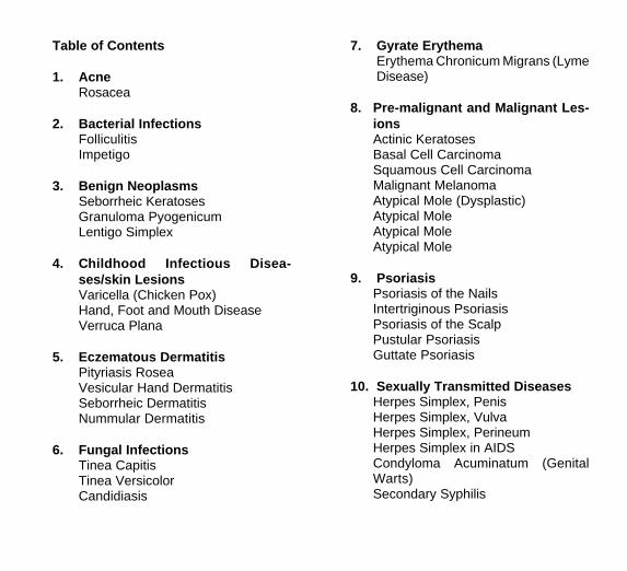
Table of Contents
1. Acne Rosacea
2. Bacterial Infections Folliculitis Impetigo
3. Benign Neoplasms Seborrheic Keratoses Granuloma Pyogenicum Lentigo Simplex
4. Childhood Infectious Diseases/skin Lesions Varicella (Chicken Pox)Hand, Foot and Mouth DiseaseVerruca Plana
5. Eczematous Dermatitis Pityriasis Rosea Vesicular Hand Dermatitis Seborrheic Dermatitis Nummular Dermatitis
6. Fungal Infections Tinea Capitis Tinea Versicolor Candidiasis
7. Gyrate Erythema Erythema Chronicum Migrans (Lyme Disease)
8. Pre-malignant and Malignant Lesions Actinic KeratosesBasal Cell CarcinomaSquamous Cell CarcinomaMalignant MelanomaAtypical Mole (Dysplastic)Atypical MoleAtypical MoleAtypical Mole
9. Psoriasis Psoriasis of the Nails Intertriginous Psoriasis Psoriasis of the Scalp Pustular Psoriasis Guttate Psoriasis
10. Sexually Transmitted Diseases Herpes Simplex, Penis Herpes Simplex, Vulva Herpes Simplex, Perineum Herpes Simplex in AIDS Condyloma Acuminatum (Genital Warts) Secondary Syphilis
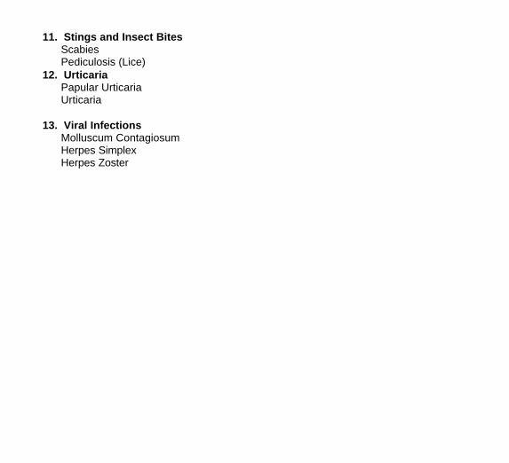
11. Stings and Insect Bites Scabies Pediculosis (Lice)
12. Urticaria Papular Urticaria Urticaria
13. Viral Infections Molluscum Contagiosum Herpes Simplex Herpes Zoster
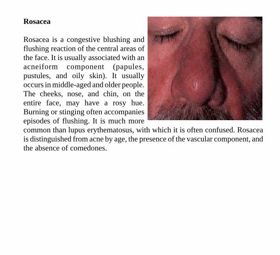
Rosacea
Rosacea is a congestive blushing andflushing reaction of the central areas ofthe face. It is usually associated with anacneiform component (papules,pustules, and oily skin). It usuallyoccurs in middle-aged and older people.The cheeks, nose, and chin, on theentire face, may have a rosy hue.Burning or stinging often accompaniesepisodes of flushing. It is much morecommon than lupus erythematosus, with which it is often confused. Rosaceais distinguished from acne by age, the presence of the vascular component, andthe absence of comedones.
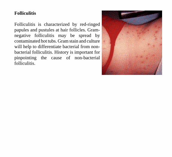
Folliculitis
Folliculitis is characterized by red-ringed papules and pustules at hair follicles. Gramnegative folliculitis may be spread by contaminated hot tubs. Gram stain and culture will help to differentiate bacterial from nonbacterial folliculitis. History is important for pinpointing the cause of non-bacterial folliculitis.
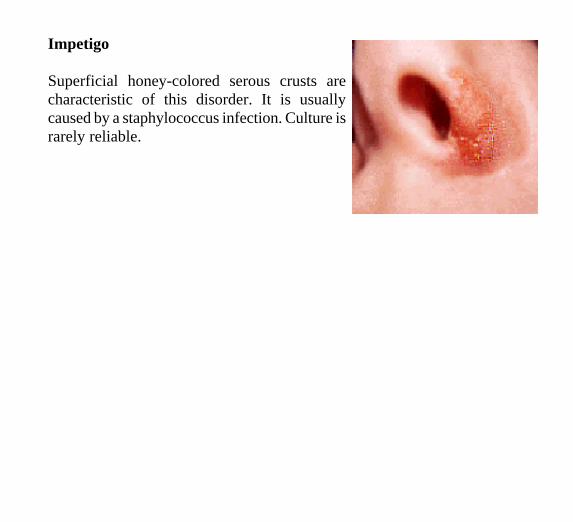
Impetigo
Superficial honey-colored serous crusts are characteristic of this disorder. It is usually caused by a staphylococcus infection. Culture is rarely reliable.
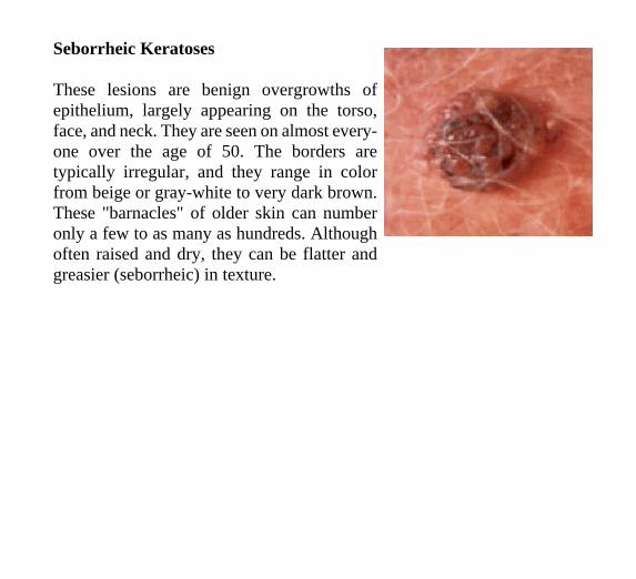
Seborrheic Keratoses
These lesions are benign overgrowths of epithelium, largely appearing on the torso, face, and neck. They are seen on almost everyone over the age of 50. The borders are typically irregular, and they range in color from beige or gray-white to very dark brown. These "barnacles" of older skin can number only a few to as many as hundreds. Although often raised and dry, they can be flatter and greasier (seborrheic) in texture.
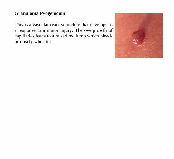
Granuloma Pyogenicum
This is a vascular reactive nodule that develops as a response to a minor injury. The overgrowth of capillaries leads to a raised red lump which bleeds profusely when torn.
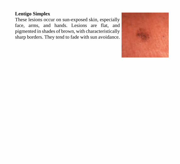
Lentigo Simplex These lesions occur on sun-exposed skin, especially face, arms, and hands. Lesions are flat, and pigmented in shades of brown, with characteristically sharp borders. They tend to fade with sun avoidance.
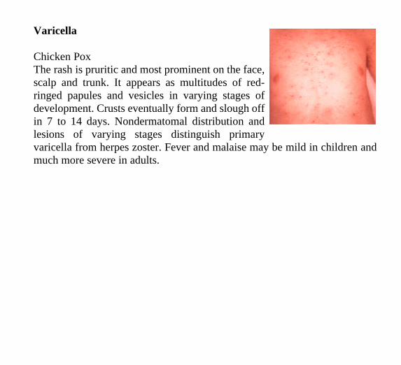
Varicella
Chicken Pox The rash is pruritic and most prominent on the face,scalp and trunk. It appears as multitudes of redringed papules and vesicles in varying stages ofdevelopment. Crusts eventually form and slough offin 7 to 14 days. Nondermatomal distribution andlesions of varying stages distinguish primaryvaricella from herpes zoster. Fever and malaise may be mild in children andmuch more severe in adults.
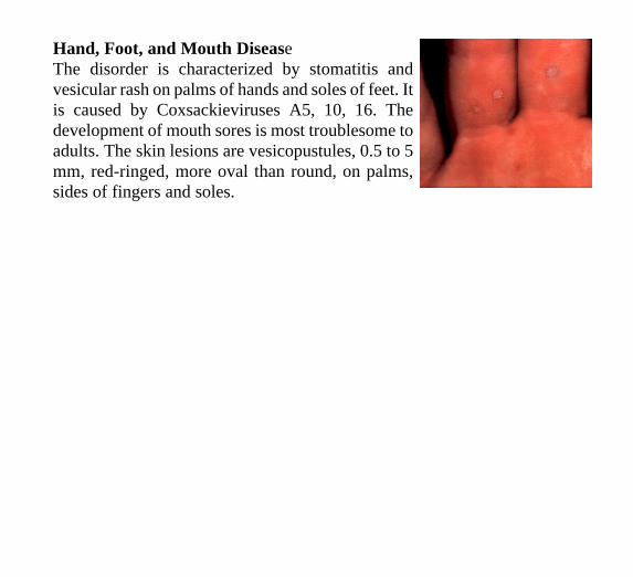
Hand, Foot, and Mouth DiseaseThe disorder is characterized by stomatitis andvesicular rash on palms of hands and soles of feet. Itis caused by Coxsackieviruses A5, 10, 16. Thedevelopment of mouth sores is most troublesome toadults. The skin lesions are vesicopustules, 0.5 to 5mm, red-ringed, more oval than round, on palms,sides of fingers and soles.
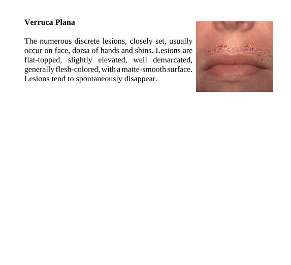
Verruca Plana
The numerous discrete lesions, closely set, usually occur on face, dorsa of hands and shins. Lesions are flat-topped, slightly elevated, well demarcated, generally flesh-colored, with a matte-smooth surface. Lesions tend to spontaneously disappear.
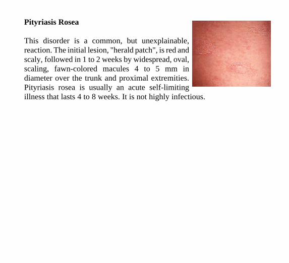
Pityriasis Rosea
This disorder is a common, but unexplainable, reaction. The initial lesion, "herald patch", is red and scaly, followed in 1 to 2 weeks by widespread, oval, scaling, fawn-colored macules 4 to 5 mm in diameter over the trunk and proximal extremities. Pityriasis rosea is usually an acute self-limiting illness that lasts 4 to 8 weeks. It is not highly infectious.
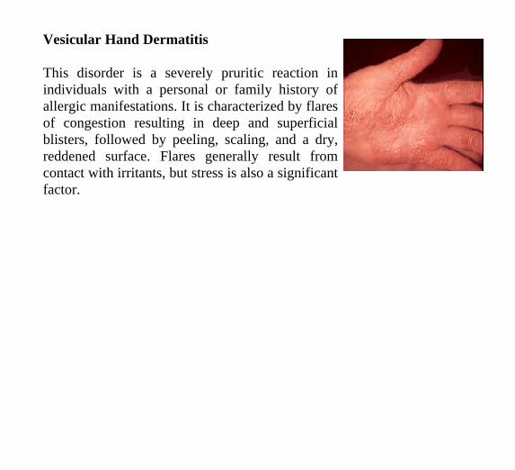
Vesicular Hand Dermatitis
This disorder is a severely pruritic reaction in individuals with a personal or family history of allergic manifestations. It is characterized by flares of congestion resulting in deep and superficial blisters, followed by peeling, scaling, and a dry, reddened surface. Flares generally result from contact with irritants, but stress is also a significant factor.
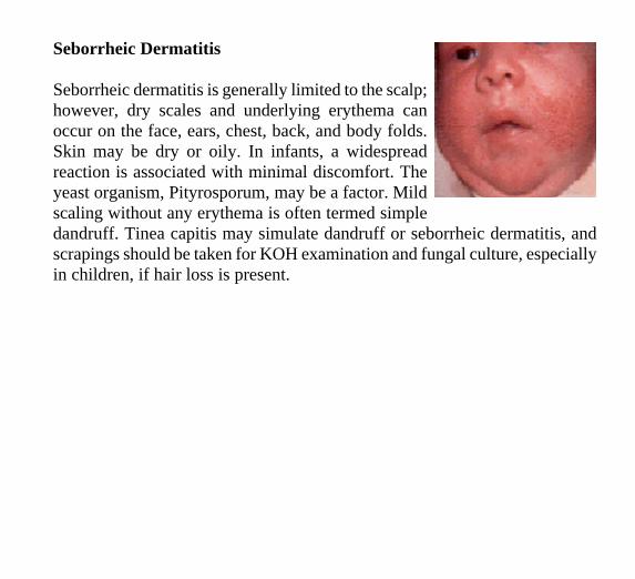
Seborrheic Dermatitis
Seborrheic dermatitis is generally limited to the scalp;however, dry scales and underlying erythema canoccur on the face, ears, chest, back, and body folds.Skin may be dry or oily. In infants, a widespreadreaction is associated with minimal discomfort. Theyeast organism, Pityrosporum, may be a factor. Mildscaling without any erythema is often termed simpledandruff. Tinea capitis may simulate dandruff or seborrheic dermatitis, andscrapings should be taken for KOH examination and fungal culture, especiallyin children, if hair loss is present.
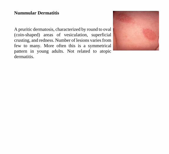
Nummular Dermatitis
A pruritic dermatosis, characterized by round to oval (coin-shaped) areas of vesiculation, superficial crusting, and redness. Number of lesions varies from few to many. More often this is a symmetrical pattern in young adults. Not related to atopic dermatitis.
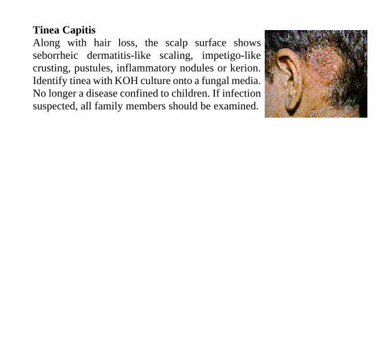
Tinea Capitis Along with hair loss, the scalp surface shows seborrheic dermatitis-like scaling, impetigo-like crusting, pustules, inflammatory nodules or kerion. Identify tinea with KOH culture onto a fungal media. No longer a disease confined to children. If infection suspected, all family members should be examined.
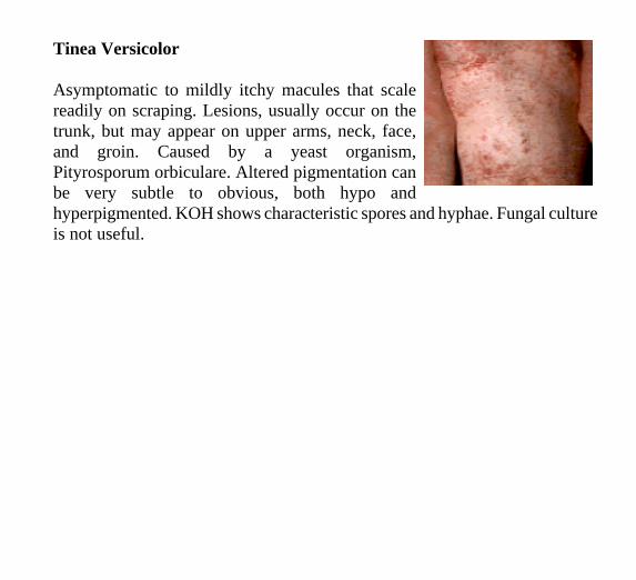
Tinea Versicolor
Asymptomatic to mildly itchy macules that scalereadily on scraping. Lesions, usually occur on thetrunk, but may appear on upper arms, neck, face,and groin. Caused by a yeast organism,Pityrosporum orbiculare. Altered pigmentation canbe very subtle to obvious, both hypo andhyperpigmented. KOH shows characteristic spores and hyphae. Fungal cultureis not useful.
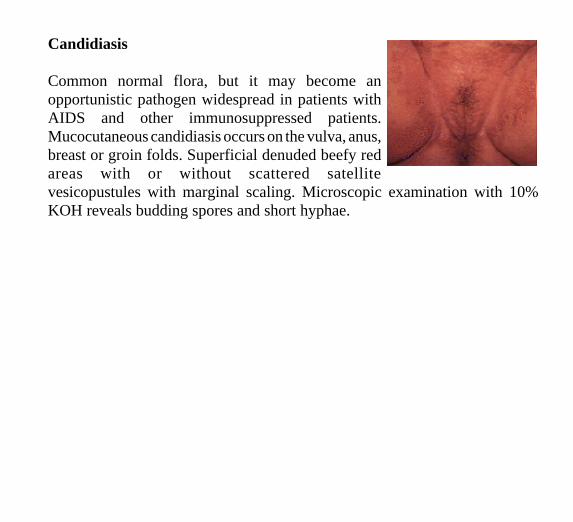
Candidiasis
Common normal flora, but it may become anopportunistic pathogen widespread in patients withAIDS and other immunosuppressed patients.Mucocutaneous candidiasis occurs on the vulva, anus,breast or groin folds. Superficial denuded beefy redareas with or without scattered satellitevesicopustules with marginal scaling. Microscopic examination with 10%KOH reveals budding spores and short hyphae.
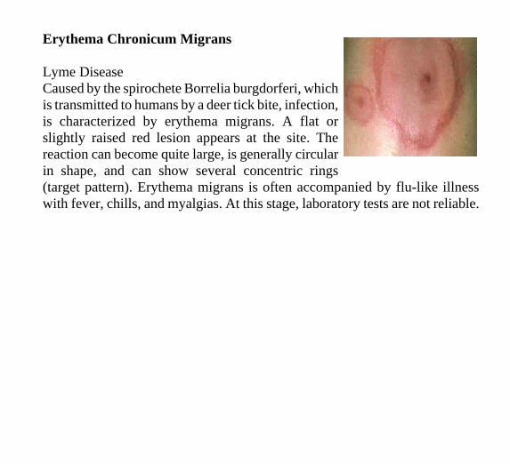
Erythema Chronicum Migrans
Lyme Disease Caused by the spirochete Borrelia burgdorferi, whichis transmitted to humans by a deer tick bite, infection,is characterized by erythema migrans. A flat orslightly raised red lesion appears at the site. Thereaction can become quite large, is generally circularin shape, and can show several concentric rings(target pattern). Erythema migrans is often accompanied by flu-like illnesswith fever, chills, and myalgias. At this stage, laboratory tests are not reliable.
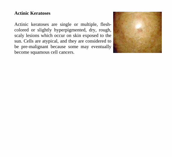
Actinic Keratoses
Actinic keratoses are single or multiple, fleshcolored or slightly hyperpigmented, dry, rough, scaly lesions which occur on skin exposed to the sun. Cells are atypical, and they are considered to be pre-malignant because some may eventually become squamous cell cancers.
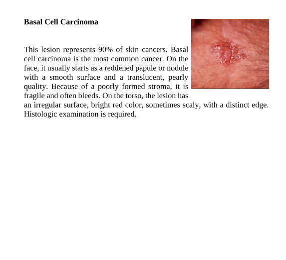
Basal Cell Carcinoma
This lesion represents 90% of skin cancers. Basalcell carcinoma is the most common cancer. On theface, it usually starts as a reddened papule or nodulewith a smooth surface and a translucent, pearlyquality. Because of a poorly formed stroma, it isfragile and often bleeds. On the torso, the lesion hasan irregular surface, bright red color, sometimes scaly, with a distinct edge.Histologic examination is required.
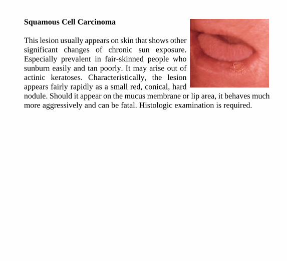
Squamous Cell Carcinoma
This lesion usually appears on skin that shows othersignificant changes of chronic sun exposure.Especially prevalent in fair-skinned people whosunburn easily and tan poorly. It may arise out ofactinic keratoses. Characteristically, the lesionappears fairly rapidly as a small red, conical, hardnodule. Should it appear on the mucus membrane or lip area, it behaves muchmore aggressively and can be fatal. Histologic examination is required.
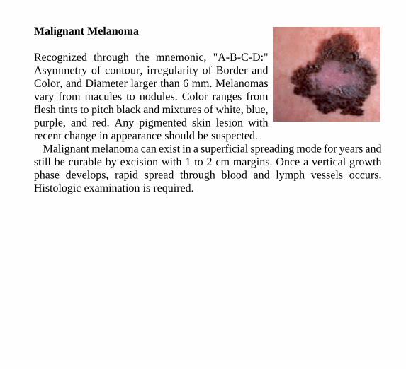
Malignant Melanoma
Recognized through the mnemonic, "A-B-C-D:" Asymmetry of contour, irregularity of Border and Color, and Diameter larger than 6 mm. Melanomas vary from macules to nodules. Color ranges from flesh tints to pitch black and mixtures of white, blue, purple, and red. Any pigmented skin lesion with recent change in appearance should be suspected.
Malignant melanoma can exist in a superficial spreading mode for years and still be curable by excision with 1 to 2 cm margins. Once a vertical growth phase develops, rapid spread through blood and lymph vessels occurs. Histologic examination is required.
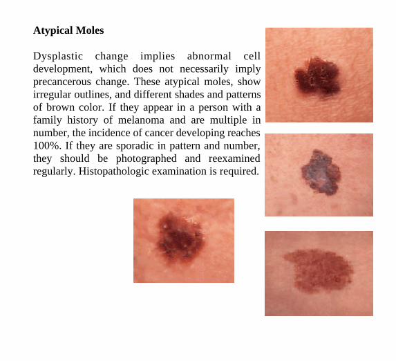
Atypical Moles
Dysplastic change implies abnormal cell development, which does not necessarily imply precancerous change. These atypical moles, show irregular outlines, and different shades and patterns of brown color. If they appear in a person with a family history of melanoma and are multiple in number, the incidence of cancer developing reaches 100%. If they are sporadic in pattern and number, they should be photographed and reexamined regularly. Histopathologic examination is required.
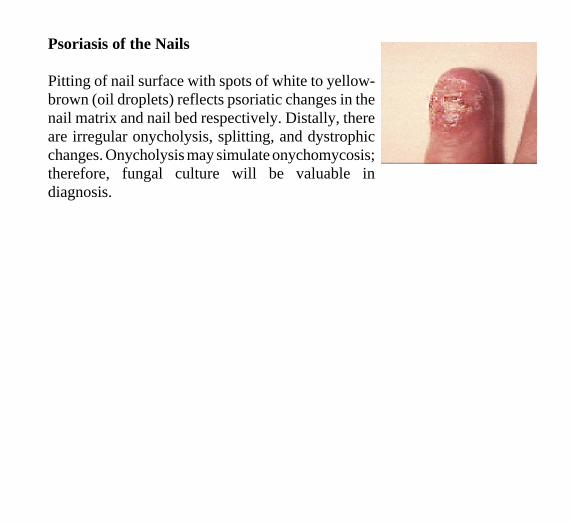
Psoriasis of the Nails
Pitting of nail surface with spots of white to yellowbrown (oil droplets) reflects psoriatic changes in the nail matrix and nail bed respectively. Distally, there are irregular onycholysis, splitting, and dystrophic changes. Onycholysis may simulate onychomycosis; therefore, fungal culture will be valuable in diagnosis.
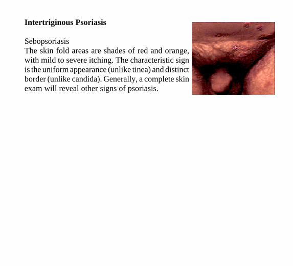
Intertriginous Psoriasis
Sebopsoriasis The skin fold areas are shades of red and orange,with mild to severe itching. The characteristic signis the uniform appearance (unlike tinea) and distinctborder (unlike candida). Generally, a complete skinexam will reveal other signs of psoriasis.
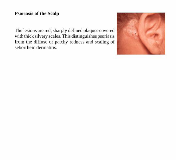
Psoriasis of the Scalp
The lesions are red, sharply defined plaques covered with thick silvery scales. This distinguishes psoriasis from the diffuse or patchy redness and scaling of seborrheic dermatitis.
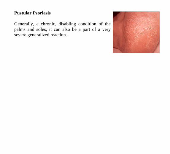
Pustular Psoriasis
Generally, a chronic, disabling condition of the palms and soles, it can also be a part of a very severe generalized reaction.
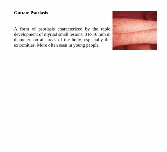
Guttate Psoriasis
A form of psoriasis characterized by the rapid development of myriad small lesions, 3 to 10 mm in diameter, on all areas of the body, especially the extremities. More often seen in young people.
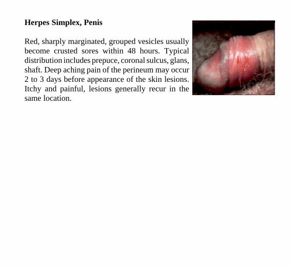
Herpes Simplex, Penis
Red, sharply marginated, grouped vesicles usually become crusted sores within 48 hours. Typical distribution includes prepuce, coronal sulcus, glans, shaft. Deep aching pain of the perineum may occur 2 to 3 days before appearance of the skin lesions. Itchy and painful, lesions generally recur in the same location.
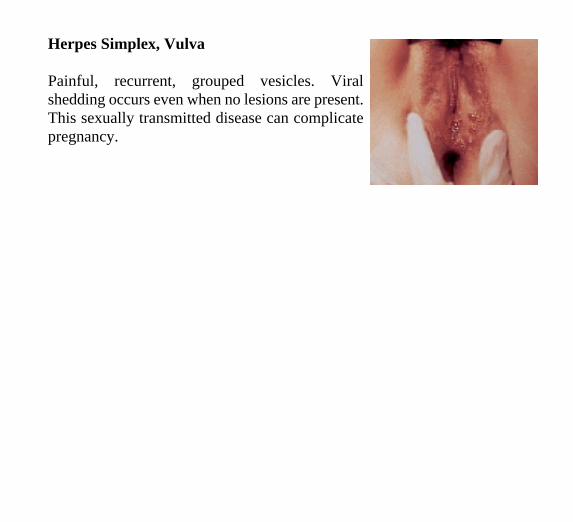
Herpes Simplex, Vulva
Painful, recurrent, grouped vesicles. Viral shedding occurs even when no lesions are present. This sexually transmitted disease can complicate pregnancy.
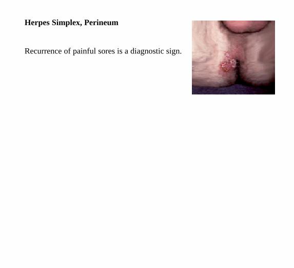
Herpes Simplex, Perineum
Recurrence of painful sores is a diagnostic sign.
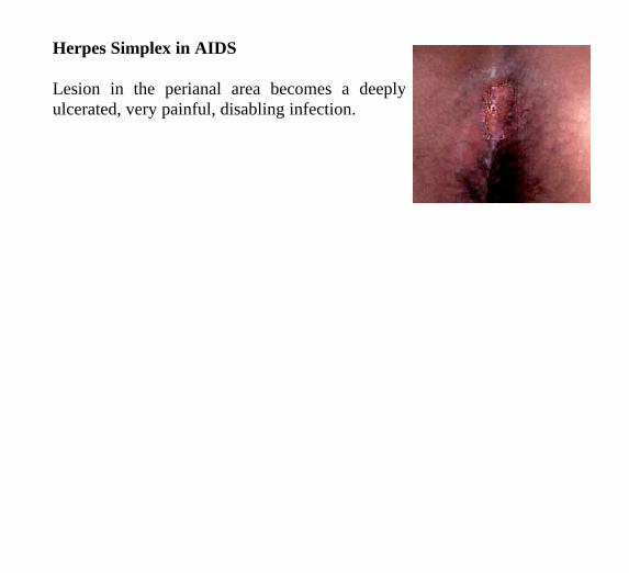
Herpes Simplex in AIDS
Lesion in the perianal area becomes a deeply ulcerated, very painful, disabling infection.
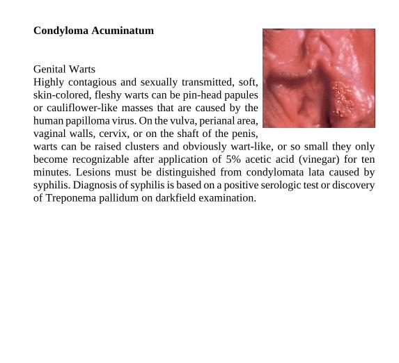
Condyloma Acuminatum
Genital Warts Highly contagious and sexually transmitted, soft,skin-colored, fleshy warts can be pin-head papulesor cauliflower-like masses that are caused by thehuman papilloma virus. On the vulva, perianal area,vaginal walls, cervix, or on the shaft of the penis,warts can be raised clusters and obviously wart-like, or so small they onlybecome recognizable after application of 5% acetic acid (vinegar) for tenminutes. Lesions must be distinguished from condylomata lata caused bysyphilis. Diagnosis of syphilis is based on a positive serologic test or discoveryof Treponema pallidum on darkfield examination.
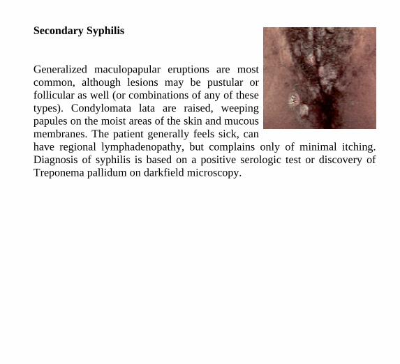
Secondary Syphilis
Generalized maculopapular eruptions are mostcommon, although lesions may be pustular orfollicular as well (or combinations of any of thesetypes). Condylomata lata are raised, weepingpapules on the moist areas of the skin and mucousmembranes. The patient generally feels sick, canhave regional lymphadenopathy, but complains only of minimal itching.Diagnosis of syphilis is based on a positive serologic test or discovery ofTreponema pallidum on darkfield microscopy.
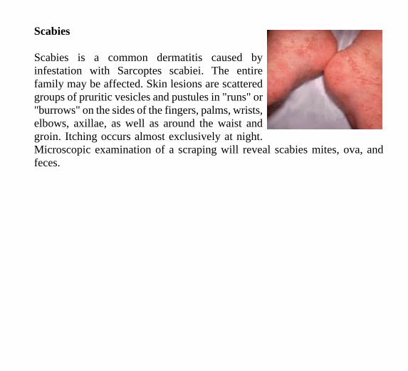
Scabies
Scabies is a common dermatitis caused byinfestation with Sarcoptes scabiei. The entirefamily may be affected. Skin lesions are scatteredgroups of pruritic vesicles and pustules in "runs" or"burrows" on the sides of the fingers, palms, wrists,elbows, axillae, as well as around the waist andgroin. Itching occurs almost exclusively at night.Microscopic examination of a scraping will reveal scabies mites, ova, andfeces.
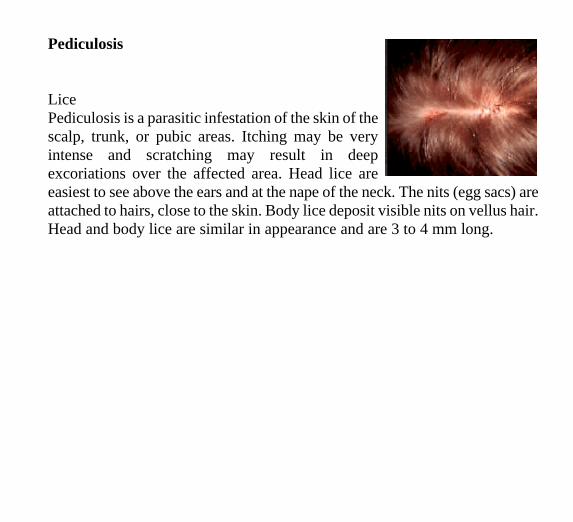
Pediculosis
LicePediculosis is a parasitic infestation of the skin of thescalp, trunk, or pubic areas. Itching may be veryintense and scratching may result in deepexcoriations over the affected area. Head lice areeasiest to see above the ears and at the nape of the neck. The nits (egg sacs) areattached to hairs, close to the skin. Body lice deposit visible nits on vellus hair.Head and body lice are similar in appearance and are 3 to 4 mm long.
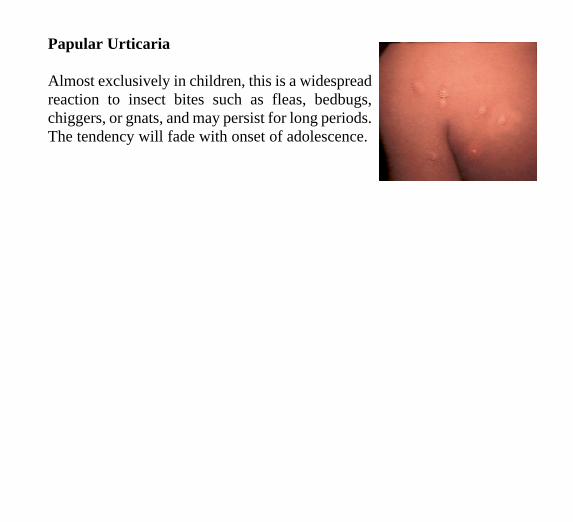
Papular Urticaria
Almost exclusively in children, this is a widespread reaction to insect bites such as fleas, bedbugs, chiggers, or gnats, and may persist for long periods. The tendency will fade with onset of adolescence.
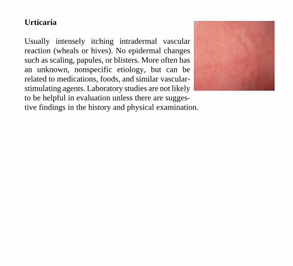
Urticaria
Usually intensely itching intradermal vascular reaction (wheals or hives). No epidermal changes such as scaling, papules, or blisters. More often has an unknown, nonspecific etiology, but can be related to medications, foods, and similar vascularstimulating agents. Laboratory studies are not likely to be helpful in evaluation unless there are suggestive findings in the history and physical examination.
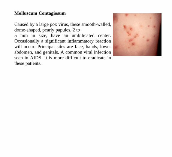
Molluscum Contagiosum
Caused by a large pox virus, these smooth-walled, dome-shaped, pearly papules, 2 to 5 mm in size, have an umbilicated center. Occasionally a significant inflammatory reaction will occur. Principal sites are face, hands, lower abdomen, and genitals. A common viral infection seen in AIDS. It is more difficult to eradicate in these patients.
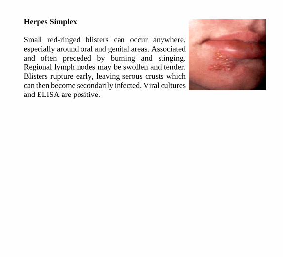
Herpes Simplex
Small red-ringed blisters can occur anywhere, especially around oral and genital areas. Associated and often preceded by burning and stinging. Regional lymph nodes may be swollen and tender. Blisters rupture early, leaving serous crusts which can then become secondarily infected. Viral cultures and ELISA are positive.
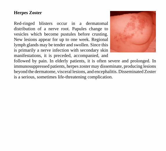
Herpes Zoster
Red-ringed blisters occur in a dermatomaldistribution of a nerve root. Papules change tovesicles which become pustules before crusting.New lesions appear for up to one week. Regionallymph glands may be tender and swollen. Since thisis primarily a nerve infection with secondary skinmanifestations, it is preceded, accompanied, andfollowed by pain. In elderly patients, it is often severe and prolonged. Inimmunosuppressed patients, herpes zoster may disseminate, producing lesionsbeyond the dermatome, visceral lesions, and encephalitis. Disseminated Zosteris a serious, sometimes life-threatening complication.