Embed Size (px)
Citation preview
1
© 2017 by the American College of Gastroenterology The American Journal of GASTROENTEROLOGY
CLINICAL GUIDELINES
Colorectal cancer (CRC) screening is the process of detecting
early-stage CRCs and precancerous lesions in asymptomatic peo-
ple with no prior history of cancer or precancerous lesions. Th e
U.S. Multi-Society Task Force of Colorectal Cancer (MSTF) is a
panel of expert gastroenterologists representing the American
College of Gastroenterology, the American Gastroenterological
Association, and the American Society for Gastrointestinal
Endoscopy. Th e MSTF, like others, has long endorsed systematic
off ers of CRC screening to average-risk persons (persons without
a high-risk family history of colorectal neoplasia) beginning at age
50 years, with general evidence supporting screening reviewed in
previous publications ( 1 ). Th is publication updates the screen-
Colorectal Cancer Screening: Recommendations for
Physicians and Patients from the U.S. Multi-Society
Task Force on Colorectal Cancer
Douglas K. Rex , MD 1 , C. Richard Boland , MD 2 , Jason A. Dominitz , MD, MHS 3 , Francis M. Giardiello , MD 4 , David A. Johnson , MD 5 ,
Tonya Kaltenbach , MD 6 , Th eodore R. Levin , MD 7 , David Lieberman , MD 8 and Douglas J. Robertson , MD, MPH 9
This document updates the colorectal cancer (CRC) screening recommendations of the U.S. Multi-Society Task
Force of Colorectal Cancer (MSTF), which represents the American College of Gastroenterology, the American
Gastroenterological Association, and The American Society for Gastrointestinal Endoscopy. CRC screening tests
are ranked in 3 tiers based on performance features, costs, and practical considerations. The fi rst-tier tests are
colonoscopy every 10 years and annual fecal immunochemical test (FIT). Colonoscopy and FIT are recommended
as the cornerstones of screening regardless of how screening is offered. Thus, in a sequential approach based on
colonoscopy offered fi rst, FIT should be offered to patients who decline colonoscopy. Colonoscopy and FIT are
recommended as tests of choice when multiple options are presented as alternatives. A risk-stratifi ed approach
is also appropriate, with FIT screening in populations with an estimated low prevalence of advanced neoplasia
and colonoscopy screening in high prevalence populations. The second-tier tests include CT colonography every
5 years, the FIT-fecal DNA test every 3 years, and fl exible sigmoidoscopy every 5 to 10 years. These tests are
appropriate screening tests, but each has disadvantages relative to the tier 1 tests. Because of limited evidence
and current obstacles to use, capsule colonoscopy every 5 years is a third-tier test. We suggest that the Septin9
serum assay (Epigenomics, Seattle, Wash) not be used for screening. Screening should begin at age 50 years in
average-risk persons, except in African Americans in whom limited evidence supports screening at 45 years. CRC
incidence is rising in persons under age 50, and thorough diagnostic evaluation of young persons with suspected
colorectal bleeding is recommended. Discontinuation of screening should be considered when persons up to date
with screening, who have prior negative screening (particularly colonoscopy), reach age 75 or have <10 years of life
expectancy. Persons without prior screening should be considered for screening up to age 85, depending on age and
comorbidities. Persons with a family history of CRC or a documented advanced adenoma in a fi rst-degree relative
age <60 years or 2 fi rst-degree relatives with these fi ndings at any age are recommended to undergo screening by
colonoscopy every 5 years, beginning 10 years before the age at diagnosis of the youngest affected relative or age
40, whichever is earlier. Persons with a single fi rst-degree relative diagnosed at ≥60 years with CRC or an advanced
adenoma can be offered average-risk screening options beginning at age 40 years.
Am J Gastroenterol advance online publication, 6 June 2017; doi: 10.1038/ajg.2017.174
1 Indiana University School of Medicine , Indianapolis , Indiana , USA ; 2 University of California San Diego , San Diego , California , USA ; 3 VA Puget Sound Health Care
System, University of Washington , Seattle , Washington , USA ; 4 Johns Hopkins University School of Medicine , Baltimore , Maryland , USA ; 5 Eastern Virginia Medical
School , Norfolk , Virginia , USA ; 6 San Francisco Veterans Affairs Medical Center , San Francisco , California , USA ; 7 Kaiser Permanente Medical Center , Walnut
Creek , California , USA ; 8 Oregon Health and Science University , Portland , Oregon , USA ; 9 VA Medical Center, White River Junction, Vermont, and Geisel School
of Medicine at Dartmouth , Hanover , New Hampshire , USA . Correspondence: Douglas K. Rex, MD , Indiana University Hospital 4100, 550 N University Blvd ,
Indianapolis , IN 46202 , USA . E-mail: [email protected]
Rex et al.
The American Journal of GASTROENTEROLOGY VOLUME XXX | XXX 2017 www.nature.com/ajg
2
ing recommendations of the MSTF for screening in average-risk
persons ( 1 ).
Screening diff ers from surveillance. Surveillance refers to the
interval use of colonoscopy in patients with previously detected
CRC or precancerous lesions and interval colonoscopy in patients
performed to detect dysplasia in persons with infl ammatory bowel
disease aff ecting the colon. Surveillance recommendations from
the MSTF on surveillance aft er cancer ( 2 ) and removal of pre-
cancerous lesions ( 3 ) are available in other documents. Screening
is also distinct from diagnostic examinations, which refer to the
investigation of patients with symptoms or positive screening tests
other than colonoscopy. Colonoscopy is generally the test of choice
for diagnostic examinations.
METHODS
Literature review
Th e English language medical literature using MEDLINE (2005
to August 1, 2016), EMBASE (2005 to third quarter 2016 update),
the Database of Abstracts of Reviews and Eff ects (2005 to third
quarter 2016 update), and the Cochrane Database of Systematic
Reviews (2005 to third quarter 2014 update) was searched. In
MEDLINE, subject headings for colorectal cancer screening were
combined with headings for fecal occult blood test, fecal immu-
nochemical test, colonoscopy, sigmoidoscopy, CT colonoscopy,
fecal DNA, serum testing, cost-eff ectiveness, and quality. Similar
searches were performed in EMBASE, the Database of Abstracts
of Reviews and Eff ects, and the Cochrane Database of System-
atic Reviews. Case reports and studies performed in patients with
infl ammatory bowel disease, prior CRC or polyps, or hereditary
CRC syndromes were excluded. Review papers, meta-analyses,
gastroenterology textbooks, and editorials were searched manu-
ally for additional pertinent references. Th e review includes studies
published since 2008 but also incorporates older evidence used
to draft the 2008 recommendations ( 1 ). Evidence-based weighted
recommendations are provided with supporting discussion to
help guide clinicians in the management of these patients.
Process and levels of evidence
Guidance statements were developed by consensus obtained
through joint teleconferences. Th e completed article was reviewed
and approved by all 3 gastroenterology societies.
Th e use of GRADE for MSTF guidance papers has been outlined
in detail elsewhere ( 2 ). GRADE involves comprehensive literature
search and summary (oft en through meta-analysis) and then a
separate review of literature quality and development of recom-
mendations. Th e MSTF uses a modifi ed qualitative approach based
on literature review (as described above for this article) but with-
out formal meta-analysis. GRADE allows for a separate assessment
of the quality of the evidence and strength of recommendation.
Th is approach explicitly recognizes the importance of literature in
informing clinical recommendations but allows latitude because
recommendations may be infl uenced by other factors, such as
patient preference, cost, and expert consensus. “Strong recom-
mendations” are those that would be chosen by most informed
patients. “Weak recommendations” are those where patient values
and preferences might play a larger role than the quality of evi-
dence. Within the document we preface strong recommendations
with phrases such as “we recommend” and weak recommenda-
tions with “we suggest.”
APPROACHES TO SCREENING
In the United States CRC screening usually results from an offi ce-
based interaction between a healthcare provider and patient.
Screening in this setting is termed opportunistic ( 4 ).
Programmatic screening (sometimes called organized screening)
refers to a system-wide, organized approach to off ering screening
to a population or members of a healthcare plan ( 4 ). Programmatic
screening has potential advantages over opportunistic screening,
including systematic off ers of screening, reduction of overscreen-
ing, superior monitoring of quality, and systematic follow-up
of testing. National CRC screening programs in Europe ( 5 ) and
Australia ( 6 ) use fecal occult blood testing and include screening
colonoscopy in Germany and Poland ( 5 ). Th e United States has
no national program for CRC screening, although several large
healthcare plans off er programmatic screening, typically with a
fecal immunochemical test (FIT) ( 7 ). Despite the potential advan-
tages of programmatic screening, the United States has achieved
the world's highest rates of CRC screening compliance at 60%
and the greatest CRC incidence and mortality reduction, using an
almost entirely opportunistic approach ( 8–12 ). Incidence reduc-
tions in the United States were 3 to 4% per year and 30% overall
in the fi rst decade of this century ( 11,12 ). High rates of screening
in the United States may refl ect widespread awareness of CRC and
insurance coverage of screening. Th e MSTF anticipates growth of
programmatic screening within healthcare systems but expects at
least short-term continued reliance on opportunistic screening in
the United States. Reliance on opportunistic screening can aff ect
the preference for CRC screening, because achieving compli-
ance with tests that should be repeated at short intervals is more
challenging in the opportunistic setting ( 13 ).
In the setting of opportunistic screening, healthcare providers
can use several broad strategies to off er screening to patients. One
approach is multiple options , in which the benefi ts, risks, and costs
of 2 or more tests are discussed and off ered to patients ( Table 1 )
( 14 ). Some evidence suggests that when patients are off ered
both colonoscopy and fecal occult blood testing, more patients
undergo screening ( 15 ). Other data suggest no benefi t in over-
all compliance when multiple options are off ered ( 16–18 ). In 1
study, off ering patients 5 options did not enhance compliance
over 2 options ( 19 ). In this regard, at least 9 diff erent screening
tests (colonoscopy, FIT, guaiac-based fecal occult blood test, FIT-
fecal DNA, sigmoidoscopy, sigmoidoscopy plus fecal occult blood
test, CT colonography, barium enema, and the Septin9 serum
assay [Epigenomics, Seattle, Wash]) are endorsed or discussed
in recent major screening guidelines ( 14,20 ). Th us, the multiple
options discussion may best be limited to 2 or 3 preferred options.
If patients decline all the off ered options, 1 or more of the other
options can be off ered.
© 2017 by the American College of Gastroenterology The American Journal of GASTROENTEROLOGY
3
Th e MSTF considers that each of the approaches outlined above
is reasonable when off ering screening in the opportunistic setting.
Th ere is insuffi cient evidence to identify one approach as superior.
Patients undergoing screening tests other than colonoscopy should
understand that colonoscopy is used to evaluate these tests when
positive. In some instances insurance coverage of colonoscopy per-
formed to evaluate other positive screening tests may be less than
coverage of primary screening colonoscopy. Awareness of the dif-
ferent approaches may assist clinicians in understanding screening
literature and in selecting an approach to off ering screening that
seems to be optimal for their practice or for an individual patient.
Recommendations
1 . We recommend that clinicians off er CRC screening begin-
ning at age 50 (strong recommendation, high-quality
evidence). (See below for adjustments in recommended age
for onset of screening based on race and family history.)
2 . We suggest that sequential off ers of screening tests, off ering
multiple screening options, and risk-stratifi ed screening
are all reasonable approaches to off ering screening (weak
recommendation, low-quality evidence).
SCREENING TARGETS
Th e object of screening is to reduce CRC incidence and mortality.
To accomplish both aims, tests need to detect early-stage (i.e.,
curable) CRCs and high-risk precancerous lesions ( 1,21 ). Detec-
tion and removal of precancerous lesions prevents CRC ( 30,31 ).
Th e 2 main classes of precancerous lesions in the colon are con-
ventional adenomas and serrated class lesions ( Table 2 ). Th ese 2
classes of precancerous lesions have distinct endoscopic features
and histology and diff erent (though overlapping) distributions
within the colorectum. Specifi c screening tests sometimes have
Th e sequential approach to screening involves an off er of a fi rst
test that is usually the provider's preferred screening option; if the
patient declines the fi rst option, a second test is off ered, and so
on. In the United States the sequential approach oft en involves an
off er of colonoscopy, followed by FIT if colonoscopy is declined,
or another screening test ( 9 ). Separate guidelines from the Ameri-
can College of Gastroenterology ( 21 ) and the American Society
for Gastrointestinal Endoscopy ( 22 ) recommend a sequential
approach with colonoscopy off ered fi rst. Sequential testing can
maximize compliance overall as well as with the test recommended
fi rst ( 23–25 ). Clinicians using the colonoscopy-fi rst sequential
approach place emphasis on the high effi cacy of colonoscopy in
preventing CRC and less emphasis on the risks of colonoscopy.
Indeed, high-quality colonoscopy has both higher single-time
testing effi cacy and greater risks than any other screening tests but
with absolute risk rates that are still very low when performed by
skilled operators ( 26 ). A variant of sequential testing oft en used in
the programmatic setting is to off er patients FIT as the initial or
preferred test and have other options such as colonoscopy available
to patients who express interest in alternatives ( 4 ).
A third approach to off ering screening to average-risk persons
is a risk-stratifi ed approach. Risk stratifi cation uses evidence that
the “average-risk” population actually represents a wide range of
risk that can be estimated based on demographic and other risk
factors. For example, older age, male gender, obesity, diabetes, and
cigarette smoking are all associated with colorectal adenomas and
cancer and therefore might be used in stratifying risk within the
average-risk population ( 21 ). Th e goal is to predict subgroups of
patients with a high prevalence of important precancerous lesions
benefi ting most from referral directly to colonoscopy, whereas the
subgroups with a predicted lower risk (prevalence) of important
precancerous lesions are referred for screening tests with less risk
and cost than colonoscopy. Risk stratifi cation has been poorly
accepted because of limited accuracy in discriminating high- and
low-prevalence subgroups ( 27 ). However, recent validated models
appear to be simple to apply and had substantial accuracy in defi n-
ing high- and low-risk groups for advanced adenomas ( 28,29 ).
Th ere are no clinical trials comparing compliance or other out-
comes using a risk-stratifi ed approach to the multiple options or
sequential approaches. Few data are currently available regarding
ease of application of a risk-stratifi ed approach in clinical practice.
Table 1 . Approaches to offering screening in the opportunistic
setting
Approach Description
Multiple options The relative benefi ts, risks, and costs of 2 or
more options are presented
Sequential testing A preferred test is offered fi rst. If the patients
decline another option(s) is offered
Risk stratifi ed approach Colonoscopy is offered to patients predicted
to have a high prevalence of advanced pre-
cancerous lesions; other tests are offered to
patients predicted at low risk
Table 2 . Histologic classifi cation of the two major classes of
colorectal polyps
I. Conventional adenomas
a. Dysplasia grade
i. High grade
ii. Low grade
b. Villousity
i. Tubular
ii. Tubulovillous
iii. Villous
II. Serrated lesions
a.Hyperplastic polyps (not considered precancerous)
b. Sessile serrated polyp
i. Without cytologic dysplasia
ii. With cytologic dysplasia
c. Traditional serrated adenoma
Rex et al.
The American Journal of GASTROENTEROLOGY VOLUME XXX | XXX 2017 www.nature.com/ajg
4
particular strengths or weaknesses detecting 1 or the other class
of precancerous lesions, particularly the serrated class. Th ere-
fore, we review here the main clinical features of the 2 classes of
precancerous lesions.
Adenomas, also known as conventional adenomas, are the pre-
cursors of perhaps 70% of all CRCs ( 32,33 ). Th e adenoma-carci-
noma sequence is believed to typically take more than 10 years
to complete in sporadic cancers, whereas much shorter intervals
occur in Lynch syndrome ( 34 ). Correspondingly, colonoscopy is
recommended at 10-year intervals in average-risk persons and at
1- to 2-year intervals in those with Lynch syndrome ( 1,34 ). Th e
distribution of adenomas is relatively even throughout the colon,
although adenomas with a fl at or depressed morphology are dis-
tributed more to the proximal colon and pedunculated lesions
more to the distal colon ( 35 ). Adenomas are by defi nition dys-
plastic, with the overwhelming majority being low grade. Th e pres-
ence of high-grade dysplasia in an adenoma should be noted by
a pathologist. Adenomas can also be characterized by tubular vs.
villous histology, with the overwhelming majority tubular. Lesions
with >25% villous elements are termed tubulovillous and those
with >75% villous elements villous . Villous elements and invasive
cancer are associated with increasing size of adenomas. Invasive
cancer in adenomas ≤5 mm in size is extremely rare, and the preva-
lence remains well below 1% in adenomas 6 to 9 mm in size ( 36 ).
Recent colonoscopic studies have identifi ed lower prevalence rates
of cancer in polyps <1 cm in size compared with early studies,
probably because improvements in colonoscope technology and
performance have led to routine detection of an array of small,
fl at, low-volume adenomas ( 36 ). Interobserver agreement in
diff erentiation of high- vs. low-grade dysplasia by pathologists
and tubular vs. tubulovillous histology is poor to moderate,
particularly in adenomas <1 cm in size ( 37 ). Conversely, inter-
observer agreement between pathologists is good to excellent
in placing lesions within the conventional adenomas vs. serrated
polyps and in identifying invasive cancer ( 38 ).
An important clinical concept is the “advanced” adenoma,
defi ned as a lesion ≥1 cm in size or having high-grade dysplasia
or villous elements ( 3 ). Because nonadvanced adenomas have
a very low prevalence of cancer and a long adenoma-cancer
sequence, screening tests can remain useful if they target cancer
and advanced adenomas and not small adenomas. Further, the
prevalence of nonadvanced adenomas is so high in modern colo-
noscopy studies that detection of such lesions by noncolonoscopic
screening tests leads to unacceptably low specifi city. Colonoscopy
has an important benefi t over other screening methods because of
its ability to detect and remove both advanced and non advanced
adenomas. Although nonadvanced adenomas have limited
clinical importance and are not the target of noncolonoscopic
screening methods, colonoscopists strive to identify and remove
non advanced adenomas. Th us, resecting lesions with any precan-
cerous potential during colonoscopy is safe, seems to be better
accepted by patients in the United States, and removes them as
a clinical concern.
Serrated colorectal lesions ( Figure 1 ) represent an emerging area
in the fi eld of precancerous colorectal lesions. Th e serrated class
of precursor lesions accounts for up to 30% of CRCs ( 33 ). Within
the serrated class, hyperplastic polyps are not currently considered
precancerous, whereas sessile serrated polyps (SSPs; also known as
sessile serrated adenoma) and traditional serrated adenomas are
considered precancerous ( Table 2 ) ( 33 ). Hyperplastic polyps are
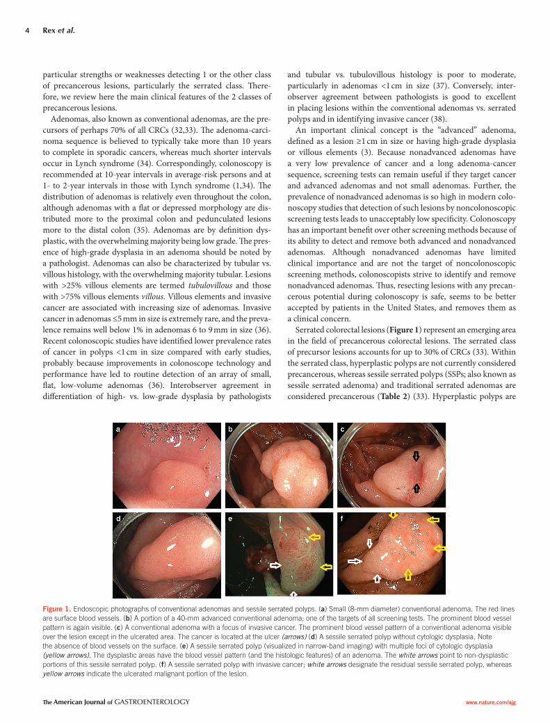
Figure 1 . Endoscopic photographs of conventional adenomas and sessile serrated polyps. ( a ) Small (8-mm diameter) conventional adenoma. The red lines
are surface blood vessels. ( b ) A portion of a 40-mm advanced conventional adenoma; one of the targets of all screening tests. The prominent blood vessel
pattern is again visible. ( c ) A conventional adenoma with a focus of invasive cancer. The prominent blood vessel pattern of a conventional adenoma visible
over the lesion except in the ulcerated area. The cancer is located at the ulcer (arrows) ( d ) A sessile serrated polyp without cytologic dysplasia. Note
the absence of blood vessels on the surface. ( e ) A sessile serrated polyp (visualized in narrow-band imaging) with multiple foci of cytologic dysplasia
(yellow arrows) . The dysplastic areas have the blood vessel pattern (and the histologic features) of an adenoma. The white arrows point to non-dysplastic
portions of this sessile serrated polyp. ( f ) A sessile serrated polyp with invasive cancer; white arrows designate the residual sessile serrated polyp, whereas
yellow arrows indicate the ulcerated malignant portion of the lesion.
© 2017 by the American College of Gastroenterology The American Journal of GASTROENTEROLOGY
5
should consider choosing colonoscopy. Although no randomized
trials of colonoscopy for screening have been completed, exten-
sive evidence from adenoma cohorts ( 30,31 ), cohort studies on
incidence and mortality ( 55,56 ), and case-control studies ( 57–64 )
support the effi cacy of colonoscopy in preventing incident CRC
and cancer deaths. One cohort study ( 56 ) and 3 case-control stud-
ies ( 58,59,64 ) were performed in screening populations. Reduc-
tions in incidence and mortality are approximately 80% in the
distal colon and 40 to 60% in the proximal colon, at least in the
United States and Germany ( 57,59,61,62,64 ). Furthermore, indi-
rect evidence from randomized trials of fecal occult blood testing
( 65 ) and sigmoidoscopy ( 66 ), as well as studies showing highly
variable cancer protection provided by diff erent colonoscopists
( 67,68 ), also supports a protective eff ect of colonoscopy against
CRC. Th ese fi ndings are consistent with the observed population
trends in the United States ( 11,12 ).
Disadvantages of colonoscopy include the need for thorough
bowel cleansing, a higher risk of perforation relative to the other
screening tests, higher risk of aspiration pneumonitis (particu-
larly when the procedure is performed with deep sedation) ( 69 ),
a small risk of splenic injury requiring splenectomy, and a greater
risk of postprocedural bleeding compared with other screening
tests. A meta-analysis of population-based studies found risks of
perforation, bleeding, and death of 0.5 per 1000, 2.6 per 1000,
and 2.9 per 100,000, respectively ( 70 ). Bleeding aft er colono-
scopy is almost entirely related to polypectomy. When electro-
cautery is used for resection of all colorectal polyps, most bleeds
occur aft er resection of small lesions. Th is relates entirely to the
high prevalence of these lesions because increasing polyp size
and proximal colon location are the major risk factors for bleed-
ing per individual resected polyp ( 71 ). Cold resection techniques
are eff ective and nearly devoid of clinically signifi cant bleeding
risk and can be generally advised for nonpedunculated lesions
<1 cm in size ( 72 ). Despite these risks, colonoscopy is the pre-
ferred approach to management of any benign colorectal polyp
regardless of size or location because the alternative of surgical
resection has higher mortality and cost compared with colono-
scopy ( 73,74 ). To the extent that other screening tests eff ectively
identify large lesions, they result in colonoscopy and do not pre-
vent adverse events related to colonoscopic resection of large
lesions.
A major disadvantage of colonoscopy is operator dependence
in performance. Operator dependence aff ects detection of can-
cer ( 67,68,75 ), adenomas ( 76,77 ), and serrated lesions ( 40,41,78 );
selection of appropriate screening and surveillance intervals aft er
colonoscopy ( 79 ); and eff ective resection of colorectal polyps
( 80 ). In general, gastroenterologists performing colonoscopy are
more eff ective than nongastroenterologists in prevention of can-
cer ( 62,81–83 ) and detection of precancerous polyps ( 84 ). How-
ever, substantial operator dependence within gastroenterologists
is consistently observed ( 42,43,76–78 ), so that selection of a colo-
noscopist by specialty is not adequate protection against subopti-
mal operator performance. Table 3 shows a list of questions that
patients can ask potential colonoscopists to judge whether perfor-
mance is likely to be at a high level. Aft erward, the colonoscopy
usually small lesions and are distributed toward the distal colon
( 39 ). SSPs are common (found in 8–9% of screening colonoscopies
performed by expert detectors) ( 40,41 ) and are distributed toward
the proximal colon compared with conventional adenomas. SSPs
are typically fl at or sessile in shape, have few or no surface blood
vessels (conventional adenomas by comparison have many surface
vessels), and are more diffi cult to detect at colonoscopy than con-
ventional adenomas ( 33,42,43 ). Because of their prevalence and
precancerous potential, SSPs are the major precancerous serrated
lesion. Th ere is poor interobserver agreement between patho-
logists in the diff erentiation of hyperplastic polyps from SSPs ( 44 ).
Consequently, clinicians can see widely varying rates of SSPs in
pathology reports, depending on the pathologist or even the center
in which they practice ( 45 ). Most SSPs are not dysplastic, and the
lesions should consistently be designated as “SSP without cytologic
dysplasia” or “SSP with cytologic dysplasia.” ( 46 ) When a dysplastic
component is present, it is oft en evident endoscopically ( Figure 1 )
and histologically is a region of conventional adenoma within an
otherwise serrated lesion ( 47 ). Microdissection studies indicate
that the dysplastic area oft en has microsatellite instability ( 48 ). Th e
SSP with cytologic dysplasia is considered a more advanced lesion
in the polyp cancer sequence than SSP without cytologic dysplasia
( 3,33,49,50 ).
Th e traditional serrated adenoma is a rare lesion, oft en in the
left colon, sessile, and uniformly dysplastic ( 33,46 ). Because tradi-
tional serrated adenoma is rare, dysplastic, and has a villous-like
growth pattern histologically, it is oft en misinterpreted as a tubu-
lovillous conventional adenoma ( 33 ).
Th e features of these 2 classes of precancerous lesions are rele-
vant to the available screening tests. Colonoscopy is the criterion
standard for the detection of all precancerous colorectal lesions.
Colonoscopy achieves its greatest superiority relative to other
screening tests in the detection of conventional adenomas <1 cm
in size and serrated class lesions. Detection of SSPs is a major defi -
ciency of fl exible sigmoidoscopy because SSPs are predominantly
in the proximal colon ( 51 ), of CT colonography because the lesions
tend to be fl at ( 52 ), and of FIT ( 53 ) probably because SSPs have no
or few surface blood vessels with less tendency to bleed than con-
ventional adenomas. Th e combined FIT-fecal DNA test achieves
its greatest relative performance compared with FIT alone in the
detection of serrated class lesions, related to the poor sensitivity
of FIT for these lesions and the inclusion of hypermethylation
markers in the DNA panel ( 53 ). Hypermethylation is a feature of
serrated lesions ( 33 ).
SPECIFIC SCREENING TESTS
Colonoscopy
Th e advantages of colonoscopy include high sensitivity for
cancer and all classes of precancerous lesions, single-session
diagnosis and treatment, and long intervals between examinations
(10 years) in subjects with normal examinations. One or 2 nega-
tive examinations may signal lifetime protection against CRC ( 54 ).
Patients who value the highest level of sensitivity in detection of
precancerous lesions and are willing to undergo invasive screening
Rex et al.
The American Journal of GASTROENTEROLOGY VOLUME XXX | XXX 2017 www.nature.com/ajg
6
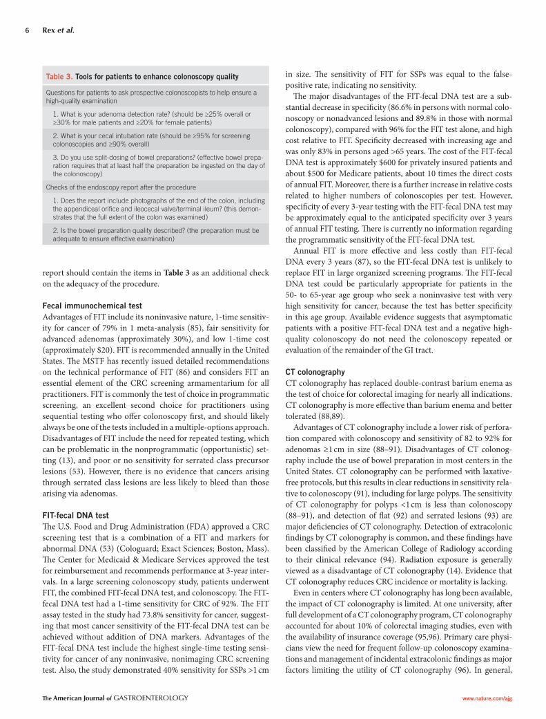
report should contain the items in Table 3 as an additional check
on the adequacy of the procedure.
Fecal immunochemical test
Advantages of FIT include its noninvasive nature, 1-time sensitiv-
ity for cancer of 79% in 1 meta-analysis ( 85 ), fair sensitivity for
advanced adenomas (approximately 30%), and low 1-time cost
(approximately $20). FIT is recommended annually in the United
States. Th e MSTF has recently issued detailed recommendations
on the technical performance of FIT ( 86 ) and considers FIT an
essential element of the CRC screening armamentarium for all
practitioners. FIT is commonly the test of choice in programmatic
screening, an excellent second choice for practitioners using
sequential testing who off er colonoscopy fi rst, and should likely
always be one of the tests included in a multiple-options approach.
Disadvantages of FIT include the need for repeated testing, which
can be problematic in the nonprogrammatic (opportunistic) set-
ting ( 13 ), and poor or no sensitivity for serrated class precursor
lesions ( 53 ). However, there is no evidence that cancers arising
through serrated class lesions are less likely to bleed than those
arising via adenomas.
FIT-fecal DNA test
Th e U.S. Food and Drug Administration (FDA) approved a CRC
screening test that is a combination of a FIT and markers for
abnormal DNA ( 53 ) (Cologuard; Exact Sciences; Boston, Mass).
Th e Center for Medicaid & Medicare Services approved the test
for reimbursement and recommends performance at 3-year inter-
vals. In a large screening colonoscopy study, patients underwent
FIT, the combined FIT-fecal DNA test, and colonoscopy. Th e FIT-
fecal DNA test had a 1-time sensitivity for CRC of 92%. Th e FIT
assay tested in the study had 73.8% sensitivity for cancer, suggest-
ing that most cancer sensitivity of the FIT-fecal DNA test can be
achieved without addition of DNA markers. Advantages of the
FIT-fecal DNA test include the highest single-time testing sensi-
tivity for cancer of any noninvasive, nonimaging CRC screening
test. Also, the study demonstrated 40% sensitivity for SSPs >1 cm
in size. Th e sensitivity of FIT for SSPs was equal to the false-
positive rate, indicating no sensitivity.
Th e major disadvantages of the FIT-fecal DNA test are a sub-
stantial decrease in specifi city (86.6% in persons with normal colo-
noscopy or nonadvanced lesions and 89.8% in those with normal
colonoscopy), compared with 96% for the FIT test alone, and high
cost relative to FIT. Specifi city decreased with increasing age and
was only 83% in persons aged >65 years. Th e cost of the FIT-fecal
DNA test is approximately $600 for privately insured patients and
about $500 for Medicare patients, about 10 times the direct costs
of annual FIT. Moreover, there is a further increase in relative costs
related to higher numbers of colonoscopies per test. However,
specifi city of every 3-year testing with the FIT-fecal DNA test may
be approximately equal to the anticipated specifi city over 3 years
of annual FIT testing. Th ere is currently no information regarding
the programmatic sensitivity of the FIT-fecal DNA test.
Annual FIT is more eff ective and less costly than FIT-fecal
DNA every 3 years ( 87 ), so the FIT-fecal DNA test is unlikely to
replace FIT in large organized screening programs. Th e FIT-fecal
DNA test could be particularly appropriate for patients in the
50- to 65-year age group who seek a noninvasive test with very
high sensitivity for cancer, because the test has better specifi city
in this age group. Available evidence suggests that asymptomatic
patients with a positive FIT-fecal DNA test and a negative high-
quality colonoscopy do not need the colonoscopy repeated or
evaluation of the remainder of the GI tract.
CT colonography
CT colonography has replaced double-contrast barium enema as
the test of choice for colorectal imaging for nearly all indications.
CT colonography is more eff ective than barium enema and better
tolerated ( 88,89 ).
Advantages of CT colonography include a lower risk of perfora-
tion compared with colonoscopy and sensitivity of 82 to 92% for
adenomas ≥1 cm in size ( 88–91 ). Disadvantages of CT colonog-
raphy include the use of bowel preparation in most centers in the
United States. CT colonography can be performed with laxative-
free protocols, but this results in clear reductions in sensitivity rela-
tive to colonoscopy ( 91 ), including for large polyps. Th e sensitivity
of CT colonography for polyps <1 cm is less than colonoscopy
( 88–91 ), and detection of fl at ( 92 ) and serrated lesions ( 93 ) are
major defi ciencies of CT colonography. Detection of extracolonic
fi ndings by CT colonography is common, and these fi ndings have
been classifi ed by the American College of Radiology according
to their clinical relevance ( 94 ). Radiation exposure is generally
viewed as a disadvantage of CT colonography ( 14 ). Evidence that
CT colonography reduces CRC incidence or mortality is lacking.
Even in centers where CT colonography has long been available,
the impact of CT colonography is limited. At one university, aft er
full development of a CT colonography program, CT colonography
accounted for about 10% of colorectal imaging studies, even with
the availability of insurance coverage ( 95,96 ). Primary care physi-
cians view the need for frequent follow-up colonoscopy examina-
tions and management of incidental extracolonic fi ndings as major
factors limiting the utility of CT colonography ( 96 ). In general,
Table 3 . Tools for patients to enhance colonoscopy quality
Questions for patients to ask prospective colonoscopists to help ensure a
high-quality examination
1. What is your adenoma detection rate? (should be ≥25% overall or
≥30% for male patients and ≥20% for female patients)
2. What is your cecal intubation rate (should be ≥95% for screening
colonoscopies and ≥90% overall)
3. Do you use split-dosing of bowel preparations? (effective bowel prepa-
ration requires that at least half the preparation be ingested on the day of
the colonoscopy)
Checks of the endoscopy report after the procedure
1. Does the report include photographs of the end of the colon, including
the appendiceal orifi ce and ileocecal valve/terminal ileum? (this demon-
strates that the full extent of the colon was examined)
2. Is the bowel preparation quality described? (the preparation must be
adequate to ensure effective examination)
© 2017 by the American College of Gastroenterology The American Journal of GASTROENTEROLOGY
7
patients with a conventional adenoma ≥6 mm in size but was
in eff ective for the detection of serrated lesions, and 9% of patients
had technically failed examinations for inadequate cleansing or
rapid transit of the capsule ( 101 ).
Overall, the burden associated with bowel preparation and the
relative superiority of colonoscopy are such that capsule colono-
scopy would be expected to appeal to a niche population con-
cerned about the risks of colonoscopy, in a fashion similar to CT
colonography. Currently, lack of FDA approval for screening and
lack of reimbursement are major obstacles to its use.
Septin9 assay
Th e fi rst FDA-approved serum test for CRC screening is the
Septin9 assay (Epigenomics, Seattle, Wash). In a large screening
colonoscopy study, this test had a sensitivity of 48% for detection
of CRC and no sensitivity for detection of precancerous polyps
( 102 ). Th e test is expensive relative to FIT.
Th e advantage of the Septin9 test is that it is a serum assay and
is at least potentially more convenient for patients. Some patients
who refused colonoscopy preferred this test over FIT ( 103 ).
Disadvantages of the Septin9 assay are markedly inferior
performance characteristics compared with FIT, including lower
sensitivity for cancer, inability to detect advanced adenomas ( 104 ),
and low cost-eff ectiveness relative to other screening tests ( 105 ).
Th e test appears to have higher sensitivity for late-stage compared
with early-stage cancer ( 102 ). Th e willingness of patients with
positive Septin9 tests to undergo colonoscopy remains uncertain.
Th e uncertainties regarding the true clinical utility of Septin 9
makes shared decision-making diffi cult. Clinicians should inform
patients of the uncertain benefi ts of this test on CRC mortality, the
inability of the assay to detect polyps, and the array of superior
alternatives. Th e best frequency for performing the test is uncer-
tain. Given these limitations, the MSTF suggests that Septin9 not
be used for screening.
COST ISSUES
A consistent fi nding is that CRC screening by any available modal-
ity is cost-eff ective compared with no screening ( 106,107 ), and in
some models screening results in cost savings. Th is fi nding relates
in part to the high costs of CRC treatment. Numerous modeling
studies have addressed the relative cost-eff ectiveness of 2 or more
screening tests. Th e conclusions of the models frequently vary,
likely depending in part on the assumptions of the respective
models. For example, diff erent models comparing colonoscopy
and CT colonography have had variable conclusions ( 108 ). Con-
sistent trends reveal that FIT performs well compared with other
screening tests ( 106,107,109 ). Colonoscopy also performs well
in most models ( 106–108,110 ), and, in general, the traditional
tests are more cost-eff ective than the newer modalities, including
CT colonography, FIT-fecal DNA, capsule colonoscopy, and the
Septin9 assay ( 105–107,111–113 ). Newer tests could reach cost-
eff ectiveness by substantially increasing compliance ( 112,113 ),
but evidence of improved compliance is lacking. Some models
support the cost-eff ectiveness of risk-stratifi ed approaches to
despite an extensive literature investigating the performance of
CT colonography, the test has limited impact on CRC screening
compliance ( 95 ). However, CT colonography appeals to a niche
of patients who are willing to undergo bowel preparation and are
concerned about the risks of colonoscopy. When used, the recom-
mended interval is 5 years in patients with normal CT colono-
graphy. We continue to recommend that patients with polyps
≥6 mm in size at CT colonography undergo colonoscopy ( 1 ).
Flexible sigmoidoscopy
Randomized controlled trials confi rm reductions in distal colon
or rectosigmoid cancer incidence and/or mortality of 29 to 76%
with fl exible sigmoidoscopy ( 66,97–99 ). Flexible sigmoidoscopy
can prevent a small fraction (14%) of proximal colon cancers, if
liberal criteria are used to indicate colonoscopy based on fl exible
sigmoidoscopy fi ndings ( 66 ). Advantages of fl exible sigmoidos-
copy include disproportionately lower cost and risk compared
with colonoscopy, a more limited bowel preparation, and no need
for sedation. Disadvantages of fl exible sigmoidoscopy include a
lower benefi t in protection against right-sided colon cancer com-
pared with the level of protection achieved in case-control and
cohort studies using colonoscopy. Also, the absence of sedation
leads to a low satisfaction experience for patients, such that they
are less willing to repeat the examination compared with colo-
noscopy ( 100 ). Further, the concept of examining only part of the
colon has been unpopular in the United States, so that screening
by fl exible sigmoidoscopy has almost disappeared from oppor-
tunistic screening settings ( 9 ). Some groups have endorsed the
combination of fl exible sigmoidoscopy plus FIT for screening
( 14 ), but compliance challenges associated with completing 2
screening tests and the dramatic decline in screening fl exible sig-
moidoscopy make signifi cant uptake of this combination unlikely.
Flexible sigmoidoscopy when used is oft en recommended at
5-year intervals. However, endoscopic screening in general is more
eff ective in the left than the right side of the colon, and there is
no clear reason why fl exible sigmoidoscopy should not be recom-
mended at 10-year intervals, similar to the recommendation for
colonoscopy. Th e MSTF considers that either 5- or 10-year inter-
vals are acceptable but favors 10-year intervals.
Capsule colonoscopy
Capsule colonoscopy has been approved by the FDA for imaging
the proximal colon in patients with previous incomplete colonos-
copies and more recently for patients who need colorectal imaging
but who are not candidates for colonoscopy or sedation. Capsule
colonoscopy is not approved by the FDA for screening average-
risk persons. Advantages of capsule colonoscopy are the achieve-
ment of endoscopic imaging without an invasive procedure and
avoiding the risks of colonoscopy. Disadvantages are that the
bowel preparation is more extensive than that for colonoscopy.
Also, because the logistics of performing same-day colonoscopy
on patients with positive capsule studies are quite diffi cult, most
patients with positive studies will require re-preparation and
colonoscopy on a separate day. In a large screening trial in 884
patients, capsule colonoscopy had 88% sensitivity for detecting
Rex et al.
The American Journal of GASTROENTEROLOGY VOLUME XXX | XXX 2017 www.nature.com/ajg
8
screening ( 114 ). Screening remains cost-eff ective in patients into
their mid-80s if they have few comorbidities and limited prior
screening ( 115,116 ).
QUALITY OF SCREENING
Variable performance of screening tests aff ects at least colono-
scopy, sigmoidoscopy, CT colonography, and FIT. Optimal results
in CRC screening cannot be achieved without optimizing the
technical performance and reporting of tests and ensuring that
patients undergo appropriate follow-up aft er testing. Th e MSTF
has made detailed recommendations regarding the technical per-
formance of FIT ( 86 ) and has previously issued quality recommen-
dations regarding the technical performance of sigmoidoscopy
( 117 ) and colonoscopy ( 118 ). Th e recommendations of the MSTF
regarding quality in the technical performance of colonoscopy
( 118 ) were largely incorporated in quality recommendations from
a combined American College of Gastroenterology-American
Society for Gastrointestinal Endoscopy Task Force on quality in
2006 ( 119 ) and 2015 ( 120 ), and the MSTF endorses the American
College of Gastroenterology-American Society for Gastrointesti-
nal Endoscopy Task Force recommendations.
Th e burden of performing high-quality FIT falls largely on
primary care physicians and/or the healthcare systems in which
they work. In the opportunistic setting there may not be resources
allocated to systematically ensure that FIT-positive patients are
referred for colonoscopy and that FIT-negative patients are off ered
repeat testing or to monitor whether compliance with quality tar-
gets is adequate ( 13 ). Inability to allocate resources to monitor the
quality of FIT testing is a factor favoring reliance on sequential
testing with colonoscopy the fi rst test off ered.
Unlike primary care physicians, the main role of gastroenterolo-
gists in the screening process is to perform colonoscopy on patients
referred for primary colonoscopy screening or for colonoscopy to
evaluate other positive screening tests. As such, a primary task of
gastroenterologists is to perform high-quality colonoscopy and
cost-eff ective follow-up. Th e adenoma detection rate, originally
proposed by the MSTF in 2002 ( 118 ), has emerged as the most
important and highly variable measure of the quality of mucosal
inspection during colonoscopy. Two large studies have validated
the adenoma detection rate as a predictor of cancer prevention
by colonoscopy ( 67,68 ). Measurement of the adenoma detection
rate is mandatory to appreciating whether a colonoscopist should
be performing screening colonoscopy. Patients should expect a
prospective colonoscopist to provide his or her adenoma detec-
tion rate, which should meet or exceed recommended minimum
thresholds ( Table 3 ).
PRACTICAL CONSIDERATIONS
No published randomized trials have directly compared and
reported the relative eff ects of diff erent tests on CRC incidence or
mortality. Several trials are ongoing, but results are not yet availa-
ble. When compared using simulation models that are dependent
on assumptions about natural history of disease, patient accept-
ance of screening, and test performance, several tests appear to
be similarly eff ective ( 121 ). Th erefore, practical considerations are
important for informing our recommendations.
A common statement made with regard to CRC screening is that
“the best test is the one that gets done.” Th e MSTF endorses this
concept because it is generally better for any person who is eligible
to be screened to undergo some screening test rather than not be
screened at all. On the other hand, the core concept underlying
sequential testing is that off ering the “best” test (s) fi rst optimizes
the sensitivity and/or cost-eff ectiveness of screening and still leaves
the opportunity to off er other tests when patients decline. As an
extreme example, the MSTF considers that equating the Septin9
assay with colonoscopy would be a disservice to patients, because
the sensitivity of colonoscopy for cancer and advanced lesions
exceeds that of Septin9 by a very large margin. Given these dispari-
ties in the performance of individual tests, the MSTF groups the
available tests into 3 tiers based on various performance features
and costs ( Table 4 ).
Th e tier 1 tests representing the cornerstone of CRC screening
are colonoscopy every 10 years and annual FIT ( Table 4 ). Th e
use of these 2 tests as the primary screening measures provides a
framework for screening that is simple and accommodates almost
every screening setting. In organized programmatic screening FIT
will oft en be off ered as the primary screen, but colonoscopy can
be considered as an alternative for patients and physicians who
prefer or request it. In the opportunistic setting, colonoscopy will
oft en be preferred when the infrastructure to ensure annual per-
formance of FIT is not available. As noted above, 1 of the most
challenging aspects of FIT screening in the opportunistic setting
is ensuring repeated annual performance. Using a tier 1 approach
that focuses on 2 tests makes the discussion of CRC screening tests
between physician and patient manageable and feasible, and, as
noted above, expanding the number of options in the initial dis-
cussion beyond 2 did not increase screening rates ( 19 ). Colono-
scopy and FIT can be adapted readily to either the sequential off er
of screening (colonoscopy is off ered fi rst with FIT reserved for
Table 4 . Multi-Society Task Force ranking of current colorectal
cancer screening tests
Tier 1
Colonoscopy every 10 years
Annual fecal immunochemical test
Tier 2
CT colonography every 5 years
FIT-fecal DNA every 3 years
Flexible sigmoidoscopy every 10 years (or every 5 years)
Tier 3
Capsule colonoscopy every 5 years
Available tests not currently recommended
Septin 9
© 2017 by the American College of Gastroenterology The American Journal of GASTROENTEROLOGY
9
those who decline colonoscopy), the multiple-options approach
(colonoscopy and FIT are each discussed with patients, and if both
are declined the discussion moves sequentially to tier 2 tests), and
the risk-stratifi ed approach (e.g., colonoscopy is off ered fi rst to
men age >60 and women age >65 with no prior screening, and FIT
is off ered to persons under these ages and persons with negative
prior colonoscopy). Risk stratifi cation may also take into account
factors such as cigarette smoking, diabetes, and obesity.
Th e rationale for placing tests in the tier 2 and 3 categories
follows from the discussion above of individual tests. Th e rank-
ing implies equivalence for tests within each category, but this is
not the intent of the MSTF. For example, fl exible sigmoidoscopy
screening has strong evidence to support its use. However, the
steady decline in the use of fl exible sigmoidoscopy in the United
States ( 9,10 ) suggests that any strong endorsement of fl exible sig-
moidoscopy is not consistent with the reality of the test’s lack of
popularity among patients and poor reimbursement for physi-
cians. Th e strong evidence base supporting fl exible sigmoidoscopy
leads us to place the test in the tier 2 category, although we expect
that most practitioners using a sequential approach would move
to CT colonography or FIT-fecal DNA in patients who decline
colonoscopy and FIT, because these tests are less invasive and
survey the entire colon.
Performance characteristics alone would place capsule colo-
noscopy in tier 2. However, off ering capsule colonoscopy as a
tier 2 test in a sequential methodology currently would oft en
lead to frustration because reimbursement for screening capsule
colonoscopy is seldom available at this time, and the test itself
is frequently not available. Th e onerous bowel preparation and
the lack of systems to accomplish same-day colonoscopy in
most patients with a positive capsule colonoscopy are additional
factors placing capsule colonoscopy in the tier 3 category at this
time.
In summary, we suggest that the tiered system ( Table 4 ) has
numerous advantages. Predominant reliance on tier 1 tests off ers
modalities with optimal eff ectiveness, cost-eff ectiveness, complete
colon screening, proven popularity with patients, and a simpli-
fi ed discussion (compared with off ering 5-7 diff erent tests) and
still leaves room to off er other tests in a sequential fashion. If
patients decline colonoscopy and FIT, all tests in tier 2 are accept-
able CRC screening tests, but each has defi ciencies relative to the
tier 1 tests.
Recommendations
1 . We recommend colonoscopy every 10 years or annual FIT
as fi rst-tier options for screening the average-risk persons
for colorectal neoplasia (strong recommendation; moderate-
quality evidence).
2 . We recommend that physicians performing screening colo-
noscopy measure quality, including the adenoma detection
rate (strong recommendation, high-quality evidence).
3 . We recommend that physicians performing FIT monitor
quality (strong recommendation, low-quality evidence). Th e
recommended quality measurements for FIT programs are
detailed in a prior publication ( 86 ).
4 . We recommend CT colonography every 5 years or FIT-fecal
DNA every 3 years (strong recommendation, low-quality
evidence) or fl exible sigmoidoscopy every 5 to 10 years
(strong recommendation, high-quality evidence) in patients
who refuse colonoscopy and FIT.
5 . We suggest that capsule colonoscopy (if available) is an ap-
propriate screening test when patients decline colonoscopy,
FIT, FIT-fecal DNA, CT colonography, and fl exible sigmoi-
doscopy (weak recommendation, low-quality evidence).
6 . We suggest against Septin9 for CRC screening (weak recom-
mendation, low-quality evidence).
FAMILY HISTORY OF CRC AND POLYPS
We recommend that screening in most average-risk persons be
initiated at age 50 years. A family history of CRC or certain pol-
yps can modify the recommended starting age and the frequency
of screening. Th e MSTF has previously issued recommendations
for screening in persons with Lynch syndrome ( 34 ), which is a
genetically defi ned inherited syndrome caused by mutations in
1 or more mismatch repair genes. Patients in families that meet
the clinical criteria for hereditary nonpolyposis CRC but have
microsatellite-stable CRCs have family colon cancer syndrome X,
which has not been genetically defi ned ( 122 ). Persons in families
with syndrome X should undergo colonoscopy at least every 3 to 5
years, beginning 10 years before the age at diagnosis of the young-
est aff ected relative.
A family history of CRC in a fi rst-degree relative increases the
risk of CRC regardless of the age at diagnosis of the aff ected rela-
tive ( 123–125 ). Th ere is a gradient of risk such that the younger
the age of the aff ected relative, the greater the risk ( 123–125 ).
Th e MSTF has previously used age 60 as a threshold of risk ele-
vation, so that a single fi rst-degree relative diagnosed with CRC
at age <60 years warrants both earlier screening and at more-
frequent intervals ( 1 ). Recent population-based studies ( 123,124 )
and reviews of risk associated with a positive family history
( 125 ) support using age at diagnosis of CRC above or below 60
years in the aff ected fi rst-degree relative as a risk stratifi er. We
continue to recommend that persons with a family history of
CRC in a fi rst-degree relative diagnosed at <60 years undergo
colonoscopy every 5 years beginning at age 40 years or 10 years
before the age the relative was diagnosed, whichever comes fi rst
( Table 5 ). In a randomized controlled trial, there was a nonsig-
nifi cant trend toward detection of more advanced neoplasia in
subjects with a positive family history who underwent colonos-
copy compared with FIT ( 126 ). Th us, patients with a positive
family history who decline colonoscopy should be off ered FIT
screening ( 126 ).
Th e greatest relative risk of CRC appears to be in persons <50
years who have a fi rst-degree relative with CRC diagnosed at <50
years ( 123–125 ). Compliance in young persons with a family his-
tory of CRC is suboptimal, and clinicians should make special
eff orts to ensure that screening occurs. Recent evidence suggests if
persons with a single fi rst-degree relative with a family history of
CRC reach the age of approximately 60 years without manifesting
Rex et al.
The American Journal of GASTROENTEROLOGY VOLUME XXX | XXX 2017 www.nature.com/ajg
10
signifi cant colorectal neoplasia ( 127 ), then they are unlikely to be
at increased risk of CRC and can be off ered the option of expand-
ing the interval between examinations.
When fi rst-degree relatives have documented advanced
serrated lesions (SSPs ≥10 mm in size, or an SSP with cytologic
dysplasia, or a traditional serrated adenoma ≥10 mm in size), there
is no clear evidence as to how to proceed, unless the relative meets
criteria for serrated polyposis ( 46 ). Currently, we recommend
that screening for fi rst-degree relatives of persons with advanced
serrated lesions should be similar to the screening of fi rst-degree
relatives of persons with advanced conventional adenomas.
Persons with a single fi rst-degree relative with CRC who was
diagnosed at age ≥60 years are recommended to begin screen-
ing at 40 years ( 128 ). However, the tests and intervals for testing
are the same as the average-risk screening recommendations
( Tables 4 and 5 ).
We no longer recommend that persons with a family history
of adenomas in a fi rst-degree relative undergo early screening,
unless there is clear documentation of an advanced adenoma
in a fi rst-degree relative. In most cases the patient has no infor-
mation regarding whether the family member’s adenoma was
advanced, and in this case we recommend that it be assumed
the adenomas or polyps were not advanced. If a colonoscopy
and/or pathology report (s) is available for a family member that
documents an advanced adenoma or there is a report of a polyp
requiring surgical resection, an advanced adenoma in a family
member is considered established. Th ese considerations regarding
adequate documentation of advanced precancerous neoplasms in
fi rst-degree relatives before intensifying screening apply to docu-
mentation of both advanced adenomas and advanced serrated
lesions. First-degree relatives with advanced adenomas are recom-
mended to be weighted the same as fi rst-degree relatives with CRC
( Table 5 ). Th e yield of colonoscopic screening in fi rst-degree
relatives of persons with advanced adenomas is substantially
increased ( 129–131 ).
Recommendations
1 . We suggest that persons with 1 fi rst-degree relative with
CRC or a documented advanced adenoma diagnosed at age
<60 years or with 2 fi rst-degree relatives with CRC and/or
documented advanced adenomas undergo colonoscopy every
5 years beginning 10 years younger than the age at which
the youngest fi rst-degree relative was diagnosed or age 40,
whichever is earlier (weak recommendation, low-quality
evidence).
2 . We suggest that persons with 1 fi rst-degree relative diagnosed
with CRC or a documented advanced adenoma at age ≥60
years begin screening at age 40. Th e options for screening
and the recommended intervals are the same as those for
average-risk persons (weak recommendation, very-low-
quality evidence).
3 . We suggest that persons with 1 or more fi rst-degree rela-
tives with a documented advanced serrated lesion (SSP or
traditional serrated adenoma ≥10 mm in size or an SSP with
cytologic dysplasia) should be screened according to above
recommendations for persons with a family history of a
documented advanced adenoma (weak recommendation,
very-low-quality evidence).
4 . We recommend that persons with 1 or more fi rst-degree
relatives with CRC or documented advanced adenomas, for
whom we recommend colonoscopy, should be off ered annual
FIT if they decline colonoscopy (strong recommendation,
moderate-quality evidence).
CONSIDERATIONS REGARDING AGE AND CRC RISK
CRC screening is recommended to begin at age 50 years in most
average-risk persons, including in prior recommendations from
the MSTF ( 1 ). Recent modeling supports this recommendation
( 121 ). Several issues related to age and CRC risk warrant specifi c
discussion.
Th e incidence of CRC is strongly age related and continues to
rise with increasing age. Partly because of widespread screening
in the United States, the incidence of CRC in falling by 3 to 4%
per year in persons age ≥50 years ( 12 ). Th e incidence of CRC in
persons under age 50 is increasing in the United States ( 132,133 ).
Although the reasons for this rising incidence remain unclear and
the relative incidence in persons under age 50 remains low, the
increasing incidence of CRC in young people is a major public
health concern.
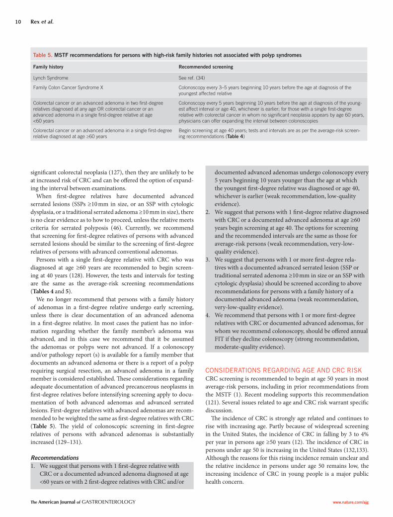
Table 5 . MSTF recommendations for persons with high-risk family histories not associated with polyp syndromes
Family history Recommended screening
Lynch Syndrome See ref. ( 34 )
Family Colon Cancer Syndrome X Colonoscopy every 3–5 years beginning 10 years before the age at diagnosis of the
youngest affected relative
Colorectal cancer or an advanced adenoma in two fi rst-degree
relatives diagnosed at any age OR colorectal cancer or an
advanced adenoma in a single fi rst-degree relative at age
<60 years
Colonoscopy every 5 years beginning 10 years before the age at diagnosis of the young-
est affect interval or age 40, whichever is earlier; for those with a single fi rst-degree
relative with colorectal cancer in whom no signifi cant neoplasia appears by age 60 years,
physicians can offer expanding the interval between colonoscopies
Colorectal cancer or an advanced adenoma in a single fi rst-degree
relative diagnosed at age ≥60 years
Begin screening at age 40 years; tests and intervals are as per the average-risk screen-
ing recommendations ( Table 4 )
© 2017 by the American College of Gastroenterology The American Journal of GASTROENTEROLOGY
11
Th e age to stop screening can be individualized. Screening
is potentially benefi cial in persons up to age 86 if there has not
been previous screening ( 116 ) but should be considered in the
context of comorbidities and life expectancy. Persons with pre-
viously nega tive screening tests, particularly negative screening
colono scopy, could consider stopping at age 75 years ( 14 ). In a
variation of this recommendation, the MSTF has recommended
that persons with previous negative screening stop when their life
expectancy is less than 10 years ( 1 ). Th us, the recommendation to
stop screening can be reasonably based on patient age and comor-
bidities.
Th e wishes of the patient should be considered in uncertain
cases. Th ese considerations do not necessarily apply in the surveil-
lance setting, where patients with advanced neoplasia may benefi t
from surveillance colonoscopy even at an advanced age, depend-
ing on comorbidities and the confi dence in neoplasia clearing at
colonoscopy.
Recommendations
1 . We recommend that screening begin in non-African American
average-risk persons at age 50 years (strong recommendation;
moderate-quality evidence).
2 . We suggest that screening begin in African Americans at age
45 years (weak recommendation, very-low-quality evidence).
3 . We recommend that adults age <50 years with colorectal
bleeding symptoms (hematochezia, unexplained iron
defi ciency anemia, melena with a negative upper endoscopy)
undergo colonoscopy or an evaluation suffi cient to determine
a bleeding cause, initiate treatment, and complete follow-up
to determine resolution of bleeding (strong recommendation,
moderate-quality evidence).
4 . We suggest that persons who are up to date with screening
and have negative prior screening tests, particularly colono-
scopy, consider stopping screening at age 75 years or when
life expectancy is less than 10 years (weak recommendation,
low-quality evidence).
5 . We suggest that persons without prior screening should be
considered for screening up to age 85, depending on consid-
eration of their age and comorbidities (weak recommenda-
tion, low-quality evidence).
SUMMARY
CRC screening should begin at age 50 years in asymptomatic per-
sons. Colonoscopy every 10 years and annual FIT are currently
the fi rst considerations for screening. Colonoscopy every 10 years
has advantages in the opportunistic screening setting. Annual FIT
is likely to be preferred in organized screening programs. Posi-
tioning of the 2 tests can be reasonably based on a sequential off er
(colonoscopy fi rst with FIT off ered to patients who decline colo-
noscopy, followed by second-tier tests for patients who decline
FIT), a multiple-options approach where both tests are discussed
with patients (followed by a sequential off er of second-tier tests
to patients who decline both colonoscopy and FIT), or a risk-
stratifi ed approach (colonoscopy is off ered to patients with a
Th e best course of action with regard to the rising incidence of
CRC in young people is currently not certain. When a young per-
son develops fatal CRC, the loss of life years is great. Th e fi rst step
in reducing CRC morbidity and mortality in persons age <50 years
is aggressive evaluation (usually colonoscopy) of patients with
colorectal symptoms, specifi cally those with bleeding symptoms:
hematochezia, iron defi ciency anemia, and/or melena with a nega-
tive upper endoscopy. Persons with bleeding symptoms evaluated
with tests other than colonoscopy (e.g., sigmoidoscopy) should
have a bleeding source identifi ed and treated, and the patient
should be followed to resolution of the symptom. Patients who
have nonbleeding symptoms (e.g., abnormal bowel habit, change
in bowel habit or shape, or abdominal pain) and who have no evi-
dence of bleeding do not have an increased risk of CRC when they
undergo colonoscopy ( 134,135 ).
Th ere is currently insuffi cient evidence to recommend system-
atic screening in asymptomatic persons <50 years old who lack
specifi c risk factors related to family history or Lynch syndrome.
Th e yield of screening colonoscopy in this age group is low in
available studies ( 136 ), and the biologic reasons for the increas-
ing incidence of CRC in persons under age 50 years are uncertain.
Additional study of the benefi ts and harms of screening in per-
sons <50 years is warranted, perhaps particularly in persons with
known colorectal risk factors such as cigarette smoking, diabetes
mellitus, and obesity ( 137–139 ).
Relative to other races, African Americans have lower
screening rates for CRC, higher incidence rates, earlier mean
age at onset, worse survival and late-stage presentation, and a
higher proportion of cancers before age 50 ( 140–143 ). Th ese
various eff ects result from both socioeconomic and biologic
factors ( 144 ). Two of the member organizations of the MSTF
endorsed beginning average-risk screening in African Ameri-
cans at age 45 ( 22,145 ), and the American College of Physi-
cians recommended beginning at age 40 ( 146 ). Th e scientifi c
rationale for beginning screening earlier includes the higher
overall incidence rates and younger mean age at onset of CRC
in African Americans ( 140–143 ). A recent joinpoint regression
analysis ( 147 ) and a MISCAN-Colon microsimulation model
( 148 ) both supported screening African Americans approxi-
mately 5 years earlier compared with whites. Th ere are few data
on the yield of screening in African Americans before age 50 or
whether earlier screening improves outcomes. In persons over
age 50, some reports have identifi ed a higher risk of advanced
polyps in African Americans ( 149 ). Th e recommendations to
begin screening earlier have served an important role in stimu-
lating discussion of and research on CRC in African Ameri-
cans, increasing awareness in physicians of an important public
health problem and racial disparity in health outcomes in the
United States, and increasing awareness of CRC in African
Americans. Increasing screening rates in African Americans
generally is an area of obvious importance. Provider recom-
mendation is key ( 150 ), and navigation can improve compli-
ance to colonoscopy screening ( 151 ). Additional study of the
yield of screening in persons under age 50 is needed, particu-
larly in African Americans.
Rex et al.
The American Journal of GASTROENTEROLOGY VOLUME XXX | XXX 2017 www.nature.com/ajg
12
higher pretest probability of neoplasia, and FIT is used in persons
with a lower pretest probability of neoplasia).
Persons with a history of CRC or a documented advanced
adenoma in a fi rst-degree relative age <60 years or 2 fi rst-degree
relatives with these fi ndings at any age are recommended to
undergo screening by colonoscopy every 5 years, beginning 10
years before the age at diagnosis of the youngest aff ected rela-
tive, or at age 40, whichever is earlier. Persons with a single fi rst-
degree relative diagnosed at ≥60 years with CRC or an advanced
adenoma can be off ered average-risk screening options begin-
ning at age 40 years.
Th e incidence of CRC is rising in persons under age 50. Patients
under age 50 with bleeding symptoms consistent with a colorectal
source should be aggressively evaluated and treated. We suggest
that screening begin at age 45 in African Americans. Discontinu-
ation of screening should be considered when patients who are
up to date with screening and have had negative screening tests,
particularly colonoscopy, reach age 75 years or when life expec-
tancy is <10 years. Persons without prior screening should be con-
sidered for screening up to age 85 years, depending on comorbid
conditions and life expectancy.
ACKNOWLEDGMENTS
Th e views expressed in this article are those of the authors and do
not necessarily refl ect the position or policy of the Department of
Veteran Aff airs.
DISCLOSURE
Th e following authors disclosed fi nancial relationships relevant
to this publication: D. K. Rex: Consultant for Olympus Corp and
Boston Scientifi c; research support recipient from Boston Scientifi c,
Endochoice, EndoAid, Medtronic, and Colonary Solutions.
T. Kaltenbach: Consultant for Olympus Corp. D. J. Robertson:
Consultant for Medtronic. All other authors disclosed no fi nancial
relationships relevant to this publication.
REFERENCES 1. Levin B , Lieberman DA , McFarland B et al. Screening and surveillance for
the early detection of colorectal cancer and adenomatous polyps . Gastro-enterology 2008 ; 134 : 1570 – 95 .
2. Kahi CJ , Boland CR , Dominitz JA et al. Colonoscopy surveillance aft er colorectal cancer resection: recommendations of the US multi-society task force on colorectal cancer . Gastrointest Endosc 2016 ; 83 : 489 – 98 .
3. Lieberman DA , Rex DK , Winawer SJ et al. Guidelines for colonos-copy surveillance aft er screening and polypectomy . Gastroenterology 2012 ; 143 : 844 – 57 .
4. Levin TR , Jamieson L , Burley DA et al. Organized colorectal cancer screening in integrated health care systems . Epidemiol Rev 2011 ; 33 : 101 – 10 .
5. Zavoral M , Suchanek S , Zavada F et al. Colorectal cancer screening in Europe . World J Gastroenterol 2009 ; 15 : 5907 – 15 .
6. Cenin DR , St John DJB , Ledger MJN et al. Optimizing the expansion of the National Bowel Cancer Screening Program . Med J Aust 2014 ; 201 : 456 – 61 .
7. Jensen CD , Corley DA , Quinn VP et al. Fecal immunochemical test pro-gram over 4 rounds of annual screening: a retrospective study . Ann Intern Med 2016 ; 164 : 456 – 63 .
8. Siegel RL , Ward EM , Jemal A . Trends in colorectal cancer incidence rates in the United States by tumor location and stage, 1992-2008 . Cancer Epidemiol Biomarkers Prev 2012 ; 21 : 411 – 6 .
9. Klabunde CN , Joseph DA , King JB et al. Vital signs: colorectal cancer screening test use—United States, 2012 . MMWR 2013 ; 62 : 881 – 8 .
10. Sabatino SS , White MC , Th ompson TT et al. Cancer screening test use—United States, 2013 . MMWR 2015 ; 64 : 464 – 8 .
11. Siegel R , Desantis C , Jemal A . Colorectal cancer statistics, 2014 . CA Cancer J Clin 2014 ; 64 : 104 – 17 .
12. Edwards BK , Noone AM , Mariotto AB et al. Annual report to the nation on the status of cancer, 1975-2010, featuring prevalence of comorbidity and impact on survival among persons with lung, colorectal, breast, or prostate cancer . Cancer 2014 ; 120 : 1290 – 314 .
13. Liang PS , Wheat CL , Abhat A et al. Adherence to competing strate-gies for colorectal cancer screening over 3 years . Am J Gastroenterol 2016 ; 111 : 105 – 14 .
14. US Preventive Services Task Force , Bibbins-Domingo K , Grossman DC , Curry SJ et al. Screening for colorectal cancer: US Preventive Services Task Force recommendation statement . JAMA 2016 ; 315 : 2564 – 75 .
15. Inadomi JM , Vijan S , Janz NK et al. Adherence to colorectal cancer screening: a randomized clinical trial of competing strategies . Arch Intern Med 2012 ; 172 : 575 – 82 .
16. Segnan N , Senore C , Andreoni B et al. Randomized trial of diff erent screening strategies for colorectal cancer: patient response and detection rates . J Natl Cancer Inst 2005 ; 97 : 147 – 57 .
17. Multicentre Australian Colorectal-neoplasia Screening (MACS) Group . A comparison of colorectal neoplasia screening tests: a multicentre community-based study of the impact of consumer choice . Med J Aust 2006 ; 184 : 546 – 50 .
18. Scott RG , Edwards JT , Fritschi L et al. Community-based screening by colonoscopy or computed tomographic colonography in asymptomatic average-risk subjects . Am J Gastroenterol 2004 ; 99 : 1145 – 51 .
19. Griffi th JM , Lewis CL , Brenner ART et al. Th e eff ect of off ering diff erent numbers of colorectal cancer screening test options in a decision aid: a pilot randomized trial . BMC Med Inform Dec Mak 2008 ; 8 : 4 .
20. Canadian Task Force on Preventive Health Care . Recommendations on screening for colorectal cancer in primary care . CMAJ 2016 ; 188 : 340 – 8 .
21. Rex DK , Johnson DA , Anderson JC et al. American College of Gastro-enterology guidelines for colorectal cancer screening . Am J Gastroenterol 2009 ; 104 : 739 – 50 .
22. ASGE Standards of Practice Committee , Davila RE , Rajan E et al. ASGE guideline: colorectal cancer screening and surveillance . Gastro-intest Endosc 2006 ; 63 : 546 – 57 .
23. Senore C , Ederle A , Benazzato L et al. Off ering people a choice for colorectal cancer screening . Gut 2013 ; 62 : 735 – 40 .
24. Symonds EL , Pedersen S , Cole SR et al. Improving participation in colorectal cancer screening: a randomized controlled trial of sequential off ers of fecal then blood based non-invasive tests . Asian Pac J Cancer Prev 2015 ; 16 : 8455 – 60 .
25. Adler A , Geiger S , Keil A et al. Improving compliance to colorectal cancer screening using blood and stool based tests in patients refusing screening colonoscopy in Germany . BMC Gastroenterol 2014 ; 14 : 183 .
26. Rex D . Colonoscopy: the current king of the hill in the USA . Dig Dis Sci 2015 ; 60 : 639 – 46 .
27. Ma GK , Ladabaum U . Personalizing colorectal cancer screening: a systematic review of models to predict risk of colorectal neoplasia . Clin Gastroenterol Hepatol 2014 ; 12 : 1624 – 34 .
28. Imperiale TF , Monahan PO , Stump TE et al. Derivation and validation of a scoring system to stratify risk for advanced colorectal neoplasia in asymp-tomatic adults: a cross-sectional study . Ann Intern Med 2015 ; 163 : 339 – 46 .
29. Chiu HM , Ching JY , Wu KC et al. A risk-scoring system combined with a fecal immunochemical test is eff ective in screening high-risk subjects for early colonoscopy to detect advanced colorectal neoplasms . Gastro-enterology 2016 ; 150 : 617 – 25 .
30. Winawer SJ , Zauber AG , Ho MN . Prevention of colorectal cancer by colonoscopic polypectomy. Th e National Polyp Study Workgroup . N Engl J Med 1993 ; 329 : 1977 – 81 .
31. Zauber AG , Winawer SJ , O'Brien MJ et al. Colonoscopic polypectomy and long-term prevention of colorectal-cancer deaths . N Engl J Med 2012 ; 366 : 687 – 96 .
32. Brenner H , Hoff meister M , Stegmaier C et al. Risk of progression of advanced adenomas to colorectal cancer by age and sex: estimates based on 840,149 screening colonoscopies . Gut 2007 ; 56 : 1585 – 9 .
33. Rex DK , Ahnen DJ , Baron JA et al. Serrated lesions of the colorectum: review and recommendations from an expert panel . Am J Gastroenterol 2012 ; 107 : 1315 – 29 .
© 2017 by the American College of Gastroenterology The American Journal of GASTROENTEROLOGY
13
58. Doubeni CA , Weinmann S , Adams K et al. Screening colonoscopy and risk for incident late-stage colorectal cancer diagnosis in average-risk adults: a nested case-control study . Ann Intern Med 2013 ; 158 : 312 – 20 .
59. Brenner H , Chang-Claude J , Jansen L et al. Reduced risk of colorectal cancer up to 10 years aft er screening, surveillance or diagnostic colono-scopy . Gastroenterology 2014 ; 146 : 709 – 17 .
60. Mulder SA , van Soest EM , Dieleman JP et al. Exposure to colorectal examinations before a colorectal cancer diagnosis: a case-control study . Eur J Gastroenterol Hepatol 2010 ; 22 : 437 – 43 .
61. Baxter NN , Goldwasser MA , Paszat LF et al. Association of colonoscopy and death from colorectal cancer . Ann Inern Med 2009 ; 150 : 1 – 8 .
62. Baxter NN , Warren JL , Barrett MJ et al. Association between colonoscopy and colorectal cancer mortality in a US cohort according to site of cancer and colonoscopist specialty . J Clin Oncol 2012 ; 30 : 2664 – 9 .
63. Nishihara R , Wu K , Lochhead P et al. Long-term colorectal cancer inci-dence and mortality aft er lower endoscopy . N Engl J Med 2013 ; 369 : 1095 – 105 .
64. Doubeni CA , Corley DA , Quinn VP et al. Eff ectiveness of screening colonoscopy in reducing the risk of death from right and left colon cancer: a large community-based study . Gut. Epub 2016 ; http://dx.doi.org/l0.1136/gutjnl-2016-312712 .
65. Mandel JS , Church TR , Bond JH . Th e eff ect of fecal occult-blood screen-ing on the incidence of colorectal cancer . N Engl J Med 2000 ; 343 : 1603 – 7 .
66. Schoen RE , Pinsky PF , Weissfeld JL et al. Colorectal-cancer incidence and mortality with fl exible sigmoidoscopy . N Engl J Med 2012 ; 366 : 2345 – 57 .
67. Kaminski MF , Regula J , Kraszewska E et al. Quality indicators for colono-scopy and the risk of interval cancer . N Engl J Med 2010 ; 362 : 1795 – 803 .
68. Corley DA , Jensen CD , Marks AR et al. Adenoma detection rate and risk of colorectal cancer and death . N Engl J Med 2014 ; 370 : 1298 – 306 .
69. Cooper GS , Kou TD , Rex DK . Complications following colonoscopy with anesthesia assistance: a population-based analysis . JAMA Intern Med 2013 ; 173 : 551 – 6 .
70. Reumkens A , Ronadagh EJ . Bakker, et al. Post-colonoscopy complica-tions: A systematic review, time trends, and meta-analysis of population based studies . Am J Gastroenterol 2016 ; 111 : 1092 – 101 .
71. Bahin FF , Rasouli KN , Byth K et al. Prediction of clinically signifi cant bleeding following wide-fi eld endoscopic resection of large sessile and laterally spreading colorectal lesions: a clinical risk score . Am J Gastro-enterol 2016 ; 111 : 1115 – 22 .
72. Ichise Y , Horiuchi A , Nakayama Y et al. Prospective randomized com-parison of cold snare polypectomy and conventional polypectomy for small colorectal polyps . Digestion 2011 ; 84 : 78 – 81 .
73. Jayanna M , Burgess NG , Singh R et al. Cost analysis of endoscopic mu-cosal resection vs surgery for large laterally spreading colorectal lesions . Clin Gastroenterol Hepatol 2016 ; 14 : 271 – 8 .
74. Law R , Das A , Gregory D et al. Endoscopic resection is cost-eff ective compared with laparoscopic resection in the management of complex co-lon polyps: an economic analysis . Gastrointest Endosc 2016 ; 83 : 1248 – 57 .
75. Shaukat A , Rector TS , Church TR et al. Longer withdrawal time is asso-ciated with a reduced incidence of interval cancer aft er screening colonos-copy . Gastroenterology 2015 ; 149 : 952 – 7 .
76. Barclay RL , Vicari JJ , Doughty AS et al. Colonoscopic withdrawal times and adenoma detection during screening colonoscopy . N Engl J Med 2006 ; 355 : 2533 – 41 .
77. Chen SC , Rex DK . Endoscopist can be more powerful than age and male gender in predicting adenoma detection at colonoscopy . Am J Gastro-enterol 2007 ; 102 : 856 – 61 .
78. Butterly L , Robinson CM , Anderson JC et al. Serrated and adenomatous polyp detection increases with longer withdrawal time: results from the New Hampshire Colonoscopy Registry . Am J Gastroenterol 2014 ; 109 : 417 – 26 .
79. Mysliwiec PA , Brown ML , Klabunde CN et al. Are physicians doing too much colonoscopy? A national survey of colorectal surveillance aft er polypectomy . Ann Intern Med 2004 ; 141 : 264 – 71 .
80. Pohl H , Srivastava A , Bensen SP et al. Incomplete polyp resection during colonoscopy-results of the complete adenoma resection (CARE) study . Gastroenterology 2013 ; 144 : 74 – 80.e1 .
81. Rex DK , Rahmani EY , Haseman JH et al. Relative sensitivity of colono-scopy and barium enema for detection of colorectal cancer in clinical practice . Gastroenterology 1997 ; 112 : 17 – 23 .
82. Rabeneck L , Paszat LF , Saskin R . Endoscopist specialty is associated with incident colorectal cancer aft er a negative colonoscopy . Clin Gastro-enterol Hepatol 2010 ; 8 : 275 – 9 .
34. Giardiello FM , Allen JI , Axilbund JE et al. Guidelines on genetic evalua-tion and management of Lynch syndrome: a consensus statement by the U.S. Multi-Society Task Force on Colorectal Cancer . Gastrointest Endosc 2014 ; 80 : 197 – 220 .
35. Soetikno R , Kaltenbach T , Rouse RV et al. Prevalence of nonpolypoid (fl at and depressed) colorectal neoplasms in asymptomatic and symptomatic adults . JAMA 2008 ; 299 : 1027 – 35 .
36. Ponugoti PL , Cummings OW , Rex DK . Risk of cancer in small and diminutive colorectal polyps . Dig Liver Dis 2017 ; 49 : 530 – 4 .
37. Lasisi F , Mouchli A , Riddell R et al. Agreement in interpreting villous elements and dysplasia in adenomas less than one centimeter in size . Dig Liver Dis 2013 ; 45 : 1049 – 55 .
38. Rex DK , Alikhan M , Cumming O et al. Accuracy of pathologic interpre-tation of colorectal polyps by general pathologists in community practice . Gastrointest Endosc 1999 ; 50 : 468 – 74 .
39. Ponugoti P , Lin J , Odze R et al. Prevalence of sessile serrated adenoma/polyp in hyperplastic appearing diminutive rectosigmoid polyps . Gastro-intest Endosc 2016 ; 85 : 622 – 7 .
40. Abdeljawad K , Vemulapalli KC , Kahi CJ et al. Sessile serrated polyp prevalence determined by a colonoscopist with a high lesion detection rate and an experienced pathologist . Gastrointest Endosc 2015 ; 81 : 517 – 24 .
41. Ijspeert JE , de Wit K , van der Vlugt M et al. Prevalence, distribution and risk of sessile serrated adenomas/polyps at a center with a high adenoma detection rate and experienced pathologists . Endoscopy 2016 ; 48 : 740 – 6 .
42. Hetzel JT , Huang CS , Coukos JA et al. Variation in the detection of serrated polyps in an average risk colorectal cancer screening cohort . Am J Gastroenterol 2010 ; 105 : 2656 – 64 .
43. Kahi CJ , Hewett DG , Norton DL et al. Prevalence and variable detection of proximal colon serrated polyps during screening colonoscopy . Clin Gastroenterol Hepatol 2011 ; 9 : 42 – 6 .
44. Tinmouth J , Henry P , Hsieh E et al. Sessile serrated polyps at screening colonoscopy: have they been underdiagnosed? Am J Gastroenterol 2014 ; 109 : 1698 – 74 .
45. Payne SR , Church TR , Wandell M et al. Endoscopic detection of proximal serrated lesions and pathologic identifi cation of sessile serrated adenomas/polyps vary on the basis of center . Clin Gastroenterol Hepatol 2014 ; 12 : 1119 – 26 .
46. Snover D , Ahnen D , Burt RW et al. Serrated polyps of the colon and rectum and serrated ("hyperplastic") polyposis . In: Bozman FT , Carneiro F , Hruban RH et al. editors. WHO classifi cation of tumours. Pathology and genetics. Tumors of the digestive system Springer-Verlag : Berlin , 2010 .
47. Nanda KS , Tutticci NG , Burgess N et al. Caught in the act: endoscopic characterization of sessile serrated adenomas with dysplasia . Gastrointest Endosc 2014 ; 79 : 864 – 70 .
48. Sheridan TB , Fenton H , Lewin MR et al. Sessile serrated adenomas with low and high grade dysplasia and early carcinomas: an immunohisto-chemical study of serrated lesions "caught in the act." . Am J Clin Pathol 2006 ; 126 : 564 – 71 .
49. Lash RH , Genta RM , Sculer CM . Sessile serrated adenomas: prevalence of dysplasia and carcinoma in 2139 patients . J Clin Pathol 2010 ; 63 : 681 – 6 .
50. Bettington M , Walker N , Rosty C et al. Clinicopathological and molecular features of sessile serrated adenomas with dysplasia or carcinoma . Gut 2017 ; 66 : 97 – 106 .
51. Kahi C , Vemulapalli KC , Snover DC et al. Findings in the distal colorectum are not associated with proximal advanced serrated lesions . Clin Gastro-enterol Hepatol 2015 ; 13 : 345 – 51 .
52. IJspeert JE , Tutein Nolthenius CJ , Kuipers EJ et al. CT-colonography vs. colonoscopy for detection of high-risk sessile serrated polyps . Am J Gastroenterol 2016 ; 111 : 516 – 22 .
53. Imperiale TF , Ransohoff DF , Itzkowitz SH et al. Multitarget stool DNA testing for colorectal-cancer screening . N Engl J Med 2014 ; 370 : 1287 – 97 .
54. Brenner H , Chang-Claude J , Seiler CM et al. Long-term risk of colorectal cancer aft er negative colonoscopy . J Clin Oncol 2011 ; 29 : 3761 – 7 .
55. Singh H , Nugent Z , Demers AA et al. Th e reduction in colorectal cancer mortality aft er colonoscopy varies by site of the cancer . Gastroenterology 2010 ; 139 : 1128 – 37 .
56. Kahi CJ , Imperiale TF , Juliar BE et al. Eff ect of screening colonoscopy on colorectal cancer incidence and mortality . Clin Gastroenterol Hepatol 2009 ; 7 : 770 – 5 .
57. Brenner H , Chang-Claude J , Seiler CM et al. Protection from colorectal cancer aft er colonoscopy: a population-based, case-control study . Ann Intern Med 2011 ; 154 : 22 – 30 .
Rex et al.
The American Journal of GASTROENTEROLOGY VOLUME XXX | XXX 2017 www.nature.com/ajg
14
83. Hassan C , Rex DK , Zullo A et al. Loss of effi cacy and cost-eff ectiveness when screening colonoscopy is performed by nongastroenterologistss . Cancer 2012 ; 118 : 4404 – 11 .
84. Ko CW , Dominitz JA , Green P et al. Specialty diff erences in polyp detec-tion, removal, and biopsy during colonoscopy . Am J Med 2010 ; 123 : 528 – 35 .
85. Lee JK , Liles EG , Bent S et al. Accuracy of fecal immunochemical tests for colorectal cancer: systematic review and meta-analysis . Ann Intern Med 2014 ; 160 : 171 – 81 .
86. Robertson DJ , Lee JK , Boland CR et al. Recommendations on fecal immunochemical testing to screen for colorectal neoplasia: a consensus statement by the US Multi-Society Task Force on colorectal cancer . Gastrointest Endosc 2016 ; 85 : 2 – 21 .
87. Ladabaum U , Mannalithara A . Comparative eff ectiveness and cost-eff ectiveness of a multi-target stool DNA test to screen for colorectal neoplasia . Gastroenterology 2016 ; 151 : 427 – 39.e6 .
88. Rosman AS , Korsten MA . Meta-analysis comparing CT colonography, air contrast barium enema, and colonoscopy meta-analysis comparing CT colonography, air contrast barium enema, and colonoscopy . Am J Med 2007 ; 120 : 203 – 10 .
89. Halligan S , Wooldrage K , Dadswell E et al. Computed tomographic colonography versus barium enema for diagnosis of colorectal cancer or large polyps in symptomatic patients (SIGGAR): a multicentre randomised trial . Lancet 2013 ; 381 : 1185 – 93 .
90. Johnson CD , Herman BA , Chen MH et al. Th e National CT Colono-graphy Trial: assessment of accuracy in participants 65 years of age and older . Radiology 2012 ; 263 : 401 – 8 .
91. Stoop EM , de Haan MC , de Wijkerslooth TR et al. Participation and yield of colonoscopy versus non-cathartic CT colonography in population-based screening for colorectal cancer: a randomized controlled trial . Lancet Oncol 2012 ; 13 : 55 – 64 .
92. Sakamoto T , Mitsuzaki K , Utsunomiya D et al. Detection of fl at colorectal polyps at screening CT colonography in comparison with conventional polypoid lesions . Acta Radiol 2012 ; 53 : 714 – 9 .
93. IJspeert JE , Tutein Nolthenius CJ , Kuipers EJ et al. CT-colonography vs. colonoscopy for detection of high-risk sessile serrated polyps . Am J Gastroenterol 2016 ; 111 : 516 – 22 .
94. Zalis ME , Barish MA , Choi JR et al. CT colonography reporting and data system: a consensus proposal . Radiology 2005 ; 236 : 3 – 9 .
95. Benson M , Pier J , Kraft S et al. Optical colonoscopy and virtual colono-scopy numbers aft er initiation of a CT colonography program: long term data . J Gastrointestin Liver Dis 2012 ; 21 : 391 – 5 .
96. Schwartz DC , Dasher KJ , Said A et al. Impact of a CT colonography screening program on endoscopic colonoscopy in clinical practice . Am J Gastroenterol 2008 ; 103 : 346 – 51 .
97. Atkin WS , Edwards R , Kralj-Hans I et al. Once-only fl exible sigmoido-scopy screening in prevention of colorectal cancer: a multicenter rando-mized controlled trial . Lancet 2010 ; 375 : 1624 – 33 .
98. Segnan N , Armaroli P , Bonelli L et al. Once-only sigmoidoscopy in colorectal cancer screening follow-up fi ndings of the Italian Randomized Controlled trial-SCORE . JNCI 2011 ; 103 : 1310 – 22 .
99. Hoff G , Grotmol T , Skovlund E et al. Risk of colorectal cancer seven years aft er fl exible sigmoidoscopy screening: randomized controlled trial . BMJ 2009 ; 338 : b1846 .
100. Zubarik R , Ganguly E , Benway D et al. Procedure-related abdominal discomfort in patients undergoing colorectal cancer screening: a com-parison of colonoscopy and fl exible sigmoidoscopy . Am J Gastroenterol 2002 ; 97 : 3056 – 61 .
101. Rex DK , Adler SN , Aisenberg J et al. Accuracy of capsule colonoscopy in detecting colorectal polyps in a screening population . Gastroenterology 2015 ; 148 : 948 – 57.e2 .
102. Church TR , Wandell M , Loft on-Day C et al. Prospective evaluation of methylated SEPT9 in plasma for detection of asymptomatic colorectal cancer . Gut 2014 ; 63 : 317 – 25 .
103. Adler A , Geiger S , Keil A et al. Improving compliance to colorectal cancer screening using blood and stool based tests in patients refusing screening colonoscopy in Germany . BMC Gastroenterology 2014 ; 14 : 183 .
104. Parikh RB , Prasad V . Blood-based screening for colon cancer: a disruptive innovation or simply a disruption? JAMA 2016 ; 315 : 2519 – 20 .
105. Ladabaum U , Alvarez-Osorio L , Rösch T et al. Cost-eff ectiveness of colorectal cancer screening in Germany: current endoscopic and fecal testing strategies versus plasma methylated Septin 9 DNA . Endosc Int Open 2014 ; 2 : e96 – 104 .
106. Patel SS , Kilgore ML . Cost eff ectiveness of colorectal cancer screening strategies . Cancer Control 2015 ; 22 : 248 – 58 .
107. Landsdorp-Vogelaar I , Knudsen AB , Brenner H . Cost-eff ectiveness of colorectal cancer screening . Epidemiol Rev 2011 ; 33 : 88 – 100 .
108. Hanly P , Skally M , Fenlon H et al. Cost-eff ectiveness of computed tomography colonography in colorectal cancer screening: a systematic review . Int J Technol Assess Health Care 2012 ; 28 : 415 – 23 .
109. Greuter MJ , Berkhof J , Fireman RJ et al. Th e potential of imaging techniques as a screening tool for colorectal cancer: a cost-eff ectiveness analysis . Br J Radiol 2016 ; 89 : 20150910 .
110. Wong MC , Ching JY , Chan VC et al. Th e comparative cost-eff ectiveness of colorectal cancer screening using fecal immunochemical test vs colono-scopy . Scientifi c Reports 2015 ; http://dx.doi.org/10.1038/srep13568 .
111. Skally M , Hanly P , Sharp L et al. Cost-eff ectiveness of fecal DNA screen-ing for colorectal cancer: a systematic review and quality appraisal of the literature . Appl Econ Health Policy 2013 ; 11 : 181 – 92 .
112. Knudsen AB , Landsdorp-Vogelaar I , Rutter LM et al. Cost-eff ectiveness of computed tomographic colonography screening for colorectal cancer in the medicare population . J Natl Cancer Inst 2010 ; 102 : 1238 – 52 .
113. Hassan C , Zullo A , Winn S . Cost-eff ectiveness of capsule endoscopy in screening for colorectal cancer . Endoscopy 2008 ; 40 : 414 – 21 .
114. Dinh T , Ladabaum U , Alperin P et al. Health benefi ts and cost-eff ective-ness of a hybrid screening strategy for colorectal cancer . Clin Gastro-enterol Hepatol 2013 ; 11 : 1158 – 66 .
115. Jenkins M . Colorectal cancer screening is cost-eff ective in the elderly who have had less intense prior screening, high baseline risk of colorectal cancer and less comorbidities . Evid Based Med 2016 ; 21 : 182 .
116. van Hees F , Habbema JD , Meester RG et al. Should colorectal cancer screening be considered in elderly persons without previous screening? A cost-eff ectiveness analysis Ann Intern Med 2014 ; 160 : 750 – 9 .
117. Levin TR , Farraye FA , Schoen RE et al. Quality in the technical perfor-mance of screening fl exible sigmoidoscopy: recommendations of an international multi-society task group . Gut 2005 ; 54 : 807 – 13 .
118. Rex DK , Bond JH , Winawer S et al. Quality in the technical performance of colonoscopy and the continuous quality improvement process for colonoscopy: recommendations of the US Multi-Society Task Force in Colorectal Cancer . Am J Gastroenterol 2002 ; 97 : 1296 – 308 .
119. Rex DK , Petrini JL , Baron TH et al. Quality indicators for colonoscopy . Gastrointest Endosc 2006 ; 63 ( 4 Suppl ): S16 – S28 .
120. Rex DK , Schoenfeld PS , Cohen J et al. Quality indicators for colonoscopy . Am J Gastroenterol 2015 ; 110 : 72 – 90 .
121. Knudsen AB , Zauber AG , Rutter CM et al. Estimation of benefi ts, burden, and harms of colorectal cancer screening strategies; modeling study for the US Preventive Services Task Force . JAMA 2016 ; 315 : 2595 – 609 .
122. Lindor NM . Familial colorectal cancer type X: the other half of hereditary nonpolyposis colon cancer syndrome . Surg Oncol Clin N Am 2009 , 637 – 45 .
123. Samadder NJ , Smith KR , Hanson H . Increased risk of colorectal cancer among family members of all ages, regardless of age of index case at diagnosis . Clin Gastroenterol Hepatol 2015 ; 13 : 2305 – 11 .
124. Samadder NJ , Curtin K , Tuohy TMF et al. Increased risk of colorectal neoplasia among family members of patients with colorectal cancer: a population-based study in Utah . Gastroenterology 2014 ; 147 : 814 – 21 .
125. Lowery JT , Ahnen DJ , Schroy PC et al. Understanding the contribution of family history to colorectal cancer risk and its clinical implications: a state-of-the-science review . Cancer 2016 ; 122 : 2633 – 45 .
126. Quintero E , Carrillo M , Gimeno-Garcia AZ et al. Equivalency of fecal immunochemical tests and colonoscopy in familial colorectal cancer screening . Gastroenterology 2014 ; 147 : 1021 – 30 .
127. Schoen RE , Razzak A , Yu KJ et al. Incidence and mortality of colorectal cancer in individuals with a family history of colorectal cancer . Gastro-enterology 2015 ; 149 : 1438 – 45 .
128. Fuchs CS , Giovannucci EL , Colditz GA et al. A prospective study of family history and the risk of colorectal cancer . N Engl J Med 1994 ; 331 : 1669 – 74 .
129. Cottet V , Pariente A , Nalet B et al. Colonoscopic screening of the fi rst-degree relatives of patients with large adenomas; increased risk of colorectal tumors . Gastroenterology 2007 ; 133 : 1086 – 92 .
130. Wong MC , Ching JY , Chiu HM et al. Risk of colorectal neoplasia in indi-viduals with self-reported family history: a prospective colonoscopy study from 16 Asia-Pacifi c regions . Am J Gastroenterol 2016 ; 111 : 1621 – 9 .
131. Ng SC , Lau JYW , Chan FKL et al. Risk of advanced adenomas in siblings of individuals with advanced adenomas: a cross-sectional study . Gastro-enterology 2016 ; 150 : 608 – 16 .
132. Ahnen DJ , Wade SW , Jones WF et al. Th e increasing incidence of young-onset colorectal cancer: a call to action . Mayo Clin Proc 2014 ; 89 : 216 – 24 .
133. Printz C . Colorectal cancer incidence increasing in young adults . Cancer 2015 ; 121 : 1912 – 3 .
© 2017 by the American College of Gastroenterology The American Journal of GASTROENTEROLOGY
15
144. Lin J , Qiu M , Xu R et al. Comparison of survival and clinicopathologic features in colorectal cancer among African American, Caucasian, and Chinese patients treated in the United States: Results from the surveillance epidemio logy and end results (SEER) database . Oncotarget 2015 ; 6 : 33935 – 43 .
145. Agrawal S , Bhupinderjit A , Bhutani MS et al. Colorectal cancer in African Americans . Am J Gastroenterol 2005 ; 100 : 515 – 23 .
146. Qaseem A , Denberg TD , Hopkins RH . Screening for colorectal cancer; a guidance statement from the American College of Physicians . Ann Intern Med 2012 ; 156 : 378 – 86 .
147. Paquette IM , Ying J , Shah SA et al. African Americans should be screened at an earlier age for colorectal cancer . Gastrointest Endosc 2015 ; 82 : 878 – 83 .
148. Landsdorp-Vogelaar I , van Ballegooijen M , Zauber AG et al. Individu-alizing colonoscopy screening by sex and race . Gsatrointest Endosc 2009 ; 70 : 96 – 108 .
149. Lieberman DA , Williams JL , Holub JL et al. Race, ethnicity, and sex aff ect risk for polyps >9 mm in average-risk individuals . Gastroenterology 2014 ; 147 : 351 – 8 .
150. Th ompson VL , Lander S , Xu S et al. Identifying key variables in African American adherence to colorectal cancer screening: the application of data mining . BMC Public Health 2014 ; 14 : 1173 .
151. Horne HN , Phelan-Emrick DF , Pollack CE et al. Eff ect of patient naviga-tion on colorectal cancer screening in a community-based randomized controlled trial of urban African American adults . Cancer Causes Control 2015 ; 26 : 239 – 46 .
134. Rex DK , Mark D , Clarke B et al. Flexible sigmoidoscopy plus air-contrast barium enema versus colonoscopy for evaluation of symptomatic patients without evidence of bleeding . Gastrointest Endosc 1995 ; 42 : 132 – 8 .
135. Lieberman DA , De Garmo PL , Fleischer DE . Colonic neoplasia in patients with nonspecifi c GI symptoms . Gastrointest Endosc 2000 ; 51 : 647 – 51 .
136. Imperiale TF , Wagner DR , Lin CY et al. Results of screening colonoscopy among persons 40 to 49 years of age . N Engl J Med 2002 ; 346 : 1781 – 5 .
137. Bardou M , Barkun AN , Martel M . Obesity and colorectal cancer . Gut 2013 ; 62 : 933 – 47 .
138. Hann LM , Jacobs EJ , Th un MJ . Th e association between cigarette smoking and risk of colorectal cancer in a large prospective cohort from the United States . Cancer Epidemiol Biomarkers Prev 2009 ; 18 : 3362 – 7 .
139. Guraya SY . Association of type 2 diabetes mellitus and the risk of colorectal cancer: A meta-analysis and systematic review . World J Gastro-enterol 2015 ; 21 : 6026 – 31 .
140. Williams R , White P , Nieto J et al. Colorectal cancer in African Americans: an update . Clin Transl Gastroenterol 2016 ; 7 : e185 .
141. QuickStats: Colorectal cancer screening among adults aged 50-75 years, by race/ethnicity—National Health interview Study, United States, 2000-2015 . MMWR Morb Mortal Wkly Rep 2016 ; 65 : 1042 .
142. Carethers JM . Screening for colorectal cancer in African-Americans: determinants and rationale for an earlier age to commence screening . Dig Dis Sci 2015 ; 60 : 711 – 21 .
143. Ashktorab H , Vimenay K , Brim H et al. Colorectal cancer in young African Americans: is it time to revisit guidelines and prevention? Dig Dis Sci 2016 ; 61 : 3026 – 30 .