1
EUROTIMES | Volume 18 | Issue 2 A ll six blind retinitis pigmentosa (RP) patients implanted with the Argus II retinal prosthesis (Second Sight, Los Angeles, California, US), including the first patient to receive the commercial version of the device regained some functional vision and half gained three or more lines of visual acuity, Stanislao Rizzo MD, Pisa, Italy, told the 12th EURETINA Congress. The best vision achieved was 20/1260, or logMAR 1.8, with two patients who previously had no light perception able to read 15cm letters 10 months after surgery. All patients report using the device in their daily lives, improving performance on visual tasks including orientation and mobility, and that it has a positive impact on their well- being, Dr Rizzo said. “One young lady who was blind, the first time we turned on the implant, she could follow the light of the floor of the corridor.” These visual acuity and functional results are broadly comparable to other long-term studies of the Argus II device (Humayun MS et al. Ophthalmol. 2012 Apr; 119(4): 779-88). No major complications have occurred with the devices, which were implanted between October 2011and May 2012, Dr Rizzo said. “Our results show this type of implant can reliably withstand long-term implantation with an acceptable safety profile.” Epi-retinal approach The visual prosthesis is the “Holy Grail” of ophthalmology because it can stimulate the visual pathway in many sites, Dr Rizzo said. Possible approaches include cortical prosthesis, optic nerve prosthesis, subretinal prosthesis and epi-retinal prosthesis, such as the Argus II. The epi-retinal approach takes advantage of remaining function in the inner retina when outer layers have been damaged by RP. The Argus II consists of an array of 60 200-micron-diameter electrodes held in contact with the macula with a surgical tack (Figure 1). They provide electrical stimulation that crosses the outer retina, where most photoreceptor cells have been destroyed in severe RP, to inner retina cells, of which about 80 per cent typically remain, and the ganglion cells, of which about 30 per cent survive. Patients are screened before surgery to ensure they still have adequate residual inner retinal and ganglion cells to respond to electrical stimulation. Electrical signals from the electrodes are interpreted by the brain as light. With intensive rehabilitation, patients learn to interpret them as movement, space and shapes. The electrode array is attached by a cable threaded through a scleral incision to a coil that wirelessly transmits both power and data to the retinal array (Figure 2). The coil is fixed to the outside of the globe by a scleral band as in a scleral buckle (Figure 3). Power is transmitted by magnetic induction from a coil attached to a pair of spectacles, which also incorporate a camera. Images from the camera are processed by a video processing unit worn on the belt (Figure 4). The arrangement allows for software and processor updates without additional surgery. Dr Rizzo said that surgical time fell from three hours 40 minutes for the first surgery to about two hours 30 minutes for the most recent procedures. He noted that the electrode array and data cable are very fragile, and recommended protecting them with a finger of a surgical glove tied over them during the coil buckling phase of surgery (Figure 5). He also recommended a pair of silicon tipped forceps for handling the cable during insertion through a 4.5mm incision in the sclera (Figure 6). Held by a corner to avoid damaging the circuits, the array must be positioned precisely over the macula with one hand and tacked in place with the other to ensure it remains stable (Figure 7). The incision should be covered to prevent leakage and exposure of the cable by pericardium (Figure 8). Promising results Early postoperative complications occurred in two of the first six patients. One was a spike in intraocular pressure to 40 mmHg, successfully treated with medications. The other was a shallow choroidal detachment that resolved spontaneously in one week. Follow-up is intense, Dr Rizzo noted. In the first week after surgery, patients spend four hours a day in rehab. First one electrode is stimulated, then four, working up to all 60 after one month. Tests used to measure results include light detection, a square localizer in which patients are asked to point to high-contrast lighted squares of variable sizes on a screen, direction of motion, orientation of a visual grating and Landolt C for visual acuity. With the system on, patients with residual light perception were able to accurately locate boxes of about 100 pixels compared with 300 or more with the system off. Half gained three or more lines of visual acuity with the system on compared with none with the system off and none in the non-implanted eye. These results also mirror those seen in longer-term trials (Dorn JD et al. Arch Ophthalmol. 2012 Oct 8:1-7. Ahuja AK et al. Br J Ophthalmol. 2011 Apr;95(4):539-43.) Field of vision is narrow, about 15 degrees, Dr Rizzo reported. Still, the visual acuity and functional results observed give hope for improving the lives of an estimated 1.2 million patients suffering from RP worldwide. There are many therapeutic strategies for RP, now under investigation, Dr Rizzo said. These include intravitreal growth factor injections, gene therapy, vitamin A supplementation, neuroretinal and retinal pigment epithelium transplantation, ozonotherapy, electrostimulation and vitamin therapy. “But all these therapies are ineffective,” said Dr Rizzo, noting that the Argus II device is among the first proven clearly effective in restoring some visual function to blind RP patients. Stanislao Rizzo [email protected] contact ARTIFICIAL RETINA Commercial Argus II implant restores some vision in blind retinitis pigmentosa patients by Howard Larkin in Milan 24 Update RETINA Our results show this type of implant can reliably withstand long-term implantation with an acceptable safety profile Stanislao Rizzo MD Figure 1: The Argus II array on the macula Figure 3: Scleral band Figure 5: Surgical glove finger protecting the array Figure 7: Tackling the array Figure 2: The Argus II system Courtesy of Stanislao Rizzo MD Figure 4: Glasses with camera connected to the video processing unit Figure 6: Insertion through a 4.5mm incision in the sclera Figure 8: Pericardium to cover the cable and the sclera incision

Commercial Argus II implant restores some vision in blind …escrs.org/publications/eurotimes/13Feb/ARTIFICIAL-RETINA.pdf · EUROTIMES | Volume 18 | Issue 2 A ll six blind retinitis

  • Upload
    lycong

  • View
    214

  • Download
    0

Embed Size (px)

Citation preview

EUROTIMES | Volume 18 | Issue 2

All six blind retinitis pigmentosa (RP) patients implanted with the Argus ii retinal prosthesis (second sight, Los Angeles, California,

Us), including the first patient to receive the commercial version of the device regained some functional vision and half gained three or more lines of visual acuity, stanislao Rizzo MD, Pisa, italy, told the 12th EURETiNA Congress.

The best vision achieved was 20/1260, or logMAR 1.8, with two patients who previously had no light perception able to read 15cm letters 10 months after surgery.

All patients report using the device in their daily lives, improving performance on visual tasks including orientation and mobility, and that it has a positive impact on their well-being, Dr Rizzo said.

“One young lady who was blind, the first time we turned on the implant, she could follow the light of the floor of the corridor.” These visual acuity and functional results are broadly comparable to other long-term studies of the Argus ii device (Humayun MS et al. Ophthalmol. 2012 Apr; 119(4): 779-88).

No major complications have occurred with the devices, which were implanted between October 2011and May 2012, Dr Rizzo said.

“Our results show this type of implant can reliably withstand long-term implantation with an acceptable safety profile.”

Epi-retinal approach The visual prosthesis is the “holy Grail” of ophthalmology because it can stimulate the visual pathway in many sites, Dr Rizzo said. Possible approaches include cortical prosthesis, optic nerve prosthesis, subretinal prosthesis and epi-retinal prosthesis, such as the Argus ii. The epi-retinal approach takes advantage of remaining function in the inner retina when outer layers have been damaged by RP.

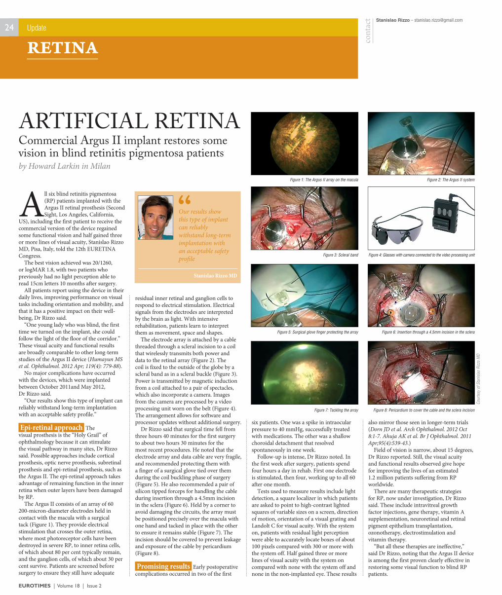
The Argus ii consists of an array of 60 200-micron-diameter electrodes held in contact with the macula with a surgical tack (Figure 1). They provide electrical stimulation that crosses the outer retina, where most photoreceptor cells have been destroyed in severe RP, to inner retina cells, of which about 80 per cent typically remain, and the ganglion cells, of which about 30 per cent survive. Patients are screened before surgery to ensure they still have adequate

residual inner retinal and ganglion cells to respond to electrical stimulation. Electrical signals from the electrodes are interpreted by the brain as light. With intensive rehabilitation, patients learn to interpret them as movement, space and shapes.

The electrode array is attached by a cable threaded through a scleral incision to a coil that wirelessly transmits both power and data to the retinal array (Figure 2). The coil is fixed to the outside of the globe by a scleral band as in a scleral buckle (Figure 3). Power is transmitted by magnetic induction from a coil attached to a pair of spectacles, which also incorporate a camera. images from the camera are processed by a video processing unit worn on the belt (Figure 4). The arrangement allows for software and processor updates without additional surgery.

Dr Rizzo said that surgical time fell from three hours 40 minutes for the first surgery to about two hours 30 minutes for the most recent procedures. he noted that the electrode array and data cable are very fragile, and recommended protecting them with a finger of a surgical glove tied over them during the coil buckling phase of surgery (Figure 5). he also recommended a pair of silicon tipped forceps for handling the cable during insertion through a 4.5mm incision in the sclera (Figure 6). held by a corner to avoid damaging the circuits, the array must be positioned precisely over the macula with one hand and tacked in place with the other to ensure it remains stable (Figure 7). The incision should be covered to prevent leakage and exposure of the cable by pericardium (Figure 8).

Promising results Early postoperative complications occurred in two of the first

six patients. One was a spike in intraocular pressure to 40 mmhg, successfully treated with medications. The other was a shallow choroidal detachment that resolved spontaneously in one week.

Follow-up is intense, Dr Rizzo noted. in the first week after surgery, patients spend four hours a day in rehab. First one electrode is stimulated, then four, working up to all 60 after one month.

Tests used to measure results include light detection, a square localizer in which patients are asked to point to high-contrast lighted squares of variable sizes on a screen, direction of motion, orientation of a visual grating and Landolt C for visual acuity. With the system on, patients with residual light perception were able to accurately locate boxes of about 100 pixels compared with 300 or more with the system off. half gained three or more lines of visual acuity with the system on compared with none with the system off and none in the non-implanted eye. These results

also mirror those seen in longer-term trials (Dorn JD et al. Arch Ophthalmol. 2012 Oct 8:1-7. Ahuja AK et al. Br J Ophthalmol. 2011 Apr;95(4):539-43.)

Field of vision is narrow, about 15 degrees, Dr Rizzo reported. still, the visual acuity and functional results observed give hope for improving the lives of an estimated 1.2 million patients suffering from RP worldwide.

There are many therapeutic strategies for RP, now under investigation, Dr Rizzo said. These include intravitreal growth factor injections, gene therapy, vitamin A supplementation, neuroretinal and retinal pigment epithelium transplantation, ozonotherapy, electrostimulation and vitamin therapy.

“But all these therapies are ineffective,” said Dr Rizzo, noting that the Argus ii device is among the first proven clearly effective in restoring some visual function to blind RP patients.

Stanislao Rizzo – [email protected]

cont

act

ARTIFICIAL RETInACommercial Argus II implant restores some vision in blind retinitis pigmentosa patientsby Howard Larkin in Milan

24 Update

RETINA

Our results show this type of implant can reliably withstand long-term implantation with an acceptable safety profile

Stanislao Rizzo MD

Figure 1: The Argus II array on the macula

Figure 3: Scleral band

Figure 5: Surgical glove finger protecting the array

Figure 7: Tackling the array

Figure 2: The Argus II system

Cour

tesy

of S

tani

slao

Rizz

o M

D

Figure 4: Glasses with camera connected to the video processing unit

Figure 6: Insertion through a 4.5mm incision in the sclera

Figure 8: Pericardium to cover the cable and the sclera incision