Embed Size (px)
Citation preview
Technology Challenges of Commercial Medical Electron
Accelerators
John Allen
Chief Engineer Elekta Ltd
Aims of this talk
• Medical Radiotherapy is already a large and profitable business
• Well established as the standard of care, for certain cancers.
• New technology needs to prove itself against the successful history of improving practice.
• Challenges remain, but physics needs to reach across disciplines in order to displace establish clinical practice.
The scale of medical accelerator business
• First systems date form the early 1950s
• Installed base of around ten thousand clinical accelerators.
• Roughly third of cancer patients treated with radiotherapy.
• Elekta treats around a third of these
• Elekta is a business with a turnover exceeding £1B.
• Both the need and the business case, require a global scale.
Why do will still have real technical demands?
• Accelerators require many disciplines
• Medical users, whilst knowledgeable, do not want exposure to the technical complexities
• Understanding both the domain and working across disciplines provides a constantly changing pallet of intellectual challenge
• Business success is clearly linked to innovation providing incentives to the owners to increase R&D funding
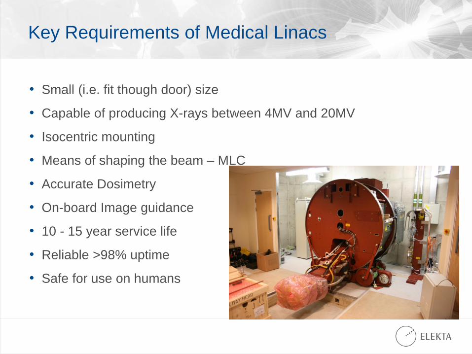
Key Requirements of Medical Linacs
• Small (i.e. fit though door) size
• Capable of producing X-rays between 4MV and 20MV
• Isocentric mounting
• Means of shaping the beam – MLC
• Accurate Dosimetry
• On-board Image guidance
• 10 - 15 year service life
• Reliable >98% uptime
• Safe for use on humans
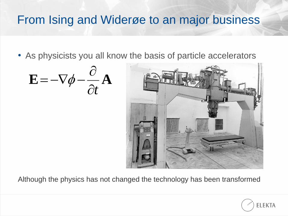
From Ising and Widerøe to an major business
• As physicists you all know the basis of particle accelerators
AEt
Although the physics has not changed the technology has been transformed
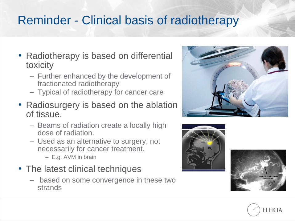
Reminder - Clinical basis of radiotherapy
• Radiotherapy is based on differential toxicity
– Further enhanced by the development of fractionated radiotherapy
– Typical of radiotherapy for cancer care
• Radiosurgery is based on the ablation of tissue.
– Beams of radiation create a locally high dose of radiation.
– Used as an alternative to surgery, not necessarily for cancer treatment.
– E.g. AVM in brain
• The latest clinical techniques
– based on some convergence in these two strands
Translating the clinical need into product
• Fractionated radiotherapy
– This requires a patient to attend daily for a few minutes of radiotherapy
– This is very cost effective, as the patient can attend as an out patient
– However 20 to 40 set ups place demands on workflow efficiency.
– Managing 30 to 80 patients a day requires good organisation.
– Software is key to managing this efficiently and safety.
• Hypo fractionated or single fraction radiosurgery.
– Depends on the accurate targeting of tumour
– Modern techniques have become ever more conformal
– The key enable has been the multi-leaf collimator
– As accuracy has increased so the for imaging to guide treatment has become vital.
– Key success factors are dose escalation and controlling toxicity.
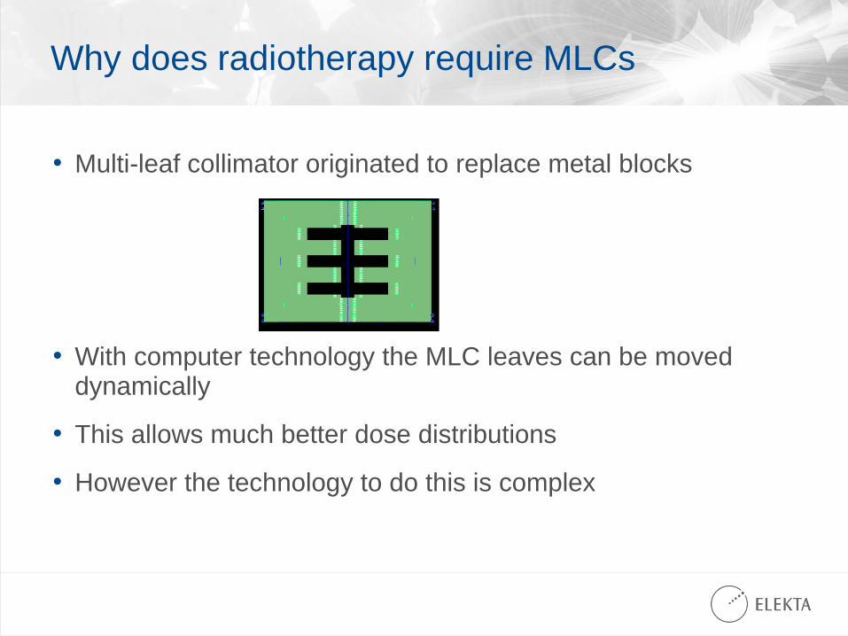
Why does radiotherapy require MLCs
• Multi-leaf collimator originated to replace metal blocks
• With computer technology the MLC leaves can be moved dynamically
• This allows much better dose distributions
• However the technology to do this is complex
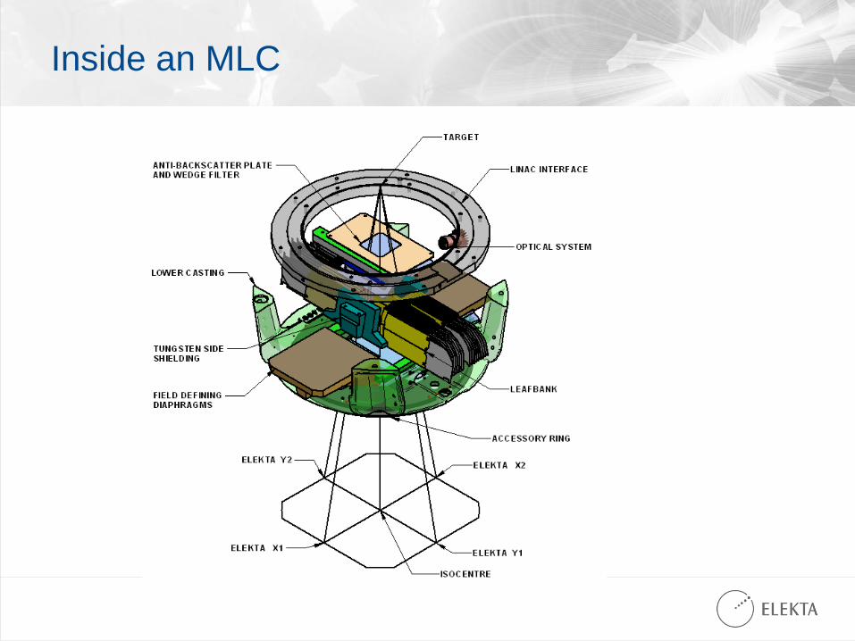
Inside an MLC
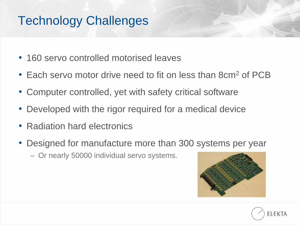
Technology Challenges
• 160 servo controlled motorised leaves
• Each servo motor drive need to fit on less than 8cm2 of PCB
• Computer controlled, yet with safety critical software
• Developed with the rigor required for a medical device
• Radiation hard electronics
• Designed for manufacture more than 300 systems per year
– Or nearly 50000 individual servo systems.
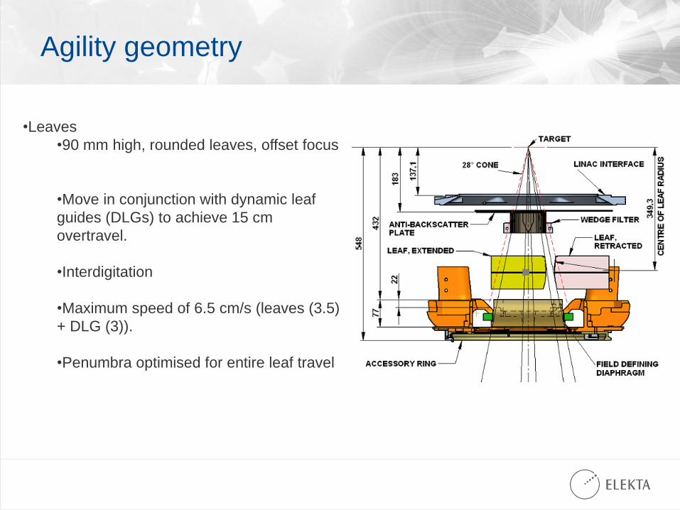
Agility geometry
•Leaves
•90 mm high, rounded leaves, offset focus
•Move in conjunction with dynamic leaf
guides (DLGs) to achieve 15 cm
overtravel.
•Interdigitation
•Maximum speed of 6.5 cm/s (leaves (3.5)
+ DLG (3)).
•Penumbra optimised for entire leaf travel
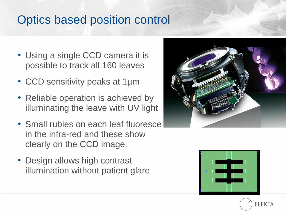
Optics based position control
• Using a single CCD camera it is possible to track all 160 leaves
• CCD sensitivity peaks at 1µm
• Reliable operation is achieved by illuminating the leave with UV light
• Small rubies on each leaf fluoresce in the infra-red and these show clearly on the CCD image.
• Design allows high contrast illumination without patient glare
Imaging
• As treatments become more conformal the need “see” the target becomes more important.
• Key enabling technologies have been amorphous silicon flat panel imaging devices and software to perform cone beam 3D reconstruction from multiple 2D images.
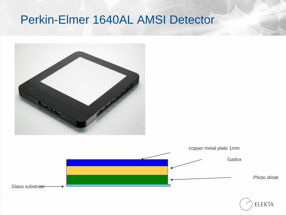
Perkin-Elmer 1640AL AMSI Detector
copper metal plate 1mm
Gadox
Photo diode
Glass substrate
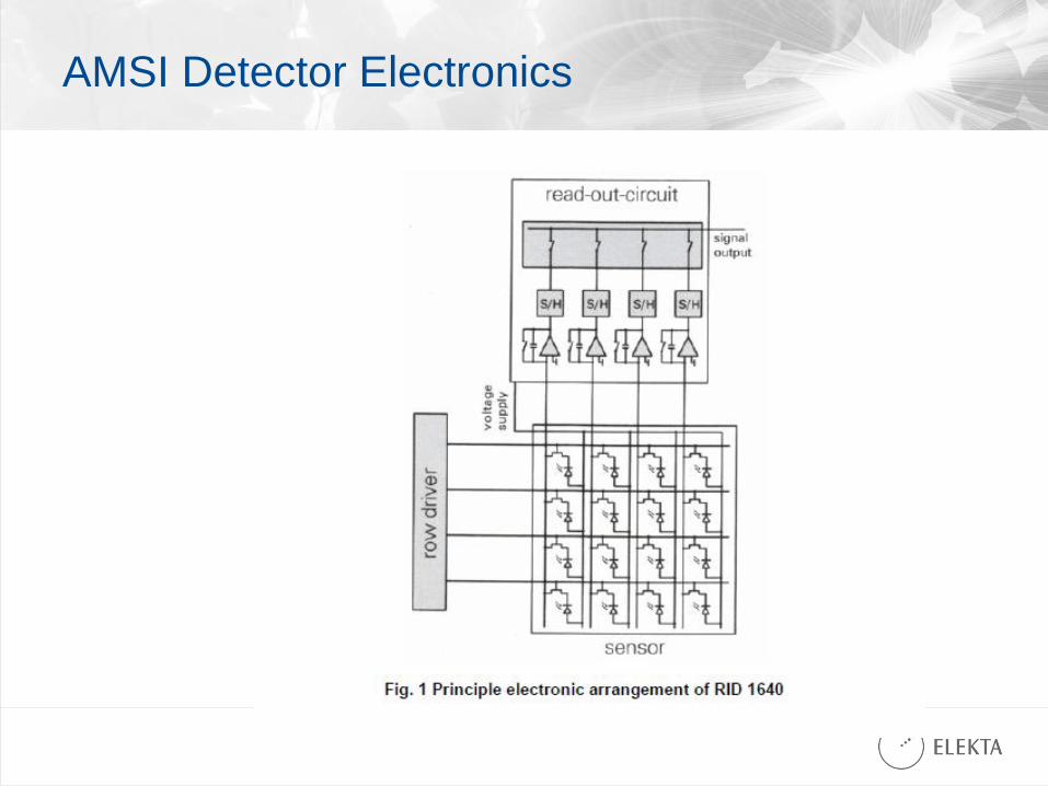
AMSI Detector Electronics
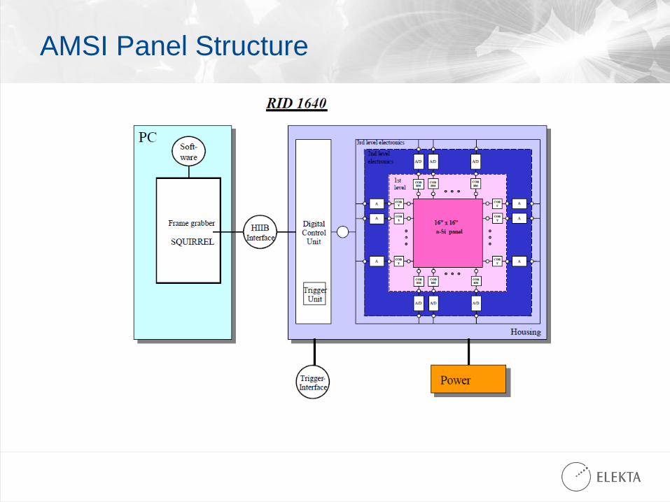
AMSI Panel Structure
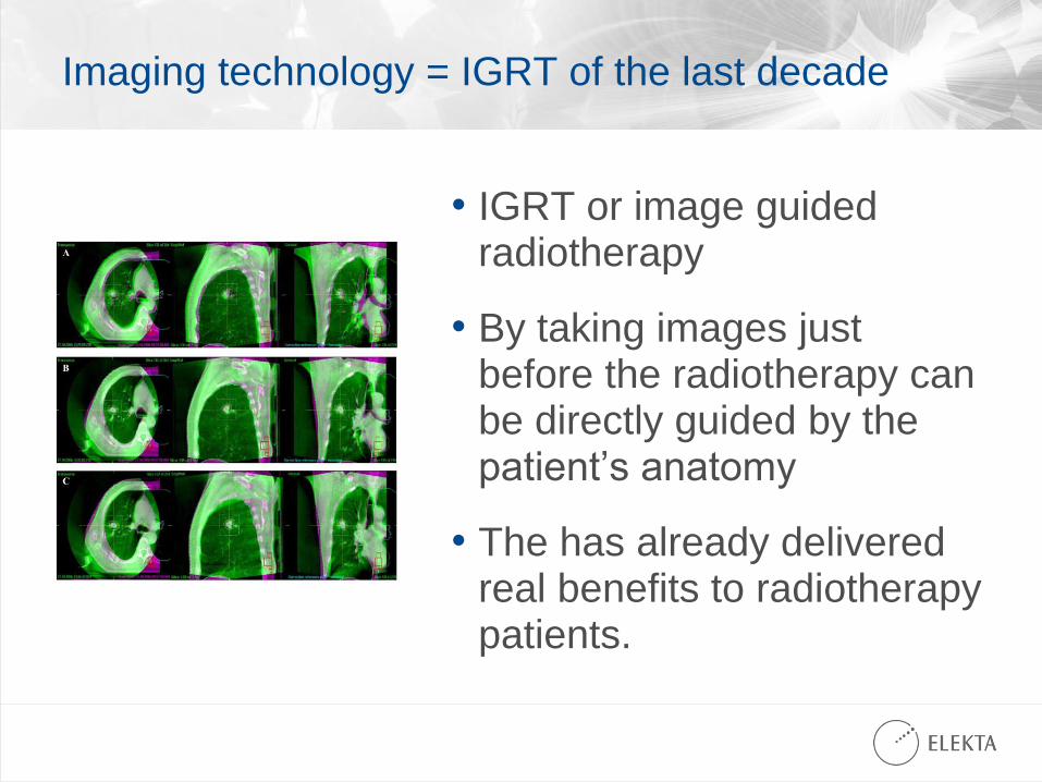
Imaging technology = IGRT of the last decade
• IGRT or image guided radiotherapy
• By taking images just before the radiotherapy can be directly guided by the patient’s anatomy
• The has already delivered real benefits to radiotherapy patients.
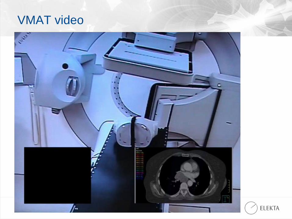
VMAT video
Agility Video
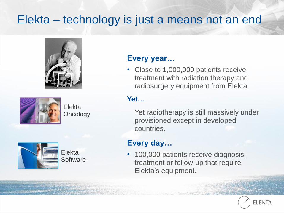
Elekta – technology is just a means not an end
Every year…
• Close to 1,000,000 patients receive treatment with radiation therapy and radiosurgery equipment from Elekta
Yet…
Yet radiotherapy is still massively under provisioned except in developed countries.
Every day…
• 100,000 patients receive diagnosis, treatment or follow-up that require Elekta’s equipment.
Elekta Oncology
Elekta Software