Embed Size (px)
Citation preview
Patient Information Handout
COMMON BENIGN TUMORS OF THE HAND AND WRIST: ENCHONDROMA:
The Latin name for the tumor means that it comes from cartilage. These are benign cartilage cells growing and expanding the bone, causing local destruction due to pressure. X-‐rays demonstrate a soap bubble appearance as the fracture expands the bone. (Figure 1). The treatment for this is surgery to remove the tumor and
replace it with bone graft. This can typically be obtained from a bone donor that has been screened to make sure that it is sterile. This does come from another human donor and a card provided after surgery, so that patients can
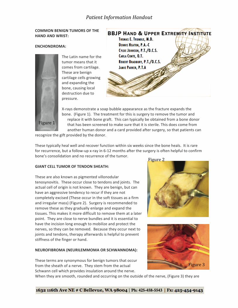
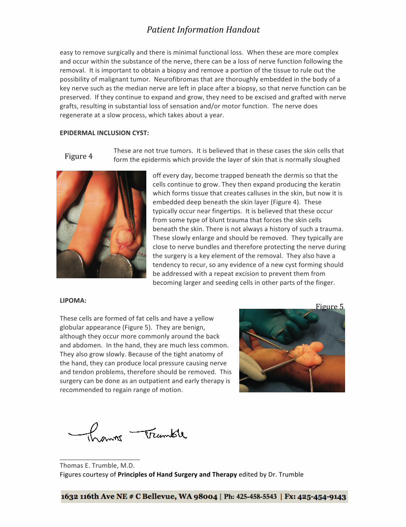
recognize the gift provided by the donor. These typically heal well and recover function within six weeks since the bone heals. It is rare for recurrence, but a follow-‐up x-‐ray in 6-‐12 months after the surgery is often helpful to confirm bone’s consolidation and no recurrence of the tumor. GIANT CELL TUMOR OF TENDON SHEATH: These are also known as pigmented villonodular tenosynovitis. These occur close to tendons and joints. The actual cell of origin is not known. They are benign, but can have an aggressive tendency to recur if they are not completely excised (These occur in the soft tissues as a firm and irregular mass) (Figure 2). Surgery is recommended to remove these as they gradually enlarge and expand the tissues. This makes it more difficult to remove them at a later point. They are close to nerve bundles and it is essential to have the incision long enough to mobilize and protect the nerves, so they can be removed. Because they occur next to joints and tendons, therapy afterwards is helpful to prevent stiffness of the finger or hand. NEUROFIBROMA (NEURILEMMOMA OR SCHWANNOMA): These terms are synonymous for benign tumors that occur from the sheath of a nerve. They stem from the actual Schwann cell which provides insulation around the nerve. When they are smooth, rounded and occurring on the outside of the nerve, (Figure 3) they are
Figure 1
Figure 2
Figure 3
Patient Information Handout
easy to remove surgically and there is minimal functional loss. When these are more complex and occur within the substance of the nerve, there can be a loss of nerve function following the removal. It is important to obtain a biopsy and remove a portion of the tissue to rule out the possibility of malignant tumor. Neurofibromas that are thoroughly embedded in the body of a key nerve such as the median nerve are left in place after a biopsy, so that nerve function can be preserved. If they continue to expand and grow, they need to be excised and grafted with nerve grafts, resulting in substantial loss of sensation and/or motor function. The nerve does regenerate at a slow process, which takes about a year. EPIDERMAL INCLUSION CYST:
These are not true tumors. It is believed that in these cases the skin cells that form the epidermis which provide the layer of skin that is normally sloughed
off every day, become trapped beneath the dermis so that the cells continue to grow. They then expand producing the keratin which forms tissue that creates calluses in the skin, but now it is embedded deep beneath the skin layer (Figure 4). These typically occur near fingertips. It is believed that these occur from some type of blunt trauma that forces the skin cells beneath the skin. There is not always a history of such a trauma. These slowly enlarge and should be removed. They typically are close to nerve bundles and therefore protecting the nerve during the surgery is a key element of the removal. They also have a tendency to recur, so any evidence of a new cyst forming should be addressed with a repeat excision to prevent them from becoming larger and seeding cells in other parts of the finger.
LIPOMA: These cells are formed of fat cells and have a yellow globular appearance (Figure 5). They are benign, although they occur more commonly around the back and abdomen. In the hand, they are much less common. They also grow slowly. Because of the tight anatomy of the hand, they can produce local pressure causing nerve and tendon problems, therefore should be removed. This surgery can be done as an outpatient and early therapy is recommended to regain range of motion.
______________________ Thomas E. Trumble, M.D. Figures courtesy of Principles of Hand Surgery and Therapy edited by Dr. Trumble
Figure 4
Figure 5