Embed Size (px)
Citation preview
Common Genetic Pathways Regulate Organ-SpecificInfection-Related Development in the Rice Blast Fungus W
Sara L. Tucker,a,1 Maria I. Besi,a,1 Rita Galhano,a Marina Franceschetti,a Stephan Goetz,a Steven Lenhert,b
Anne Osbourn,c and Ane Sesmaa,2
a Department of Disease and Stress Biology, John Innes Centre, Norwich NR4 7UH, United Kingdomb Institut fur Nanotechnologie, Forschungszentrum Karlsruhe, Eggenstein-Leopoldshafen 76344, Germanyc Department of Metabolic Biology, John Innes Centre, Norwich NR4 7UH, United Kingdom
Magnaporthe oryzae is the most important fungal pathogen of rice (Oryza sativa). Under laboratory conditions, it is able to
colonize both aerial and underground plant organs using different mechanisms. Here, we characterize an infection-related
development in M. oryzae produced on hydrophilic polystyrene (PHIL-PS) and on roots. We show that fungal spores develop
preinvasive hyphae (pre-IH) from hyphopodia (root penetration structures) or germ tubes and that pre-IH also enter root
cells. Changes in fungal cell wall structure accompanying pre-IH are seen on both artificial and root surfaces. Using
characterized mutants, we show that the PMK1 (for pathogenicity mitogen-activated protein kinase 1) pathway is required
for pre-IH development. Twenty mutants with altered pre-IH differentiation on PHIL-PS identified from an insertional library
of 2885 M. oryzae T-DNA transformants were found to be defective in pathogenicity. The phenotypic analysis of these
mutants revealed that appressorium, hyphopodium, and pre-IH formation are genetically linked fungal developmental
processes. We further characterized one of these mutants, M1373, which lacked theM. oryzae ortholog of exportin-5/Msn5p
(EXP5). Mutants lacking EXP5 were much less virulent on roots, suggesting an important involvement of proteins and/or
RNAs transported by EXP5 during M. oryzae root infection.
INTRODUCTION
Rice blast disease caused by the hemibiotroph fungal pathogen
Magnaporthe oryzae is one of themost important diseases of rice
(Oryzae sativa) worldwide. During leaf infection, the fungus forms
melanized structures known as appressoria, which enable host
penetration through a combination ofmechanical force and plant
cell wall degradation (Bourett and Howard, 1990; Talbot, 2003;
Skamnioti and Gurr, 2007). Two important signaling cascades
regulated by themitogen-activated protein kinase PMK1 and the
cAMP-dependent kinase CPKA regulate appressorium differen-
tiation and function (Xu and Hamer, 1996; Xu et al., 1997). M.
oryzae has also been shown to infect roots, although the epide-
miological significance of this infection route in the field is not yet
known (Dufresne and Osbourn, 2001; Sesma and Osbourn,
2004). M. oryzae has a number of genetically intractable close
relatives that are important root-infecting pathogens, such as the
turfgrass pathogen Magnaporthe poae and the causal agent of
take-all disease of cerealsGaeumannomyces graminis (Cannon,
1994; Bryan et al., 1995; Bunting et al., 1996; Besi et al., 2009).M.
oryzae undergoes a very different set of pathogenesis-related
developmental events on roots versus leaves. Root infection
does not depend on the melanized appressoria associated with
leaf infection and instead is mediated by simple penetration
structures known as hyphopodia (Sesma and Osbourn, 2004).
Also, root penetration is not dependent on the cAMP-mediated
pathway or on melanin synthesis (Sesma and Osbourn, 2004).
Fungal hyphae growing within the plant cell (i.e., postpenetra-
tion fungal growth) are known as invasive hyphae (IH). On leaves,
several types of IH have been described based on their distinct
biological features (Kankanala et al., 2007). Once the appresso-
rium has formed on the leaf surface, an infection peg generated
from the center of the appressorium crosses the host cell wall,
reaches the plant cell lumen, and then undergoes differentiation
to a short filamentous primary hypha. This narrow primary hypha
expands to form a thicker and biotrophic intracellular pseudo-
hypha known as a bulbous invasive hypha, though sometimes
the bulbous IH is in fact in filamentous form (Kankanala et al.,
2007). The bulbous IH fills the first infected cell (32 to 36 h),
moves into the adjacent cells by swelling and producing con-
stricted infection pegs at specific points of infection (pit fields),
and then differentiates into a biotrophic filamentous invasive
hypha. The filamentous IH grows more rapidly and requires only
a few hours to move into neighboring cells (Kankanala et al.,
2007). On roots, primary hyphae, bulbous IH, and filamentous IH
are observed, and bulbous and filamentous IH are also capable
of swelling and producing highly constricted infection pegs to
colonize adjacent cells (Sesma and Osbourn, 2004). It is also
common to find intercellular hyphae growing between cortical
root cells (Sesma and Osbourn, 2004). Necrotrophic intercellular
hyphae are also seen during leaf colonization (Kankanala et al.,
2007).
1 These authors contributed equally to this work.2 Address correspondence to [email protected] author responsible for distribution of materials integral to thefindings presented in this article in accordance with the policy describedin the Instructions for Authors (www.plantcell.org) is: Ane Sesma ([email protected]).WOnline version contains Web-only data.www.plantcell.org/cgi/doi/10.1105/tpc.109.066340
The Plant Cell, Vol. 22: 953–972, March 2010, www.plantcell.org ã 2010 American Society of Plant Biologists
Little is known about the cell wall properties of fungal IH. InM.
oryzae, a reduction in chitin and glycosyl/mannosyl residues in
the cell wall of the penetration peg have been described following
differential labeling with wheat germ agglutinin (WGA) and con-
canavalin A (ConA) during in vitro appressorium development
(Bourett and Howard, 1990). WGA is a lectin with binding
specificity for N-acetylglucosamine residues (NAcGlc, the con-
stituent monomer of fungal chitin), while ConA has an affinity for
nonreducing a-D-mannosyl and a-D-glucosyl residues present
oncellwallmannoproteins andglycoproteins (Weis andDrickamer,
1996). Immunogold-labeled WGA also revealed the presence of
chitin in the cell wall of the filamentous IH within leaf cells
(Rodrigues et al., 2003). Live-cell imaging using the endocytotic
tracker FM4-64 and transmission electron microscopy has
allowed the identification of a plant-derived extrainvasive hyphal
membrane surrounding both bulbous and filamentous IH
(Kankanala et al., 2007). This extrainvasive hyphal membrane is
tightly linked to the IH and therefore makes it difficult to identify
and/or quantify cell wall components of fungal IH using fluores-
cent markers. A reduction in chitin content (through modification
to chitosan by de-N-acetylation) has been identified in infection
structures of different fungal plant pathogens; i.e., Colletotri-
chum, Puccinia, and Uromyces during plant colonization (El
Gueddari et al., 2002).
Hydrophobic surfaces have been used extensively to investi-
gate the M. oryzae appressorium differentiation (Tucker and
Talbot, 2001), which is known to be regulated by physical and
chemical cues such as hydrophobicity and light (Jelitto et al.,
1994), plant-derived compounds including cutin monomers and
lipids (Gilbert et al., 1996), and surface hardness (Xiao et al.,
1994; Liu et al., 2007). Previously, we used a range of artificial
surfaces with grooves of different dimensions to investigate
the influence of surface topography on M. oryzae germ tubes
(Lenhert et al., 2007).
Here, we show that hydrophilic polystyrene (PHIL-PS) can
induce M. oryzae to form hyphopodium-like structures, which
then go on to differentiate into thick and filamentous preinvasive
hyphae (pre-IH). The pre-IH (hyphal development produced by
hyphopodia or germ tubes that penetrate roots) is observed on
the root surface prior to penetration. Several approaches, in-
cluding lectin cytochemistry, gene expression analysis of infec-
tion-related genes, and cytological studies of well-characterized
mutants, were used for a detailed investigation of this morpho-
genetic development. We then used the PHIL-PS surface to
identify 33 mutants with altered pre-IH formation. Twenty of
these mutants were not severely compromised in their ability to
grow in vitro but were compromised in their ability to cause
disease on both leaves and roots, suggesting that this differen-
tiation process may be significant for fungal infection. The
inability of these mutants to infect aerial and underground plant
tissues also suggests that appressorium formation and the
distinct pathogenesis-related developmental processes induced
on PHIL-PS and roots (hyphopodia and pre-IH) share common
genetic requirements.
This screen allowed us to identify six mutants with an organ-
specific phenotype (five mutants unable to infect leaves and one
mutant unable to infect roots). One of thesemutants (M1373) was
selected for further characterization.M1373was tagged in theM.
oryzae ortholog of exportin-5/MSN5 (EXP5) and showed strong
defects in its ability to produce necrotic symptoms on roots.
Dexp5mutants also showed moderate pathogenicity defects on
leaves. Overall, our results suggest common genetic require-
ments for appressorium, hyphopodium, and pre-IH differentia-
tion and reveal an important function of the karyopherin EXP5
during M. oryzae colonization of underground plant tissues.
RESULTS
Hyphopodium-Like Structures and Preinvasive Growth Are
Induced on Hydrophilic Polystyrene and Root Surfaces
We germinated M. oryzae spores on polystyrene surfaces that
differed in hydrophobicity (polystyrene with and without plasma
treatment). Polystyrene without plasma treatment is hydropho-
bic, but following plasma treatment it becomes more hydrophilic
(van Kooten et al., 2004). Glass and untreated polystyrene
(PHOB-PS) were used as controls. On glass, M. oryzae spores
germinated but the germ tubes did not differentiate into infection
structures (Figure 1A). On PHOB-PS surfaces, appressoria were
observedwithin 6 h, as expected (Bourett andHoward, 1990). By
contrast, on the hydrophilic polystyrene surface (PHIL-PS), germ
tubes formed hyphal tip swellings similar to hyphopodia, which
then differentiated into thick hyphae that resembled those typ-
ically associated with invasive growth in planta (Figures 1B and
1C). We called this novel type of hypha a pre-IH given that it is
also visualized on root surfaces, penetrates roots, and differen-
tiates into IH (Figures 1C, 1D, and 2; see Supplemental Figures
2A and 2B online).
We then used immunocytochemical methods to examine the
cell wall composition of differentM. oryzae structures in vitro and
in planta. In our experiments, spores were germinated on differ-
ent surfaces in vitro (glass, PHOB-PS, and PHIL-PS) and also on
leaves and roots. Twenty-four hours after inoculation, images
were captured by confocal microscopy and the fluorescence of
the hyphal walls quantified by measuring pixel intensity after
treatment with WGA or ConA to visualize components of fungal
cell walls. M. oryzae germ tubes showed different fluorescent
levels of WGA and ConA on all the surfaces tested, indicating the
ability of the fungus to sense and respond differently to the
surfaces used in our study (Figure 2; see Supplemental Figure
1 online). We consistently observed a reduction in WGA and
ConA binding on the cell wall of the pre-IH compared with the
germ tube on PHIL-PS (Figures 2A and 2B). By analogy with what
is known of M. oryzae and other plant pathogenic fungi (Bourett
andHoward, 1990; El Gueddari et al., 2002), we inferred from this
difference in chitin content and glucosyl-mannosyl residues that
a developmental switch occurs when germ tubes develop into
pre-IH on PHIL-PS. We found that a similar switch takes place
when the fungus grows on the root surface (Figures 1D and 2; see
Supplemental Figure 2 online). This difference in WGA and ConA
binding on the pre-IH seen on PHIL-PS remained up to 7 d (see
Supplemental Figure 2D online), indicating that the observed
reduction in chitinwas not temporarily caused by the early stages
of cell wall formation. In addition, branching germ tubes on glass
were unaffected, indicating that branching was also not
954 The Plant Cell
implicated in the observed fluorescent change on the pre-IH
(Figure 2A, glass).
Chitosan is not synthesized de novo; it is produced by
deacetylation of chitin (Tsigos et al., 2000). To determine if the
reduction in chitin content in pre-IH was due to its deacetylation
to chitosan, a cell wall modification that occurs during plant
colonization in other fungal pathogens (El Gueddari et al., 2002),
we also analyzed the chitosan content of M. oryzae cell walls
using specific antichitosan antibodies. We observed an in-
creased chitosan signal in germ tubes compared with the pre-
IH seen on PHIL-PS and roots (Figure 2C; see Supplemental
Figure 1C online), similar to the results with WGA binding,
suggesting that the reduction in chitin content is not due to
chitosan conversion.
Overall, the relative abundance of cell wall components in
germ tubes and pre-IH during M. oryzae in vitro and in planta
growth enabled us to differentiate three developmental cate-
gories related to pathogenesis: (1) leaves and PHOB-PS
(appressoria), (2) roots and PHIL-PS (hyphopodia and develop-
mental switch leading to pre-IH), and (3) glass (undifferentiated
germ tubes). These results suggest a correlation between M.
oryzae infection-related structures developed in vitro and in
planta. These data also show thatM. oryzae pre-IH observed on
the PHIL-PS surface do not specifically require the recognition of
the host plant because it appears that chemical and/or topo-
graphical components of the PHIL-PS were sufficient to induce
this root infection-related development.
In summary, our cytological analysis indicates that M. oryzae
root penetration is mediated by simple hyphopodia (Figure 1C,
left germ tube in this image) or pre-IH (Figure 2A, root; see
Supplemental Figures 2A and 2B online). Pre-IH develop either
(1) from the bottom or the side of hyphopodia (Figure 1D; see
Supplemental Figures 2A and 2B online) or (2) from germ tubes
that produce prehyphopodia at their apex (Figure 2C; see Sup-
plemental Figure 2C online). On PHIL-PS, pre-IH develops a
septum near the prehyphopodia and hyphopodia (see Supple-
mental Figures 2A to 2C online). Pre-IH–mediated penetration
starts in intercellular or proximate regions of the root epidermis
(Figures 1C, right germ tube in this image, and 2A; see Supple-
mental Figures 2A and 2B online). Germ tubes and pre-IH have
significant differences in cell wall composition (chitin, chitosan,
and mannosyl-glycosyl residues; Figure 2), and the higher
calcofluor fluorescence observed in prehyphopodia and hypho-
podia on PHIL-PS also suggests that these structures have
distinct cell wall morphology (see Supplemental Figures 2A to 2C
on line).
Although pre-IH–mediated penetration of roots has not been
described in other members of the Magnaporthaceae family,
including G. graminis and M. poae, it is known that G. graminis
produces two types of hyphae during root surface colonization:
melanized, thick-walled macrohyphae known as runner hyphae
that usually do not produce hyphopodia, and hyaline thin-walled
microhyphae known as infection hyphae fromwhich hyphopodia
are produced terminally, laterally or intercalary (Skou, 1981).
Alternative penetration mechanisms for G. graminis and other
Magnaporthe species have not yet been characterized. How-
ever, hyphae-mediated root penetration has been observed in
the pathogenic fungus Fusarium oxysporum f. sp radicis-lycopersici
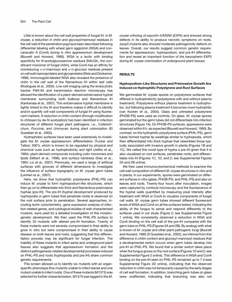
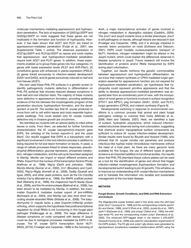
Figure 1. Characterization of M. oryzae Development on PHIL-PS
Surfaces and Roots.
(A) Time-course images of M. oryzae conidia (CO) germinated on
different surfaces: glass, untreated polystyrene (PHOB-PS), or hydro-
philic polystyrene (PHIL-PS). AP, appressoria; ds, developmental switch;
GT, germ tubes; HY, hyphopodia.
(B) Cross section of a leaf sheath (plant cell wall autofluorescence in
yellow) infected with a GFP-tagged M. oryzae strain (shown in green) in
which IH can be seen.
(C) A GFP-taggedM. oryzae conidium (shown in green) on a root surface
(plant cell wall autofluorescence in red); hyphopodia are observed on the
surface and IH can be seen emerging from pre-IH within epidermal cells.
(D) Scanning electron micrograph of a germinating conidium growing on
a root surface. A magnification of the developmental transition from germ
tube to pre-IH (asterisk) is inset in the right-hand corner. ds, develop-
mental switch.
Bars = 10 mm.
Infection-Related Development in M. oryzae 955
and the fungal symbiont Trichoderma spp (Lagopodi et al., 2002;
Harman et al., 2004), indicating that other fungal species
have also developed (or maintained) mechanisms to enter roots
without the formation of noticeable penetration structures.
Hyphopodia and Pre-IH Differentiation on PHIL-PS and
Roots Are Regulated by the PMK1-Dependent
Signaling Pathway
We then investigated whether the cAMP-dependent protein
kinase A (PKA) and the mitogen-activated protein kinase
(MAPK) PMK1 pathways were involved in hyphopodium and
pre-IH formation. Mutants affected in CPKA (encoding the cat-
alytic subunit of PKA), PMK1, andMST12 (encoding a transcrip-
tion factor regulated by PMK1) genes were tested for their
behavior on the PHIL-PS surface. These mutants grow normally
in both rich and minimal media, indicating that they are not
affected in general growth processes (Xu and Hamer, 1996; Xu
et al., 1997; see Supplemental Table 1 online). Spores of all three
mutants, like those of thewild type, were able to form germ tubes
on glass. On the PHIL-PS surface, the Dcpka mutant behaved
like the wild type, producing hyphopodia and developing pre-IH
(Figures 3A and 3B), indicating that the cAMP-dependent sig-
naling pathway mediated by CPKA is not required for this
differentiation process. By contrast, the Dpmk1 mutant failed to
form pre-IH on PHIL-PS (Figure 3C). TheDpmk1mutant was also
severely impaired in its ability to grow on roots (Figure 3D),
producingshort germ tubesandnopre-IH. TheDpmk1mutantwas
found to be impaired in production of root disease symptoms
(Figure 3I) as previously shown (Dufresne and Osbourn, 2001).
TheDmst12mutant was impaired in pre-IH formation on PHIL-
PS compared with the wild-type strain (Figure 3E), confirming
that the PMK1 pathway is required forM. oryzae development on
these surfaces. Interestingly, 29% of Dmst12 conidia (176 out of
600 conidia; see Supplemental Table 2 online) were able to
produce hyphopodia and pre-IH on PHIL-PS at 36 h (Figures 3F
and 3H), suggesting that additional components downstream of
the PMK1 pathway are also required for this differentiation
process. Some of theDmst12 conidia (33%; 27 out of 81 conidia)
were able to germinate and produce hyphopodia and pre-IH on
roots (Figures 3G and 3H). The Dmst12 mutant produced ne-
crotic symptoms on roots, although to a lesser extent than the
wild type (Figure 3I), which indicated a correlation between the
Dmst12defects in producing pre-IH and the formation of necrotic
symptoms on roots. Quantification of the production of pre-IH on
PHIL-PS and roots by Dpmk1, Dmst12, and wild-type strains
confirmed the capacity of these artificial surfaces to induce the
M. oryzae infection-related development that is seen on under-
ground tissues (Figure 3H; see Supplemental Table 2 online).
Previously, it was shown that Dpmk1 and Dmst12mutants fail to
grow invasively in rice leaves (Xu and Hamer, 1996; Park et al.,
2002; see Supplemental Table 1 online). The involvement of the
PMK1 pathway in the formation of pre-IH on roots and PHIL-PS
strongly suggests that the observed development on PHIL-PS is
part of the M. oryzae infection-related morphogenesis.
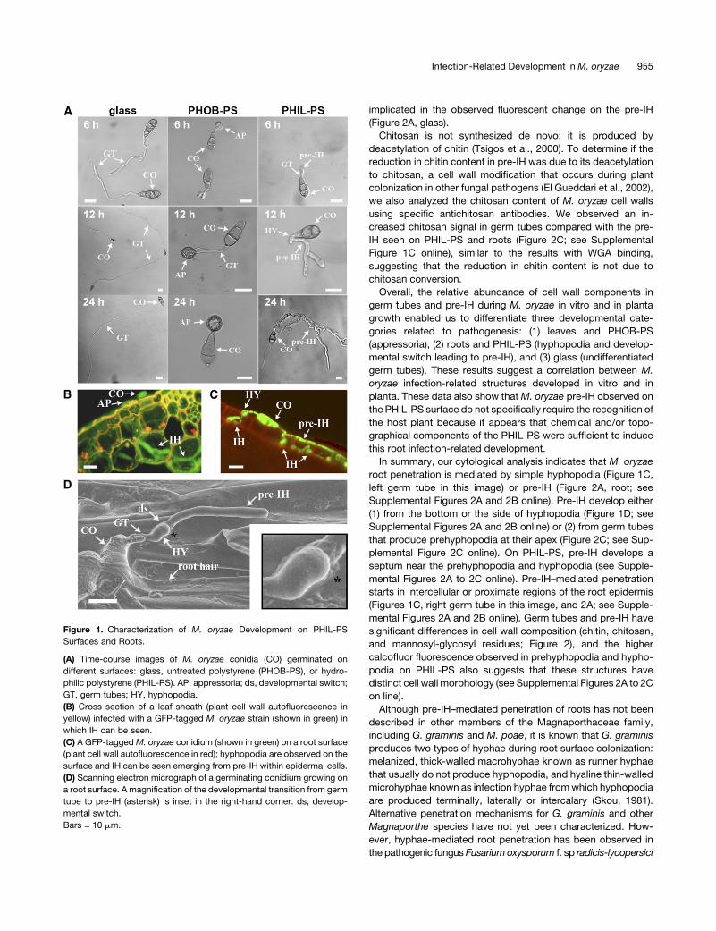
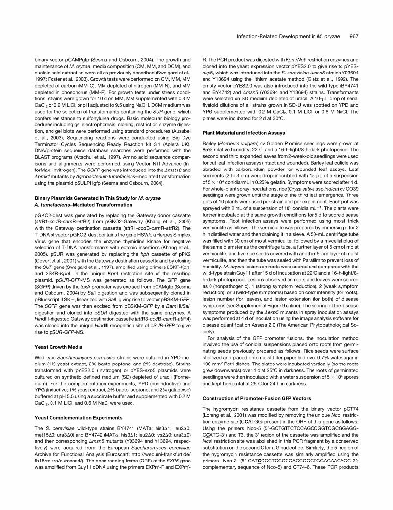
Figure 2. Detection of Carbohydrates and Glycoproteins in M. oryzae Cell Wall by Lectin Binding Assays.
Confocal imaging of M. oryzae conidia on various surfaces at 24 h using tetramethylrhodamine isothiocyanate (TRITC)-labeled WGA (antichitin lectin)
(A), Alexa594-ConA (ConA, antimannosyl and antiglucosyl lectin) (B), or antichitosan antibodies (C). Fungal cell walls in (A) and (C) show chitin and
chitosan content in red. Fungal cell walls in (B) show Alexa594-ConA fluorescence in yellow. A GFP-tagged M. oryzae strain (shown in green) was used
on roots. AP, appressoria; CO, conidia; ds, developmental switch; GT, germ tubes; HY, hyphopodia. Bars = 10 mm.
956 The Plant Cell
We then tested M. oryzae mutants affected in surface recog-
nition and appressorium formation (Dpth11, Dmpg1, and Dcbp1;
see Supplemental Table 1 online) to further characterize the
genetic requirements for the infection-related fungal develop-
ment observed on PHIL-PS. First, we examined the Dpth11
mutant, which is defective in a gene predicted to encode a
CFEM-like G-protein coupled receptor (DeZwaan et al., 1999).
The Dpth11mutant cannot efficiently develop appressoria and is
reduced in pathogenicity on leaves. These defects can be
restored by exogenous cAMP and diacylglycerol, suggesting
that PTH11 acts as an upstream activator of PKA and PKC
signaling pathways upon recognition of surface signals (DeZwaan
et al., 1999). The Dpth11 mutant produced pre-IH on PHIL-PS
surfaces and wild-type necrotic symptoms on roots (see Sup-
plemental Figures 2E and 2F online), indicating that PTH11 is not
involved in the PHIL-PS–dependent developmental switch.
Next, we tested theDmpg1mutant for its ability to differentiate
and form bulbous hyphae on PHIL-PS. The Dmpg1 mutant is
unable to produce the hydrophobinMPG1, a cell wall component
required for formation of functional appressoria (Talbot et al.,
1993). Like Dpth11 mutants, Dmpg1 mutants were able to
differentiate normally, producing hyphopodia and exhibiting
pre-IH formation on PHIL-PS and wild-type necrotic symptoms
on roots (see Supplemental Figures 2E and 2F online). This result
demonstrates that MPG1 is dispensable for the developmental
switch and pre-IH formation on PHIL-PS surfaces, suggesting
that this hyphal growth is not regulated by MPG1.
Finally, we tested mutants affected in CBP1, which encodes a
chitin binding protein containing a deacetylase domain. CBP1 is
required for recognition of physical and/or chemical factors on
synthetic surfaces that induce appressorium development
(Kamakura et al., 2002). The Dcbp1 mutant is unable to form
appressoria on hydrophobic cover slips, although it is able to
differentiate functional appressoria and to cause disease on rice
leaves (Kamakura et al., 2002). The molecular mechanism that
links the CBP1 chitin binding protein to appressorium develop-
ment is unknown. On PHIL-PS surfaces, the Dcbp1 mutant was
impaired in its ability to form hyphopodium-like structures and to
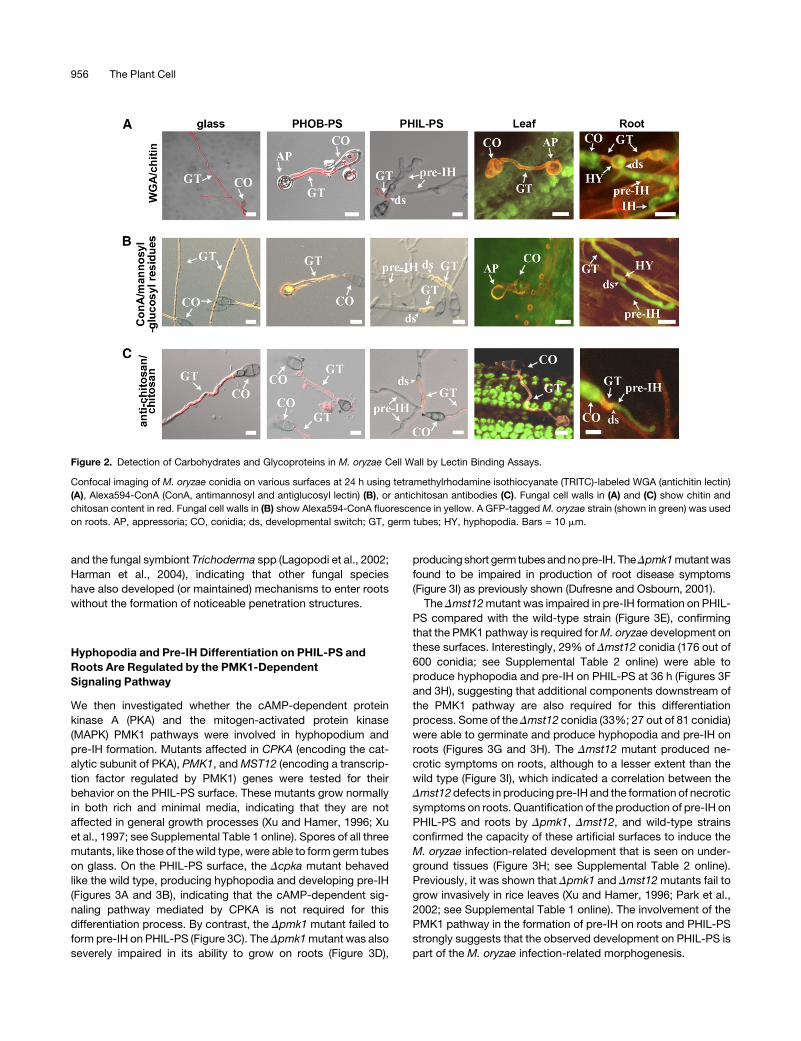
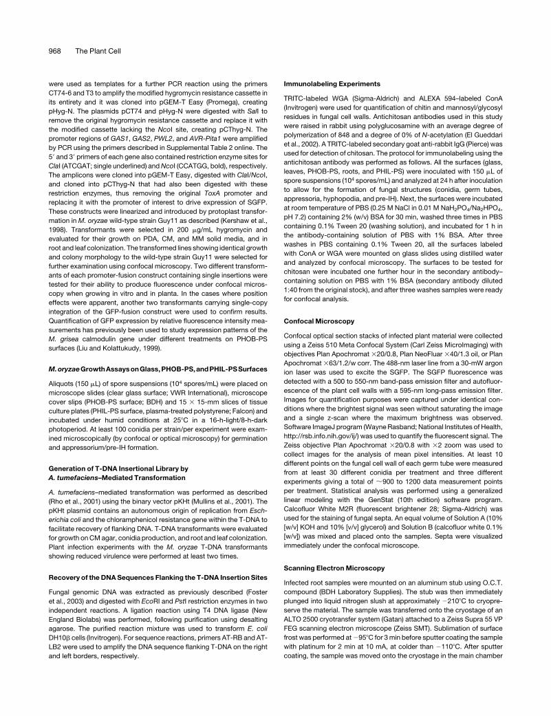
Figure 3. Development of M. oryzae Mutants on Roots and Artificial Surfaces.
(A) to (B) M. oryzae wild-type strain Guy11 (A) and the Dcpka mutant (B) on hydrophilic polystyrene (PHIL-PS).
(C) and (D) Dpmk1 germinating conidia (CO) grown on either PHIL-PS (C) or on roots (D).
(E) to (G) Dmst12 mutant on either PHIL-PS ([E] and [F]), on which both types of growth (with and without pre-IH formation) are visualized, or on roots (G).
(H) Plot of time versus mean percentage of conidia of wild-type strain (diamonds), Dmst12 mutant (squares), and Dpmk1 mutant (triangles) developing
pre-IH on PHIL-PS and roots (mean 6 SD; n = 600 on PHIL-PS and 41 to 81 on roots; three experiments).
(I) Rice roots infected with wild-type strain or with Dpmk1 and Dmst12 mutants.
Fungal cell walls in (A) to (C), (E), and (F) show chitin content in red by TRITC-WGA fluorescence. CO, conidia; GT, germ tubes; HY, hyphopodia. Bars =
20 mm.
Infection-Related Development in M. oryzae 957
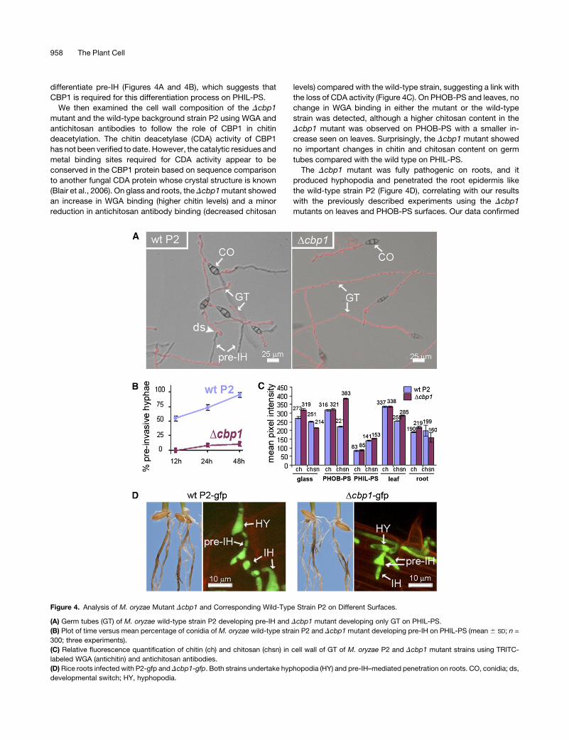
differentiate pre-IH (Figures 4A and 4B), which suggests that
CBP1 is required for this differentiation process on PHIL-PS.
We then examined the cell wall composition of the Dcbp1
mutant and the wild-type background strain P2 using WGA and
antichitosan antibodies to follow the role of CBP1 in chitin
deacetylation. The chitin deacetylase (CDA) activity of CBP1
has not been verified to date. However, the catalytic residues and
metal binding sites required for CDA activity appear to be
conserved in the CBP1 protein based on sequence comparison
to another fungal CDA protein whose crystal structure is known
(Blair et al., 2006). On glass and roots, the Dcbp1mutant showed
an increase in WGA binding (higher chitin levels) and a minor
reduction in antichitosan antibody binding (decreased chitosan
levels) compared with the wild-type strain, suggesting a link with
the loss of CDA activity (Figure 4C). On PHOB-PS and leaves, no
change in WGA binding in either the mutant or the wild-type
strain was detected, although a higher chitosan content in the
Dcbp1 mutant was observed on PHOB-PS with a smaller in-
crease seen on leaves. Surprisingly, the Dcbp1 mutant showed
no important changes in chitin and chitosan content on germ
tubes compared with the wild type on PHIL-PS.
The Dcbp1 mutant was fully pathogenic on roots, and it
produced hyphopodia and penetrated the root epidermis like
the wild-type strain P2 (Figure 4D), correlating with our results
with the previously described experiments using the Dcbp1
mutants on leaves and PHOB-PS surfaces. Our data confirmed
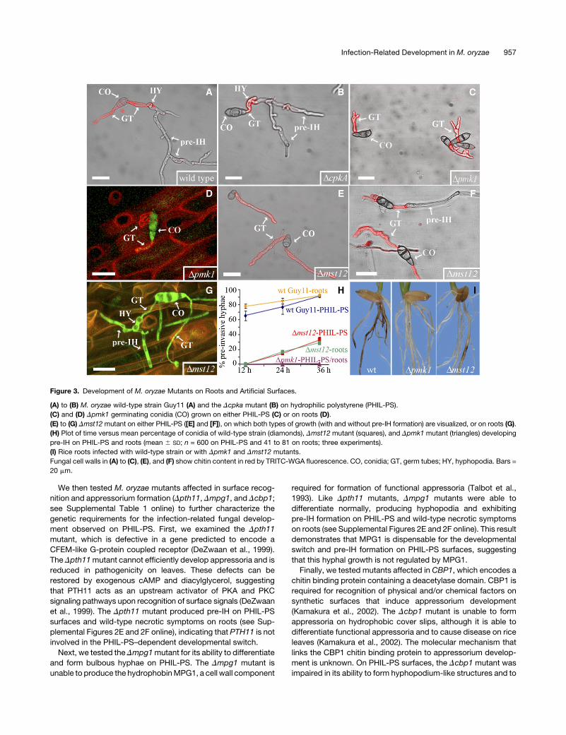
Figure 4. Analysis of M. oryzae Mutant Dcbp1 and Corresponding Wild-Type Strain P2 on Different Surfaces.
(A) Germ tubes (GT) of M. oryzae wild-type strain P2 developing pre-IH and Dcbp1 mutant developing only GT on PHIL-PS.
(B) Plot of time versus mean percentage of conidia of M. oryzae wild-type strain P2 and Dcbp1 mutant developing pre-IH on PHIL-PS (mean 6 SD; n =
300; three experiments).
(C) Relative fluorescence quantification of chitin (ch) and chitosan (chsn) in cell wall of GT of M. oryzae P2 and Dcbp1 mutant strains using TRITC-
labeled WGA (antichitin) and antichitosan antibodies.
(D) Rice roots infected with P2-gfp and Dcbp1-gfp. Both strains undertake hyphopodia (HY) and pre-IH–mediated penetration on roots. CO, conidia; ds,
developmental switch; HY, hyphopodia.
958 The Plant Cell
the involvement of CBP1 in the recognition of specific compo-
nents present in artificial substrates (PHOB-PS and PHIL-PS)
that trigger infection-related development (appressorium, hy-
phopodium, and pre-IH). The altered chitin/chitosan ratio ob-
served in the Dcbp1 mutant compared with the wild type during
growth on PHOB-PS could interfere with appressorium differen-
tiation. The lack of significant differences in the chitin/chitosan
ratio during growth onPHIL-PS suggests that the physical and/or
chemical signals that contribute to cell wall morphogenesis on
this surface could mask the deficiency of CBP1-dependent CDA
activity. The virulence shown by Dcbp1 indicates, as we might
expect, that in vitro surfaces do not perfectly mimic plant
surfaces and reveals the complexity ofM. oryzae sensing mech-
anisms. These results also show that distinct upstream signals
regulate the PMK1-dependent signaling pathway during appres-
sorium and pre-IH development. Some of these signals are
present on the PHIL-PS and PHOB surfaces. Therefore, these
surfaces could serve as an additional tool to understand the role
of genes that otherwise would not show a phenotype in planta.
Importantly, the behavior of theDcbp1mutant on artificial surfaces
also suggests a genetic link between appressorium/hyphopodium
morphogenesis and pre-IH differentiation in M. oryzae.
The Regulation of the Expression ofM. oryzae
Infection-Related Genes Is Surface Dependent
Our data so far indicate that there are structural and genetic
commonalities between the hyphopodium and the pre-IH ob-
served on PHIL-PS and roots. To further investigate this, we
looked at the regulation of characterized infection-related genes
(see Supplemental Table 1 online). For this purpose, we gener-
ated a series of green fluorescent protein (GFP)-promoter fusions
for genes encoding the effectors PWL2 (Sweigard et al., 1995)
and AVR-Pita1 (Orbach et al., 2000; these two effectors are
known to be expressed during growth in planta); and for GAS1
and GAS2, which are specifically induced during appressorium
development (Xue et al., 2002). Other previously published GFP
fusion constructs for genes required forM. oryzae pathogenicity
were also included in our experiments (see Supplemental Table
1 online). These included promoter reporter fusions for ACE1
(encoding a polyketide synthase that determines avirulence and
that is expressed in appressoria during fungal penetration;
Bohnert et al., 2004; Fudal et al., 2007) and the tetraspanin-
encoding gene PLS1 (Clergeot et al., 2001). The M. oryzae
transformant constitutively expressing SGFP fused to the TOXA
promoter from Pyrenophora tritici-repentis [TOXA(p):SGFP] was
used as a positive control strain (Sesma and Osbourn, 2004).
Conidia of the different fungal strains were germinated on glass,
PHOB-PS, and PHIL-PS synthetic surfaces and on rice leaves
and roots and then examined for formation of appressorium/
hyphopodium structures and pre-IH.
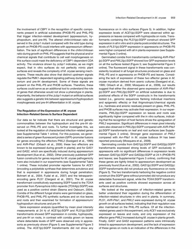
Gene expression analysis (quantified by mean pixel intensity
measurements at 24 h) of ACE1(p):SGFP and PLS1(p):SGFP
transformants showed GFP expression in conidia, hyphopodia,
and pre-IH on roots, in contrast with conidia grown on leaves
where detectable levels of GFP were observed only in appres-
soria as previously shown (Figure 5; see Supplemental Figure 3
online). The ACE1(p):SGFP transformants did not show any
fluorescence on in vitro surfaces (Figure 5). In addition, higher
expression levels of ACE1(p):SGFP were observed within ap-
pressoria on leaves compared with hyphopodia on roots. Trans-
formants containing the PLS1(p):SGFP construct showed GFP
expression in all in vitro and in planta postconidial structures. The
levels of PLS1(p):SGFP expression in appressoria on PHOB-PS
were higher compared with all in planta expression (see Supple-
mental Figure 3 online).
Germinated conidia from transformants containing AVR-Pita1
(p):SGFP and PWL2(p):SGFP showed lowGFP expression levels
on all the surfaces tested (Figure 5; see Supplemental Figure 3
online). The fluorescent signal in these transformants was con-
sistently localized in all fungal structures produced on glass and
PHIL-PS and in appressoria on PHOB-PS and leaves. Consid-
ering the lack of expression of these two effector genes in M.
oryzae mycelium derived from axenic cultures (Sweigard et al.,
1995; Orbach et al., 2000; Mosquera et al., 2009), our results
suggest that either the observed gene expression of AVR-Pita1
(p):SGFP and PWL2(p):SGFP on artificial substrates is due to
positional effects of the SGFP constructs in the transformants
tested (i.e., genomic sequences flanking individual insertions
and epigenetic effects) or that thigmotropic/chemical signals
(i.e., hardness and anionic residues) present on glass, PHIL-PS,
and PHOB surfaces may activate their expression. In our exper-
iments, PWL2(p):SGFP expression on leaves and roots was
significantly higher compared with the in vitro surfaces, indicat-
ing that the recognition of host factors drives the upregulation of
PWL2 expression. Significant differences in mean pixel intensity
were also observed in PWL2(p):SGFP compared with AVR-Pita1
(p):SGFP transformants on leaf and root surfaces (see Supple-
mental Figure 3 online). Stronger gene expression of PWL2
compared with Avr-Pita1 in M. oryzae bulbous IH has been
reported previously (Mosquera et al., 2009).
Germinating conidia from GAS1(p):SGFP and GAS2(p):SGFP
transformants expressed strong levels of GFP exclusively in
appressoria with no significant differences in expression levels
between GAS1(p):SGFP and GAS2(p):SGFP at 24 h (PHOB-PS
and leaves; see Supplemental Figure 3 online), confirming that
these genes are tightly linked to appressorium development as
previously found (Xue et al., 2002). GAS1(p):SGFP and GAS2(p):
SGFP transformants did not produce any fluorescence on roots
(Figure 5). The two transformants harboring the negative control
construct (theSGFP genewithout promoter) did not produce any
detectable fluorescent signal, while the TOXA:SGFP transform-
ants used as positive controls showed expression across all
surfaces and structures.
We looked at the expression of infection-related genes to
better understand their regulation during the differentiation of
infection-related structures in vitro and in planta. In summary,
PLS1, AVR-Pita1, and PWL2 were expressed during M. oryzae
growth on all surfaces tested, indicating that their regulation was
also maintained on the PHIL-PS surface. Of all the infection-
related genes used in this experiment, onlyACE1was exclusively
expressed on leaves and roots, and only expression of the
effector gene PWL2 increased duringM. oryzae in planta growth.
Finally, GAS1 and GAS2 expression patterns were intrinsically
linked to appressorium development, and the lack of expression
of these genes on roots is an indication of the differences in the
Infection-Related Development in M. oryzae 959
genetic pathways that regulate appressorium and hyphopodium
formation and function.
PHIL-PS Surfaces Allow the Identification of Genes
Required for Plant Infection
To identify genes involved inM. oryzae hyphopodium and pre-IH
development, we generated a library of 2885 random T-DNA
insertion mutants using Agrobacterium tumefaciens–mediated
transformation and screened these for their ability to differentiate
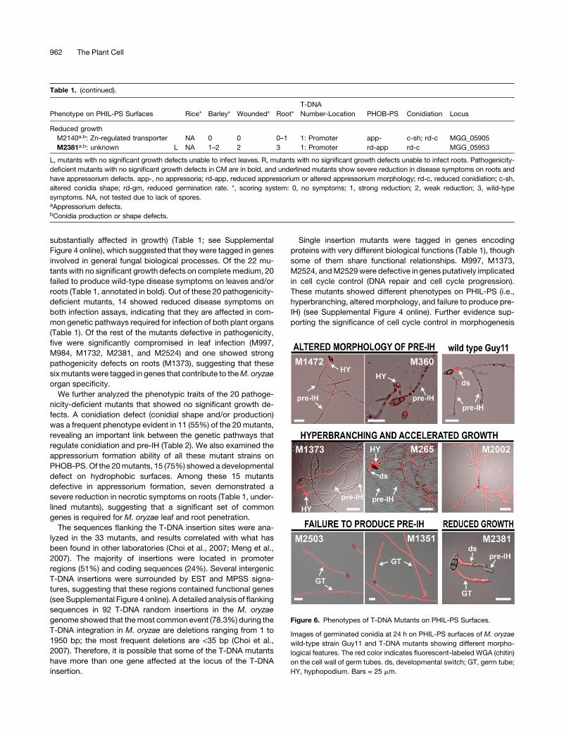
pre-IH on PHIL-PS. We identified 33mutants with altered growth
on this artificial surface (Table 1; see Supplemental Figure 4
online). We grouped thesemutants into four categories based on
their morphological features on the PHIL-PS surface (Figure 6). A
hyperbranching and accelerated growth phenotype was the
most common defect, present in 15 of the mutants. An altered
morphology of the pre-IH was found in another 12 mutants; this
included subtypes of grainy, poorly septated, overswollen, and
thin pre-IH. In addition, four mutants (M1351, M1876, M2503,
and M2529) were categorized based on their failure to undergo
the developmental switch. Finally, two mutants (M2140 and
M2381) showed a reduced hyphal growth rate on PHIL-PS.
M2002was the onlymutant with a hyperbranching phenotype on
PHIL-PS that did not show a change inWGA binding on PHIL-PS
(Figure 6; see Supplemental Figure 4 online).
We then tested these mutants for their ability to infect rice (and
barley [Hordeum vulgare]) in leaf and root pathogenicity assays.
Eleven of the 33 mutants showed strong pleiotropic effects (i.e.,
Figure 5. Gene Expression Analysis of M. oryzae Pathogenesis-Related Genes.
Confocal images of germinated conidia at 24 h containing promoter:SGFP fusion constructs (as shown on left) of infection-related genes. Negative
control:M. oryzae transformants containing a promoterless SGFP gene construct. The level of SGFP fluorescence in fungal structures (shown in green)
correlates with the mean pixel intensity values in Supplemental Figure 3 online.
960 The Plant Cell
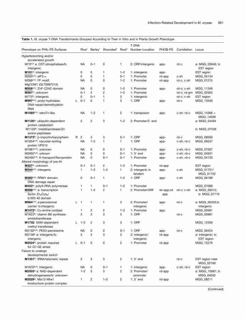
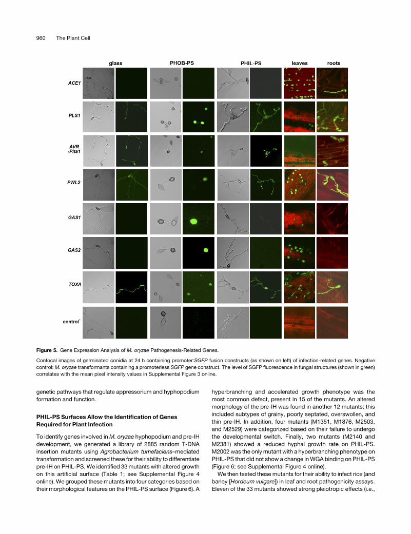
Table 1. M. oryzae T-DNA Transformants Grouped According to Their in Vitro and in Planta Growth Phenotype
Phenotype on PHIL-PS Surfaces Rice* Barley* Wounded* Root*
T-DNA
Number-Location PHOB-PS Conidiation Locus
Hyperbranching and/or
accelerated growth
M73a,b a: CDT-phosphatase/b:
intergenic
NA 0–1 0 1 2: ORF/intergenic app- rd-c a: MGG_03646; b:
EST region
M161a: intergenic 0 0 1 1–2 1: intergenic app- EST region
M265a,b: eIF3-a 0 0 1 0–1 1: Promoter rd-app c-sh MGG_05134
M288a,b: FF motif;
MgCOM1 (GI:70997212)
NA 0 0 1–2 1: Promoter rd-app rd-c, c-sh MGG_01215
M558a,b: ZnF-C2H2 domain NA 0 0 1–2 1: Promoter app- rd-c; c-sh MGG_11346
M581b: unknown 0–1 2 2 1–2 1: Promoter rd-c; rd-gm MGG_02925
M775b: intergenic 0 0–1 1 3 1: intergenic rd-c; c-sh EST region
M997a,b: prolyl-hydrolase;
DNA-repair/demethylation
RNA
L 0–1 0 1 3 1: ORF app- rd-c MGG_13340
M1085a,b: retroTn-like NA 1–2 1 2 1: transposon app- c-sh; rd-c MGG_14366 =
MGG_14039
M1120a: ubiquitin-dependent
protein catabolism
2 2 3 1–2 2: Promoter/39 end a: MGG_04494
M1120b: metalloprotease/Zn
amino-peptidase
b: MGG_07536
M1373b: b-importin/karyopherin R 2 3 3 0–1 1: ORF app- rd-c MGG_09560
M1854a,b: vacuolar-sorting
protein VPS13
NA 1–2 1 1 1: ORF app- c-sh; rd-c MGG_06537
M1981a,b: unknown NA 0 0 0–1 1: Promoter app- c-sh; rd-c MGG_07267
M2002a,b: unkown NA 0 0 0–1 1: 39 end app- c-sh; rd-c MGG_04931
M2495a,b: K-transport/flavoprotein NA 0 0–1 0–1 1: Promoter app- c-sh, rd-c MGG_04120
Altered morphology of pre-IH
M327a: unknown 0–1 0–1 2 1–2 1: Promoter rd-app EST region
M344a,b: intergenic 1 1–2 1–2 1 2: intergenic in
tandem
app- c-sh MGG_01751/
MGG_01752
M360a,b: RRM1 domain;
DNA damage repair
0 0–1 1 1–2 1: ORF app- c-sh MGG_06188
M423a: polyA-RNA polymerase 1 1 0–1 1–2 1: Promoter MGG_07089
M598a,b: a: transcription
factor Zn2Cys6/
b:WD-40 domain
1 1–2 2 1 2: Promoter/ORF sh-app,rd-
app
rd-c; c-sh a: MGG_09312;
b: MGG_07710
M984a,b: a:peroxisomal
carrier/ b:intergenic
L 1 1 1 3 2: Promoter/
intergenic
app- rd-c a: MGG_06332;b:
intergenic
M1472a: Cu-amine-oxidase 1 2 2 1–2 1: Promoter app- MGG_02681
M1623b: vitamin B6 synthesis-
amidotransferase
3 3 3 3 1: ORF rd-c MGG_05981
M1732: SAM-dependent
methyl transferase
L 1–2 2 3 3 1: ORF MGG_12356
M2102a,b: PEX3-peroxisome NA 0 0 0–1 1: ORF app- rd-c MGG_06424
M2138a a: intergenic/b:
intergenic
3 3 3 3 2: intergenic/
intergenic
rd-app a: intergenic; b:
EST region
M2524a: protein required
for G1-G2 arrest
L 0–1 0 0 2 1: Promoter rd-app MGG_12276
Failure to undergo
developmental switch
M1351b: tRNA/telomeric repeat 2 3 3 2 1: 39 end rd-c EST region near
MGG_02160
M1876a,b: intergenic NA 0 0–1 1 1: intergenic app- c-sh; rd-c EST region
M2503a a: NAD-dependent
dehydrogenases/b: unknown
1–2 3 3 2 2: Promoter/
promoter
rd-app a: MGG_10087; b:
MGG_04032
M2529a: Mis12-Mtw1
kinetochore protein complex
1 2 1–2 2 1: 39 end rd-app MGG_08211
(Continued)
Infection-Related Development in M. oryzae 961
substantially affected in growth) (Table 1; see Supplemental
Figure 4 online), which suggested that they were tagged in genes
involved in general fungal biological processes. Of the 22 mu-
tants with no significant growth defects on complete medium, 20
failed to produce wild-type disease symptoms on leaves and/or
roots (Table 1, annotated in bold). Out of these 20 pathogenicity-
deficient mutants, 14 showed reduced disease symptoms on
both infection assays, indicating that they are affected in com-
mon genetic pathways required for infection of both plant organs
(Table 1). Of the rest of the mutants defective in pathogenicity,
five were significantly compromised in leaf infection (M997,
M984, M1732, M2381, and M2524) and one showed strong
pathogenicity defects on roots (M1373), suggesting that these
sixmutants were tagged in genes that contribute to theM. oryzae
organ specificity.
We further analyzed the phenotypic traits of the 20 pathoge-
nicity-deficient mutants that showed no significant growth de-
fects. A conidiation defect (conidial shape and/or production)
was a frequent phenotype evident in 11 (55%) of the 20 mutants,
revealing an important link between the genetic pathways that
regulate conidiation and pre-IH (Table 2). We also examined the
appressorium formation ability of all these mutant strains on
PHOB-PS. Of the 20mutants, 15 (75%) showed a developmental
defect on hydrophobic surfaces. Among these 15 mutants
defective in appressorium formation, seven demonstrated a
severe reduction in necrotic symptoms on roots (Table 1, under-
lined mutants), suggesting that a significant set of common
genes is required for M. oryzae leaf and root penetration.
The sequences flanking the T-DNA insertion sites were ana-
lyzed in the 33 mutants, and results correlated with what has
been found in other laboratories (Choi et al., 2007; Meng et al.,
2007). The majority of insertions were located in promoter
regions (51%) and coding sequences (24%). Several intergenic
T-DNA insertions were surrounded by EST and MPSS signa-
tures, suggesting that these regions contained functional genes
(see Supplemental Figure 4 online). A detailed analysis of flanking
sequences in 92 T-DNA random insertions in the M. oryzae
genome showed that themost common event (78.3%) during the
T-DNA integration in M. oryzae are deletions ranging from 1 to
1950 bp; the most frequent deletions are <35 bp (Choi et al.,
2007). Therefore, it is possible that some of the T-DNA mutants
have more than one gene affected at the locus of the T-DNA
insertion.
Single insertion mutants were tagged in genes encoding
proteins with very different biological functions (Table 1), though
some of them share functional relationships. M997, M1373,
M2524, andM2529were defective in genes putatively implicated
in cell cycle control (DNA repair and cell cycle progression).
These mutants showed different phenotypes on PHIL-PS (i.e.,
hyperbranching, altered morphology, and failure to produce pre-
IH) (see Supplemental Figure 4 online). Further evidence sup-
porting the significance of cell cycle control in morphogenesis
Table 1. (continued).
Phenotype on PHIL-PS Surfaces Rice* Barley* Wounded* Root*
T-DNA
Number-Location PHOB-PS Conidiation Locus
Reduced growth
M2140a,b: Zn-regulated transporter NA 0 0 0–1 1: Promoter app- c-sh; rd-c MGG_05905
M2381a,b: unknown L NA 1–2 2 3 1: Promoter rd-app rd-c MGG_05953
L, mutants with no significant growth defects unable to infect leaves. R, mutants with no significant growth defects unable to infect roots. Pathogenicity-
deficient mutants with no significant growth defects in CM are in bold, and underlined mutants show severe reduction in disease symptoms on roots and
have appressorium defects. app-, no appressoria; rd-app, reduced appressorium or altered appressorium morphology; rd-c, reduced conidiation; c-sh,
altered conidia shape; rd-gm, reduced germination rate. *, scoring system: 0, no symptoms; 1, strong reduction; 2, weak reduction; 3, wild-type
symptoms. NA, not tested due to lack of spores.aAppressorium defects.bConidia production or shape defects.
Figure 6. Phenotypes of T-DNA Mutants on PHIL-PS Surfaces.
Images of germinated conidia at 24 h on PHIL-PS surfaces of M. oryzae
wild-type strain Guy11 and T-DNA mutants showing different morpho-
logical features. The red color indicates fluorescent-labeled WGA (chitin)
on the cell wall of germ tubes. ds, developmental switch; GT, germ tube;
HY, hyphopodium. Bars = 25 mm.
962 The Plant Cell
and fungal plant virulence comes from recent studies on the cell
cycle–regulated autophagy process in M. oryzae (Veneault-
Fourrey et al., 2006) and on the cyclin-dependent kinase Cdk5
that is necessary for polarized growth and pathogenicity in
Ustilago maydis (Castillo-Lluva et al., 2007). The potential func-
tions of the candidate genes identified in the PHIL-PS screen
also correlate with some of the expected roles of the PMK1
pathway during fungal morphogenesis. The M. oryzae PMK1 is
the functional homolog of the MAPKs FUS3 and KSS1 in yeast.
The FUS3-dependent pathway regulates the expression of nu-
merous mating-specific genes implicated in polarized cell
growth, cell cycle arrest, and changes in fungal plasma mem-
branes and cell walls (Chen and Thorner, 2007).
Overall, we observed different types of developmental defects
during M. oryzae pre-IH differentiation on PHIL-PS. The pheno-
typic analysis of the mutants demonstrated the existence of
common genetic links between appressorium and hyphopodium-
mediated plant penetration, based on the number of mutants
identified unable to infect both leaves and roots. These mutants
were not able to produce normal pre-IH. Taken together, these
data suggest that appressorium/hyphopodium differentiation
and pre-IH are infection-related processes that share common
genetic pathways. The pathogenicity tests also allowed us to
identify sixmutantswith reduced disease symptoms on leaves or
roots, indicating that they are defective in genes required for
organ-specific infection in M. oryzae. The mutant M1373 was
selected for further characterization because it was the only
mutant that showed a clear root-specific pathogenicity-deficient
phenotype.
TheM.oryzaeKaryopherinEXP5 IsRequired forFullDisease
Symptom Development on Leaves and Roots
M1373 showed hyperbranching and accelerated growth on
PHIL-PS and was severely impaired in root infection (Figure 6;
see Supplemental Figure 4 online). The T-DNA insertion of this
mutant was located within the coding sequence of M. oryzae
EXP5 (MGG_09560), the ortholog of the yeast MSN5, and the
human exportin-5 (EXP5/XPO5; see Supplemental Table 2 on-
line). Karyopherins belong to a conserved protein family repre-
senting the largest group of nuclear transport receptors found
across organisms from yeast to humans (Harel and Forbes,
2004). In Saccharomyces cerevisiae, 14 karyopherins have been
identified, and the corresponding gene orthologs are present in
M. oryzae (see Supplemental Table 3 online). Msn5p is the only
yeast karyopherin identified to date with roles in both import and
export of different cargoes (RNA and proteins) between the
cytoplasm and the nucleus.
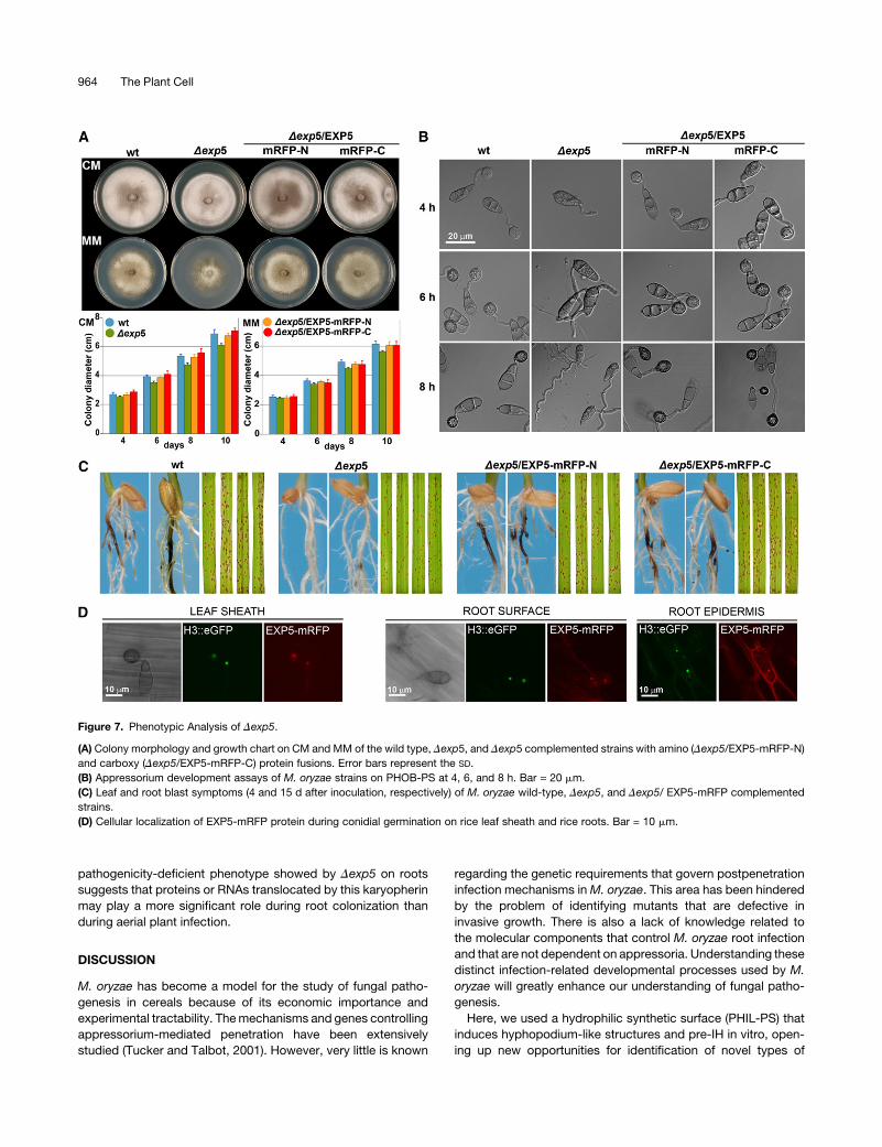
Two independent deletion mutants, Dexp5-2 and Dexp5-6,
were generated by targeted gene disruption in the EXP5 gene to
confirm the M1373 phenotype (see Supplemental Figure 5 on-
line). Colony morphology and growth of Dexp5 mutants on
complete (CM) and minimal (MM) media were slightly different
than those of the wild-type strain (Figure 7A). The Dexp5mutants
did not produce appressoria on PHOB-PS (Figure 7B). However,
Dexp5 did infect leaves producing a similar number of lesions
(Figure 7C; see Supplemental Figure 6A online), indicating that
the appressorium formation defects were restored in planta.
However, the perimeter of the lesions produced by the Dexp5
mutants was smaller, suggesting deficiencies in invasive growth
(see Supplemental Figure 6 online). The Dexp5 mutants were
strongly reduced in the ability to produce necrotic symptoms on
roots (Figure 7C; see Supplemental Figure 6B online), indicating
that the EXP5-mediated export/import pathways are important
for M. oryzae root colonization. We generated amino and
carboxy EXP5-monomeric red fluorescent protein (mRFP) trans-
lational fusion constructs to seewhere the EXP5 protein localizes
during leaf and root infection. The amino (EXP5-mRFP-N) and
carboxy (EXP5-mRFP-C) constructs restored conidiation,
growth, and appressorium formation defects of Dexp5 mutants,
indicating that these fusion proteins were fully functional (Figures
7A and 7B). A strong fluorescent signal of both amino and
carboxy EXP5-mRFP protein fusions was seen in the nucleus
during leaf and root colonization (Figure 7D).
To determine the role of EXP5 in cellular adaption to stress
conditions, as has been described for Msn5p in yeast (Alepuz
et al., 1999), we tested the resistance of the Dexp5 mutants to
nutrient deprivation (nitrogen, carbon, and phosphate), pH, and
salt-related stress (NaCl, CaCl2, and LiCl) (see Supplemental
Figure 7 online). TheDexp5mutants only showeddifferent colony
morphology and growth under pH 9.5 and 0.3MLiCl, indicating a
link between EXP5-dependent nucleocytoplasmic transport
pathways and these two stress-related responses in M. oryzae.
We also validated the putative nucleocytoplasmic transport
activity of EXP5 by testing its ability to complement the yeast
Dmsn5 mutant. We cloned the EXP5 cDNA into a yeast expres-
sion vector under the control of a galactose-inducible promoter
and introduced it intoDmsn5. The EXP5 gene complemented the
Dmsn5 growth defects in the presence of CaCl2 and galactose
(see Supplemental Figure 8 online), suggesting that EXP5 rep-
resents a functional karyopherin protein.
In summary, the PHIL-PS screen allowed us to identify EXP5,
which represents a novel infection-related gene required for full
disease symptom production on leaves and roots. The strong
Table 2. Phenotypic Traits of the 20 Pathogenicity-Deficient M. oryzae
Mutants That Showed No Significant Growth Defects
Phenotype on PHIL-PS PHOB-PSa Conidiationb
Hyperbranching and/or accelerated
growth (seven mutants)
5 5
Altered morphology of pre-IH
(nine mutants)
7 4
Failure to undergo developmental
transition (three mutants)
2 1
Reduced growth on PHIL-PS
(one mutant)
1 1
Total: 20 15 11
Number of mutants with altered phenotype on PHIL-PS is shown in
parentheses. Numbers in each column represent the number of mutants
with additional defects on appressorium formation (PHOB-PS column)
and/or conidiation.aNo appressorium, reduced appressorium, or altered appressorium
morphology.bReduced conidia production and/or altered conidial shape.
Infection-Related Development in M. oryzae 963
pathogenicity-deficient phenotype showed by Dexp5 on roots
suggests that proteins or RNAs translocated by this karyopherin
may play a more significant role during root colonization than
during aerial plant infection.
DISCUSSION
M. oryzae has become a model for the study of fungal patho-
genesis in cereals because of its economic importance and
experimental tractability. Themechanisms and genes controlling
appressorium-mediated penetration have been extensively
studied (Tucker and Talbot, 2001). However, very little is known
regarding the genetic requirements that govern postpenetration
infection mechanisms inM. oryzae. This area has been hindered
by the problem of identifying mutants that are defective in
invasive growth. There is also a lack of knowledge related to
the molecular components that control M. oryzae root infection
and that are not dependent on appressoria. Understanding these
distinct infection-related developmental processes used by M.
oryzae will greatly enhance our understanding of fungal patho-
genesis.
Here, we used a hydrophilic synthetic surface (PHIL-PS) that
induces hyphopodium-like structures and pre-IH in vitro, open-
ing up new opportunities for identification of novel types of
Figure 7. Phenotypic Analysis of Dexp5.
(A) Colony morphology and growth chart on CM and MM of the wild type, Dexp5, and Dexp5 complemented strains with amino (Dexp5/EXP5-mRFP-N)
and carboxy (Dexp5/EXP5-mRFP-C) protein fusions. Error bars represent the SD.
(B) Appressorium development assays of M. oryzae strains on PHOB-PS at 4, 6, and 8 h. Bar = 20 mm.
(C) Leaf and root blast symptoms (4 and 15 d after inoculation, respectively) of M. oryzae wild-type, Dexp5, and Dexp5/ EXP5-mRFP complemented
strains.
(D) Cellular localization of EXP5-mRFP protein during conidial germination on rice leaf sheath and rice roots. Bar = 10 mm.
964 The Plant Cell
pathogenicity genes. To determine if the differentiation pro-
cesses observed on PHIL-PS correlate withM. oryzae growth on
roots, we first monitored the distribution of cell wall structural
components of spores germinating on different in vitro and in
planta surfaces. We observed a developmental switch in growth
followed by changes in morphology and cell wall content (i.e.,
reduction in chitin, chitosan, and mannosyl/glucosyl residues) in
M. oryzae germ tubes grown on PHIL-PS and roots. Cell wall
modification has also been identified on fungal structures of
phytopathogenic fungi when colonizing their hosts (El Gueddari
et al., 2002). Chitin is perceived by the plant as a fungal path-
ogen-associated molecular pattern (Boller, 1995; Zipfel, 2008).
Chitosan also induces plant immune responses, including pro-
duction of hydrogen peroxide, and increases defense-related
enzyme activities (phenylalanine ammonialyase and chitinase),
transcription of defense-related genes, such as b-1,3-glucana-
ses and chitinases, and accumulation of the pathogen-related
protein PR1 (Shibuya and Minami, 2001; Zuppini et al., 2004).
This could explain why we observed a reduction in both chitin
and chitosan levels duringM. oryzae pre-IH differentiation as the
fungus seeks to avoid detection and induction of immune sig-
naling pathways in the host plant.
Next, we examined well-characterized M. oryzae mutants to
see if previously identified genes required for infection-related
development were also involved in the formation of hyphopo-
dium-like structures and pre-IH observed on PHIL-PS. We
demonstrated that the M. oryzae developmental switch and
pre-IH formationwere dependent on theMAPKPMK1, indicating
a direct link between this developmental program and plant
infection. The partial ability of Dmst12 mutant to produce pre-IH
indicates that additional genetic components regulated by the
PMK1 pathway are required for the complete invasive growth
differentiation by M. oryzae. The Dcpka and Dpth11 mutants
were able to undergo the developmental switch and to produce
pre-IH on PHIL-PS, suggesting that neither PKA nor PTH11-
dependent PKC pathways have a role during these develop-
mental processes. How the chitin deacetylase CBP1 influences
appressorium differentiation and pre-IH development exclu-
sively on synthetic surfaces remains unclear. These results
suggest that artificial surfaces are unlikely to fully mimic plant
tissues. Interestingly, our findings link the function of this protein
directly to the PMK1-dependent signaling pathway. The lack of
significant changes in chitin and chitosan content between the
wild-type strain and the Dcbp1 mutant could reflect a compen-
satory effect of CDA activity by one or both of the other two
predicted chitin binding proteins that contain a predicted CDA
domain, MGG_09159 andMGG_05023, present in theM. oryzae
genome. Alternatively, CBP1 expression in the fungal cell wall
might be necessary for appressorium/pre-IH formation on
PHOB/ PHIL-PS similar to what is seen with the expression of
FLO11 in S. cerevisiae, a flocculin-related gene activated by the
Kss1/Fus1 MAPK cascade whose expression is necessary to
enable the developmental transition from yeast-like to pseudo-
hyphal and filamentous growth (Lo and Dranginis, 1998). Either
way, the presence of 36 genes encoding proteins with chitin
recognition domains inM. oryzae reflects the complexity of chitin
metabolism in this organism (Dean et al., 2005).
The expression analysis of GFP-promoter fusions of pathoge-
nicity genes during M. oryzae growth on in vitro and in planta
surfaces revealed commonalities and differences between the
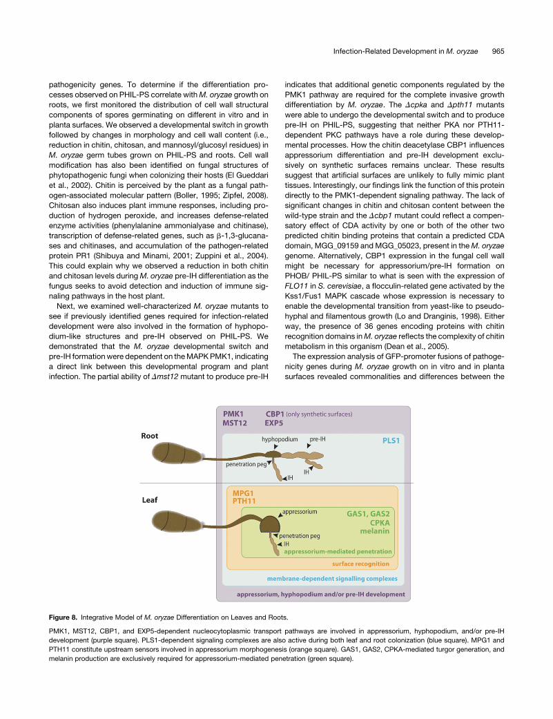
Figure 8. Integrative Model of M. oryzae Differentiation on Leaves and Roots.
PMK1, MST12, CBP1, and EXP5-dependent nucleocytoplasmic transport pathways are involved in appressorium, hyphopodium, and/or pre-IH
development (purple square). PLS1-dependent signaling complexes are also active during both leaf and root colonization (blue square). MPG1 and
PTH11 constitute upstream sensors involved in appressorium morphogenesis (orange square). GAS1, GAS2, CPKA-mediated turgor generation, and
melanin production are exclusively required for appressorium-mediated penetration (green square).
Infection-Related Development in M. oryzae 965
molecular mechanisms mediating appressorium and hyphopo-
dium penetration. The lack of expression of GAS1(p):SGFP and
GAS2(p):SGFP on roots suggests that these genes are not
implicated in the formation and function of hyphopodia. How-
ever, it has been shown that ACE1 and PLS1 are linked to
appressorium-mediated penetration (Fudal et al., 2007; see
Supplemental Table 1 online). The observed expression of
ACE1(p):SGFP and PLS1(p):SGFP on leaves and roots implies
that appressorium- and hyphopodium-mediated penetration
require both ACE1 and PLS1 genes. In addition, these exper-
iments enabled us to group these genes into four categories: (1)
genes with basal expression levels (PLS1 and AVR-Pita1), (2)
constitutive genes with enhanced in planta expression (PWL2),
(3) genes linked exclusively to infection-related development
(GAS1 andGAS2), and (4) genes exclusively induced on leaf and
root tissues (ACE1).
We next used these PHIL-PS surfaces in a genetic screen to
identify pathogenicity mutants defective in differentiation on
PHIL-PS surfaces that showed reduced disease symptoms in
both leaf and root infection tests. The majority of these mutants
were defective in appressorium formation, providing additional
evidence of the link between the morphogenetic program of this
penetration structure, hyphopodium formation, and the devel-
opment of pre-IH. The swollen tips that M. oryzae produces to
move from cell to cell within leaves and roots resemble hypho-
podia swellings. This could explain why M. oryzae mutants
defective only in invasive growth are uncommon.
We identified six mutants with an altered ability to infect either
leaves or roots. From them, we selected for further functional
characterization the M. oryzae karyopherin/b-importin gene
EXP5, the ortholog of the human exportin-5, and the yeast
Msn5. Our results suggest that EXP5 is a karyopherin with a
significant involvement in the root infection process in addition to
being required for full size lesion formation on leaves. In yeast, a
range of cellular processes linked to stress responses, pseudo-
phyphal differentiation, glucose repression, phosphate metabo-
lism, nitrogenmetabolism, and the cell cycle have been assigned
to Msn5p. Msn5p can import or export different proteins and
RNAs. Export from the nucleus of the transcription factors Pho4p
(Kaffman et al., 1998), Mig1p (DeVit and Johnston, 1999),
Msn2p-Msn4p (Gorner et al., 2002), Crz1 (Boustany and Cyert,
2002), Rtg1p-Rtg3p (Komeili et al., 2000), Swi6p (Queralt and
Igual, 2003), and other yeast proteins, such as the Cln-Cdc28p
inhibitor Far1p (Blondel et al., 1999), the MAPK scaffold protein
Ste5 (Mahanty et al., 1999), the chaperone hsp70-Ssa4p (Quan
et al., 2006), and the Ho endonuclease (Bakhrat et al., 2008), has
been shown to be mediated by Msn5p. In addition, the mam-
malian Exportin-5 mediates nuclear export of pre-miRNAs
(Bohnsack et al., 2004), and Msn5p can also bind short non-
coding double-stranded RNAs (Shibata et al., 2006). The basy-
diomycete U. maydis lacks a clear Exportin-5/Msn5p protein
ortholog, which supports the hypothesis that posttranscriptional
regulation by small RNAs is not functioning in this fungal plant
pathogen (Feldbrugge et al., 2008). The large difference in
disease symptoms on roots compared with leaves of Dexp5
could be due to biological processes mediated by EXP5 in M.
oryzae. The M. oryzae GATA transcription factor NUT1
(MGG_02755; Froeliger and Carpenter, 1996) is the homolog of
AreA, a major transcriptional activator of genes involved in
nitrogen metabolism in Aspergillus nidulans (Caddick, 2004).
The Dnut1 and Dexp5 mutants show a similar phenotype: Dnut1
is still pathogenic on leaves, although lesions are reduced in size
(Froeliger and Carpenter, 1996), and it is strongly impaired in
necrotic lesion production on roots (Dufresne and Osbourn,
2001). EXP5 could mediate nucleocytoplasmic transport of
NUT1; therefore, nitrogen metabolism might be altered in the
Dexp5 mutant, which could explain the differences observed in
disease symptoms in Dexp5. Future research will involve the
identification of proteins and/or RNAs transported by EXP5
during plant colonization.
Together, our results reinforce the genetic commonalities
between appressorium and hyphopodium differentiation. As
we know that melanin synthesis or CPKA-mediated turgor gen-
eration essential for appressorium function are not required for
hyphopodium-mediated penetration, we hypothesize that hy-
phopodia could represent primitive appressoria and that the
ability to develop appressorium-mediated penetration was ac-
quired later from an evolutionary point of view, with the incorpo-
ration of novel genes or functions involved in surface recognition
(PTH11 and MPG1), peg formation (GAS1, GAS2, and PLS1),
turgor generation (CPKA), and melanin synthesis (Figure 8).
Developmental switching in response to environmental con-
ditions is a common process that animal and plant fungal
pathogens undergo to colonize their hosts (Mehrabi et al.,
2006; Klein and Tebbets, 2007). Here, we identified a type
of surface, hydrophilic polystyrene, which induced infection-
related structures observed during root colonization, suggesting
that chemical and/or topographical surface components are
sufficient to induce M. oryzae infection-related development.
Similar results were found by Bourett and Howard (1990), who
showed that M. oryzae conidia can penetrate and produce
infectious-like hyphae inside nitrocellulose membranes without
the need of a host plant. As there are many genomic tools
available for this fungus, the use of different types of genetic
screens is an important addition to functional studies. Our results
show that PHIL-PS (standard tissue culture plates) can be used
as a tool for the identification of genes and stimuli that trigger
infection-related morphogenesis (hyphopodium formation and
developmental switch leading to pre-IH growth). Thiswill allow us
to improve our understanding ofM. oryzae infectionmechanisms
and to translate this information into durable and sustainable
management of the rice blast disease.
METHODS
Fungal Strains, Growth Conditions, and DNA and RNA Extraction
and Analysis
The Magnaporthe oryzae isolates used in this study were the wild-type
strain Guy11 (Leung et al., 1988) and the corresponding mutants Dpmk1
(Xu and Hamer, 1996), Dmst12 (Park et al., 2002), Dcpka (Xu et al., 1997),
Dpth11 (DeZwaan et al., 1999), and Dmpg1 (Talbot et al., 1993); the wild-
type strain P2, and the corresponding mutant Dcbp1 (Kamakura et al.,
2002). The enhanced GFP-tagged strain in the histone 3 (H3:eGFP;
Veneault-Fourrey et al., 2006) was used for colocalization experiments of
the EXP5 protein. The P2-gfp and Dcbp1-gfp strains were generated by
Agrobacterium tumefaciens-mediated transformation (ATMT) using the
966 The Plant Cell
binary vector pCAMPgfp (Sesma and Osbourn, 2004). The growth and
maintenance ofM. oryzae, media composition (CM, MM, and DCM), and
nucleic acid extraction were all as previously described (Sweigard et al.,
1997; Foster et al., 2003). Growth tests were performed on CM, MM, MM
depleted of carbon (MM-C), MM depleted of nitrogen (MM-N), and MM
depleted in phosphorus (MM-P). For growth tests under stress condi-
tions, strains were grown for 10 d on MM, MM supplemented with 0.3 M
CaCl2 or 0.2M LiCl, or pH adjusted to 9.5 using NaOH. DCMmediumwas
used for the selection of transformants containing the SUR gene, which
confers resistance to sulfonylurea drugs. Basic molecular biology pro-
cedures including gel electrophoresis, cloning, restriction enzyme diges-
tion, and gel blots were performed using standard procedures (Ausubel
et al., 2003). Sequencing reactions were conducted using Big Dye
Terminator Cycles Sequencing Ready Reaction kit 3.1 (Aplera UK).
DNA/protein sequence database searches were performed with the
BLAST programs (Altschul et al., 1997). Amino acid sequence compar-
isons and alignments were performed using Vector NTI Advance (In-
forMax; Invitrogen). The SGFP gene was introduced into the Dmst12 and
Dpmk1mutants byAgrobacterium tumefaciens–mediated transformation
using the plasmid pSULPHgfp (Sesma and Osbourn, 2004).
Binary Plasmids Generated in This Study forM. oryzae
A. tumefaciens–Mediated Transformation
pGKO2-dest was generated by replacing the Gateway donor cassette
(attB1-ccdB-camR-attB2) from pGKO2-Gateway (Khang et al., 2005)
with the Gateway destination cassette (attR1-ccdB-camR-attR2). The
T-DNA of vector pGKO2-dest contains the geneHSVtk, a Herpes Simplex
Virus gene that encodes the enzyme thymidine kinase for negative
selection of T-DNA transformants with ectopic insertions (Khang et al.,
2005). pSUR was generated by replacing the hph cassette of pPK2
(Covert et al., 2001) with the Gateway destination cassette and by cloning
the SUR gene (Sweigard et al., 1997), amplified using primers 2SKF-KpnI
and 2SKR-KpnI, in the unique KpnI restriction site of the resulting
plasmid. pSUR-GFP-MS was generated as follows. The GFP gene
(SGFP) driven by the toxA promoter was excised from pCAMgfp (Sesma
and Osbourn, 2004) by SalI digestion and was subsequently cloned in
pBluescript II SK2, linearized withSalI, giving rise to vector pBSKM-GFP.
The SGFP gene was then excised from pBSKM-GFP by a BamHI/SalI
digestion and cloned into pSUR digested with the same enzymes. A
HindIII-digested Gateway destination cassette (attR3-ccdB-camR-attR4)
was cloned into the unique HindIII recognition site of pSUR-GFP to give
rise to pSUR-GFP-MS.
Yeast Growth Media
Wild-type Saccharomyces cerevisiae strains were cultured in YPD me-
dium (1% yeast extract, 2% bacto-peptone, and 2% dextrose). Strains
transformed with pYES2.0 (Invitrogen) or pYES-exp5 plasmids were
cultured on synthetic defined medium (SD) depleted of uracil (Forme-
dium). For the complementation experiments, YPD (noninductive) and
YPG (inductive; 1% yeast extract, 2%bacto-peptone, and 2%galactose)
buffered at pH 5.5 using a succinate buffer and supplemented with 0.2 M
CaCl2, 0.1 M LiCl, and 0.6 M NaCl were used.
Yeast Complementation Experiments
The S. cerevisiae wild-type strains BY4741 (MATa; his3D1; leu2D0;
met15D0; ura3D0) and BY4742 (MATa; his3D1; leu2D0; lys2D0; ura3D0)
and their corresponding Dmsn5 mutants (Y03694 and Y13694, respec-
tively) were acquired from the European Saccharomyces cerevisiae
Archive for Functional Analysis (Euroscarf; http://web.uni-frankfurt.de/
fb15/mikro/euroscarf/). The open reading frame (ORF) of the EXP5 gene
was amplified from Guy11 cDNA using the primers EXPrY-F and EXPrY-
R. The PCR product was digested withKpnI/NotI restriction enzymes and
cloned into the yeast expression vector pYES2.0 to give rise to pYES-
exp5, which was introduced into the S. cerevisiae Dmsn5 strains Y03694
and Y13694 using the lithium acetate method (Gietz et al., 1992). The
empty vector pYES2.0 was also introduced into the wild type (BY4741
and BY4742) and Dmsn5 (Y03694 and Y13694) strains. Transformants
were selected on SD medium depleted of uracil. A 10-mL drop of serial
fivefold dilutions of all strains grown in SD-U was spotted on YPD and
YPG supplemented with 0.2 M CaCl2, 0.1 M LiCl, or 0.6 M NaCl. The
plates were incubated for 2 d at 308C.
Plant Material and Infection Assays
Barley (Hordeum vulgare) cv Golden Promise seedlings were grown at
85% relative humidity, 228C, and a 16-h-light/8-h-dark photoperiod. The
second and third expanded leaves from 2-week-old seedlings were used
for cut leaf infection assays (intact and wounded). Barley leaf cuticle was
abraded with carborundum powder for wounded leaf assays. Leaf
segments (2 to 3 cm) were drop-inoculated with 15 mL of a suspension
of 53 104 conidia/mL in 0.25% gelatin. Symptoms were scored after 4 d.
For whole-plant spray inoculations, rice (Oryza sativa ssp indica) cv CO39
seedlings were grown until the stage of the third leaf emergence. Three
pots of 10 plants were used per strain and per experiment. Each pot was
sprayed with 2 mL of a suspension of 105 conidia mL21. The plants were
further incubated at the same growth conditions for 5 d to score disease
symptoms. Root infection assays were performed using moist thick
vermiculite as follows. The vermiculite was prepared by immersing it for 2
h in distilled water and then draining it in a sieve. A 50-mL centrifuge tube
was filled with 30 cm of moist vermiculite, followed by a mycelial plug of
the same diameter as the centrifuge tube, a further layer of 5 cm of moist
vermiculite, and five rice seeds covered with another 5-cm layer of moist
vermiculite, and then the tube was sealed with Parafilm to prevent loss of
humidity.M. oryzae lesions on roots were scored and compared with the
wild-type strain Guy11 after 15 d of incubation at 228C and a 16-h-light/8-
h-dark photoperiod. Lesions observed on roots and leaves were scored
as 0 (nonpathogenic), 1 (strong symptom reduction), 2 (weak symptom
reduction), or 3 (wild-type symptoms) based on color intensity (for roots),
lesion number (for leaves), and lesion extension (for both) of disease
symptoms (see Supplemental Figure 9 online). The scoring of the disease
symptoms produced by the Dexp5 mutants in spray inoculation assays
was performed at 4 d of inoculation using the image analysis software for
disease quantification Assess 2.0 (The American Phytopathological So-
ciety).
For analysis of the GFP promoter fusions, the inoculation method
involved the use of conidial suspensions placed onto roots from germi-
nating seeds previously prepared as follows. Rice seeds were surface
sterilized and placed onto moist filter paper laid over 0.7% water agar in
100-mm2 Petri dishes. The plates were incubated vertically (so the roots
grew downwards) over 4 d at 258C in darkness. The roots of germinated
seedlingswere then inoculatedwith awater suspension of 53 104 spores
and kept horizontal at 258C for 24 h in darkness.
Construction of Promoter-Fusion GFP Vectors
The hygromycin resistance cassette from the binary vector pCT74
(Lorang et al., 2001) was modified by removing the unique NcoI restric-
tion enzyme site (CCATGG) present in the ORF of this gene as follows.
Using the primers Nco-5 (59-GCTGTTCTCCAGCCGGTCGCGGAGG-
CGATG-39) and T3, the 39 region of the cassette was amplified and the
NcoI restriction site was abolished in this PCR fragment by a conserved
substitution on the second C for a G nucleotide. Similarly, the 59 region of
the hygromycin resistance cassette was similarly amplified using the
primers Nco-3 (59-CATCGCCTCCGCGACCGGCTGGAGAACAGC-39;
complementary sequence of Nco-5) and CT74-6. These PCR products
Infection-Related Development in M. oryzae 967
were used as templates for a further PCR reaction using the primers
CT74-6 and T3 to amplify the modified hygromycin resistance cassette in
its entirety and it was cloned into pGEM-T Easy (Promega), creating
pHyg-N. The plasmids pCT74 and pHyg-N were digested with SalI to
remove the original hygromycin resistance cassette and replace it with
the modified cassette lacking the NcoI site, creating pCThyg-N. The
promoter regions of GAS1, GAS2, PWL2, and AVR-Pita1 were amplified
by PCR using the primers described in Supplemental Table 2 online. The
59 and 39 primers of each gene also contained restriction enzyme sites for
ClaI (ATCGAT; single underlined) andNcoI (CCATGG, bold), respectively.
The amplicons were cloned into pGEM-T Easy, digested with ClaI/NcoI,
and cloned into pCThyg-N that had also been digested with these
restriction enzymes, thus removing the original ToxA promoter and
replacing it with the promoter of interest to drive expression of SGFP.
These constructs were linearized and introduced by protoplast transfor-
mation in M. oryzae wild-type strain Guy11 as described (Kershaw et al.,
1998). Transformants were selected in 200 mg/mL hygromycin and
evaluated for their growth on PDA, CM, and MM solid media, and in
root and leaf colonization. The transformed lines showing identical growth
and colony morphology to the wild-type strain Guy11 were selected for
further examination using confocal microscopy. Two different transform-
ants of each promoter-fusion construct containing single insertions were
tested for their ability to produce fluorescence under confocal micros-
copy when growing in vitro and in planta. In the cases where position
effects were apparent, another two transformants carrying single-copy
integration of the GFP-fusion construct were used to confirm results.
Quantification of GFP expression by relative fluorescence intensity mea-
surements has previously been used to study expression patterns of the
M. grisea calmodulin gene under different treatments on PHOB-PS
surfaces (Liu and Kolattukudy, 1999).
M.oryzaeGrowthAssaysonGlass,PHOB-PS,andPHIL-PSSurfaces
Aliquots (150 mL) of spore suspensions (104 spores/mL) were placed on
microscope slides (clear glass surface; VWR International), microscope
cover slips (PHOB-PS surface; BDH) and 15 3 15-mm slices of tissue
culture plates (PHIL-PS surface, plasma-treated polystyrene; Falcon) and
incubated under humid conditions at 258C in a 16-h-light/8-h-dark
photoperiod. At least 100 conidia per strain/per experiment were exam-
ined microscopically (by confocal or optical microscopy) for germination
and appressorium/pre-IH formation.
Generation of T-DNA Insertional Library by
A. tumefaciens–Mediated Transformation
A. tumefaciens–mediated transformation was performed as described
(Rho et al., 2001) using the binary vector pKHt (Mullins et al., 2001). The
pKHt plasmid contains an autonomous origin of replication from Esch-
erichia coli and the chloramphenicol resistance gene within the T-DNA to
facilitate recovery of flanking DNA. T-DNA transformants were evaluated
for growth onCMagar, conidia production, and root and leaf colonization.
Plant infection experiments with the M. oryzae T-DNA transformants
showing reduced virulence were performed at least two times.
Recovery of the DNASequences Flanking the T-DNA Insertion Sites
Fungal genomic DNA was extracted as previously described (Foster
et al., 2003) and digested with EcoRI and PstI restriction enzymes in two
independent reactions. A ligation reaction using T4 DNA ligase (New
England Biolabs) was performed, following purification using desalting
agarose. The purified reaction mixture was used to transform E. coli
DH10b cells (Invitrogen). For sequence reactions, primers AT-RB and AT-
LB2 were used to amplify the DNA sequence flanking T-DNA on the right
and left borders, respectively.
Immunolabeling Experiments
TRITC-labeled WGA (Sigma-Aldrich) and ALEXA 594–labeled ConA
(Invitrogen) were used for quantification of chitin and mannosyl/glycosyl
residues in fungal cell walls. Antichitosan antibodies used in this study
were raised in rabbit using polyglucosamine with an average degree of
polymerization of 848 and a degree of 0% of N-acetylation (El Gueddari
et al., 2002). A TRITC-labeled secondary goat anti-rabbit IgG (Pierce) was
used for detection of chitosan. The protocol for immunolabeling using the
antichitosan antibody was performed as follows. All the surfaces (glass,
leaves, PHOB-PS, roots, and PHIL-PS) were inoculated with 150 mL of
spore suspensions (104 spores/mL) and analyzed at 24 h after inoculation
to allow for the formation of fungal structures (conidia, germ tubes,
appressoria, hyphopodia, and pre-IH). Next, the surfaces were incubated
at room temperature of PBS (0.25 M NaCl in 0.01 M NaH2PO4/Na2HPO4,
pH 7.2) containing 2% (w/v) BSA for 30 min, washed three times in PBS
containing 0.1% Tween 20 (washing solution), and incubated for 1 h in
the antibody-containing solution of PBS with 1% BSA. After three
washes in PBS containing 0.1% Tween 20, all the surfaces labeled
with ConA or WGA were mounted on glass slides using distilled water
and analyzed by confocal microscopy. The surfaces to be tested for
chitosan were incubated one further hour in the secondary antibody–
containing solution on PBS with 1% BSA (secondary antibody diluted
1:40 from the original stock), and after three washes samples were ready
for confocal analysis.
Confocal Microscopy
Confocal optical section stacks of infected plant material were collected
using a Zeiss 510 Meta Confocal System (Carl Zeiss MicroImaging) with
objectives Plan Apochromat 320/0.8, Plan NeoFluar 340/1.3 oil, or Plan
Apochromat363/1.2/w corr. The 488-nm laser line from a 30-mW argon
ion laser was used to excite the SGFP. The SGFP fluorescence was
detected with a 500 to 550-nm band-pass emission filter and autofluor-
escence of the plant cell walls with a 595-nm long-pass emission filter.
Images for quantification purposes were captured under identical con-
ditions where the brightest signal was seen without saturating the image
and a single z-scan where the maximum brightness was observed.
Software ImageJ program (WayneRasband; National Institutes of Health,
http://rsb.info.nih.gov/ij/) was used to quantify the fluorescent signal. The
Zeiss objective Plan Apochromat 320/0.8 with 32 zoom was used to
collect images for the analysis of mean pixel intensities. At least 10
different points on the fungal cell wall of each germ tube were measured
from at least 30 different conidia per treatment and three different
experiments giving a total of ;900 to 1200 data measurement points
per treatment. Statistical analysis was performed using a generalized
linear modeling with the GenStat (10th edition) software program.
Calcofluor White M2R (fluorescent brightener 28; Sigma-Aldrich) was
used for the staining of fungal septa. An equal volume of Solution A (10%
[w/v] KOH and 10% [v/v] glycerol) and Solution B (calcofluor white 0.1%
[w/v]) was mixed and placed onto the samples. Septa were visualized
immediately under the confocal microscope.
Scanning Electron Microscopy
Infected root samples were mounted on an aluminum stub using O.C.T.
compound (BDH Laboratory Supplies). The stub was then immediately
plunged into liquid nitrogen slush at approximately 22108C to cryopre-
serve the material. The sample was transferred onto the cryostage of an
ALTO 2500 cryotransfer system (Gatan) attached to a Zeiss Supra 55 VP
FEG scanning electron microscope (Zeiss SMT). Sublimation of surface
frost was performed at2958C for 3min before sputter coating the sample
with platinum for 2 min at 10 mA, at colder than 21108C. After sputter
coating, the sample was moved onto the cryostage in the main chamber
968 The Plant Cell
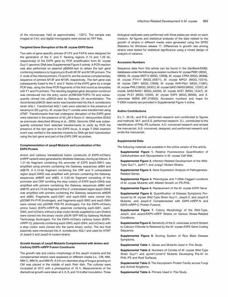
of the microscope, held at approximately 21308C. The sample was
imaged at 3 kV, and digital micrographs were stored as TIFF files.
Targeted Gene Disruption of theM. oryzae EXP5 Gene
Two sets of gene-specific primers (F1/F2 and F3/F4) were designed for
the generation of the 59 and 39 flanking regions (1.15 and 1.29 kb,
respectively) of the EXP5 gene by PCR amplification from M. oryzae
Guy11 genomic DNA (see Supplemental Figure 5 online). A PCR reaction
was also performed on plasmid pBSKM-hph to obtain the hph gene
conferring resistance to hygromycin Bwith M13F andM13R primers. The
59 ends of the internal primers, F3 and F4, are the reverse complementary
sequence of primers M13F and M13R, respectively. The hph gene was
subsequently fused to the 59 and 39 flanks of the EXP5 gene by a single
PCR step, using the three PCR fragments of the first round as templates
with F1 and F4 primers. The resulting targeted gene disruption construct
was introduced into the entry vector pCR8/GW/TOPO-TA and subse-
quently cloned into pGKO2-dest by Gateway LR recombination. The
recombined pGKO2-dest vector was transformed into theA. tumefaciens
strain AGL1. Transformed AGL1 cells were selected in the presence of
kanamycin (50 mg/mL).M. oryzae Guy11 conidia were transformed using
ATMT. Transformants that had undergone disruption of the EXP5 gene
were selected in the presence of 50 mM 5-fluoro-29-deoxyuridine (f2dU)
as previously described (Khang et al., 2005). Genomic DNA was subse-
quently extracted from selected transformants to verify by PCR the
presence of the hph gene in the EXP5 locus. A single T-DNA insertion
event was verified in the selected mutants by DNA gel blot hybridization
using the hph gene and part of the EXP5 ORF as probes.
Complementation of Dexp5Mutants and Localization of the
EXP5 Protein
Amino and carboxy translational fusion constructs of EXP5-mCherry
(mRFP variant) were generated byMultisite Gateway cloning as follows. A
1.21-kb fragment containing the promoter of EXP5 (exp5-GW1) was
amplified using primers containing the Gateway sequences attB4 and
attB1R. A 5.14-kb fragment containing the ORF and 39-untranslated
region (exp5-GW2) was amplified with primers containing the Gateway
sequences attB2R and attB3. A 5.62-kb fragment consisting of the
promoter and ORF (omitting the stop codon) of EXP5 (exp5-GW3) was
amplified with primers containing the Gateway sequences attB4 and
attB1R, and a 0.73-kb fragment of the 39-untranslated region (exp5-GW4)
was amplified with primers containing the Gateway sequences attB2R
and attB3. Fragments exp5-GW1 and exp5-GW3 were cloned into
pDONR P4-P1R (Invitrogen), and fragments exp5-GW2 and exp5-GW4
were cloned into pDONR P2R-P3 (Invitrogen). For the EXP5-mCherry
amino fusion (EXP5-mRFP-N), plasmids containing exp5-GW1, exp5-
GW2, andmCherry without a stop codon (kindly supplied by LiamDolam)
were cloned into the binary vector pSUR-GFP-MS by Gateway Multisite
Technology (Invitrogen). For the EXP5-mCherry carboxy fusion (EXP5-
mRFP-C), plasmids containing exp5-GW3, exp5-GW4, andmCherrywith
a stop codon were cloned into the same binary vector. The two final
plasmids were introduced into A. tumefaciens AGL1 and used for ATMT
of Dexp5-2 and Dexp5-6 mutant strains.
Growth Assays of Dexp5Mutants Complemented with Amino and
Carboxy EXP5-mRFP Fusion Constructs
The growth rate and colony morphology of the Dexp5 mutants and the
complemented strains were assessed on different media (i.e., CM, MM,
MM-C, MM-N, andMM-P). A 0.6-cm-diameter plug of fungus growing on
CM was placed in the middle of each Petri dish. The cultures were
incubated at 258C with a photoperiod of 16 h. Measurements of the
diametrical growth were taken at 4, 6, 8, and 10 d after inoculation. Three
biological replicates were performed with three plates per strain on each
medium. All figures and statistical analyses of the data related to the
growth of strains in different media were generated using the SPSS
Statistics for Windows release 17. Differences in growth rate among
strains were tested for statistical significance using a mixed design of
analysis of variance.
Accession Numbers
Sequence data from this article can be found in the GenBank/EMBL
databasesunder the following accession numbers:M.oryzaePMK1 (MGG_
09565), M. oryzae MST12 (MGG_12958), M. oryzae CPKA (MGG_06368),
M. oryzae PTH11 (MGG_05871), M. oryzae MPG1 (MGG_10315),
M. oryzae CBP1 (MGG_12939), M. oryzae AVR-Pita1 (MGG_11081),
M. oryzaePWL2 (MGG_04301),M. oryzaeGAS1/MAS3 (MGG_12337),M.
oryzae GAS2/MAS1 (MGG_04202), M. oryzae ACE1 (MGG_12447), M.
oryzae PLS1 (MGG_12594), M. oryzae EXP5 (MGG_09560), and S.
cerevisiae MSN5 (NP_010622). Accession numbers and maps for
T-DNA mutants are provided in Supplemental Figure 4 online.
Author Contributions
S.L.T., M.I.B., and R.G. performed research and contributed to figures
and methods. M.F. and S.G. performed research. S.L. contributed to the
identification of PHIL-PS surfaces. A.O. conceived research and revised
the manuscript. A.S. conceived, designed, and performed research and
wrote the manuscript.
Supplemental Data
The following materials are available in the online version of this article.
Supplemental Figure 1. Relative Fluorescence Quantification of
Carbohydrates and Glycoproteins in M. oryzae Cell Wall.
Supplemental Figure 2. Infection-Related Development of the Wild-
Type Guy11, Dpth11, and Dmpg1 Mutant Strains.
Supplemental Figure 3. Gene Expression Analysis of Pathogenesis-
Related Genes.
Supplemental Figure 4. Phenotypes and T-DNA–Tagged Locations
of M. oryzae Mutants with Altered Growth on PS-PHIL.
Supplemental Figure 5. Replacement of the M. oryzae EXP5 Gene.
Supplemental Figure 6. Quantification of Disease Symptoms Pro-
duced by M. oryzae Wild-Type Strain Guy11, Dexp5-2, and Dexp5-6
Mutants, and Dexp5-6 Complemented with EXP5-mRFP-N and
EXP5-mRFP-C Protein Fusions.
Supplemental Figure 7. Colony Morphology of the Wild-Type,
Dexp5, and Dexp5/EXP5-mRFP Strains on Various Stress-Related
Conditions.
Supplemental Figure 8. Sensitivity of the S. cerevisiae Dmsn5 Mutant
to Calcium Chloride Is Restored by the M. oryzae EXP5 Gene Coding
Sequence.
Supplemental Figure 9. Scoring System of Rice Blast Disease
Symptoms.
Supplemental Table 1. Genes and Mutants Used in This Study.
Supplemental Table 2. Numbers of Conidia of M. oryzae Wild-Type
Strain Guy11 and Dpmk1/Dmst12 Mutants Developing Pre-IH on
PHIL-PS and Root Surfaces.
Supplemental Table 3. The Karyopherin Protein Family across Fungi
and Animal Kingdoms.
Supplemental Table 4. Primers Used in This Study.
Infection-Related Development in M. oryzae 969
ACKNOWLEDGMENTS
We thank B. Moerschbacher for antichitosan antibodies. We also thank
N.J. Talbot, J.-R. Xu, M.-H. Lebrun, B. Valent, and T. Kamakura for
supplying M. oryzae strains and S. Kang for supplying pKHt and
pGKO2-Gateway plasmids. Thanks are also extended to Allan Downie,
Andy Maule, Giles Oldroyd, and Monica Pernas-Ochoa for critical
reading of the manuscript, Kim Findlay for the assistance with the
scanning electron micrographs, and Panagiotis Tourlomousis for his
help with the statistics of the Dexp5 mutants. M.I.B. and S.G. were
supported by a Marie Curie Early Stage Training Fellowship (019727).
R.G. is supported by a PhD grant from Fundacao para a Ciencia e
Tecnologia. This work was supported by the Biotechnology and Bio-
logical Sciences Research Council (Grant BB/C520720/1).
Received February 16, 2009; revised February 25, 2010; accepted March
10, 2010; published March 26, 2010.
REFERENCES
Alepuz, P.M., Matheos, D., Cunningham, K.W., and Estruch, F.
(1999). The Saccharomyces cerevisiae RanGTP-binding protein
Msn5p is involved in different signal transduction pathways. Genetics
153: 1219–1231.
Altschul, S.F., Madden, T.L., Schaeffer, A.A., Zhang, J., Zhang, Z.,
Miller, W., and Lipman, D.J. (1997). Gapped BLAST and PSI-BLAST:
A new generation of protein database search programs. Nucleic Acids
Res. 25: 3389–3402.
Ausubel, F.M., Brent, R., Kingston, R.E., Moore, D.D., Seidman, J.G.,
Smith, J.A., and Struhl, K. (2003). Current Protocols in Molecular
Biology. (New York: John Wiley & Sons).
Bakhrat, A., Baranes-Bachar, K., Reshef, D., Voloshin, O.,
Krichevsky, O., and Raveh, D. (2008). Nuclear export of Ho endo-
nuclease of yeast via Msn5. Curr. Genet. 54: 271–281.
Besi, M.I., Tucker, S.L., and Sesma, A. (2009). Magnaporthe and Its
Relatives. In Encyclopedia of Life Sciences (Chichester, UK: John
Wiley & Sons, Ltd), doi/10.1002/9780470015902.a0021311.
Blair, D.E., Hekmat, O., Schuttelkopf, A.W., Shrestha, B., Tokuyasu,
K., Withers, S.G., and van Aalten, D.M.F. (2006). Structure and
mechanism of chitin deacetylase from the fungal pathogen Colleto-
trichum lindemuthianum. Biochemistry (Mosc.) 45: 9416–9426.
Blondel, M., Alepuz, P.M., Huang, L.S., Shaham, S., Ammerer, G.,
and Peter, M. (1999). Nuclear export of Far1p in response to
pheromones requires the export receptor Msn5p/Ste21p. Genes
Dev. 13: 2284–2300.
Bohnert, H.U., Fudal, I., Dioh, W., Tharreau, D., Notteghem, J.-L.,
and Lebrun, M.-H. (2004). A putative polyketide synthase/peptide
synthetase from Magnaporthe grisea signals pathogen attack to
resistant rice. Plant Cell 16: 2499–2513.
Bohnsack, M.T., Czaplinski, K., and Gorlich, D. (2004). Exportin 5 is a
RanGTP-dependent dsRNA-binding protein that mediates nuclear
export of pre-miRNAs. RNA 10: 185–191.
Boller, T. (1995). Chemoperception of microbial signals in plant cells.
Annu. Rev. Plant Physiol. Plant Mol. Biol. 46: 189–214.
Bourett, T.M., and Howard, R.J. (1990). In vitro development of
penetration structures in the rice blast fungus Magnaporthe grisea.
Can. J. Bot. 68: 329–342.
Boustany, L.M., and Cyert, M.S. (2002). Calcineurin-dependent regu-
lation of Crz1p nuclear export requires Msn5p and a conserved
calcineurin docking site. Genes Dev. 16: 608–619.
Bryan, G.T., Daniels, M.J., and Osbourn, A.E. (1995). Comparison of
fungi within the Gaeumannomyces phialophora complex by analysis
of ribosomal DNA sequences. Appl. Environ. Microbiol. 61: 681–689.
Bunting, T.E., Plumley, K.A., Clarke, B.B., and Hillman, B.I. (1996).
Identification of Magnaporthe poae by PCR and examination of its
relationship to other fungi by analysis of their nuclear rDNA ITS-1
regions. Phytopathology 86: 398–404.
Caddick, M. (2004). Nitrogen regulation in mycelial fungi. In The Mycota
III. Biochemistry and Molecular Biology, R. Brambl and G.A. Marzluf,
eds (Berlin-Heidelberg: Springer-Verlag), pp. 349–368.
Cannon, P.F. (1994). The newly recognized family Magnaporthaceae
and its interrelationships. Systema Ascomycetum 13: 25–42.
Castillo-Lluva, S., Alvarez-Tabares, I., Weber, I., Steinberg, G., and
Perez-Martin, J. (2007). Sustained cell polarity and virulence in the
phytopathogenic fungus Ustilago maydis depends on an essential
cyclin-dependent kinase from the Cdk5/Pho85 family. J. Cell Sci. 120:
1584–1595.
Chen, R.E., and Thorner, J. (2007). Function and regulation in MAPK
signaling pathways: Lessons learned from the yeast Saccharomyces
cerevisiae. Biochim. Biophys. Acta 1773: 1311–1340.
Choi, J., Park, J., Jeon, J., Chi, M.H., Goh, J., Yoo, S.Y., Jung, K.,
Kim, H., and Park, S.Y. (2007). Genome-wide analysis of T-DNA
integration into the chromosomes of Magnaporthe oryzae. Mol.
Microbiol. 66: 371–382.
Clergeot, P.H., Gourgues, M., Cots, J., Laurans, F., Latorse, M.P.,
Pepin, R., Tharreau, D., Notteghem, J.L., and Lebrun, M.H. (2001).
PLS1, a gene encoding a tetraspanin-like protein, is required for
penetration of rice leaf by the fungal pathogen Magnaporthe grisea.
Proc. Natl. Acad. Sci. USA 98: 6963–6968.
Covert, S.F., Kapoor, P., Lee, M.H., Briley, A., and Nairn, C.J. (2001).
Agrobacterium tumefaciens-mediated transformation of Fusarium
circinatum. Mycol. Res. 105: 259–264.
Dean, R.A., Talbot, N.J., Ebbole, D.J., Farman, M.L., Mitchell, T.K.,
Orbach, M.J., Thon, M., Kulkarni, R., Xu, J.R., and Pan, H. (2005).
The genome sequence of the rice blast fungus Magnaporthe grisea.
Nature 434: 980–986.
DeVit, M.J., and Johnston, M. (1999). The nuclear exportin Msn5 is
required for nuclear export of the Mig1 glucose repressor of Saccha-
romyces cerevisiae. Curr. Biol. 9: 1231–1242.
DeZwaan, T.M., Carroll, A.M., Valent, B., and Sweigard, J.A. (1999).
Magnaporthe grisea Pth11p is a novel plasma membrane protein that
mediates appressorium differentiation in response to inductive sub-
strate cues. Plant Cell 11: 2013–2030.
Dufresne, M., and Osbourn, A.E. (2001). Definition of tissue-specific
and general requirements for plant infection in a phytopathogenic
fungus. Mol. Plant Microbe Interact. 14: 300–307.
El Gueddari, N.E., Rauchhaus, U., Moerschbacher, B.M., and
Deising, H.B. (2002). Developmentally regulated conversion of sur-
face-exposed chitin to chitosan in cell walls of plant pathogenic fungi.
New Phytol. 156: 103–112.
Feldbrugge, M., Zarnack, K., Vollmeister, E., Baumann, S., Koepke,
J., Koenig, J., Munsterkotter, M., and Mannhaupt, G. (2008). The
posttranscriptional machinery of Ustilago maydis. Fungal Genet. Biol.
45: S40–S46.
Foster, A.J., Jenkinson, J.M., and Talbot, N.J. (2003). Trehalose
synthesis and metabolism are required at different stages of plant
infection by Magnaporthe grisea. EMBO J. 22: 225–235.
Froeliger, E.H., and Carpenter, B.E. (1996). NUT1, a major nitrogen
regulatory gene in Magnaporthe grisea, is dispensable for pathoge-
nicity. Mol. Gen. Genet. 251: 647–656.
Fudal, I., Collemare, J., Bohnert, H.U., Melayah, D., and Lebrun,
M.H. (2007). Expression of Magnaporthe grisea avirulence gene ACE1
is connected to the initiation of appressorium-mediated penetration.
Eukaryot. Cell 6: 546–554.
970 The Plant Cell
Gietz, D., Stjean, A., Woods, R.A., and Schiestl, R.H. (1992). Improved
method for high-efficiency transformation of intact yeast cells. Nucleic
Acids Res. 20: 1425.
Gilbert, R.D., Johnson, A.M., and Dean, R.A. (1996). Chemical signals
responsible for appressorium formation in the rice blast fungus
Magnaporthe grisea. Physiol. Mol. Plant Pathol. 48: 335–346.
Gorner, W., Durchschlag, E., Wolf, J., Brown, E.L., Ammerer, G.,
Ruis, H., and Schuller, C. (2002). Acute glucose starvation activates
the nuclear localization signal of a stress-specific yeast transcription
factor. EMBO J. 21: 135–144.
Harel, A., and Forbes, D.J. (2004). Importin beta. Mol. Cell 16:
319–330.
Harman, G.E., Howell, C.R., Viterbo, A., Chet, I., and Lorito, M.
(2004). Trichoderma species - Opportunistic, avirulent plant symbi-
onts. Nat. Rev. Microbiol. 2: 43–56.
Jelitto, T.C., Page, H.A., and Read, N.D. (1994). Role of external
signals in regulating the pre-penetration phase of infection by the rice
blast fungus, Magnaporthe grisea. Planta 194: 471–477.
Kaffman, A., Rank, N.M., O’Neill, E.M., Huang, L.S., and O’Shea, E.K.
(1998). The receptor Msn5 exports the phosphorylated transcription
factor Pho4 out of the nucleus. Nature 396: 482–486.
Kamakura, T., Yamaguchi, S., Saitoh, K.I., Teraoka, T., and
Yamaguchi, I. (2002). A novel gene, CBP1, encoding a putative
extracellular chitin-binding protein, may play an important role in the
hydrophobic surface sensing of Magnaporthe grisea during appres-
sorium differentiation. Mol. Plant Microbe Interact. 15: 437–444.
Kankanala, P., Czymmek, K., and Valent, B. (2007). Roles for rice
membrane dynamics and plasmodesmata during biotrophic invasion
by the blast fungus. Plant Cell 19: 706–724.
Kershaw, M.J., Wakley, G., and Talbot, N.J. (1998). Complementation
of the Mpg1 mutant phenotype in Magnaporthe grisea reveals
functional relationships between fungal hydrophobins. EMBO J. 17:
3838–3849.
Khang, C.H., Park, S.Y., Lee, Y.H., and Kang, S. (2005). A dual
selection based, targeted gene replacement tool for Magnaporthe
grisea and Fusarium oxysporum. Fungal Genet. Biol. 42: 483–492.
Klein, B.S., and Tebbets, B. (2007). Dimorphism and virulence in fungi.
Curr. Opin. Microbiol. 10: 314–319.
Komeili, A., Wedaman, K.P., O’Shea, E.K., and Powers, T. (2000).
Mechanism of metabolic control: Target of rapamycin signaling links
nitrogen quality to the activity of the Rtg1 and Rtg3 transcription
factors. J. Cell Biol. 151: 863–878.
Lagopodi, A.L., Ram, A.F.J., Lamers, G.E.M., Punt, P.J., Van den
Hondel, C.A.M.J.J., Lugtenberg, B.J.J., and Bloemberg, G.V.
(2002). Novel aspects of tomato root colonization and infection by
Fusarium oxysporum f. sp. radicis-lycopersici revealed by confocal
laser scanning microscopic analysis using the green fluorescent
protein as a marker. Mol. Plant Microbe Interact. 15: 172–179.
Lenhert, S., Sesma, A., Hirtz, M., Chi, L., Fuchs, H., Wiesmann, H.P.,
Osbourn, A.E., and Moerschbacher, B.M. (2007). Capillary-induced
contact guidance. Langmuir 23: 10216–10223.
Leung, H., Borromeo, E., Bernardo, M., and Notteghem, J.L. (1988).
Genetic analysis of virulence in the rice blast fungus Magnaporthe
grisea. Phytopathology 78: 1227–1233.
Liu, H., Suresh, A., Willard, F.S., Siderovski, D.P., Lu, S., and Naqvi,
N.I. (2007). Rgs1 regulates multiple G alpha subunits in Magnaporthe
pathogenesis, asexual growth and thigmotropism. EMBO J. 26:
690–700.
Liu, Z.M., and Kolattukudy, P.E. (1999). Early expression of the
calmodulin gene, which precedes appressorium formation in Magna-
porthe grisea, is inhibited by self-inhibitors and requires surface
attachment. J. Bacteriol. 181: 3571–3577.
Lo, W.S., and Dranginis, A.M. (1998). The cell surface flocculin Flo11 is
required for pseudohyphae formation and invasion by Saccharomy-
ces cerevisiae. Mol. Biol. Cell 9: 161–171.
Lorang, J.M., Tuori, R.P., Martinez, J.P., Sawyer, T.L., Redman, R.S.,
Rollins, J.A., Wolpert, T.J., Johnson, K.B., Rodriguez, R.J.,
Dickman, M.B., and Ciuffetti, L.M. (2001). Green fluorescent protein
is lighting up fungal biology. Appl. Environ. Microbiol. 67: 1987–1994.
Mahanty, S.K., Wang, Y., Farley, F.W., and Elion, E.A. (1999). Nuclear
shuttling of yeast scaffold Ste5 is required for its recruitment to the
plasma membrane and activation of the mating MAPK cascade. Cell
98: 501–512.
Mehrabi, R., Zwiers, L.H., de Waard, M.A., and Kema, G.H.J. (2006).
MgHogI regulates dimorphism and pathogenicity in the fungal wheat
pathogen Mycosphaerella graminicola. Mol. Plant Microbe Interact.
19: 1262–1269.
Meng, Y., et al. (2007). A systematic analysis of T-DNA insertion events
in Magnaporthe oryzae. Fungal Genet. Biol. 44: 1050–1064.
Mosquera, G., Giraldo, M.C., Khang, C.H., Coughlan, S., and Valent,
B. (2009). Interaction transcriptome analysis identifies Magnaporthe
oryzae BAS1-4 as biotrophy-associated secreted proteins in rice blast
disease. Plant Cell 21: 1273–1290.
Mullins, E.D., Chen, X., Romaine, P., Raina, R., Geiser, D.M., and
Kang, S. (2001). Agrobacterium-mediated transformation of Fusarium
oxysporum: An efficient tool for insertional mutagenesis and gene
transfer. Phytopathology 91: 173–180.
Orbach, M.J., Farrall, L., Sweigard, J.A., Chumley, F.G., and Valent,
B. (2000). A telomeric avirulence gene determines efficacy for the rice
blast resistance gene Pi-ta. Plant Cell 12: 2019–2032.
Park, G., Xue, C., Zheng, L., Lam, S., and Xu, J.R. (2002). MST12
regulates infectious growth but not appressorium formation in the rice
blast fungus Magnaporthe grisea. Mol. Plant Microbe Interact. 15:
183–192.
Quan, X., Tsoulos, P., Kuritzky, A., Zhang, R., and Stochaj, U. (2006).
The carrier Msn5p/Kap142p promotes nuclear export of the
hsp70 Ssa4p and relocates in response to stress. Mol. Microbiol.
62: 592–609.
Queralt, E., and Igual, J.C. (2003). Cell cycle activation of the Swi6p
transcription factor is linked to nucleocytoplasmic shuttling. Mol. Cell.
Biol. 23: 3126–3140.
Rho, H.S., Kang, S., and Lee, Y.H. (2001). Agrobacterium tumefaciens-
mediated transformation of the plant pathogenic fungus, Magna-
porthe grisea. Mol. Cells 12: 407–411.
Rodrigues, F.A., Benhamou, N., Datnoff, L.E., Jones, J.B., and
Belanger, R.R. (2003). Ultrastructural and cytochemical aspects of
silicon-mediated rice blast resistance. Phytopathology 93: 535–546.
Sesma, A., and Osbourn, A.E. (2004). The rice leaf blast pathogen
undergoes developmental processes typical of root-infecting fungi.
Nature 431: 582–586.
Shibata, S., Sasaki, M., Miki, T., Shimamoto, A., Furuichi, Y.,
Katahira, J., and Yoneda, Y. (2006). Exportin-5 orthologues are
functionally divergent among species. Nucleic Acids Res. 34:
4711–4721.
Shibuya, N., and Minami, E. (2001). Oligosaccharide signalling for
defence responses in plant. Physiol. Mol. Plant Pathol. 59: 223–233.
Skamnioti, P., and Gurr, S.J. (2007). Magnaporthe grisea Cutinase2
mediates appressorium differentiation and host penetration and is
required for full virulence. Plant Cell 19: 2674–2689.
Skou, J.P. (1981). Morphology and cytology of the infection process. In
Biology and Control of Take-All, M.C. Asher and P.J. Shipton, eds
(London: Academic Press), pp. 175–198.
Sweigard, J.A., Chumley, F., Carroll, A., Farrall, L., and Valent, B.
(1997). A series of vectors for fungal transformation. Fungal Genet.
Newsl. 44: 52–53.
Sweigard, J.A., Chumley, F.G., Carroll, A.M., Farrall, L., and Valent,
Infection-Related Development in M. oryzae 971
B. (1995). Identification, cloning, and characterization of PWL2, a
gene for host species specificity in the rice blast fungus. Plant Cell 7:
1221–1233.
Talbot, N.J. (2003). On the trail of a cereal killer: Exploring the biology of
Magnaporthe grisea. Annu. Rev. Microbiol. 57: 177–202.
Talbot, N.J., Ebbole, D.J., and Hamer, J.E. (1993). Identification and
characterization of MPG1, a gene involved in pathogenicity from the
rice blast fungus Magnaporthe grisea. Plant Cell 5: 1575–1590.
Tsigos, I., Martinou, A., Kafetzopoulos, D., and Bouriotis, V. (2000).
Chitin deacetylases: New, versatile tools in biotechnology. Trends
Biotechnol. 18: 305–312.
Tucker, S.L., and Talbot, N.J. (2001). Surface attachment and pre-
penetration stage development by plant pathogenic fungi. Annu. Rev.
Phytopathol. 39: 385–418.
van Kooten, T.G., Spijker, H.T., and Busscher, H.J. (2004). Plasma-
treated polystyrene surfaces: Model surfaces for studying cell-
biomaterial interactions. Biomaterials 25: 1735–1747.
Veneault-Fourrey, C., Barooah, M., Egan, M., Wakley, G., and
Talbot, N.J. (2006). Autophagic fungal cell death is necessary for
infection by the rice blast fungus. Science 312: 580–583.
Weis, W.I., and Drickamer, K. (1996). Structural basis of lectin-
carbohydrate recognition. Annu. Rev. Biochem. 65: 441–474.
Xiao, J.Z., Watanabe, T., Kamakura, T., and Ohshima, A. (1994).
Studies on cellular differentiation of Magnaporthe grisea. Physico-
chemical aspects of substratum surfaces in relation to appressorium
formation. Physiol. Mol. Plant Pathol. 44: 227–236.
Xu, J.R., and Hamer, J.E. (1996). MAP kinase and cAMP signalling
regulate infection structure formation and pathogenic growth in the
rice blast fungus Magnaporthe grisea. Genes Dev. 10: 2696–2706.
Xu, J.R., Urban, M., Sweigard, J.A., and Hamer, J.E. (1997). The
CPKA gene of Magnaporthe grisea is essential for appressorial
penetration. Mol. Plant Microbe Interact. 10: 187–194.
Xue, C., Park, G., Choi, W., Zheng, L., Dean, R.A., and Xu, J.-R.
(2002). Two novel fungal virulence genes specifically expressed in
appressoria of the rice blast fungus. Plant Cell 14: 2107–2119.
Zipfel, C. (2008). Pattern-recognition receptors in plant innate immunity.
Curr. Opin. Immunol. 20: 10–16.
Zuppini, A., Baldan, B., Millioni, R., Favaron, F., Navazio, L., and
Mariani, P. (2004). Chitosan induces Ca2+-mediated programmed cell
death in soybean cells. New Phytol. 161: 557–568.
972 The Plant Cell
DOI 10.1105/tpc.109.066340; originally published online March 26, 2010; 2010;22;953-972Plant Cell
Osbourn and Ane SesmaSara L. Tucker, Maria I. Besi, Rita Galhano, Marina Franceschetti, Stephan Goetz, Steven Lenhert, Anne
Blast FungusCommon Genetic Pathways Regulate Organ-Specific Infection-Related Development in the Rice
This information is current as of September 8, 2018
Supplemental Data /content/suppl/2010/03/15/tpc.109.066340.DC1.html
References /content/22/3/953.full.html#ref-list-1
This article cites 77 articles, 26 of which can be accessed free at:
Permissions https://www.copyright.com/ccc/openurl.do?sid=pd_hw1532298X&issn=1532298X&WT.mc_id=pd_hw1532298X
eTOCs http://www.plantcell.org/cgi/alerts/ctmain
Sign up for eTOCs at:
CiteTrack Alerts http://www.plantcell.org/cgi/alerts/ctmain
Sign up for CiteTrack Alerts at:
Subscription Information http://www.aspb.org/publications/subscriptions.cfm
is available at:Plant Physiology and The Plant CellSubscription Information for
ADVANCING THE SCIENCE OF PLANT BIOLOGY © American Society of Plant Biologists