Embed Size (px)
Citation preview

Common Hand Problems: Things You’ll See All Day
Marci Dara Jones, M.D. Associate Professor Department of Orthopedic Surgery and Rehabilitation University of Massachusetts

Disclosure
“I have no actual or potential conflict of interest in relation to this program/presentation.”

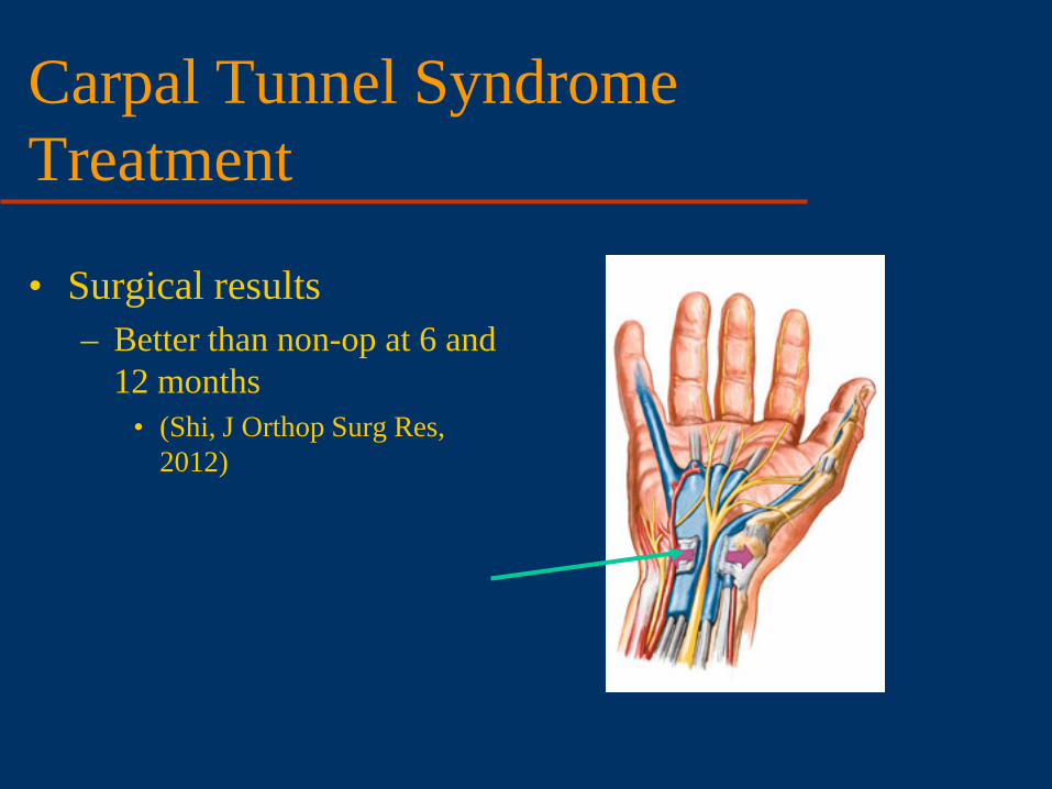
Carpal Tunnel Syndrome Median Nerve Compression • Carpal tunnel
Anatomy – Carpal bones – Transverse carpal
ligament • Contents
– Finger flexor tendons – Median nerve

Carpal Tunnel Syndrome Median Nerve Compression • Carpal tunnel
Anatomy – Carpal bones – Transverse carpal
ligament • Contents
– Finger flexor tendons – Median nerve

Carpal Tunnel Syndrome Median Nerve Compression • Carpal tunnel
Anatomy – Carpal bones – Transverse carpal
ligament • Contents
– Finger flexor tendons – Median nerve

Carpal Tunnel Syndrome Symptoms • Numbness in palmar
thumb, index, long and ½ of ring finger
• Awake at night with numbness
• Tingling with driving, typing, reading

Carpal Tunnel Syndrome Signs
• Thenar (base of thumb) atrophy – Late finding – hope to
avoid this! • Decreased sensation in
median distribution
Less muscle mass
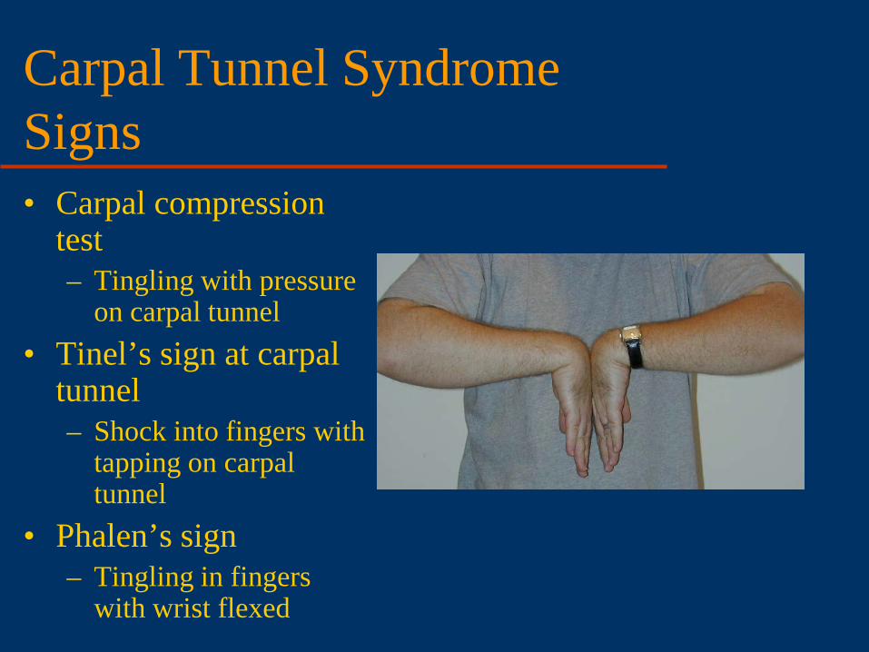
Carpal Tunnel Syndrome Signs • Carpal compression
test – Tingling with pressure
on carpal tunnel • Tinel’s sign at carpal
tunnel – Shock into fingers with
tapping on carpal tunnel
• Phalen’s sign – Tingling in fingers
with wrist flexed

Carpal Tunnel Syndrome Diagnostic Criteria • Graham et al J Hand 2006
– Statistically significant probability of being associated with consensus diagnosis by MD panel
1. Numbness and tingling in median nerve distribution 2. Nocturnal numbness 3. Weakness and/or atrophy of the thenar musculature 4. Tinel sign 5. Phalen’s test 6. Loss of 2 point discrimination
Carpal Tunnel Syndrome Diagnostic Criteria (??) • Graham et al J Hand 2006
– Statistically significant probability of being associated with consensus diagnosis by MD panel
1. Numbness and tingling in median nerve distribution 2. Nocturnal numbness 3. Weakness and/or atrophy of the thenar musculature 4. Tinel sign 5. Phalen’s test 6. Loss of 2 point discrimination

Carpal Tunnel Syndrome
• Prolonged nerve conduction velocity

Carpal Tunnel Syndrome Etiology • Idiopathic • Pregnancy • Diabetes • Hypothyroid • Deformity of carpal
canal – Mass – DJD – Prior fracture

Carpal Tunnel Syndrome Treatment • Volar wrist splint
– Increases space in tunnel – Wear at night
• Level II studies – More effective than
nothing at 3 months • Graham, J Hand 2009
– Short term relief • Cochrane 2012

Carpal Tunnel Syndrome Treatment • Oral steroids
– Improvement at 8 weeks – Not worth the risks
• NSAIDS – No evidence
• Vitamin B – No good study

Carpal Tunnel Syndrome Treatment • Cochrane 2012 – limited and very low
quality studies – Work/activity modification – Exercises – Yoga – Ultrasound – Magnets – Acupuncture – Laser/cold laser – Weight reduction – Smoking cessation – Cognitive behavioral therapy

Carpal Tunnel Syndrome Treatment • Steroid injection • Level II evidence
– Improvement at 1 mo vs placebo
– Improvement to 6 months vs splint
– 1/3 with continued improvement at 1 year
• (Berger, 2012)

Carpal Tunnel Syndrome Treatment
• Surgical Indications – Failure of non-operative
treatment – Positive Nerve Conduction
test (false negative rate 5-30%)
• Transection of ligament – Increases space in tunnel
Carpal Tunnel Syndrome Treatment
• Surgical results – Better than non-op at 6 and
12 months • (Shi, J Orthop Surg Res,
2012)
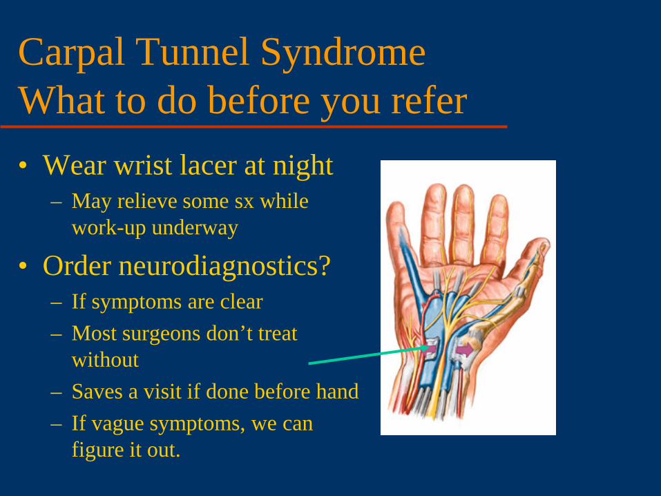
Carpal Tunnel Syndrome What to do before you refer • Wear wrist lacer at night
– May relieve some sx while work-up underway
• Order neurodiagnostics? – If symptoms are clear – Most surgeons don’t treat
without – Saves a visit if done before hand – If vague symptoms, we can
figure it out.

Cubital Tunnel Syndrome Ulnar Nerve Compression
• Cubital tunnel anatomy – “funny bone” – Nerve travels in groove
behind medial elbow

Cubital Tunnel Syndrome Symptoms
• Tingling in small and ulnar ½ of ring finger
• Awake at night with numbness
• Tingling with elbow bent

Cubital Tunnel Syndrome Signs
• Decreased sensation in ulnar distribution
• Tinel’s sign in cubital tunnel
• Hypothenar (base of small finger) and interosseous (between metacarpals) atrophy

Cubital Tunnel Syndrome Treatment
• Elbow extension at night – Soft elbow pad
backwards – Towel around arm – Splint – less well
tolerated • NSAIDS
Flip around so padding is in cubital fossa

Cubital Tunnel Syndrome Treatment
• Injection not used – too much danger to nerve
• Surgery – Release tunnel – Transpose nerve
anteriorly

Cubital Tunnel Syndrome Surgical Treatment
• Insuffucient evidence for best treatment
• Decompression vs transposition ??
• Endoscopic decompression – new procedure, limited good results

Cubital Tunnel Syndrome What to do before you refer
• Intermittant symptoms, no weakness or atrophy – Few months expectant
management – Night elbow pad – Activity modifications
• Refer if continued symptoms
• Constant symptoms, + weakness or atrpohy – Neurodiagnostic
studies – Referral

Thumb CMC arthritis
• Carpometacarpal joint – Very common
• 40% women • 25% men
– Young age (40s +)

Thumb CMC arthritis
• Carpometacarpal joint – X-ray osteoarthritis – Sclerosis – Spurs – Space narrowing – Cysts
** Stage 1 disease = normal x-ray
Thumb CMC arthritis
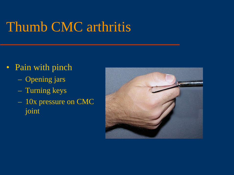
• Pain with pinch – Opening jars – Turning keys – 10x pressure on CMC
joint

Thumb CMC arthritis
• Treatment – Symptom relief – Splint – Immobilize MCP, CMC – IP and wrist free – Wear as needed
• OT order – Short opponens splint – Small joint preservation
exercises
Thumb CMC arthritis
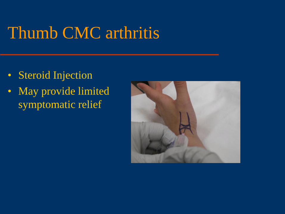
• Steroid Injection • May provide limited
symptomatic relief

Thumb CMC arthritis
• Surgery • Many different yet similar
options • Excise trapezium +/- interposition of tendon
+/- resuspension of MC
Thumb CMC arthritis
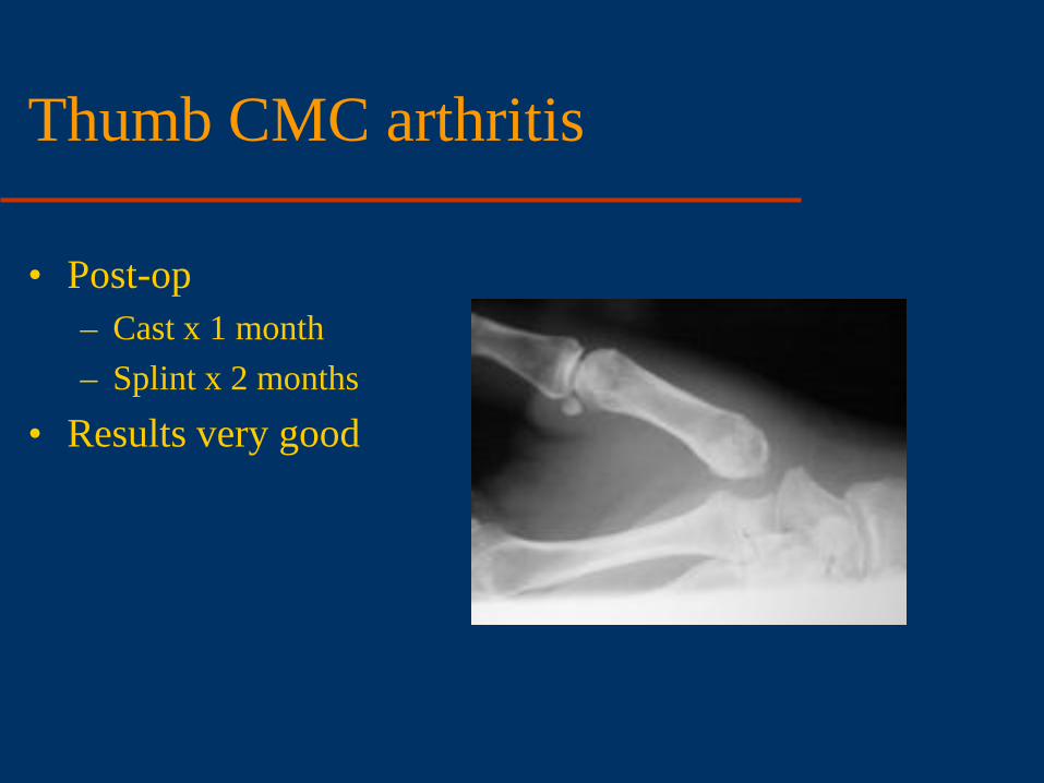
• Post-op – Cast x 1 month – Splint x 2 months
• Results very good

Thumb CMC arthritis What can you do before you refer?
• Clinical diagnosis • NSAIDS • OT splint and exercises • X-ray
– Normal does not preclude

Trigger Finger Anatomy
• Flexor tendons run through a sheath with pulleys
• Tendon gets stuck trying to go through A1 pulley
A1 pulley

Trigger Finger Symptoms
• Finger stuck in flexed position, need to use other hand to straighten
• Pain in palm over metacarpal head
• Palpable nodule on tendon
nodule

Trigger Finger Treatment
• Splint less effective – Wear continuously x 6
weeks – Approximately 25%

Trigger Finger Treatment
• Steroid injection – Review of level I and
II studies – 57% effective (Fleish,
JAAOS 2007)

Trigger Finger Treatment
• Surgery – Release A1 pulley – 1-2 cm incision – Local +/- sedation – No immobilzation

Trigger Finger What to do before you refer
• Nothing – just send them over!

de Quervain Tenosynovitis Anatomy • 1st extensor
compartment – Contains APL and
EPB – Radial border of
anatomic snuffbox • Held to radius through
tunnel

de Quervain Tenosynovitis Anatomy • 1st extensor
compartment – Contains APL and
EPB – Radial border of
anatomic snuffbox • Held to radius through
tunnel

de Quervain Tenosynovitis Symptoms and Signs • Pain in radial forearm
with thumb ROM • Tenderness over
tendons • Pain with Finkelstein’s
maneuver – Thumb grasped and
ulnar deviation

de Quervain Tenosynovitis Treatment • Majority is self-limited • Splint
– Forearm based thumb spica, IP free
– 20%-88% improvement – Better for symptom
improvement than disease modification
(Ilyas J Hand 2009)

de Quervain Tenosynovitis Treatment • NSAIDS
– No good studies when used alone
• Steroid Injection – Low level evidence 62%-
93% effective – Cochrane 2009 – better
than splint

de Quervain Tenosynovitis Treatment Surgery
– Release 1st dorsal compartment
– Uncommonly necessary – if sx persist >3-6 mos
– Release 1st dorsal compartment
– Local +/- sedation – 1-2 cm incision – +/- post-op splint

de Quervain Tenosynovitis What to do before you refer • Forearm based thumb spica
splint, IP free • Activity modifications
– Avoid cutting, texting, pinching, scissors

Ganglion Cysts Anatomy • Outpouching of joint
capsule with one-way valve
• Filled with synovial fluid
• Most commonly – dorsal scapho-lunate joint – Palmar radiocarpal joint

Ganglion Cysts Symptoms and Signs
• Volar retinacular cyst • Firm, well
circumscribed, fixed – Feels like a BB
• May be painful with grasping

Ganglion Cysts Treatment
• Aspiration – Usually recurs – 65-90%
• Rollins 2013
– Rarely for palmar ganglions – radial artery
– Rarely for retinacular cysts – NV bundle

Ganglion Cysts Treatment • Surgical excision
– Still high recurrence rate – “rule of thumb” 10% – Recent 10 year f/u 42%
• Lidder 2009
• Expectant management – 58% resolve over 5 years
• Dias 2007

Ganglion Cysts What to do before you refer • Clinical diagnosis **mass in any other area should be referred • Education
– no long term deleterious effect of no treatment

Mucous Cyst Anatomy
• Ganglion cyst from the DIP joint
• Due to arthritis • Looks like a blister

Mucous Cyst Symptoms and Signs
• Asymmetric mass on dorsal finger
• May ulcerate skin • Firm, fixed • Pain, decreased ROM
due to DJD • X-ray shows DJD

Mucous Cyst Treatment • Cyst excision indications
– Local symptoms from cyst – Recurrent ulceration
• Prevent osteomyelitis
• If arthritis symptomatic – NSAIDS – Steroid injection – DIP fusion

Mucous Cyst What to do before you refer
• Clinical diagnosis • Education • Refer if cyst related
symptoms with activities or ulcerating

Mallet Finger Anatomy
• Avulsion or fracture of terminal slip of extensor tendon

Mallet Finger Signs and Symptoms • Extension lag at DIP
joint • Usually history of
trauma • Tenderness over
dorsal DIP joint

Mallet Finger Signs and Symptoms • X-ray to look for
avulsion fracture • Surgical indication if
subluxated

Mallet Finger Treatment • Extension splint x 6-8
weeks • No removal without
supporting finger • Can begin up to 6
months post-injury

Mallet Finger What to do before you refer • X -ray
– Evaluate for subluxation
• DIP extension splint – PIP free – 24/7 use

Proximal Phalangeal Joint Injury Anatomy
• Collateral ligaments – Stabilize joint in
radial/ulnar direction • Often trivial injury • Long lasting
symptoms

Proximal Phalangeal Joint Injury Signs and Symptoms • Tenderness and
swelling at PIP • Decreased ROM • Pain with radial or
ulnar deviation • Instability
(uncommon) – Test in flexion and
extension

Proximal Phalangeal Joint Injury Signs and Symptoms • Radiographs
– True lateral – Evaluate alignment of
joint – Look for fracture

Proximal Phalangeal Joint Injury Treatment – Stable Injuries
• Buddy tape x 3 weeks and begin ROM
• Hand therapy indicated if motion not progressing rapidly
Proximal Phalangeal Joint Injury Treatment – Stable Injuries
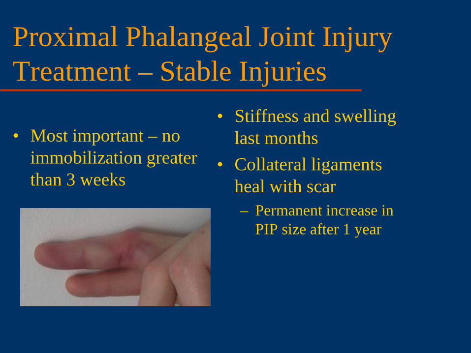
• Most important – no immobilization greater than 3 weeks
• Stiffness and swelling last months
• Collateral ligaments heal with scar – Permanent increase in
PIP size after 1 year

• Very difficult! • Unlikely to regain full
ROM • Immobilize and
prompt referral
Proximal Phalangeal Joint Injury Treatment – Unstable Injuries

• X-ray unstable – Refer!
• X-ray stable – Buddy tape – Hand therapy
• Both – education – Long course of sx – Painful to regain
motion
Proximal Phalangeal Joint Injury What to do before you refer

Thank you!!