Embed Size (px)
Citation preview

Common
Neuro-ophthalmologic Conditions
William L Hills, MD
Neuro-ophthalmology
Oregon Neurology Associates
Affiliate Assistant Professor of Neurology and Ophthalmology
Oregon Health & Science University

DisclosuresDisclosures
•• no disclosures no disclosures
Objectives Review presentation and differential diagnosis of more
common neuro-ophthalmic conditions
Optic neuritis
Diplopia
Anisocoria
Idiopathic intracranial hypertension

31yo woman
Right periobital pain/HA
Right eye pain with eye
movement
Photophobia
“no vision right eye”
Decreased over 1-2 days
Visual acuity
OD: Hand Motion
OS: 20/20
Color vision
OD: unable
OS: 14/14 ishihara plates
EOMI – painful OD
Pupils: right RAPD

Funduscopic Exam
Right eye Left eye

MRI brain wwo
T1 coronal wwo, with fat suppression

MRI brain wwo
T2 coronal, with fat suppression

Clinical presentation of Optic
Neuritis• Pain
– 92% report peri/retro-orbital
– Worse with eye movement
– Typically presents before vision loss
• Monocular vision loss
– 20/20 to NLP
– 1/3 experience photopsias
– Decreased color vision
• Red desaturation
– Visual field defects
• Central
• Peripheral
• Pupils• +Relative afferent pupillary
defect
Digre K, Corbett J. 2003. chapter2/2-39a.jpg
- 2/3 retro-bulbar-“Patient sees nothing”-“Doctor sees nothing”- 1/3 anterior

Causes of Optic neuritis
Most Common
Idiopathic demyelination
Other causes:
Viral/bacterial
Auto-immune
Inflammatory
Other

Optic Neuritis Treatment Trial
(1992)
• Optic Neuritis Treatment Trial (1992)
• Compared the speed and extent of visual recovery
Patients treated with:
Oral prednisone
increased risk of recurrent optic neuritis (35%)
IV methylprednisolone
Risk of recurrent optic neuritis 16%
Decreased risk of developing clinically definite MS at 2 yrs
(however, no difference at 3 yrs)
Placebo
Risk of recurrent optic neuritis 17% Beck et al 1992
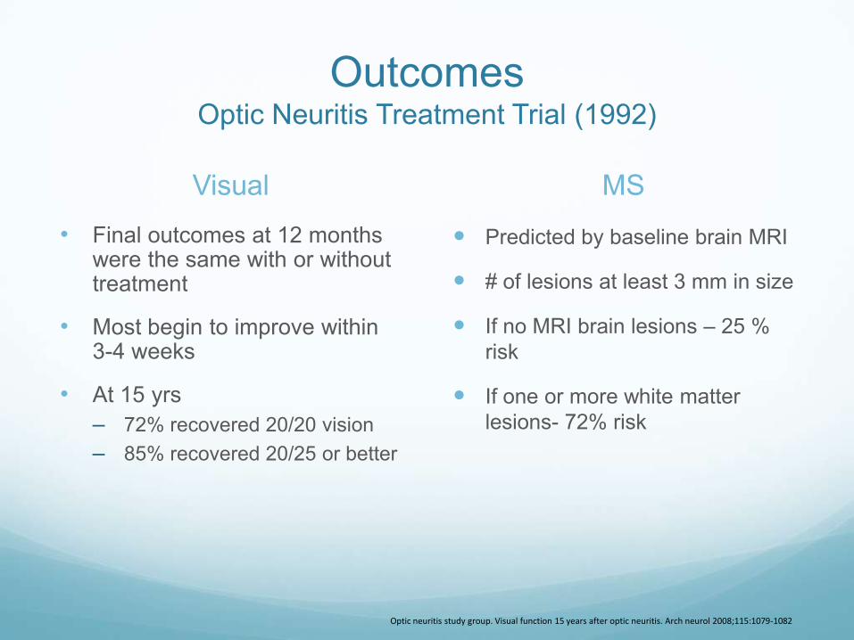
OutcomesOptic Neuritis Treatment Trial (1992)
Visual
• Final outcomes at 12 months were the same with or without treatment
• Most begin to improve within 3-4 weeks
• At 15 yrs
– 72% recovered 20/20 vision
– 85% recovered 20/25 or better
MS
Predicted by baseline brain MRI
# of lesions at least 3 mm in size
If no MRI brain lesions – 25 %
risk
If one or more white matter
lesions- 72% risk
Optic neuritis study group. Visual function 15 years after optic neuritis. Arch neurol 2008;115:1079-1082
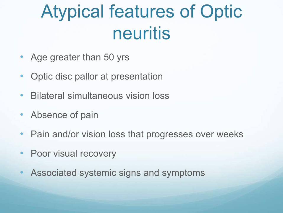
Atypical features of Optic
neuritis• Age greater than 50 yrs
• Optic disc pallor at presentation
• Bilateral simultaneous vision loss
• Absence of pain
• Pain and/or vision loss that progresses over weeks
• Poor visual recovery
• Associated systemic signs and symptoms
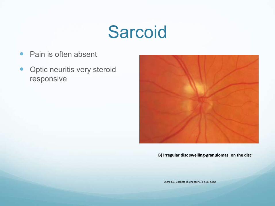
Sarcoid Pain is often absent
Optic neuritis very steroid
responsive
Digre KB, Corbett JJ. chapter3/3-56a-b.jpg
B) Irregular disc swelling-granulomason the disc

Neuromyelitis Opticaaka Devic’s disease
• NMO
– Optic neuritis + spinal cord disease
– Severe vision loss
– CSF:
• Neg OCB
• Elevated Prot and WBC
– Symptoms rarely improve
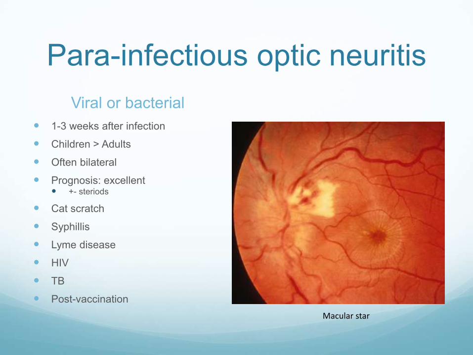
Para-infectious optic neuritis
Viral or bacterial
1-3 weeks after infection
Children > Adults
Often bilateral
Prognosis: excellent +- steriods
Cat scratch
Syphillis
Lyme disease
HIV
TB
Post-vaccination
Macular star

Diplopia
http://toughpigs.com/uploaded_images/martyfeldman-735756.jpg

Diplopia
Monocular
Ocular
Dry eye syndrome
Corneal
Refractive
Lenticular – cataract
Retinal
Psychiatric/functional
Binocular
Cranial nerve paresis
Brainstem lesion
Cerebeller lesion
Intra-orbital mass

Cranial Nerve III Palsy
Appearance of palsy
Extraocular movements
Eye goes down and out
Drooping eyelid
Dilated pupil

Cranial Nerve III Palsy
CN III palsy has several
potential etiologies
Intracranial process
Aneurysm!!!
Tumor
Brain herniation etc
Ischemia
Two Types
Pupil Involving
Pupil Sparing
BCSC Neuro-Ophthalmology 1999

Cranial Nerve IV Palsy
Appearance of CN IV palsy
One eye is higher than the other
Patients will often compensate with a head tilt
Patient will describe vertical double vision
May also note that 1 image is rotated

Cranial Nerve IV Palsy
CN IV palsy potential
etiologies
Trauma
Tumor
Function of CN IV
Incyclotorsion
Infraducts eye

Cranial Nerve VI Palsy Function of CN VI
Abduction (moves eye out)
Eyes cross
Effected eye can not move out
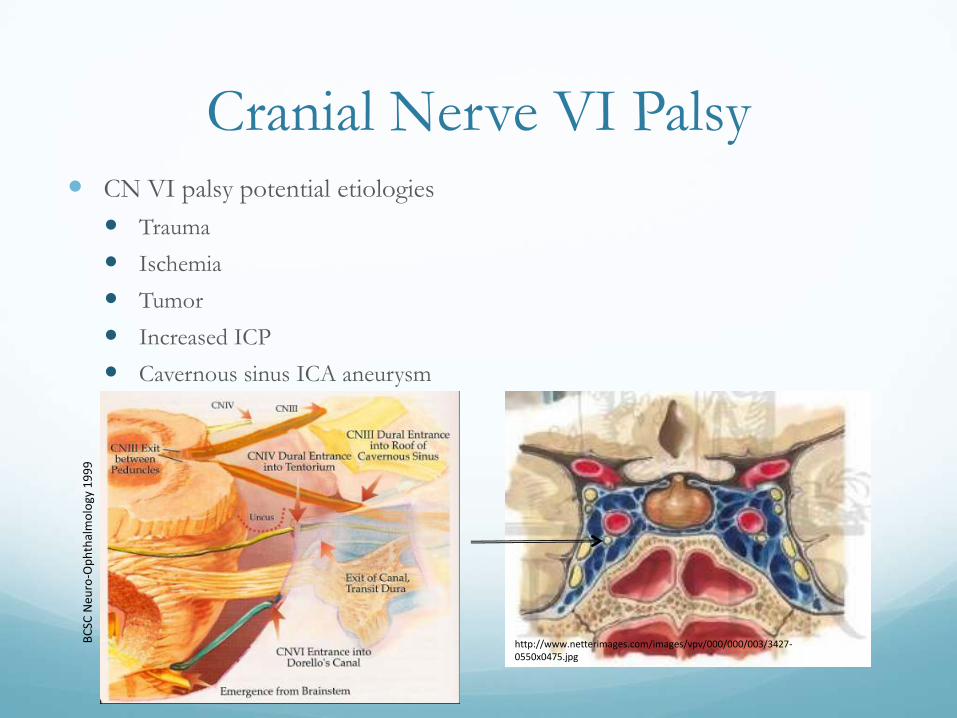
Cranial Nerve VI Palsy CN VI palsy potential etiologies
Trauma
Ischemia
Tumor
Increased ICP
Cavernous sinus ICA aneurysm
BC
SC N
euro
-Op
hth
alm
olo
gy 1
99
9
http://www.netterimages.com/images/vpv/000/000/003/3427-0550x0475.jpg

Cranial Nerve Palsy
Testing/Treatment
CT/CTA or MRI/A if suspicion for Intracranial process
Headache
Other neurologic symptoms
No microvascular disease
History of recent Trauma
If patient with microvascular disease (DM, HTN, Cholesterol, smoking)
No imagining required
Observation for 6-8 weeks

Anisocoria
http://www.maybebabyblog.com/images/2009/10/davidbowiegb0.jpg

Anisocoria greater in:
Darkness
Physiologic (simple)
anisocoria
Horner’s syndrome
Bright light
Adie’s (tonic) pupil
Oculomotor (third) nerve
palsy
Pharmacologic mydriasis
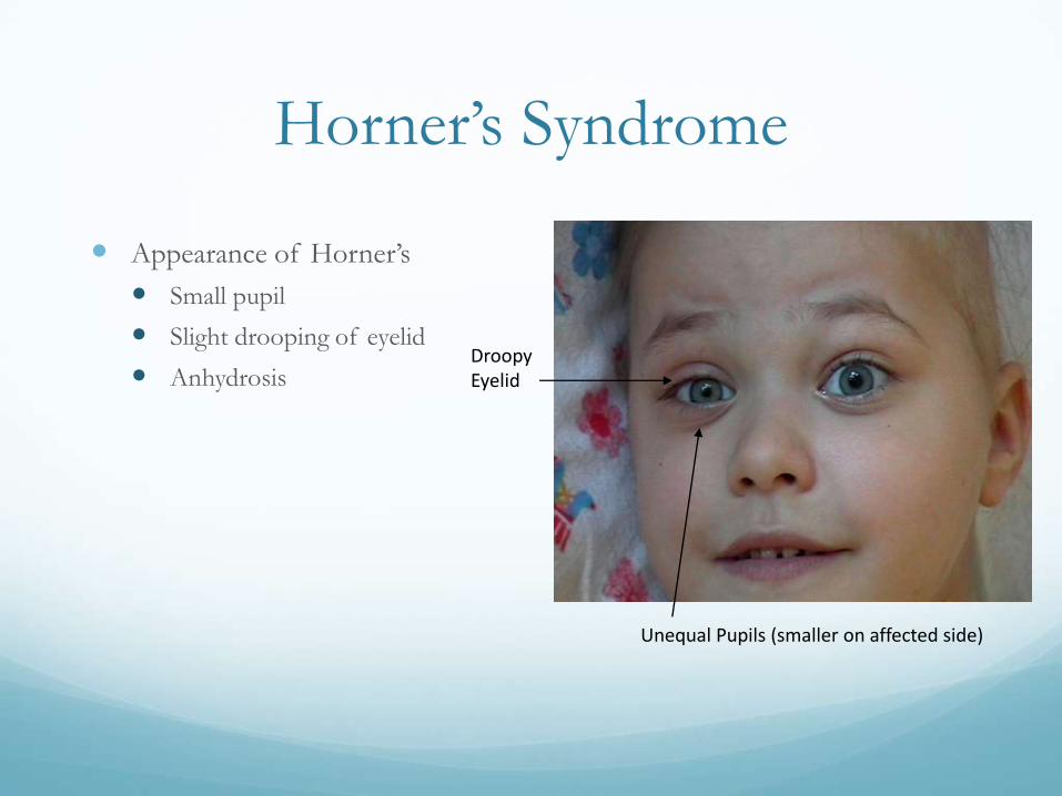
Horner’s Syndrome
Appearance of Horner’s
Small pupil
Slight drooping of eyelid
AnhydrosisDroopy Eyelid
Unequal Pupils (smaller on affected side)

Horner’s Syndrome
Sympathetic Pathway
Etiology - variable Lung tumor (Pancoast
tumor)
Carotid dissection
Neck trauma
Cervical cord lesion
Lateral medullary syndrome (Wallenberg)
Viral/idiopathic

Adie’s (tonic) pupil
• Acute denervation of the postganglionic parasympathetic fibers
• Initially dilated
• Sluggish, segemental pupillary responses
• Better response to near effort

Causes of Adie’s pupil Idiopathic
Viral ganglionitis
migrainous vasospasm
Trauma
Ocular surgery (e.g. scleral buckle)
Tumors

Bilateral simultaneous tonic
pupils
• systemic/autonoimic peripheral neuropathy
• diabetes mellitus
• amyloidsosis
• syphilis
• paraneoplastic syndromes
• Sjogren’s syndrome
• polyarteritis nodosa
IDIOPATHIC INTRACRANIAL
HYPERTENSION
Idiopathic intracranial hypertension (IIH)
Benign intracranial hypertension
Pseudotumor cerebri
Intracranial hypertension secondary to…

Clinical manifestations Headache
Transient visual obscurations
Pulse synchronous tinnitus
Diplopia
Visual loss
Papilledema
6th nerve palsy
Visual field defects

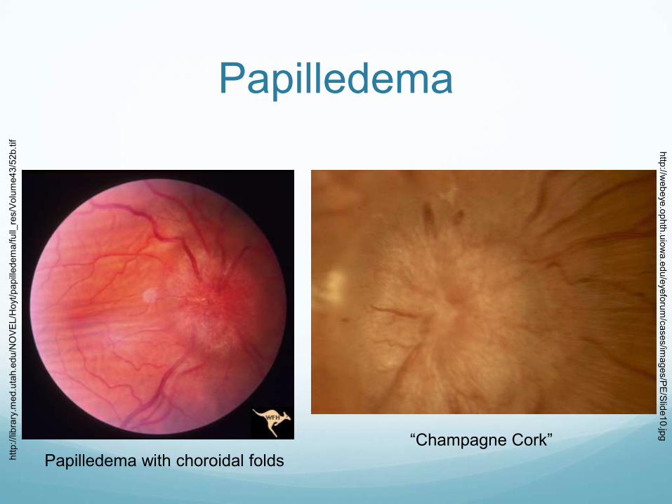
Papilledema
http://webeye.ophth.uiowa.edu/eyeforum/cases/case30/case30-Figure1.jpg

Papilledema
Normal Disc
Poorly Defined Borders
Papilledema
http://lib
rary
.med.u
tah.e
du/N
OV
EL/H
oyt/papill
edem
a/f
ull_
res/V
olu
me43/5
2b.tif
“Champagne Cork”
http
://webeye.o
phth
.uio
wa.e
du/e
yefo
rum
/cases/im
ages/P
E/S
lide10.jp
g
Papilledema with choroidal folds
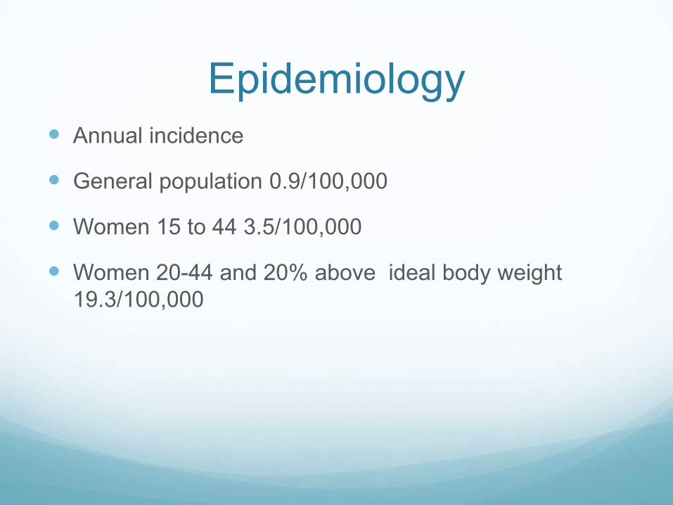
Epidemiology Annual incidence
General population 0.9/100,000
Women 15 to 44 3.5/100,000
Women 20-44 and 20% above ideal body weight
19.3/100,000

Epidemiology Before puberty boys = girls
After puberty women affected 9 times as often as men
Rarely develops in patients over 45

Headache Almost all patients with IIH
Daily, retro-bulbar, worse with eye movement
Neck and back pain are prominent features
Throbbing, nausea, vomiting, photophobia
Often worse supine

Transient visual obscurations Brief episodes of monocular or binocular vision loss
Partial or complete
Likely due to disc edema leading to ischemia of the
optic nerve head

Pulse Synchronous Tinnitus Pulsatile tinnitus 60%
Unilateral or bilateral
Abolished with LP or jugular venous compression
Transmission of intensified vascular pulsations via CSF

Diplopia Unilateral or bilateral sixth
nerve palsy
Secondary to increased ICP
Binocular horizontal diplopia
Resolves when ICP lowered

Visual Loss Blurred vision
Temporal dark spot
Tunnel vision
Profound or complete blindness
Tempo variable: as soon as days

Other symptoms: Paresthesias
Neck stiffness
Arthralgia shoulders, wrists, knees
Ataxia
Facial palsy- rare
Radicular pain
Depression
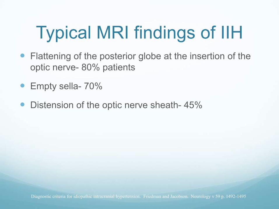
Typical MRI findings of IIH Flattening of the posterior globe at the insertion of the
optic nerve- 80% patients
Empty sella- 70%
Distension of the optic nerve sheath- 45%
Diagnostic criteria for idiopathic intracranial hypertension. Friedman and Jacobson. Neurology v 59 p. 1492-1495

Idiopathic intracranial hypertension
Hassan et al. Teaching NeuroImages:
Idiopathic intracranial hypertension.
Neurology. v 74 (7) Feb 2010, p e 24Emma Burbank, MD

Lumbar Puncture Lateral decubitus position with legs relaxed
18- to 20- gauge spinal needle
Document elevated CSF pressure
Opening pressure > 250mm H20
201 – 249 mm H20 are nondiagnostic
Repeat LP may be necessary if initial OP nondiagnostic
Rarely need 24 hour transducer monitoring through lumbar drain to diagnose
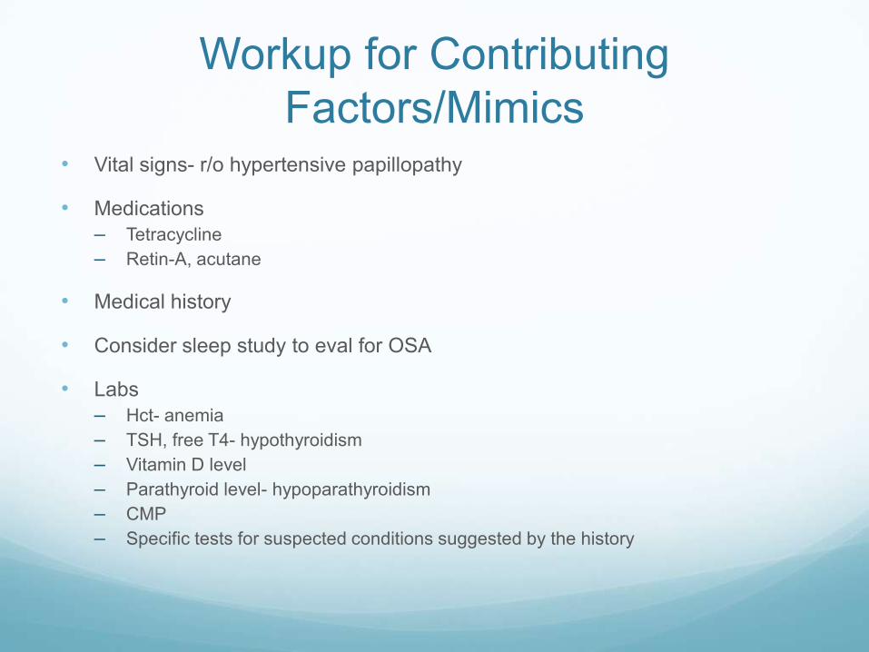
Workup for Contributing
Factors/Mimics• Vital signs- r/o hypertensive papillopathy
• Medications
– Tetracycline
– Retin-A, acutane
• Medical history
• Consider sleep study to eval for OSA
• Labs
– Hct- anemia
– TSH, free T4- hypothyroidism
– Vitamin D level
– Parathyroid level- hypoparathyroidism
– CMP
– Specific tests for suspected conditions suggested by the history
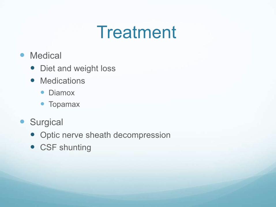
Treatment Medical
Diet and weight loss
Medications
Diamox
Topamax
Surgical
Optic nerve sheath decompression
CSF shunting