Embed Size (px)
Citation preview
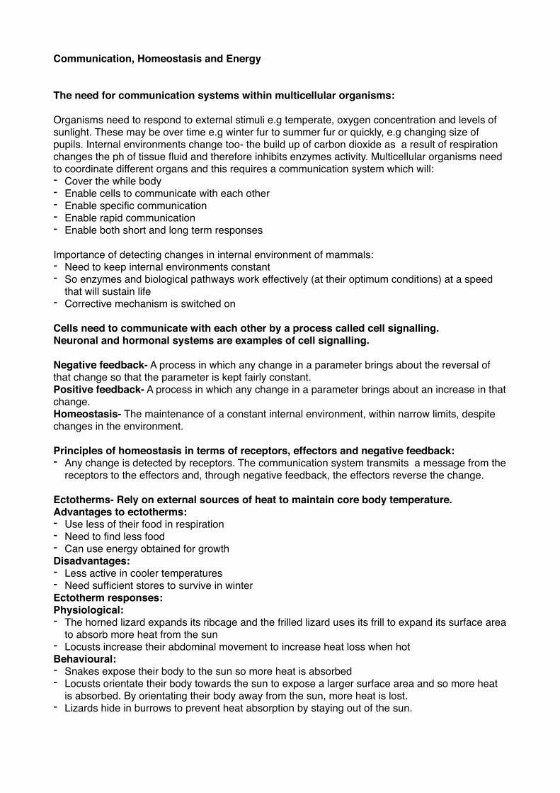
Communication, Homeostasis and Energy
The need for communication systems within multicellular organisms:
Organisms need to respond to external stimuli e.g temperate, oxygen concentration and levels of sunlight. These may be over time e.g winter fur to summer fur or quickly, e.g changing size of pupils. Internal environments change too- the build up of carbon dioxide as a result of respiration changes the ph of tissue fluid and therefore inhibits enzymes activity. Multicellular organisms need to coordinate different organs and this requires a communication system which will:- Cover the while body - Enable cells to communicate with each other - Enable specific communication- Enable rapid communication - Enable both short and long term responses
Importance of detecting changes in internal environment of mammals:- Need to keep internal environments constant - So enzymes and biological pathways work effectively (at their optimum conditions) at a speed
that will sustain life- Corrective mechanism is switched on
Cells need to communicate with each other by a process called cell signalling. Neuronal and hormonal systems are examples of cell signalling.
Negative feedback- A process in which any change in a parameter brings about the reversal of that change so that the parameter is kept fairly constant.Positive feedback- A process in which any change in a parameter brings about an increase in that change.Homeostasis- The maintenance of a constant internal environment, within narrow limits, despite changes in the environment.
Principles of homeostasis in terms of receptors, effectors and negative feedback:- Any change is detected by receptors. The communication system transmits a message from the
receptors to the effectors and, through negative feedback, the effectors reverse the change.
Ectotherms- Rely on external sources of heat to maintain core body temperature. Advantages to ectotherms:- Use less of their food in respiration - Need to find less food - Can use energy obtained for growth Disadvantages:- Less active in cooler temperatures - Need sufficient stores to survive in winter Ectotherm responses:Physiological:- The horned lizard expands its ribcage and the frilled lizard uses its frill to expand its surface area
to absorb more heat from the sun - Locusts increase their abdominal movement to increase heat loss when hot Behavioural:- Snakes expose their body to the sun so more heat is absorbed - Locusts orientate their body towards the sun to expose a larger surface area and so more heat
is absorbed. By orientating their body away from the sun, more heat is lost. - Lizards hide in burrows to prevent heat absorption by staying out of the sun.
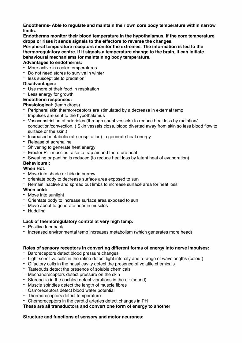
Endotherms- Able to regulate and maintain their own core body temperature within narrow limits.Endotherms monitor their blood temperature in the hypothalamus. If the core temperature drops or rises it sends signals to the effectors to reverse the changes.Peripheral temperature receptors monitor the extremes. The information is fed to the thermoregulatory centre. If it signals a temperature change to the brain, it can initiate behavioural mechanisms for maintaining body temperature. Advantages to endotherms:- More active in cooler temperatures - Do not need stores to survive in winter - less susceptible to predation Disadvantages: - Use more of their food in respiration - Less energy for growth Endotherm responses:Physiological: (temp drops)- Peripheral skin thermoreceptors are stimulated by a decrease in external temp- Impulses are sent to the hypothalamus - Vasoconstriction of arterioles (through shunt vessels) to reduce heat loss by radiation/
conduction/convection. ( Skin vessels close, blood diverted away from skin so less blood flow to surface or the skin.)
- Increased metabolic rate (respiration) to generate heat energy - Release of adrenaline- Shivering to generate heat energy - Erector Pilli muscles raise to trap air and therefore heat - Sweating or panting is reduced (to reduce heat loss by latent heat of evaporation) Behavioural: When Hot: - Move into shade or hide in burrow - orientate body to decrease surface area exposed to sun - Remain inactive and spread out limbs to increase surface area for heat lossWhen cold:- Move into sunlight - Orientate body to increase surface area exposed to sun - Move about to generate hear in muscles - Huddling
Lack of thermoregulatory control at very high temp:- Positive feedback - Increased environmental temp increases metabolism (which generates more head)
Roles of sensory receptors in converting different forms of energy into nerve impulses:- Baroreceptors detect blood pressure changes- Light sensitive cells in the retina detect light intercity and a range of wavelengths (colour)- Olfactory cells in the nasal cavity detect the presence of volatile chemicals - Tastebuds detect the presence of soluble chemicals - Mechanoreceptors detect pressure on the skin- Stereocilia in the cochlea detect vibrations in the air (sound)- Muscle spindles detect the length of muscle fibres - Osmoreceptors detect blood water potential - Thermoreceptors detect temperature - Chemoreceptors in the carotid arteries detect changes in PHThese are all transductors and convert one form of energy to another
Structure and functions of sensory and motor neurones:
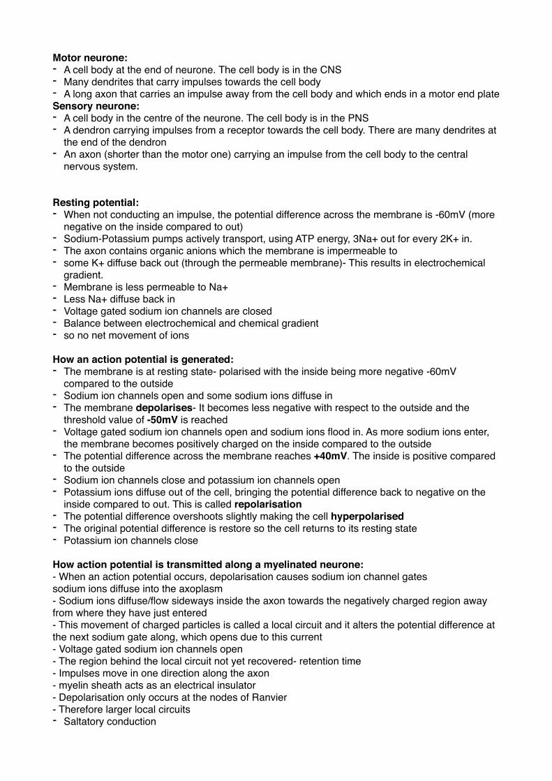
Motor neurone:- A cell body at the end of neurone. The cell body is in the CNS- Many dendrites that carry impulses towards the cell body - A long axon that carries an impulse away from the cell body and which ends in a motor end plate Sensory neurone:- A cell body in the centre of the neurone. The cell body is in the PNS- A dendron carrying impulses from a receptor towards the cell body. There are many dendrites at
the end of the dendron- An axon (shorter than the motor one) carrying an impulse from the cell body to the central
nervous system.
Resting potential:- When not conducting an impulse, the potential difference across the membrane is -60mV (more
negative on the inside compared to out)- Sodium-Potassium pumps actively transport, using ATP energy, 3Na+ out for every 2K+ in.- The axon contains organic anions which the membrane is impermeable to - some K+ diffuse back out (through the permeable membrane)- This results in electrochemical
gradient.- Membrane is less permeable to Na+- Less Na+ diffuse back in - Voltage gated sodium ion channels are closed- Balance between electrochemical and chemical gradient - so no net movement of ions
How an action potential is generated:- The membrane is at resting state- polarised with the inside being more negative -60mV
compared to the outside- Sodium ion channels open and some sodium ions diffuse in - The membrane depolarises- It becomes less negative with respect to the outside and the
threshold value of -50mV is reached- Voltage gated sodium ion channels open and sodium ions flood in. As more sodium ions enter,
the membrane becomes positively charged on the inside compared to the outside- The potential difference across the membrane reaches +40mV. The inside is positive compared
to the outside- Sodium ion channels close and potassium ion channels open- Potassium ions diffuse out of the cell, bringing the potential difference back to negative on the
inside compared to out. This is called repolarisation- The potential difference overshoots slightly making the cell hyperpolarised- The original potential difference is restore so the cell returns to its resting state- Potassium ion channels close
How action potential is transmitted along a myelinated neurone: - When an action potential occurs, depolarisation causes sodium ion channel gatessodium ions diffuse into the axoplasm - Sodium ions diffuse/flow sideways inside the axon towards the negatively charged region away from where they have just entered - This movement of charged particles is called a local circuit and it alters the potential difference at the next sodium gate along, which opens due to this current - Voltage gated sodium ion channels open- The region behind the local circuit not yet recovered- retention time- Impulses move in one direction along the axon - myelin sheath acts as an electrical insulator - Depolarisation only occurs at the nodes of Ranvier - Therefore larger local circuits - Saltatory conduction
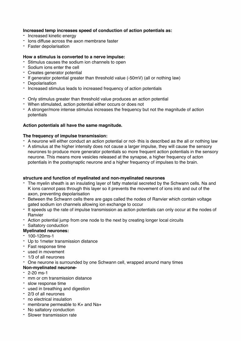
Increased temp increases speed of conduction of action potentials as:- Increased kinetic energy - Ions diffuse across the axon membrane faster - Faster depolarisation
How a stimulus is converted to a nerve impulse:- Stimulus causes the sodium ion channels to open - Sodium ions enter the cell- Creates generator potential - If generator potential greater than threshold value (-50mV) (all or nothing law)- Depolarisation - Increased stimulus leads to increased frequency of action potentials
- Only stimulus greater than threshold value produces an action potential - When stimulated, action potential either occurs or does not- A stronger/more intense stimulus increases the frequency but not the magnitude of action
potentials
Action potentials all have the same magnitude.
The frequency of impulse transmission:- A neurone will either conduct an action potential or not- this is described as the all or nothing law - A stimulus at the higher intensity does not cause a larger impulse, they will cause the sensory
neurones to produce more generator potentials so more frequent action potentials in the sensory neurone. This means more vesicles released at the synapse, a higher frequency of acton potentials in the postsynaptic neurone and a higher frequency of impulses to the brain.
structure and function of myelinated and non-myelinated neurones- The myelin sheath is an insulating layer of fatty material secreted by the Schwann cells. Na and
K ions cannot pass through this layer so it prevents the movement of ions into and out of the axon, preventing depolarisation
- Between the Schwann cells there are gaps called the nodes of Ranvier which contain voltage gated sodium ion channels allowing ion exchange to occur
- It speeds up the rate of impulse transmission as action potentials can only occur at the nodes of Ranvier
- Action potential jump from one node to the next by creating longer local circuits- Saltatory conductionMyelinated neurones:- 100-120ms-1- Up to 1meter transmission distance - Fast response time- used in movement- 1/3 of all neurones- One neurone is surrounded by one Schwann cell, wrapped around many times Non-myelinated neurone-- 2-20 ms-1- mm or cm transmission distance - slow response time - used in breathing and digestion - 2/3 of all neurones- no electrical insulation - membrane permeable to K+ and Na+ - No saltatory conduction- Slower transmission rate
So myelinated fibres transmit impulses quicker than non-myelinated.
Thicker axons transmit impulses faster than thin ones as:- Less leakage of ions - Less resistance - Greater surface area to volume ratio
Refractory period: - following an action potential, the membrane needs to restore resting potential. - Voltage gated sodium ion channels are closed- During which another impulse cannot be generated - This ensures that impulses are separated - Determines maximum frequency of impulse transmission - Impulse passes along the axon in one direction only
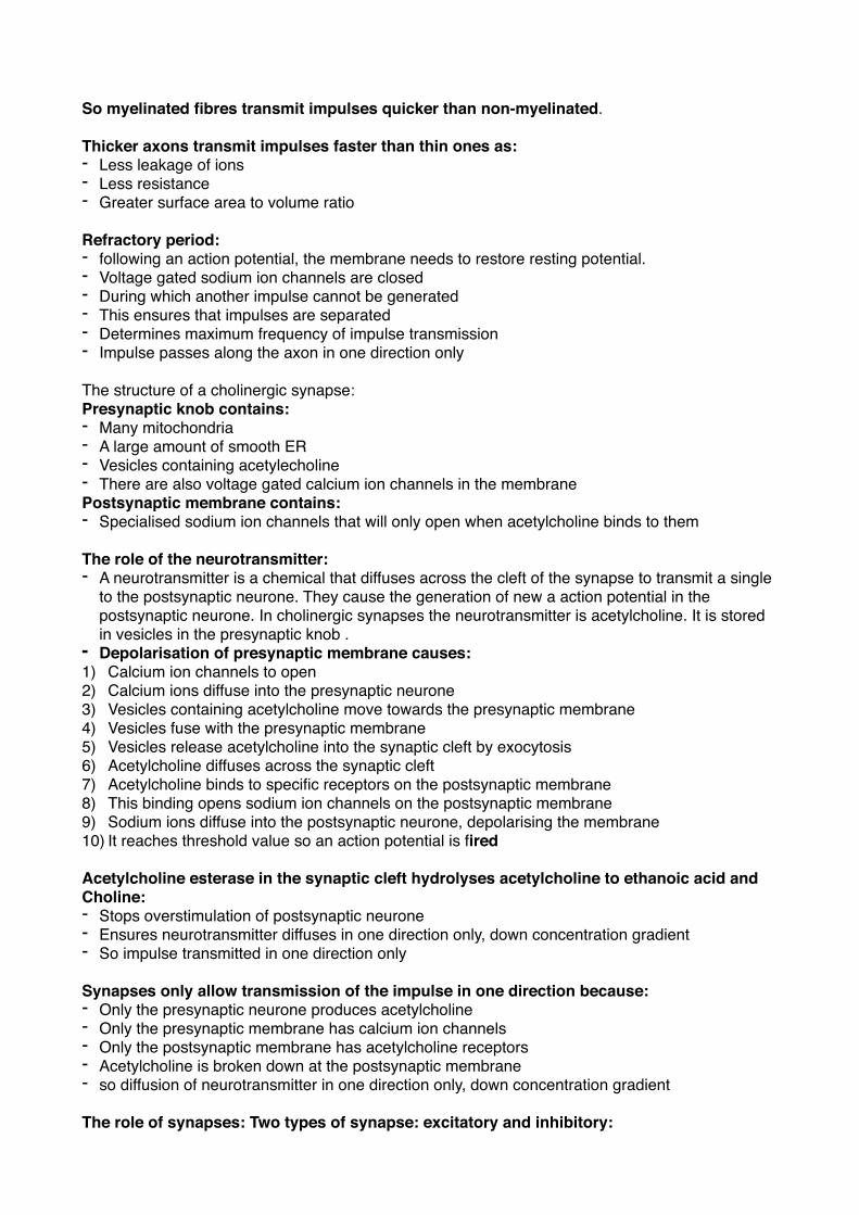
The structure of a cholinergic synapse:Presynaptic knob contains:- Many mitochondria - A large amount of smooth ER- Vesicles containing acetylecholine- There are also voltage gated calcium ion channels in the membrane Postsynaptic membrane contains:- Specialised sodium ion channels that will only open when acetylcholine binds to them
The role of the neurotransmitter:- A neurotransmitter is a chemical that diffuses across the cleft of the synapse to transmit a single
to the postsynaptic neurone. They cause the generation of new a action potential in the postsynaptic neurone. In cholinergic synapses the neurotransmitter is acetylcholine. It is stored in vesicles in the presynaptic knob .
- Depolarisation of presynaptic membrane causes:1) Calcium ion channels to open 2) Calcium ions diffuse into the presynaptic neurone 3) Vesicles containing acetylcholine move towards the presynaptic membrane 4) Vesicles fuse with the presynaptic membrane 5) Vesicles release acetylcholine into the synaptic cleft by exocytosis 6) Acetylcholine diffuses across the synaptic cleft7) Acetylcholine binds to specific receptors on the postsynaptic membrane 8) This binding opens sodium ion channels on the postsynaptic membrane 9) Sodium ions diffuse into the postsynaptic neurone, depolarising the membrane 10) It reaches threshold value so an action potential is fired
Acetylcholine esterase in the synaptic cleft hydrolyses acetylcholine to ethanoic acid and Choline:- Stops overstimulation of postsynaptic neurone - Ensures neurotransmitter diffuses in one direction only, down concentration gradient - So impulse transmitted in one direction only
Synapses only allow transmission of the impulse in one direction because:- Only the presynaptic neurone produces acetylcholine - Only the presynaptic membrane has calcium ion channels - Only the postsynaptic membrane has acetylcholine receptors - Acetylcholine is broken down at the postsynaptic membrane - so diffusion of neurotransmitter in one direction only, down concentration gradient
The role of synapses: Two types of synapse: excitatory and inhibitory:
- Primarily the role of the synapse is to connect two neurones together to pass an impulse from one to the other, but they have other functions:
- Integration:1) Several presynaptic neurones ay converge together to allow signals from different parts of the
nervous system to create the same response 2) One presynaptic neurone may diverge to several postsynaptic neurones to allow one signal to
be transmitted to several parts of the nervous system. one may elicit a response and one may inform the brain
- They ensure that impulses are transferred in only one direction- only the presynaptic knob contains acetylcholine in vesicles
- They can filter out unwanted low-level signals possibly caused by low level stimuli. Several vesicles of acetylcholine must be released for an action potential to be created in the post synaptic neurone
- Low-level signals can be amplified by summation- When several small potential charges combine to produce one larger charge in the potential membrane. If a low level stimuli is persistent it can generate several successive action potentials in the presynaptic neurone. The release of many vesicles of acetylcholine in a short space of time will enable the postsynaptic generator potentials to combine together to produce an action potential.
- Two types of Summation: 1) Spatial- Combination of action potentials all at once 2) Temporal- Combination of action potentials after one another - Acclimatisation- after repeated stimulation a synapse my run out of vesicles containing the
neurotransmitter substance. The synapse is said to be fatigues. This helps avoid overstimulation of an effector, which could damage it.
- The creation of specific pathways in the nervous system is thought to be the basis of conscious thought and memory.
Competitive inhibitors (with complementary shape for binding site) bind to Acetylcholine receptors on the postsynaptic membrane without causing depolarisation:- They reduce the effects of acetylcholine Non-competitive inhibitors do not bind to receptor site, they compete by binding to something else e.g Acetylcholine esterase (stop breakdown of acetylcholine)
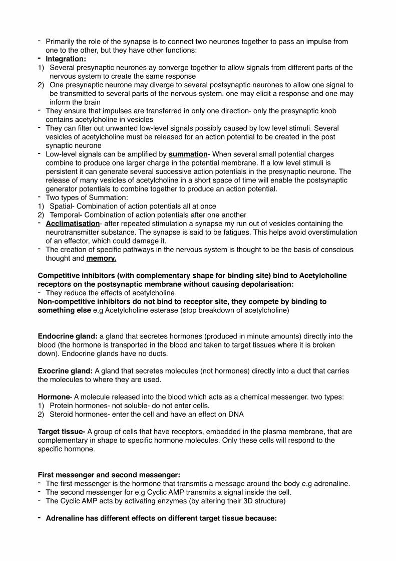
Endocrine gland: a gland that secretes hormones (produced in minute amounts) directly into the blood (the hormone is transported in the blood and taken to target tissues where it is broken down). Endocrine glands have no ducts.
Exocrine gland: A gland that secretes molecules (not hormones) directly into a duct that carries the molecules to where they are used.
Hormone- A molecule released into the blood which acts as a chemical messenger. two types:1) Protein hormones- not soluble- do not enter cells. 2) Steroid hormones- enter the cell and have an effect on DNA
Target tissue- A group of cells that have receptors, embedded in the plasma membrane, that are complementary in shape to specific hormone molecules. Only these cells will respond to the specific hormone.
First messenger and second messenger:- The first messenger is the hormone that transmits a message around the body e.g adrenaline. - The second messenger for e.g Cyclic AMP transmits a signal inside the cell.- The Cyclic AMP acts by activating enzymes (by altering their 3D structure)
- Adrenaline has different effects on different target tissue because:
1) Different tissues have different types of adrenaline receptors causing cAMP concentrations to increase or decrease.
2) cAMP activates different enzymes in different target cells3) The second messenger may be different, causing different effects.
Functions of the adrenal glands: The adrenal glands have two distinct regions- the cortex region and the medulla region.The adrenal medulla releases adrenaline which:- Relaxes smooth muscle in the bronchioles - Increases the stroke volume of the heart- Increases heart rate - Causes general vasoconstriction- raising blood pressure - Causes increased activity of glycogen phosphorylase- Stimulates conversion of glycogen to glucose (promotes glycogenolysis) - Dilates pupils- Increases mental awareness - Inhibits the action of the gut (to save energy)- Decreases peristalsis - Causes body hair to erect The cortex medulla releases corticosteroid hormones which are made from cholesterol:- Mineralocorticoids (e.g Aldosterone cortisol) help control the concentrations of sodium and
potassium in the blood- Glucocorticoids help control the metabolism of carbohydrates and proteins in the liver
How adrenaline causes effect in target cells:- Adrenaline receptor site has shape complementary to adrenaline (1st messenger)- Adenyl cyclase enzyme is inactive - Adrenaline binds to receptor site. Adrenaline-receptor complex forms - This activates Adenyl Cyclase enzyme - The now active Adenyl cyclase converts ATP to cAMP (2nd messenger)- cAMP can then activate enzymes inside the cell (by altering their 3D structure)
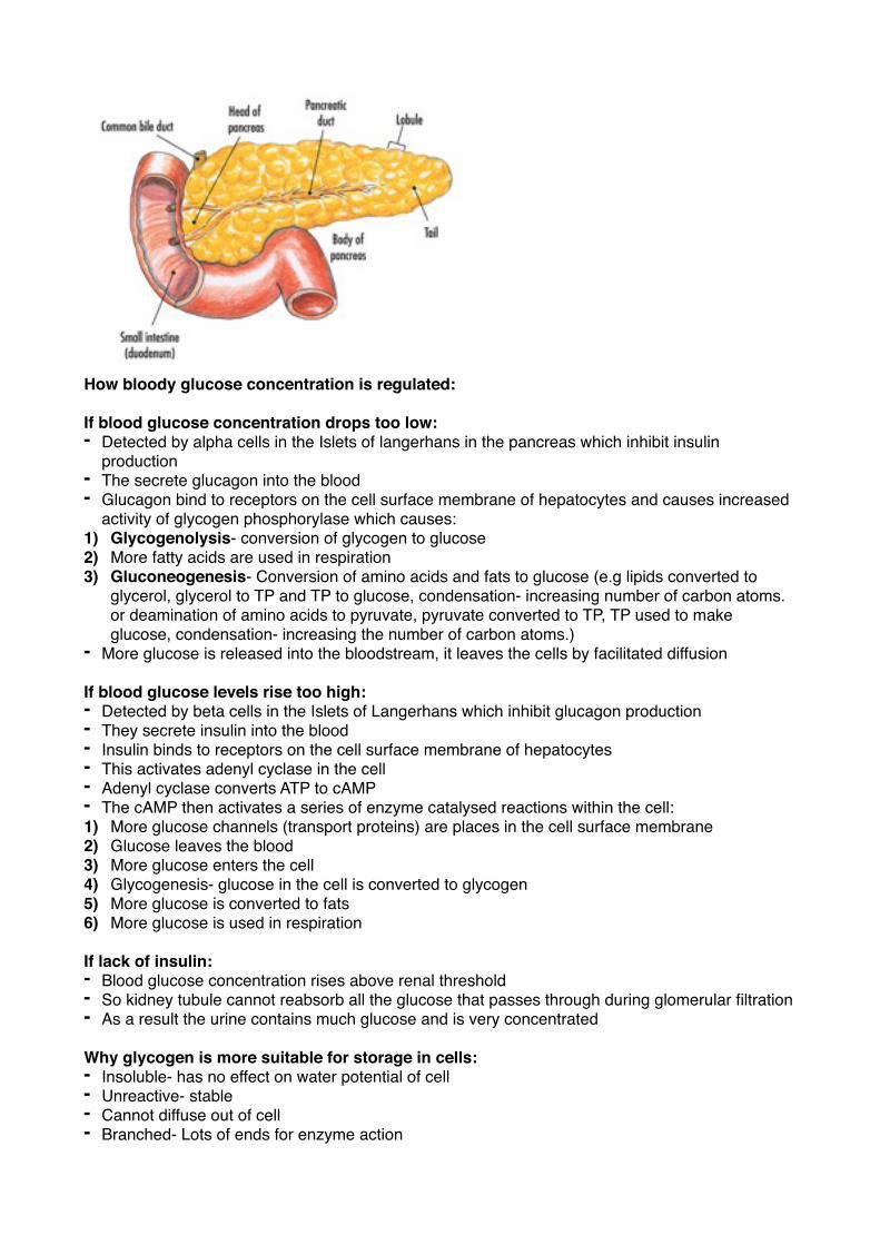
Histology of the pancreas: The exocrine cells of the pancreas secrete digestive enzymes (e.g trypsinogen which forms trypsin) directly into the pancreatic duct, which transports them to the small intestine. (Pancreatic secretions into the duct are triggered by nerve stimulation.) These cells make up the majority of the pancreas.The endocrine cells of the pancreas are found in the Islets of Langerhans and consist of alpha and beta cells. The alpha cells manufacture and secrete glucagon whereas the beta cells manufacture and secrete insulin. They are involved in the regulation of blood glucose levels.
�How bloody glucose concentration is regulated:
If blood glucose concentration drops too low:- Detected by alpha cells in the Islets of langerhans in the pancreas which inhibit insulin
production- The secrete glucagon into the blood - Glucagon bind to receptors on the cell surface membrane of hepatocytes and causes increased
activity of glycogen phosphorylase which causes:1) Glycogenolysis- conversion of glycogen to glucose2) More fatty acids are used in respiration 3) Gluconeogenesis- Conversion of amino acids and fats to glucose (e.g lipids converted to
glycerol, glycerol to TP and TP to glucose, condensation- increasing number of carbon atoms. or deamination of amino acids to pyruvate, pyruvate converted to TP, TP used to make glucose, condensation- increasing the number of carbon atoms.)
- More glucose is released into the bloodstream, it leaves the cells by facilitated diffusion
If blood glucose levels rise too high:- Detected by beta cells in the Islets of Langerhans which inhibit glucagon production - They secrete insulin into the blood- Insulin binds to receptors on the cell surface membrane of hepatocytes - This activates adenyl cyclase in the cell- Adenyl cyclase converts ATP to cAMP - The cAMP then activates a series of enzyme catalysed reactions within the cell:1) More glucose channels (transport proteins) are places in the cell surface membrane 2) Glucose leaves the blood3) More glucose enters the cell 4) Glycogenesis- glucose in the cell is converted to glycogen 5) More glucose is converted to fats6) More glucose is used in respiration
If lack of insulin:- Blood glucose concentration rises above renal threshold - So kidney tubule cannot reabsorb all the glucose that passes through during glomerular filtration- As a result the urine contains much glucose and is very concentrated
Why glycogen is more suitable for storage in cells:- Insoluble- has no effect on water potential of cell- Unreactive- stable - Cannot diffuse out of cell- Branched- Lots of ends for enzyme action
- Easy to hydrolyse to glucose
Glycogen is a carbohydrate made in the liver which can undergo glycogenolysis Glucagon is a hormone made in the pancreas which stimulates glycogenolysis
How insulin secretion is controlled:- The cell membranes of the beta cells contain calcium and potassium ion channels - The potassium ion channels are normally open and the calcium ion channels are normally shut- Potassium ions diffuse out of the cell, making the inside more negative (compared to the
outside)- When glucose concentration outside of the cells is high, more glucose molecules diffuse into the
cell - The glucose is quickly metabolised to ATP (glucose enters the glycolytic pathway and ATP is
produced)- The extra ATP causes the potassium ion channels to close - The potassium ions can no longer diffuse out, so the cell becomes more positive on the inside- This change in potential difference opens calcium ion channels- Calcium ions enter the cell and cause the secretion of insulin by making the vesicles containing
insulin move towards the cell surface membrane and fuse with it, releasing insulin by exocytosis.
Normal blood glucose concentration is 90mg 100cm3
Type 1 diabetes:- Juvenile onset - The body is unable to produce enough insulin/ does not secrete insulin/ Produces insufficient
insulin - The insulin producing cells (beta cells) are destroyed by the body’s own immune system - This is an auto-immune disease (immune system mistakes beta cells as foreign) - It can be genetic - It can be triggered by a virus Type 2 diabetes: - The body can produce insulin but the insulin receptors lose the ability to detect and respond to
insulin- Treatment- Monitoring and controlling diet- Late onset (more prevalent over 40)- Risk increased by:1) increasing age 2) Family history, genetic, hereditary 3) Being males 4) Being african, afro-caribbean, asian, hispanic, oceanic 5) being obese, overweight, having fat around the abdomen6) High, frequent intake of sugar, highly processed food, high GI food 7) Lack of physical activity, sedentary lifestyle 8) High blood pressure9) Excessive alcohol intake
Treatments of diabetes:Insulin produced by genetically modified bacteria:- Exact copy of human insulin - So faster acting - More effective - More rapid response - Less chance of developing immune response/ rejection- Cheaper to produce - More adaptable to demand - Less likely to have moral objections- sensitive issue to kill cattle
- Ideal for people who do not respond to animal insulin- Less chance of infection/disease Potential use of stem cells: - Undifferentiated cells - Could be used to produce new beta cells - Scientists have found stem cells in the pancreas of adult mice Benefits:- Have potential to cure the condition (diabetes)- Long term effect as no need to inject insulin regularly
Hormonal and nervous mechanisms involved in the control of heart rate in humans:- Action potentials sent down the accelerator nerve (sympathetic) to the heart from the
cardiovascular centre of the medulla oblongata to the SAN cause the heart rate to increase. As the SNA controls the frequency of waves of depolarisation in the heart these impulses will speed up the heart rate. An increase may be needed because of:
- A drop in PH detected by chemoreceptors in the carotid arteries, aorta and the brain. (when we exercise we produce CO2, this dissolves in the blood plasma and combines with H2O to form carbonic acid which dissociated, reducing the PH.)
- CO2+H2O—>H2CO3- H2CO3—> HCO3- + H+
- Action potentials sent down the vagus nerve (parasympathetic) decrease the heart rate. A decrease may be needed because of:
- Blood pressure rising which is detected by baroreceptors
- The presence of adrenaline increases the heart rate to prepare the body for action
Excretion- The removal of metabolic wastes from the body- Metabolic waste or toxin or harmful substance- Substance is to be removed from the body- Does not use vesicles- E.g Urea, Carbon dioxide and waterSecretion - - Useful product/ used in cell communication - Substance remains in body- Uses vesicles - Released from glands, ducts or ductlessBoth excretion and secretion:- require ATP- involved in homeostasis- produced by cells - produced by metabolism - need to cross membrane- Need to leave cells - transported in blood
Importance of removing metabolic wastes:Carbon dioxide: Must be removed as it causes respiratory acidosis; breathing difficulties, headaches, drowsiness, restlessness etc caused by carbon dioxide dissolving in the blood plasma and combining with water to produce carbonic acid which dissociates to release hydrogen ions, This lowers the PH. CO2 also combines with haemoglobin which reduces oxygen affinity. Nitrogenous waste: Must be removed because the amino group is highly toxic. But proteins and amino acids are very high in energy so it would be wasteful to excrete them. In the Ornithine cycle the amino group is removed to form ammonia, which forms urea, water and a veto acid when added to oxygen and carbon dioxide. The veto acid can then be used in respiration and the urea
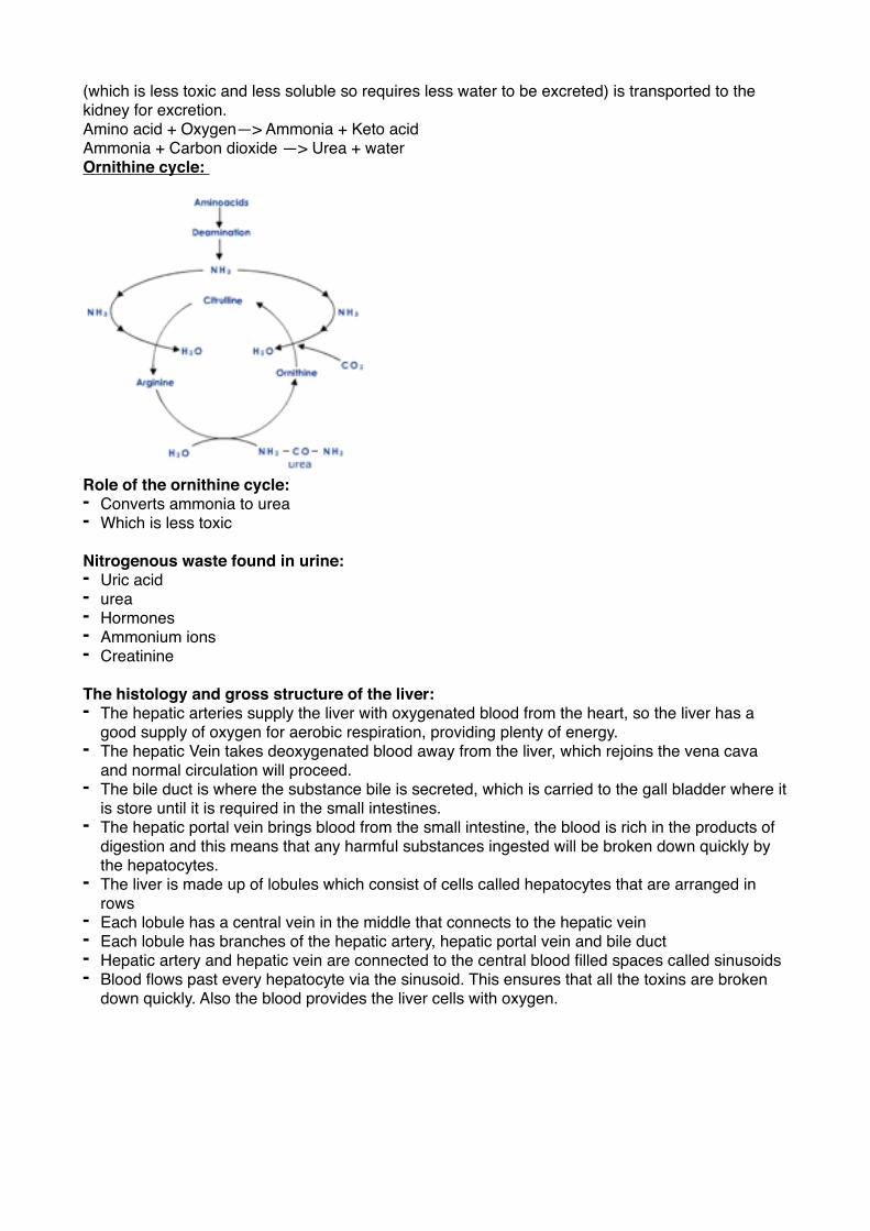
(which is less toxic and less soluble so requires less water to be excreted) is transported to the kidney for excretion.Amino acid + Oxygen—> Ammonia + Keto acidAmmonia + Carbon dioxide —> Urea + waterOrnithine cycle:
�Role of the ornithine cycle:- Converts ammonia to urea - Which is less toxic
Nitrogenous waste found in urine:- Uric acid - urea- Hormones- Ammonium ions- Creatinine
The histology and gross structure of the liver:- The hepatic arteries supply the liver with oxygenated blood from the heart, so the liver has a
good supply of oxygen for aerobic respiration, providing plenty of energy. - The hepatic Vein takes deoxygenated blood away from the liver, which rejoins the vena cava
and normal circulation will proceed. - The bile duct is where the substance bile is secreted, which is carried to the gall bladder where it
is store until it is required in the small intestines.- The hepatic portal vein brings blood from the small intestine, the blood is rich in the products of
digestion and this means that any harmful substances ingested will be broken down quickly by the hepatocytes.
- The liver is made up of lobules which consist of cells called hepatocytes that are arranged in rows
- Each lobule has a central vein in the middle that connects to the hepatic vein - Each lobule has branches of the hepatic artery, hepatic portal vein and bile duct- Hepatic artery and hepatic vein are connected to the central blood filled spaces called sinusoids- Blood flows past every hepatocyte via the sinusoid. This ensures that all the toxins are broken
down quickly. Also the blood provides the liver cells with oxygen.
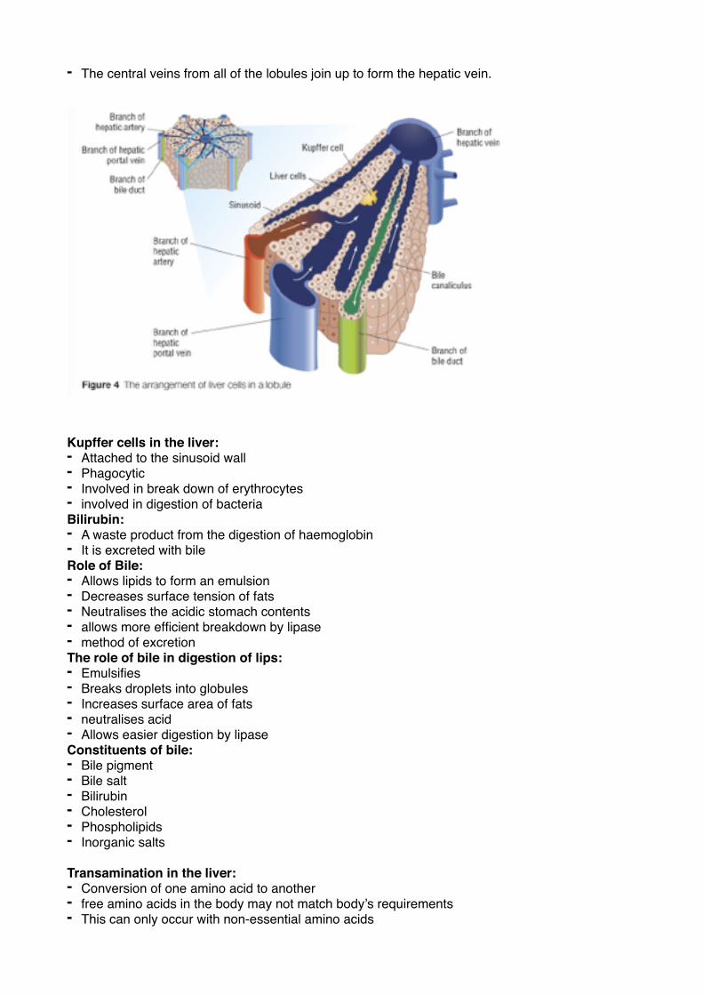
- The central veins from all of the lobules join up to form the hepatic vein.
Kupffer cells in the liver:- Attached to the sinusoid wall- Phagocytic - Involved in break down of erythrocytes- involved in digestion of bacteria Bilirubin: - A waste product from the digestion of haemoglobin - It is excreted with bile Role of Bile:- Allows lipids to form an emulsion - Decreases surface tension of fats- Neutralises the acidic stomach contents - allows more efficient breakdown by lipase - method of excretion The role of bile in digestion of lips:- Emulsifies- Breaks droplets into globules - Increases surface area of fats- neutralises acid- Allows easier digestion by lipase Constituents of bile:- Bile pigment - Bile salt- Bilirubin - Cholesterol - Phospholipids - Inorganic salts
Transamination in the liver:- Conversion of one amino acid to another- free amino acids in the body may not match body’s requirements - This can only occur with non-essential amino acids
Functions of the liver:- Control of concentrations of blood glucose, amino acids and lipids- Synthesis of red blood cells, bile, cholesterol and plasma proteins (e.g albumin: transports fatty
acids, Prothrombin- clotting factor, and Globulin: transports hormones)- Storage of vitamin A, D, B12, Iron,- Destruction of red blood cells- Detoxification of alcohol and drugs
Role of liver in detoxification: - Catalase can convert 5 million molecules of H2O2 into harmless substances in a minute.- Alcohol contains a lot of chemical potential energy which can be used in respiration - Ethanol dehydrogenase catalyses the detoxification of alcohol in hepatocytes - Ethanol—> Ethanal—> Ethanoic acid (ethanoate/ acetate)—> Acetyl CoA - Ethanal and ethanoic acid are dehydrogenated (using ethanol dehydrogenase and
acetaldehyde) and the hydrogen reduces NAD. If too many NAD are busy detoxifying alcohol, there will too few NAD (they are all reduced) to break down fatty acids for use in respiration. So the fatty acids are converted back to Lipids, which are stored in hepatocytes, making the liver enlarged- fatty liver (hepatitis, inflammation)
so NAD—> reduced NAD:- This interrupts normal metabolic reactions - unused fatty acids/lipids build upHow ethanoate/ acetate (from alcohol detoxification) is metabolise by hepatocytes:- Acetate combines with coenzyme A to form Acetyl CoA - Acetyl CoA is carried to the Krebs cycle where Acetate is offloaded again - Acetate combines with Oxaloacetate to form citrate - Citrate undergoes dehydrogenation and decarboxylation - ATP is produced - Acetyl CoA May be involved in synthesis of fatty acids
What happens when liver becomes inflamed: (Cirrhosis) - White blood cells attack and destroy hepatocytes - So hepatocytes replaced by scar tissue - Liver becomes unable to function - This is called Cirrhosis - It is irreversible/ permanent
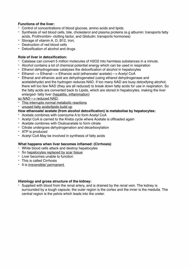
Histology and gross structure of the kidney:- Supplied with blood from the renal artery, and is drained by the renal vein. The kidney is
surrounded by a tough capsule, the outer region is the cortex and the inner is the medulla. The central region is the pelvis which leads into the ureter.
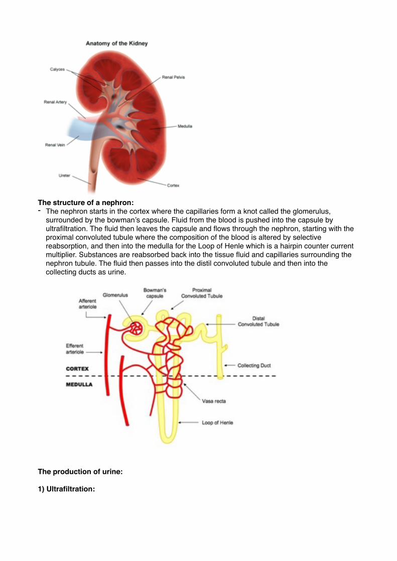
�The structure of a nephron:- The nephron starts in the cortex where the capillaries form a knot called the glomerulus,
surrounded by the bowman’s capsule. Fluid from the blood is pushed into the capsule by ultrafiltration. The fluid then leaves the capsule and flows through the nephron, starting with the proximal convoluted tubule where the composition of the blood is altered by selective reabsorption, and then into the medulla for the Loop of Henle which is a hairpin counter current multiplier. Substances are reabsorbed back into the tissue fluid and capillaries surrounding the nephron tubule. The fluid then passes into the distil convoluted tubule and then into the collecting ducts as urine.
�
The production of urine:
1) Ultrafiltration:
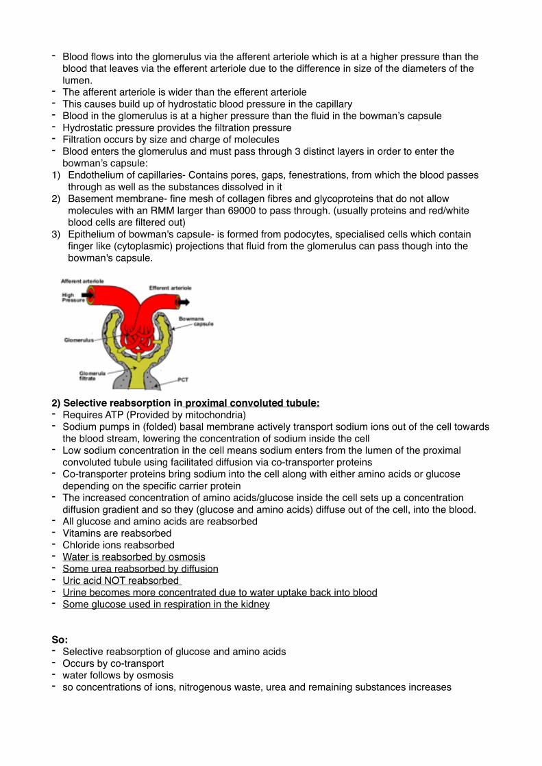
- Blood flows into the glomerulus via the afferent arteriole which is at a higher pressure than the blood that leaves via the efferent arteriole due to the difference in size of the diameters of the lumen.
- The afferent arteriole is wider than the efferent arteriole - This causes build up of hydrostatic blood pressure in the capillary- Blood in the glomerulus is at a higher pressure than the fluid in the bowman’s capsule - Hydrostatic pressure provides the filtration pressure - Filtration occurs by size and charge of molecules - Blood enters the glomerulus and must pass through 3 distinct layers in order to enter the
bowman’s capsule:1) Endothelium of capillaries- Contains pores, gaps, fenestrations, from which the blood passes
through as well as the substances dissolved in it 2) Basement membrane- fine mesh of collagen fibres and glycoproteins that do not allow
molecules with an RMM larger than 69000 to pass through. (usually proteins and red/white blood cells are filtered out)
3) Epithelium of bowman's capsule- is formed from podocytes, specialised cells which contain finger like (cytoplasmic) projections that fluid from the glomerulus can pass though into the bowman's capsule.
�2) Selective reabsorption in proximal convoluted tubule:- Requires ATP (Provided by mitochondria) - Sodium pumps in (folded) basal membrane actively transport sodium ions out of the cell towards
the blood stream, lowering the concentration of sodium inside the cell- Low sodium concentration in the cell means sodium enters from the lumen of the proximal
convoluted tubule using facilitated diffusion via co-transporter proteins- Co-transporter proteins bring sodium into the cell along with either amino acids or glucose
depending on the specific carrier protein- The increased concentration of amino acids/glucose inside the cell sets up a concentration
diffusion gradient and so they (glucose and amino acids) diffuse out of the cell, into the blood. - All glucose and amino acids are reabsorbed- Vitamins are reabsorbed - Chloride ions reabsorbed - Water is reabsorbed by osmosis - Some urea reabsorbed by diffusion- Uric acid NOT reabsorbed - Urine becomes more concentrated due to water uptake back into blood- Some glucose used in respiration in the kidney
So:- Selective reabsorption of glucose and amino acids- Occurs by co-transport - water follows by osmosis - so concentrations of ions, nitrogenous waste, urea and remaining substances increases
Structure of cells of the proximal convoluted tubule:- Microvilli- Increase the surface area for selective reabsorption - Co-transporter proteins- contained in the cell surface membrane that is in contact with the tubule
fluid. Transports glucose or amino acids. - Sodium pumps- contained in the cell surface membrane opposite to the fluid tubule. Actively
transports sodium ions against their concentration gradient. - Many mitochondria- Provides the energy needed to drive the selective reabsorption process.
Many mitochondria = Lots of ATP - Folded basal membrane - Rough ER/ Ribosomes
Most water is reabsorbed into the blood at proximal convoluted tubule.
The control of water content of the blood:
- The role of the loop of henle is to cause a decrease in water potential in the medulla. - In the loop of henle, salts (sodium and chloride ions) are transferred out of the ascending limb
and into the descending limb. - This means that the tissue fluid in the medulla has a very negative water potential - The Ascending limb is impermeable to water - The walls of the descending limb are permeable to water - Water is removed from the deciding limb - The collecting ducts pass through the medulla- So water is removed from the collecting ducts by osmosis
- The water potential of the blood is monitored by osmoreceptors in the hypothalamus of the brain- When the water potential is very low, they shrink and stimulate neurosecretory cells in the
hypothalamus. - These produce and release Anti diuretic hormone (ADH) which flows down their axon to the
posterior pituitary gland where they are stored until needed. - When the neurosecretory cells are stimulated, they send action potentials down their axons and
cause the release of ADH - ADH enters the capillaries running through the posterior pituitary gland. It is transported around
the body and acts on the cells of the collecting ducts. - When it binds to the receptors, It causes a chain of enzyme catalysed reactions (activates
Adenyl Cyclase which converts ATP to cAMP, cAMP activates Kinase enzyme) the end result of which is the insertion of vesicles containing water permeable channels (aquaporins) in the membranes of the cells, so the cells of the collecting ducts become more permeable to water.
- More water is reabsorbed, by osmosis down the water potential gradient, back into the blood.- Water potential of blood is restored to normal - Less urine, with a lower water potential, is excreted- Less ADH is released when the water potential rises again - ADH is slowly broken down and the collecting ducts receive less stimulus.
How aquaporin channels only allow passage of water and not other substances such as ions:- the ions are too large to pass through the channel - Shapes not compatible - Positive charge in the channel repels the positively charged ions
Problems that arise from kidney failure:- Unable to remove excess water and excess waste products from the body e.g urea and excess
salts - Inability to regulate urea and salt level
- Death
Renal dialysis as treatment for kidney failure:- waste, excess fluids and salts are removed from the body by passing the blood over a dialysis
membrane. This allows the exchange of substance between the blood and the dialysis fluid (dialysate), which has the same concentration of substances as the blood plasma. Substances diffuse from both sides to create the correct concentrations of substances.
Two types of dialysis:- Haemodialysis:Blood is taken from an artery or vein and is passed through a machine, that contains an artificial dialysis membrane, before returning via a vein. Heparin (anticoagulant) is used to void clotting of the blood in the machine. Thrice weekly trips to the hospital lasting several hours. Anticoagulant not added towards end of treatment so that blood clots normally after treatment- prevents low blood pressure.Blood and dialysate flow in different directions to maintain diffusion gradient. - Peritoneal dialysis:The body’s own abdominal membrane is used as a filter.
Kidney transplant as treatment for kidney failure:Advantages:- No dialysis- Less limited diet - Better physical feeling - Better quality life - No longer “Chronically ill”Disadvantages - Need immunosuppressants for life of Kidney - Major surgery - Risk of infection - Need frequent check in case of rejection- Side effects of medication e.g Weak immune system/ Increased susceptibility to infectionIf donated kidney not closely matched:- Donated kidney recognised as foreign - Antigens on kidney will be different - Causing infection by immune system - Use of immunosuppressant
How urine samples can be used to test for pregnancy:- A human embryo secretes Human chorionic gonadotrophin (hCG) as soon as it is implanted on
the uterine lining. The hormone is small so can pass from the blood into the filtrate and can be detected in the mother’s urine after as few as 6 days.
- Pregnancy tests contain monoclonal antibodies that are tagged with a blue bead (attached to marker) and bind only to hCG
- The hCG-antibody complex moves along the strip until it binds to a band of immobilised antibodies specific to the hCG-antibody complex
- The blue bead line up to form a blue line - The unbound antibodies bind to another band of immobilised antibodies specific to them.- One blue line shows the test is working so two lines mean pregnancy
How urine samples can be used to detect misuse of anabolic steroids:- Urine samples are tested using gas chromatography - The sample is vaporised in the presence of a gaseous solvent - It is passed down a long tube lined with an absorbing agent - Each substance dissolves differently in the gas and so stays there for a unique specific time- the
retention time. - Eventually the substance leaves the gas and is absorbed by the lining
- It is then analysed to make a chromatogram - Standard samples of drugs and urine samples are run so drugs can be identified and quantified
in the chromatogram Testing for anabolic steroids is done because:- of fairness- drug gives unfair advantage - There are ideas of health risks e.g depression/aggression/liver damage/heart attach. - Outstanding performances may be distrusted- Does not reflect athlete’s natural talent- There is pressure to keep up with rival competitors - Those using can train longer, respire for longer, can recovery from injury quicker and can build
up muscle mass
Photosynthesis:
Autotroph- An organism that uses light or chemical energy and an inorganic molecule to synthesise complex organic molecules.Heterotrophs- Organisms that ingest and digest complex organic molecules releasing the chemical potential energy stored in them.
How respiration in plants and animals depends upon the products of photosynthesis: Photoautotrophs and heterotrophs can release the chemical energy in complex organic molecules, which were made during photosynthesis, in respiration. They use oxygen, which was first released into the atmosphere as a product of photosynthesis, for aerobic respiration.
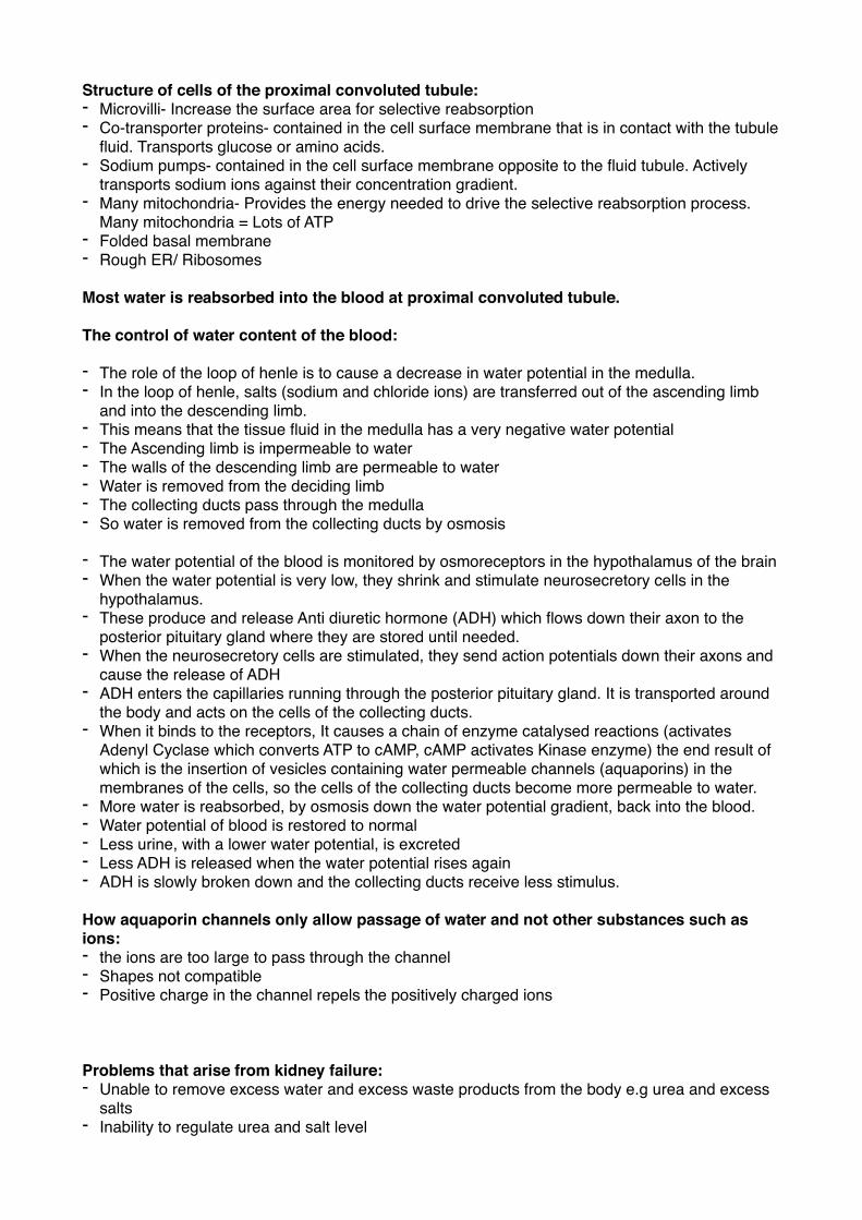
In plants photosynthesis is a two stage process taking place in chloroplasts.
How the structure of chloroplasts allow them to carry out their functions:
�- Lamellae- Made of thylakoid membranes - Grana- Stacks of lamellae - Intergranal lamellae- Lamellae between the grana - Intermembrane space- between outer and inner membrane - Stroma- The matrix of chloroplast
- The inner membrane contains transport proteins which control the entry and exit of substances between the cytoplasm and the stroma.
- The grana (sites of light absorption and ATP synthase) prove a large surface area for photosynthetic pigment, electron carriers and ATP synthase, all involved in the light dependent reaction.
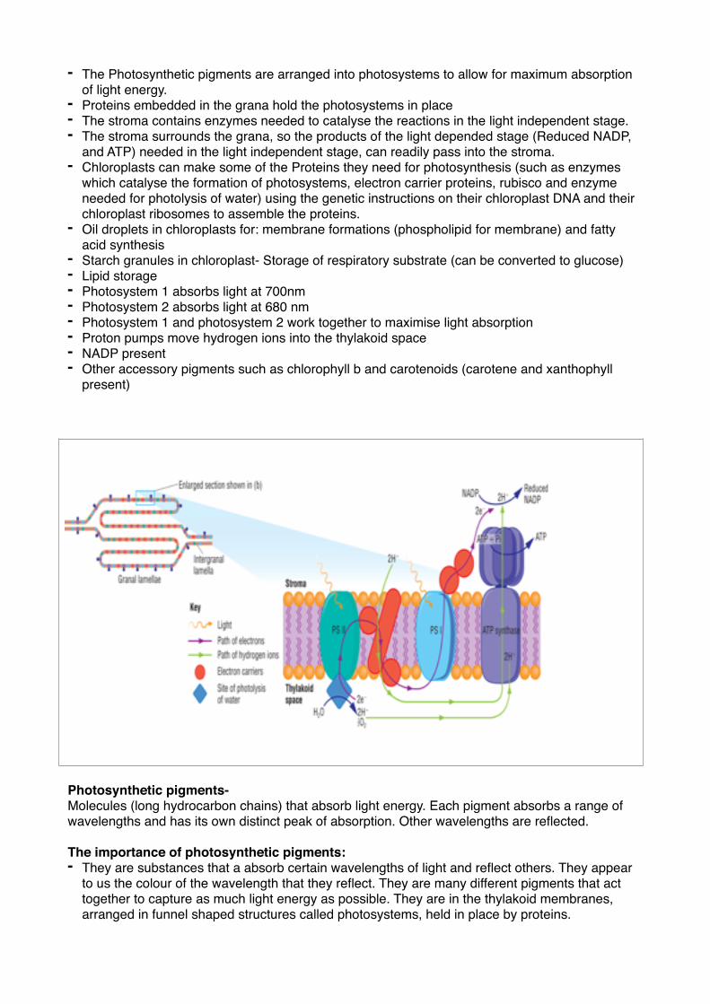
- The Photosynthetic pigments are arranged into photosystems to allow for maximum absorption of light energy.
- Proteins embedded in the grana hold the photosystems in place- The stroma contains enzymes needed to catalyse the reactions in the light independent stage.- The stroma surrounds the grana, so the products of the light depended stage (Reduced NADP,
and ATP) needed in the light independent stage, can readily pass into the stroma. - Chloroplasts can make some of the Proteins they need for photosynthesis (such as enzymes
which catalyse the formation of photosystems, electron carrier proteins, rubisco and enzyme needed for photolysis of water) using the genetic instructions on their chloroplast DNA and their chloroplast ribosomes to assemble the proteins.
- Oil droplets in chloroplasts for: membrane formations (phospholipid for membrane) and fatty acid synthesis
- Starch granules in chloroplast- Storage of respiratory substrate (can be converted to glucose) - Lipid storage - Photosystem 1 absorbs light at 700nm- Photosystem 2 absorbs light at 680 nm- Photosystem 1 and photosystem 2 work together to maximise light absorption- Proton pumps move hydrogen ions into the thylakoid space - NADP present - Other accessory pigments such as chlorophyll b and carotenoids (carotene and xanthophyll
present)
Photosynthetic pigments-Molecules (long hydrocarbon chains) that absorb light energy. Each pigment absorbs a range of wavelengths and has its own distinct peak of absorption. Other wavelengths are reflected.
The importance of photosynthetic pigments:- They are substances that a absorb certain wavelengths of light and reflect others. They appear
to us the colour of the wavelength that they reflect. They are many different pigments that act together to capture as much light energy as possible. They are in the thylakoid membranes, arranged in funnel shaped structures called photosystems, held in place by proteins.
- Chlorophyll in a leaf reflects and so does not absorb green light. So in green light there is little photosynthesis as there is no light dependent reaction, no photolysis.
- In an experimental situation using pond weed, this would mean that no CO2 is fixed and becomes some CO2 is produced during respiration, there would be an increase in CO2, increasing the acidity and therefore reducing the pH.
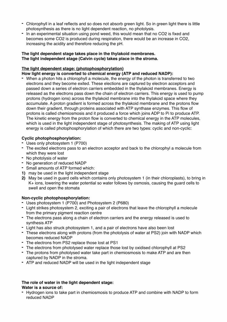
The light dependent stage takes place in the thylakoid membranes.The light independent stage (Calvin cycle) takes place in the stroma.
The light dependent stage: (photophosphorylation)How light energy is converted to chemical energy (ATP and reduced NADP):- When a photon hits a chlorophyll a molecule, the energy of the photon is transferred to two
electrons and they become exited. These elections are captured by electron acceptors and passed down a series of electron carriers embedded in the thylakoid membranes. Energy is released as the electrons pass down the chain of electron carriers. This energy is used to pump protons (hydrogen ions) across the thylakoid membrane into the thylakoid space where they accumulate. A proton gradient is formed across the thylakoid membrane and the protons flow down their gradient, through proteins associated with ATP synthase enzymes. This flow of protons is called chemiosmosis and it produced a force which joins ADP to Pi to produce ATP. The kinetic energy from the proton flow is converted to chemical energy in the ATP molecules, which is used in the light independent stage of photosynthesis. The making of ATP using light energy is called photophosphorylation of which there are two types: cyclic and non-cyclic:
Cyclic photophosphorylation:- Uses only photosystem 1 (P700)- The excited electrons pass to an electron acceptor and back to the chlorophyl a molecule from
which they were lost - No photolysis of water- No generation of reduced NADP- Small amounts of ATP formed which:1) may be used in the light independent stage2) May be used in guard cells which contains only photosystem 1 (in their chloroplasts), to bring in
K+ ions, lowering the water potential so water follows by osmosis, causing the guard cells to swell and open the stomata
Non-cyclic photophosphorylation:- Uses photosystem 1 (P700) and Photosystem 2 (P680)- Light strikes photosystem 2, exciting a pair of electrons that leave the chlorophyll a molecule
from the primary pigment reaction centre - The electrons pass along a chain of electron carriers and the energy released is used to
synthesis ATP- Light has also struck photosystem 1, and a pair of electrons have also been lost- These electrons along with protons (from the photolysis of water at PS2) join with NADP which
becomes reduced NADP- The electrons from PS2 replace those lost at PS1 - The electrons from photolysed water replace those lost by oxidised chlorophyll at PS2 - The protons from photolysed water take part in chemiosmosis to make ATP and are then
captured by NADP in the stroma. - ATP and reduced NADP will be used in the light independent stage
The role of water in the light dependent stage:Water is a source of:- Hydrogen ions to take part in chemiosmosis to produce ATP and combine with NADP to form
reduced NADP
- Electrons to replace those lost by oxidised chlorophyll at photosystem 2 - The oxygen produced comes from water
Light independent stage- Calvin cycle:- CO2 diffused into the leaf through the open stomata - CO2 combines with 5C Ribulose Biphosphate, catalysed by Rubisco- This forms two molecules of Glycerate-3-phosphate (GP) 3C- GP is reduced (using reduced NADP from the light dependent stage) and phosphorylated (using
ATP from the light dependent stage) to to form Triose Phosphate (TP) 3C- 5/6 molecules of TP are recycled by phosphorylation (using ATP from the light dependent stage)
to three molecules of Ribulose Biphosphate (RuBP) and the rest for production- Rearrangement of carbons to form pentose sugars from TP
The role of CO2 in the light independent stage:Carbon dioxide is the source of carbon and oxygen for the production of all large complex organic molecules.
The fate of TP is that is:- Regenerates Ribulose biphosphate so the cycle can continue for further CO2 fixation - It forms sugar/glucose/starch/fructose/hexose/cellulose/sucrose- It forms fats/triglycerides/fatty acids/amino acids/ glycerol/ nucleotides/ protein/ nucleic acids/
lipid- Most TP is used to produce RuBP and the rest for production
The effect of changing light intensity, carbon dioxide concentration, and temperature on the rate of photosynthesis:
Light intensity- Affects light dependent stage. (light provides energy for excitation of electrons and photolysis of water, it also causes stomata to open so CO2 can diffuse in) 1) lots of light- More light energy - More excitation of electrons - More photophosphorylation - More reduced NADP and ATP produced - More GP reduced and phosphorylated to TP- More TP phosphorylated to RuBP- More CO2 fixed - Overall increase in rate of photosynthesis 2) Little light - Less light energy - Less excitation of electrons - Less photophosphorylation - Less ATP and reduced NADP formed - GP cannot be changed to TP- Levels of TP will fall - GP will accumulate- Less RuBP- Less CO2 fixed - Less GP formed
Carbon dioxide concentration-Affects light-independent stage1) Lots of CO2- More CO2 fixation- More GP- More TP- More regeneration of RuBP- However, Open stomata may lead to increased transpiration, so the plant may wilt if water loss
exceeds water uptake. This leads to a stress response and following the release of abscisic acid, the stomata close, reducing the CO2 uptake and therefore reducing the rate of photosynthesis.
2) Little CO2- Less CO2 fixation- Less GP- Less TP - Less sugars formed (reduction in rate of photosynthesis)
Temperature- - Has little effect on light dependent stage as the reactions in this stage are not dependent on
enzymes except for photolysis of water - The light independent stage is a series of biochemical steps, each catalysed by a specific
enzyme 1)- Between 0 and 25 degrees Celsius the rate of photosynthesis doubles every 10 degrees celsius
rise in temperature. 2) - Above 25 degrees celsius the enzymes work less effectively and oxygen successfully competes
for the active site of rubisco preventing it from accepting carbon dioxide.
- Photorespiration exceeds photosynthesis as the oxygenase activity of rubisco increases more than the carboxylase activity.
- Less CO2 fixation- Less GP- Less TP- Less regeneration of RuBP- ATP and reduced NADP from the light dependent stage are dissipated and wasted - Reduces the overall rate of photosynthesis - High temperature may also denature proteins (such as electron carriers, and Rubisco)- High temperature= high water loss- This may lead to stomata closure, and the reduction in rate of photosynthesis due to less CO2
uptake.
The benefit of having more stomata on lower surface of leaves:- Reduces transpiration as stomata sheltered from heat.
The benefits of having more stomata:- Allows entry of more CO2- More CO2 for light independent stage (Calvin cycle)- CO2 not as limiting
Nitrogen is present in the air in the plant. It leaves the plant with oxygen during photosynthesis.
Limiting factors in photosynthesis
1) Carbon dioxide: - Growers can increase the concentration of CO2 in their greenhouses by burning fuel/gas/
paraffin - This will increase the rate of photosynthesis providing that nothing else is limiting the process - As CO2 is no longer a limiting factor - Increased CO2 increases CO2 fixation in the calvin cycle
2) Light intensity:Light:- Causes stomata to open so CO2 can diffuse in- Trapped by chlorophyll and excited elections - Splits water molecules (photolysis) to produce protons for chemiosmosis and generation of
reduced NADP. The electrons and protons are used in photophosphorylation to produce ATP to fix CO2.
3) Temperature:- The calvin cycle is very much effected by temperature as it is enzyme catalysed. - Above 25 degrees celsius the enzymes work less effectively and oxygen successfully competes
for the active site of rubisco preventing it from accepting carbon dioxide- Less CO2 fixation- Less GP- Less TP- Less regeneration of RuBP- High temp= high water loss/ increased transpiration- This leads to stomata closure and a reduction in the rate of photosynthesis due to less CO2
uptake.
- Temperature can be:- Increased by heater - Reduced by ventilation (or fan)- Maintained by air conditioning
Growing plants in greenhouses has other advantages:
- It is easier to control water supply (prevent transpiration), Humidity, minerals and fertilisers - It is easier to control the use of pesticides, pest control, biological control - The gas/paraffin heater supplies heat and CO2- It prevents the damage of plants by wind chill/frost/wind/hail
How to investigate the factors that affect the rate of photosynthesis:
could measure:- Volume of O2 produced- Rate of uptake of CO2- Rate of increase in dry mass of plant
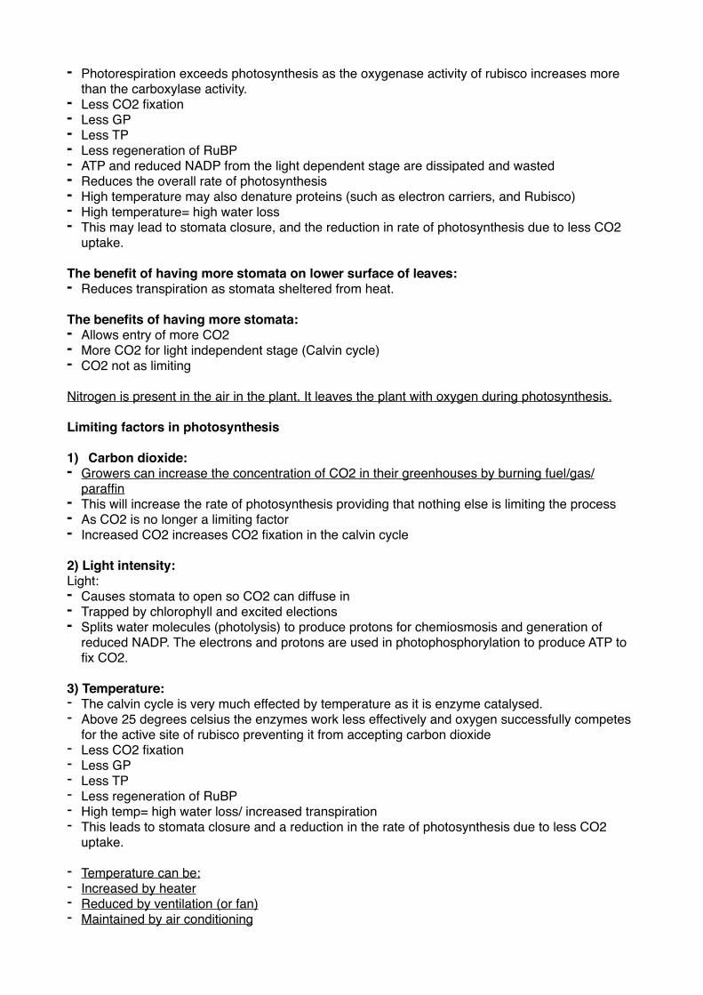
Use a photosynthometer/audus microburette
- set up, air tight, ensuring no air bubbles are present- Gas given off by the plant over time collects in the flared end of the capillary tube- The syringe can be used to move the air bubble into the part of the capillary tube against the
scale - Distance moved by the air bubble at each light intensity can be used to work out the volume (pi x
radius squared x height) and essentially the rate (by dividing the volume by the time taken)- The experiment should be repeated at the same light intensity and average values used- The apparatus should be left to acclimatise for 5 minutes - All other factors should be kept constant e.g a water bath to keep the temperature constant
Glass beads used to make the volume of contents the same in the photosynthometer/respirometer. Without beads there would be more oxygen/ carbon dioxide in one and less in the other.To determine the quantity of glass beads needed, find the difference in volume of plants/seads the difference represents the volume of beads required.
DiscsCut disks from leaves:- the same size so the same surface area
- From the same part of plant so they have same amount of chloroplasts
Place 5/6 discs in a syringe and half fill the syringe with dilute sodium hydrogen carbonate solution Hold the syringe upright, placing finger over the end and gently pulling on the plunger (air is extracted from the spongey mesophyll in the leaf discs) as density of leaf discs increases, they sink to the bottom After all leaf discs have sunk, transfer the contents of the syringe into a beaker. Illuminate using bright light and time how long it takes for one leaf disc to float to the surface of the solution.Repeat at the same light intensity and use average valuesRepeat at different light intensitiesrecord results in a table The leaf discs rise as they become less dense due to them photosynthesising and releasing oxygen Both methods can also be used to change carbon dioxide concentration by using sodium hydrogen carbonate solution of differing concentrations keeping light intensity constant.
Methods can be adapted to investigate the effects of temperature, carbon dioxide concentration and light wavelength on the rate of photosynthesis.
Investigating rate of photosynthesis by measuring rate of uptake of carbon dioxide:Using hydrogen carbonate indicator solutionwhich is:Red- when neutralYellow/orange- pH6 (more acidic)Purple/red- above pH7 (more alkali)
Change in colour of indicator solution can be measure using a colorimeter.Change in absorption/time taken= indication of rate of uptake of CO2
In light—> indicator solution turns purple as plant is photosynthesising so taking in CO2 therefore reducing the acidity of surrounding solution and therefore the ph increase above 7
In dark—> indicator solution turns yellow as plant is not photosynthesising, only respiring so releasing CO2 therefore increasing the acidity of surrounding solution therefore the pH decreased to around 6
Other sources of CO2:- Bubbling CO2 - Dry ice - Hydrogen carbonate
Respiration
Why plants, animals and microorganisms need to respire:- All living organisms need to respire as they need energy to drive their biological processes.- Metabolic reactions that require energy include:1) Active transport- most of and organisms energy is used for this 2) Endocytosis- bulk movement of larger molecules into the cell3) Secretion- large molecules made in some cells are released by exocytosis4) Metabolic reactions- the synthesis of large molecules from smaller one or vice versa. e.g
proteins from amino acids, steroids from cholesterol, cellulose from beta glucose, these are all anabolic. (catabolic metabolic reactions are the breakdown of larger molecules into smaller ones)
5) Replication of DNA and synthesis of organelles before a cell divides (meiosis an mitosis)6) Movement
- Bacterial flagella - Eukaryotic cilia and undulipodia - Muscles contraction7) activation of chemicals e.g phosphorylation of glucose (so that it becomes less stable and can be broken down to release energy)8) Protein synthesis
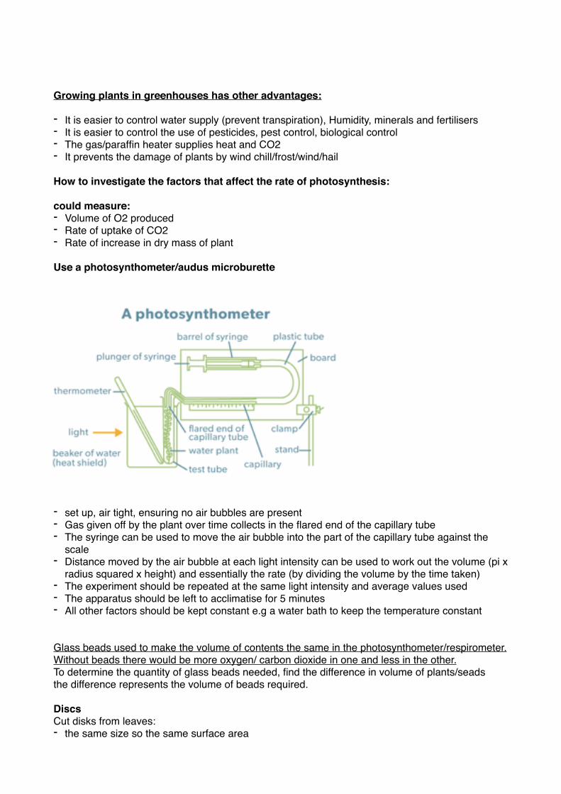
Structure of ATP:- An adenine group, attached to a ribose sugar and three phosphate molecules.
�
The hydrolysis of ATP provides the immediate source of energy for biological processes:ATP :- Transfers energy - It is the universal energy molecule and provides an immediate source of energy - The phosphate(s) can be removed by hydrolysis to release 30 Jkmol-1 of energy - It releases energy for metabolism such as:1) Muscle contraction2) Active transport3) Phosphorylation4) Glycolysis5) In movement, biding to protein to change their shape - ADP can be attached to an inorganic phosphate to form ATP during respirating/photosynthesis- Energy is released in small “packets” to prevent cell damage
Importance of co-enzymes in respiration:- Coenzymes aid in the oxidation and reduction of reactions- NAD and FAD accept hydrogen atoms and are reduced- Reduced NAD and reduced FAD carry electrons to the electron transport chain for oxidative
phosphorylation - Reduced NAD and reduced FAD carry hydrogen ions for chemiosmosis (oxidative
phosphorylation)- Coenzyme A carries acetate to the krebs cycle
Glycolysis takes place in the cytoplasm:Process of glycolysis:- An ATP molecule is hydrolysed and he phosphate attached to the glucose molecule at C-6- Glucose-6-phosphate is turned into fructose-6-phosphate- Another ATP molecule is hydrolysed and the phosphate attached to C-1- The hexose sugar is activated by the energy release from hydrolysed ATP, it now cannot leave
the cell and is known as Hexose-1,6-biphosphate- Hexose-1,6-biphosphate is split into two molecules of triose phosphate - Two hydrogen atoms are removed from each Triose phosphate which involves dehydrogenase
enzymes- NAD combines with the hydrogen atoms to become reduced NAD
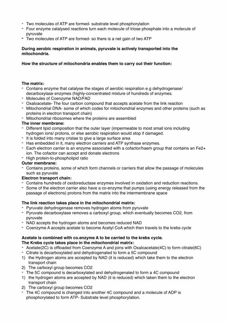
- Two molecules of ATP are formed- substrate level phosphorylation - Four enzyme catalysed reactions turn each molecule of triose phosphate into a molecule of
pyruvate- Two molecules of ATP are formed- so there is a net gain of two ATP
During aerobic respiration in animals, pyruvate is actively transported into the mitochondria.
How the structure of mitochondria enables them to carry out their function:
The matrix:- Contains enzyme that catalyse the stages of aerobic respiration e.g dehydrogenase/
decarboxylase enzymes (highly-concentrated mixture of hundreds of enzymes.- Molecules of Coenzyme NAD/FAD- Oxaloacetate- The four carbon compound that accepts acetate from the link reaction - Mitochondrial DNA- some of which codes for mitochondrial enzymes and other proteins (such as
proteins in electron transport chain) - Mitochondrial ribosomes where the proteins are assembledThe inner membrane:- Different lipid composition that the outer layer (impermeable to most small ions including
hydrogen ions/ protons, or else aerobic respiration would stop if damaged.- It is folded into many cristae to give a large surface area - Has embedded in it, many electron carriers and ATP synthase enzymes.- Each electron carrier is an enzyme associated with a cofactor/haem group that contains an Fe2+
ion. The cofactor can accept and donate electrons - High protein-to-phospholipid ratio Outer membrane:- Contains proteins, some of which form channels or carriers that allow the passage of molecules
such as pyruvate Electron transport chain:- Contains hundreds of oxidoreductase enzymes involved in oxidation and reduction reactions. - Some of the electron carrier also have a co-enzyme that pumps (using energy released from the
passage of electrons) protons from the matrix into the intermembrane space
The link reaction takes place in the mitochondrial matrix:- Pyruvate dehydrogenase removes hydrogen atoms from pyruvate - Pyruvate decarboxylase removes a carboxyl group, which eventually becomes CO2, from
pyruvate - NAD accepts the hydrogen atoms and becomes reduced NAD - Coenzyme A accepts acetate to become Acetyl CoA which then travels to the krebs cycle
Acetate is combined with co.enzyme A to be carried to the krebs cycle.The Krebs cycle takes place in the mitochondrial matrix:- Acetate(2C) is offloaded from Coenzyme A and joins with Oxaloacetate(4C) to form citrate(6C)- Citrate is decarboxylated and dehydrogenated to form a 5C compound1) the Hydrogen atoms are accepted by NAD (it is reduced) which take them to the electron
transport chain 2) The carboxyl group becomes CO2- The 5C compound is decarboxylated and dehydrogenated to form a 4C compound 1) the hydrogen atoms are accepted by NAD (it is reduced) which taken them to the electron
transport chain 2) The carboxyl group becomes CO2- The 4C compound is changed into another 4C compound and a molecule of ADP is
phosphorylated to form ATP- Substrate level phosphorylation.
- The second 4C compound is changed into a third 4C compound and a pair of hydrogen atoms are removed- dehydrogenation, reducing FAD.
- The third 4C compound is further dehydrogenated (reducing NAD) to regenerate oxaloacetate(4C).
Oxidative phosphorylation takes place in the inner mitochondrial membrane (electron transport chain):- The final stage of aerobic respiration involves electron carriers embedded in the inner
mitochondrial membrane - The membranes are folded into cristae which increases the surface area for electron carriers
and ATP synthase enzymes - Oxidative phosphorylation is the formation of ATP by the addition of an inorganic phosphate to
ADP in the presence of oxygen - As the protons flow through ATP synthase (as a result of chemiosmosis) they drive the rotation
part of the enzyme and join ADP to Pi to form ATP. - The electrons are passed from the final electron carrier to molecular oxygen which is final
electron acceptor- Hydrogen ions also join so oxygen is reduced to water - The products are: ATP, water and oxidised NAD/FAD - 4H+ +4e- + O2 —> 2H2O
Overall products of aerobic respiration:ATP, Water, CO2, Oxidised NAD/FAD
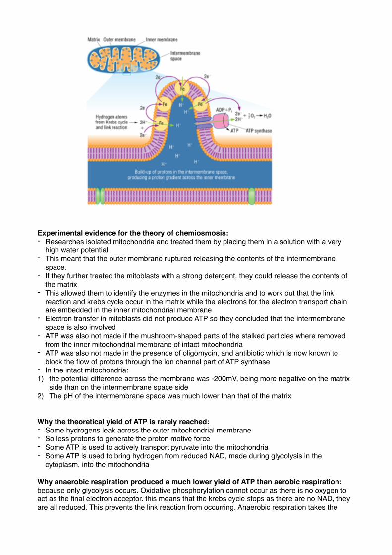
Chemiosmosis:- Reduced NAD and reduced FAD donate hydrogen atoms which are split into protons and
electrons to the electron carriers- The protons are pumped across the inner mitochondrial membrane from the matrix into the
intermembrane space using the energy released from the passing of electrons down the electron transport chain
- this builds up a proton gradient which is also a ph gradient and an electrochemical gradient - Thus potential energy builds up - The hydrogen ions cannot diffuse through the lipid part of the inner membrane but can diffuse
down their gradient through ATP synthase- an ion channel in the membrane. - The flow of hydrogen ions is chemiosmosis.
Oxygen is the final electron acceptor in aerobic respiration.
Experimental evidence for the theory of chemiosmosis:- Researches isolated mitochondria and treated them by placing them in a solution with a very
high water potential- This meant that the outer membrane ruptured releasing the contents of the intermembrane
space. - If they further treated the mitoblasts with a strong detergent, they could release the contents of
the matrix - This allowed them to identify the enzymes in the mitochondria and to work out that the link
reaction and krebs cycle occur in the matrix while the electrons for the electron transport chain are embedded in the inner mitochondrial membrane
- Electron transfer in mitoblasts did not produce ATP so they concluded that the intermembrane space is also involved
- ATP was also not made if the mushroom-shaped parts of the stalked particles where removed from the inner mitochondrial membrane of intact mitochondria
- ATP was also not made in the presence of oligomycin, and antibiotic which is now known to block the flow of protons through the ion channel part of ATP synthase
- In the intact mitochondria:1) the potential difference across the membrane was -200mV, being more negative on the matrix
side than on the intermembrane space side 2) The pH of the intermembrane space was much lower than that of the matrix
Why the theoretical yield of ATP is rarely reached:- Some hydrogens leak across the outer mitochondrial membrane- So less protons to generate the proton motive force - Some ATP is used to actively transport pyruvate into the mitochondria - Some ATP is used to bring hydrogen from reduced NAD, made during glycolysis in the
cytoplasm, into the mitochondria
Why anaerobic respiration produced a much lower yield of ATP than aerobic respiration:because only glycolysis occurs. Oxidative phosphorylation cannot occur as there is no oxygen to act as the final electron acceptor. this means that the krebs cycle stops as there are no NAD, they are all reduced. This prevents the link reaction from occurring. Anaerobic respiration takes the
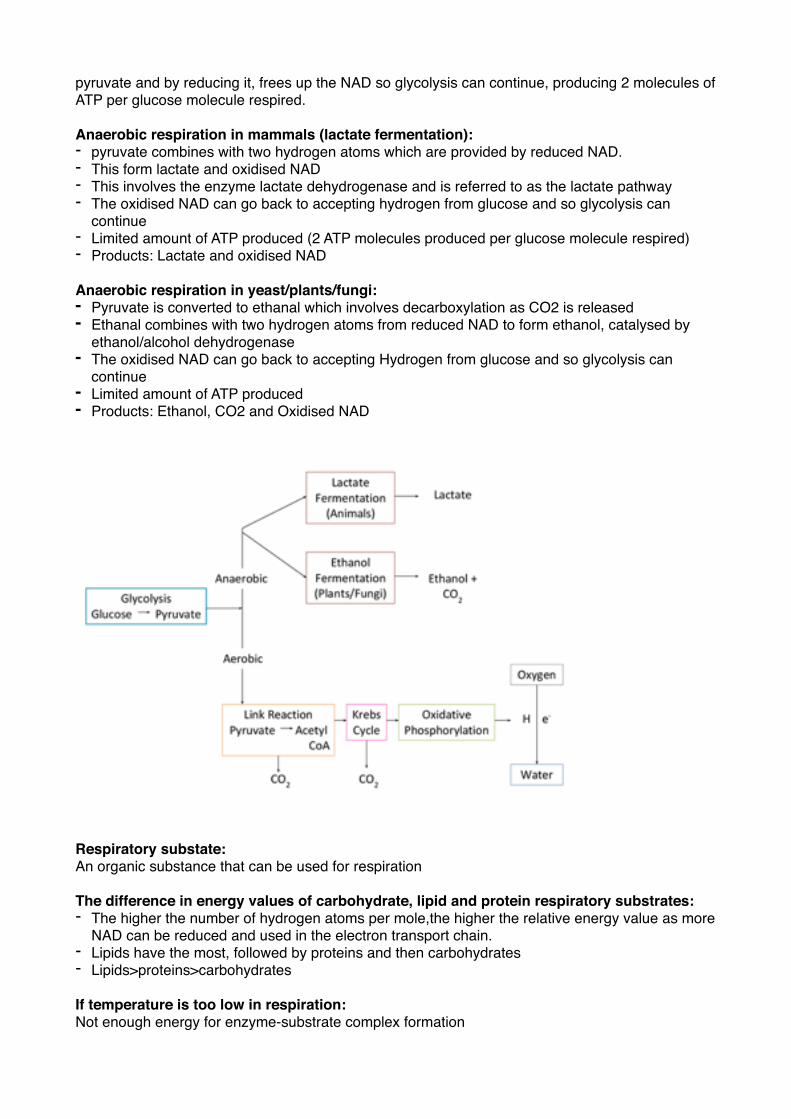
pyruvate and by reducing it, frees up the NAD so glycolysis can continue, producing 2 molecules of ATP per glucose molecule respired.
Anaerobic respiration in mammals (lactate fermentation):- pyruvate combines with two hydrogen atoms which are provided by reduced NAD.- This form lactate and oxidised NAD- This involves the enzyme lactate dehydrogenase and is referred to as the lactate pathway - The oxidised NAD can go back to accepting hydrogen from glucose and so glycolysis can
continue- Limited amount of ATP produced (2 ATP molecules produced per glucose molecule respired) - Products: Lactate and oxidised NAD
Anaerobic respiration in yeast/plants/fungi:- Pyruvate is converted to ethanal which involves decarboxylation as CO2 is released- Ethanal combines with two hydrogen atoms from reduced NAD to form ethanol, catalysed by
ethanol/alcohol dehydrogenase - The oxidised NAD can go back to accepting Hydrogen from glucose and so glycolysis can
continue- Limited amount of ATP produced- Products: Ethanol, CO2 and Oxidised NAD
�
Respiratory substate:An organic substance that can be used for respiration
The difference in energy values of carbohydrate, lipid and protein respiratory substrates:- The higher the number of hydrogen atoms per mole,the higher the relative energy value as more
NAD can be reduced and used in the electron transport chain. - Lipids have the most, followed by proteins and then carbohydrates- Lipids>proteins>carbohydrates
If temperature is too low in respiration:Not enough energy for enzyme-substrate complex formation
If temperature is too high in respiration:Enzymes denatured (proteins in electron transport chain and electron carriers as well as decarboxylase/ dehydrogenase enzymes denatured)
Specialisation of neurones:- The cells are very long (large surface area)- The cell surface membrane contains many gate ion channels to control the entry and exit of ions - The cell surface membrane contains sodium potassium pumps that use ATP to pump Na+ out
and K+ in- active transport- Many are surrounded by a myelin sheath of Schwann cells which insulate the neurones from
surrounding neurones - The gaps in between the Schwann cells are called the nodes of Ranvier - The cell body contains the nucleus, many mitochondria and ribosomes
Ways in which the structure of leaf is adapted for gaseous exchange:- Stomata allow gases to diffuse into cell- Thin cell walls- Large surface area of palisade- Cylindrical palisade cells