Embed Size (px)
Citation preview

148 © 2016 The Journal of Indian Prosthodontic Society | Published by Wolters Kluwer - Medknow
Comparative analysis of sagittal condylar guidance by protrusive interocclusal records with panoramic and lateral cephalogram radiographs in dentulous population: A clinico‑radiographic study
Girish Galagali, Sufiyan M. Kalekhan, Prakash Nidawani, Jagadeesh Naik, Sidhartha BeheraDepartment of Prosthodontics, Navodaya Dental College, Raichur, Karnataka, India
Original Article
Aim: To evaluate and compare the correlation between sagittal condylar guidance obtained by protrusive interocclusal records mounted on semi-adjustable articulator with panoramic and Lateral cephalogram radiograph tracings in dentulous population.Materials and Methods: One-hundred twenty dentulous subjects between age group of 20–40 years, free of signs and symptoms of any temporomandibular diseases were selected. The sagittal condylar guidance was determined by protrusive interocclusal records and transferred to a semi-adjustable articulator through a face bow. The condylar guidance angles obtained were tabulated. The sagittal outline of the articular eminence and glenoid fossa were traced on panoramic and lateral cephalogram radiographs. The sagittal condylar path inclination was constructed. This was related to the Frankfurt’s horizontal plane on radiograph to determine the radiographic angle of sagittal condylar guidance. The comparison of protrusive interocclusal records was done with the angles obtained by panoramic and lateral cephalogram radiograph tracings.Results: The mean difference between the condylar guidance values obtained using panoramic and lateral cephalogram radiographs was 0.13° and 0.67° on right side and 1.0° and 1.54° on left side with the radiographic values being higher. A Significant positive correlation was observed with P values 0.001 and 0.005 in left and right respectively.Conclusion: This study highlighted on the correlation between protrusive interocclusal records and the lateral cephalogram radiograph tracings which were more positively related than the panoramic radiograph. The values of lateral cephalogram radiograph tracings are closer as separate radiographs for left and right side were taken, causing the amount and quality of image distortion less. Lateral cephalogram radiograph may be taken as an important tool to rely on for recording the Sagittal condylar guidance angle.
Key Words: Lateral cephalogram, panoramic radiograph, protrusive interocclusal records, sagittal condylar guidance
Abstract
Address for correspondence: Dr. Girish Galagali, Department of Prosthodontics, 1st Floor, Navodaya Dental College, Raichur, Karnataka, India. E‑mail: [email protected]: 17th May, 2015, Accepted: 07th January, 2016
Access this article onlineQuick Response Code:
Website:
www.j‑ips.org
DOI:
10.4103/0972‑4052.179322
How to cite this article: Galagali G, Kalekhan SM, Nidawani P, Naik J, Behera S. Comparative analysis of sagittal condylar guidance by protrusive interocclusal records with panoramic and lateral cephalogram radiographs in dentulous population: A clinico‑radiographic study. J Indian Prosthodont Soc 2016;16:148‑53.
This is an open access article distributed under the terms of the Creative Commons Attribution‑NonCommercial‑ShareAlike 3.0 License, which allows others to remix, tweak, and build upon the work non‑commercially, as long as the author is credited and the new creations are licensed under the identical terms.
For reprints contact: [email protected]
[Downloaded free from http://www.j-ips.org on Saturday, April 02, 2016, IP: 49.206.1.43]

Galagali, et al.: Analysis of sagittal condylar guidance by panoramic and lateral cephalogram radiographs
The Journal of Indian Prosthodontic Society | Apr-Jun 2016 | Vol 16 | Issue 2 149
INTRODUCTION
The goal of a prosthodontic rehabilitation is to fabricate a prosthesis which is in harmony with the patient’s stomatognathic system. The most essential consideration in the oral rehabilitation of any patient is the inclination of the condylar path.[1] Success of complex prosthodontic procedures is enhanced by accurate simulation of the condylar path of the patient on an articulator. It enables the clinician to estimate the correlation between the path traced by the condyle during mandibular movements and the morphology of the occlusal surfaces which in turn aids in restoring the occlusion without interferences.[2] Condylar guidance by definition (GPT‑8) is the mandibular guidance generated by the condyle and articular disc traversing the contour of the glenoid fossa.[3] Condylar path is a path traversed by the condyle in relation to the articular eminence when the mandible is moved either protrusively or laterally from centric relation. If condylar guidance is not recorded accurately, it will lead to occlusal interferences during mandibular movements. This may also increase chairside denture adjustment time, which can be frustrating for both the patient and the dentist.[4] The protrusive jaw relation is important to record and to set the condylar elements of the articulator so that they will reproduce inclinations, which are similar or comparable to that of the patient’s temporomandibular articulation.
Various intraoral and extraoral methods have been used to register the path of condyle and adjust the condyle accordingly. Extraoral methods are generally exemplified and are mainly used in edentulous patients.[5,6] Interocclusal protrusive wax records, Lucia jig, leaf gauge, and intraoral tracers are the most commonly used intraoral methods. However, many rely on average values of condylar guidance, which range from 22° to 65°.[1,7,8] Centric and eccentric relations of the mandible can be recorded through intraoral or positional wax method. Despite accurate registration methods, sources of error arise in laboratory procedures as a result of instability of materials and changes during their setting or polymerization. Moreover, if the patient moves the jaw laterally in the protrusive movement, the registration of the condyle will be changed.
Literature indicates the use of lateral cephalogram, pantomographs, and tomography for recording condylar guidance. Studies have shown that radiographic methods can record condylar guidance more accurately.[9‑12] In addition, there is little evidence in literature to suggest radiographic method in comparison with other methods, mainly using lateral cephalogram radiographs. Recently, digital computed tomography scans have made them safer, more accurate and comparatively cheaper resulting in their widespread application in many areas of dentistry. It can be argued that application of advanced imaging is unwarranted in prosthodontics. Yet, the
higher levels of safety, accuracy, and ultimate patient benefit from advanced digital imaging suggest that time may be ripe for its introduction into prosthodontics.
The use of supplementary aids such as imaging may help in resolving the above problems and establish accurate registration of sagittal condylar guidance. The outline of the articular eminence and the glenoid fossa of the temporal bone have been evaluated on panoramic radiographs and also on the lateral cephalometric radiographs and are of valuable aid in setting the condylar guidance in semi‑adjustable articulators. This study is sought to evaluate the two radiographs for recording and determining sagittal condylar guidance and to correlate with the protrusive interocclusal records by the patient.
MATERIALS AND METHODS
Patient selectionA total of 120 dentulous subjects participated in the study after written informed consent was obtained. The study was given clearance by the Institutional Ethical Review Committee and was completed over a period of 3 months after collection of records. Subjects within the age group of 20–40 years having at least three teeth in each posterior quadrants were included while subjects with less than three teeth in each of the posterior quadrant, facial or skeletal malformations, temporomandibular disorders, and poor neuromuscular co‑ordination were excluded from the study.
Interocclusal recordsMaxillary and mandibular impressions were made using irreversible hydrocolloid (Algitex 2012; DPI, India) impression material, disinfected, and casts were made using Type III dental stone (Dental Stone, Kalabhai, India). Each patient was instructed to move his/her mandible forward by approximately 6 mm and/or edge to edge contact in anterior using Aluwax, and a protrusive record was obtained [Figure 1]. Using face‑bow transfer, the maxillary cast was mounted on the semi‑adjustable articulator (HANAUTM Wide‑Vue, Whip Mix Corporation, USA) and the mandibular cast was mounted using the patient’s maximum intercuspation record [Figure 2]. The condylar guidance angles recorded on the articulator were tabulated.
Panoramic and lateral cephalogram radiographsPanoramic radiographs [Figure 3] and right and left lateral cephalogram radiographs [Figure 4] of last 6 months were obtained from the Department of Oral Medicine and Radiology. These radiographs are made for the patients’ who have visited for different treatment like Single tooth Implant in any of the posterior quadrant or for surgical removal of impacted teeth. The radiograph of each patient was made with personal protection barrier with the Frankfurt horizontal
[Downloaded free from http://www.j-ips.org on Saturday, April 02, 2016, IP: 49.206.1.43]

Galagali, et al.: Analysis of sagittal condylar guidance by panoramic and lateral cephalogram radiographs
150 The Journal of Indian Prosthodontic Society | Apr-Jun 2016 | Vol 16 | Issue 2
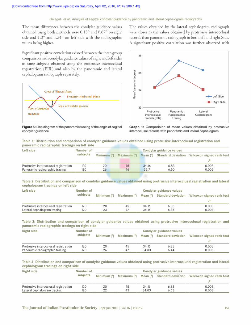
plane parallel to the floor of the mouth. All radiographs were made by the same panoramic radiographic unit. The images were acquired at 74 kVp and 10 mA. Two radio‑opaque lines are consistently apparent on the radiographs in the region of the temporal bone. One depicts the outline of the articular eminence and fossa, the second, the inferior border of the zygomatic arch. Tracings of the radiographic images were made on a transparent sheet. A horizontal reference line was marked by joining the orbitale and porion. The most superior and the most inferior points of the curvatures were identified. These two lines were connected by a straight line representing the mean curvature line. Condylar Guidance angles made by the intersection of the mean curvature line and the horizontal reference line traced were measured [Figure 5].
Statistical analysisThe results obtained were subjected to statistical analysis in SPSS 16 software (Statistical Package for the Social Sciences, IBM Software Group, USA). The Paired t‑test was applied for intergroup analysis. Spearman’s‑Rho test was used to find the correlation between the sagittal condylar guidance between two radiographic methods on both sides and also between left
and right sides. Further, Wilcoxon signed rank test was applied to check the significance between the right and left condylar guidance determined by two radiographic methods.
RESULTS
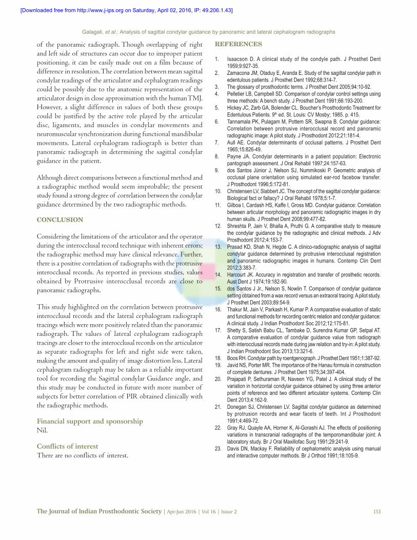
A total of 120 dentulous subjects participated in the study. Tables 1 and 2 summarize left whereas Tables 3 and 4 summarize right mean and standard deviation of condylar guidance values. The angles of the condylar guidance and standard deviations were measured by the protrusive interocclusal record and with panoramic and lateral cephalogram radiographic methods. The correlation between the condylar guidance angles measured by protrusive interocclusal record and panoramic radiographic image were compared with condylar guidance angles measured by protrusive interocclusal record and lateral cephalometric radiographs on both right and left separately [Graph 1].
Figure 1: Protrusive interocclusal records Figure 2: Mounting of casts on semi-adjustable articulator
Figure 3: Panaromic radiograph of the subject showing the tracing of the angle of sagittal condylar guidance. Red line: Outline of articular fossa and eminence. Yellow line: Frankfurt horizontal plane. Blue line: Sagittal condylar path inclination
Figure 4: Lateral cephalogram radiograph of the subject showing the tracing of the angle of sagittal condylar guidance. Yellow line: Frankfurt horizontal plane Blue line: Sagittal condylar path inclination
[Downloaded free from http://www.j-ips.org on Saturday, April 02, 2016, IP: 49.206.1.43]
Galagali, et al.: Analysis of sagittal condylar guidance by panoramic and lateral cephalogram radiographs
The Journal of Indian Prosthodontic Society | Apr-Jun 2016 | Vol 16 | Issue 2 151
The mean differences between the condylar guidance values obtained using both methods were 0.13° and 0.67° on right side and 1.0° and 1.54° on left side with the radiographic values being higher.
Significant positive correlation existed between the inter‑group comparison with condylar guidance values of right and left sides in same subjects obtained using the protrusive interocclusal registration (PIR) and also by the panoramic and lateral cephalogram radiograph separately.
The values obtained by the lateral cephalogram radiograph were closer to the values obtained by protrusive interocclusal records than panoramic radiograph in both left and right Side. A significant positive correlation was further observed with
Table 1: Distribution and comparison of condylar guidance values obtained using protrusive interocclusal registration and panoramic radiographic tracings on left sideLeft side Number of
subjectsCondylar guidance values
Minimum (°) Maximum (°) Mean (°) Standard deviation Wilcoxon signed rank test
P
Protrusive interocclusal registration 120 20 45 34.16 6.83 0.003Panoramic radiographic tracing 120 26 46 35.7 6.50 0.005
Table 2: Distribution and comparison of condylar guidance values obtained using protrusive interocclusal registration and lateral cephalogram tracings on left sideLeft side Number of
subjectsCondylar guidance values
Minimum (°) Maximum (°) Mean (°) Standard deviation Wilcoxon signed rank test
P
Protrusive interocclusal registration 120 20 45 34.16 6.83 0.003Lateral cephalogram tracing 120 23 47 35.16 5.85 0.003
Table 3: Distribution and comparison of condylar guidance values obtained using protrusive interocclusal registration and panoramic radiographic tracings on right sideRight side Number of
subjectsCondylar guidance values
Minimum (°) Maximum (°) Mean (°) Standard deviation Wilcoxon signed rank test
P
Protrusive interocclusal registration 120 20 45 34.16 6.83 0.003Panoramic radiographic tracing 120 26 47 34.83 6.44 0.005
Table 4: Distribution and comparison of condylar guidance values obtained using protrusive interocclusal registration and lateral cephalogram tracings on right sideRight side Number of
subjectsCondylar guidance values
Minimum (°) Maximum (°) Mean (°) Standard deviation Wilcoxon signed rank test
P
Protrusive interocclusal registration 120 20 45 34.16 6.83 0.003Lateral cephalogram tracing 120 22 43 34.03 6.63 0.003
Figure 5: Line diagram of the panoramic tracing of the angle of sagittal condylar guidance
33
34
35
36
Protrusiveinterocclusalrecords (PIR)
PanoramicRadiographic
Tracing
LateralCephalogram
Mea
n V
alue
s in
deg
rees
Left Side
Right Side
Graph 1: Comparision of mean values obtained by protrusive interocclusal records with panoramic and lateral cephalogram
[Downloaded free from http://www.j-ips.org on Saturday, April 02, 2016, IP: 49.206.1.43]

Galagali, et al.: Analysis of sagittal condylar guidance by panoramic and lateral cephalogram radiographs
152 The Journal of Indian Prosthodontic Society | Apr-Jun 2016 | Vol 16 | Issue 2
P values obtained by Wilcoxon signed rank test were 0.003 and 0.005 in left and right side, respectively with left side being highly significant.
DISCUSSION
During any prosthodontic rehabilitation, it is of utmost importance to restore the patient’s occlusion which coincides with centric relation and to provide an occlusion free of interference.[13] Sometimes, frictional inhibition of movement of the condylar components of the articulator also introduces errors in the values of the condylar guidance.[14‑16]
Condylar guidance is described as the mandibular guidance generated by the condyle and articular disc traversing the contour of the glenoid fossae or, synonymously, as the mechanical form located in the upper posterior region of an articulator that controls movement of the mobile member.[17]
The angle of condylar guidance was determined by relating the sagittal condylar path inclination to the Camper’s plane (ala of the nose to the tragus).[18] However, the reproducibility of these temporomandibular joint (TMJ) specific views is questionable, as also the use of a reference plane which does not rely on stable bony landmarks which can lead to further incorporation of errors. The HANAU™ Wide‑Vue Articulator with fixed intercondylar distance can be set using the protrusive interocclusal registration.[14,19]
Hence, the present study utilized Hanau springbow which relies on the Frankfurt’s Horizontal plane to transfer the patients’ relation to the articulator. The same plane is readily demonstrable on a panoramic and cephalometric radiograph by joining the porion and the orbitale landmarks [Figure 5].
The panoramic and lateral cephalogram radiographic image of the sagittal outline of the articular eminence and glenoid fossae was clearly identified in all the 120 subjects. Inter‑examiner reliability in identification of the radiographic outline of the articular eminence and determination of condylar guidance by the radiographic method showed suitable mean value difference of 0.13° and 0.67° on the right side and 1.0° and 1.54° on the left side.
The average condylar guidance by the interocclusal method was 34.16° on both right and left side. The mean condylar guidance values obtained using the panoramic radiographic method was 34.83° on the right side and 35.7° on the left side. Whereas condylar guidance values obtained using the lateral cephalogram radiographic method was 34.03° on the right side and 35.16° on the left side. Literature suggests that the right and left eminences seldom have exactly the same slants,
contours, and declivities.[7] Similarly, the present study showed a lesser mean difference between the right and left sides by the protrusive interocclusal record method than the panoramic radiograph and lateral cephalogram method, highlighting the inherent differences in the method of determination of condylar guidance.
Before making of the prosthesis, the jaws are to be positioned on an articulator in the three dimensions of space. This needs the recording of the orientation, vertical and horizontal relation.[20]
A significant positive correlation was observed between the condylar guidance acquired using protrusive interocclusal records and panoramic images for both right and left sides was P = 0.005. Further more significant positive correlation was observed between the condylar guidance acquired using protrusive interocclusal records and lateral cephalogram images for right and left side P = 0.003. A study comparing the radiographic image of the sagittal condylar path inclination and its actual anatomic outline in dry skulls found that the radiographic values were on an average 7° greater than the skull values.[11] The present study found that the condylar guidance values by both the radiographic method were greater than the protrusive interocclusal method for the right and left sides.
There are some limitations of the radiographic method concerning panoramic distortion, head and reference plane orientation, and difficulty in distinguishing the articular eminence outline from the zygomatic arch. Further, the articular eminence inclination in the radiographic image was traced. This may be different from the guiding inclination with approximately 4–6 mm of protrusion, which is the clinically significant range of protrusion and condylar guidance.[21]
Despite these drawbacks, the panoramic radiograph is useful for comparison between right and left sides since it shows both the TMJs with relatively same magnification errors (×1.2). It is a reproducible radiograph unlike the other TMJ specific radiographs which are subject to projection errors.[22]
Digital lateral cephalogram was selected in this study to obtain individual sagittal condylar guidance value which was taken as a standard for critical comparison with two groups. According to Davis and Mackay,[23] digital imaging with interactive computer processing have added benefits of high‑quality images, speed of application, low radiation dosage, direct analysis and as accurate as manual technique. Lateral cephalogram reveals accurate morphology of the articular eminence and anatomic landmarks to draw reference planes, i.e. Frankfort horizontal plane in single image. The exposure time for lateral cephalogram radiograph is 6–7 times less than that
[Downloaded free from http://www.j-ips.org on Saturday, April 02, 2016, IP: 49.206.1.43]
Galagali, et al.: Analysis of sagittal condylar guidance by panoramic and lateral cephalogram radiographs
The Journal of Indian Prosthodontic Society | Apr-Jun 2016 | Vol 16 | Issue 2 153
of the panoramic radiograph. Though overlapping of right and left side of structures can occur due to improper patient positioning, it can be easily made out on a film because of difference in resolution. The correlation between mean sagittal condylar readings of the articulator and cephalogram readings could be possibly due to the anatomic representation of the articulator design in close approximation with the human TMJ. However, a slight difference in values of both these groups could be justified by the active role played by the articular disc, ligaments, and muscles in condylar movements and neuromuscular synchronization during functional mandibular movements. Lateral cephalogram radiograph is better than panoramic radiograph in determining the sagittal condylar guidance in the patient.
Although direct comparisons between a functional method and a radiographic method would seem improbable; the present study found a strong degree of correlation between the condylar guidance determined by the two radiographic methods.
CONCLUSION
Considering the limitations of the articulator and the operator during the interocclusal record technique with inherent errors; the radiographic method may have clinical relevance. Further, there is a positive correlation of radiographs with the protrusive interocclusal records. As reported in previous studies, values obtained by Protrusive interocclusal records are close to panoramic radiographs.
This study highlighted on the correlation between protrusive interocclusal records and the lateral cephalogram radiograph tracings which were more positively related than the panoramic radiograph. The values of lateral cephalogram radiograph tracings are closer to the interocclusal records on the articulator as separate radiographs for left and right side were taken, making the amount and quality of image distortion less. Lateral cephalogram radiograph may be taken as a reliable important tool for recording the Sagittal condylar Guidance angle, and this study may be conducted in future with more number of subjects for better correlation of PIR obtained clinically with the radiographic methods.
Financial support and sponsorshipNil.
Conflicts of interestThere are no conflicts of interest.
REFERENCES
1. Isaacson D. A clinical study of the condyle path. J Prosthet Dent 1959;9:927‑35.
2. Zamacona JM, Otaduy E, Aranda E. Study of the sagittal condylar path in edentulous patients. J Prosthet Dent 1992;68:314‑7.
3. The glossary of prosthodontic terms. J Prosthet Dent 2005;94:10‑92.4. Pelletier LB, Campbell SD. Comparison of condylar control settings using
three methods: A bench study. J Prosthet Dent 1991;66:193‑200.5. Hickey JC, Zarb GA, Bolender CL. Boucher’s Prosthodontic Treatment for
Edentulous Patients. 9th ed. St. Louis: CV Mosby; 1985. p. 415.6. Tannamala PK, Pulagam M, Pottem SR, Swapna B. Condylar guidance:
Correlation between protrusive interocclusal record and panoramic radiographic image: A pilot study. J Prosthodont 2012;21:181‑4.
7. Aull AE. Condylar determinants of occlusal patterns. J Prosthet Dent 1965;15:826‑49.
8. Payne JA. Condylar determinants in a patient population: Electronic pantograph assessment. J Oral Rehabil 1997;24:157‑63.
9. dos Santos Júnior J, Nelson SJ, Nummikoski P. Geometric analysis of occlusal plane orientation using simulated ear‑rod facebow transfer. J Prosthodont 1996;5:172‑81.
10. Christensen LV, Slabbert JC. The concept of the sagittal condylar guidance: Biological fact or fallacy? J Oral Rehabil 1978;5:1‑7.
11. Gilboa I, Cardash HS, Kaffe I, Gross MD. Condylar guidance: Correlation between articular morphology and panoramic radiographic images in dry human skulls. J Prosthet Dent 2008;99:477‑82.
12. Shreshta P, Jain V, Bhalla A, Pruthi G. A comparative study to measure the condylar guidance by the radiographic and clinical methods. J Adv Prosthodont 2012;4:153‑7.
13. Prasad KD, Shah N, Hegde C. A clinico‑radiographic analysis of sagittal condylar guidance determined by protrusive interocclusal registration and panoramic radiographic images in humans. Contemp Clin Dent 2012;3:383‑7.
14. Harcourt JK. Accuracy in registration and transfer of prosthetic records. Aust Dent J 1974;19:182‑90.
15. dos Santos J Jr., Nelson S, Nowlin T. Comparison of condylar guidance setting obtained from a wax record versus an extraoral tracing: A pilot study. J Prosthet Dent 2003;89:54‑9.
16. Thakur M, Jain V, Parkash H, Kumar P. A comparative evaluation of static and functional methods for recording centric relation and condylar guidance: A clinical study. J Indian Prosthodont Soc 2012;12:175‑81.
17. Shetty S, Satish Babu CL, Tambake D, Surendra Kumar GP, Setpal AT. A comparative evaluation of condylar guidance value from radiograph with interocclusal records made during jaw relation and try‑in: A pilot study. J Indian Prosthodont Soc 2013;13:321‑6.
18. Boos RH. Condylar path by roentgenograph. J Prosthet Dent 1951;1:387‑92.19. Javid NS, Porter MR. The importance of the Hanau formula in construction
of complete dentures. J Prosthet Dent 1975;34:397‑404.20. Prajapati P, Sethuraman R, Naveen YG, Patel J. A clinical study of the
variation in horizontal condylar guidance obtained by using three anterior points of reference and two different articulator systems. Contemp Clin Dent 2013;4:162‑9.
21. Donegan SJ, Christensen LV. Sagittal condylar guidance as determined by protrusion records and wear facets of teeth. Int J Prosthodont 1991;4:469‑72.
22. Gray RJ, Quayle AA, Horner K, Al‑Gorashi AJ. The effects of positioning variations in transcranial radiographs of the temporomandibular joint: A laboratory study. Br J Oral Maxillofac Surg 1991;29:241‑9.
23. Davis DN, Mackay F. Reliability of cephalometric analysis using manual and interactive computer methods. Br J Orthod 1991;18:105‑9.
[Downloaded free from http://www.j-ips.org on Saturday, April 02, 2016, IP: 49.206.1.43]
![Conservative Approach to Unilateral Condylar Fracture in a … · 2016-10-09 · of condylar fractures [7]. It appears that pediatric condylar fractures could be managed by closed](https://img.pdfslide.net/doc/110x75/5f48360e47a39a42e102f2f1/conservative-approach-to-unilateral-condylar-fracture-in-a-2016-10-09-of-condylar.jpg)


![Oral & Maxillofacial Surgeryopenaccessebooks.com/oral-maxillofacial-surgery/condylar-fractures.pdftreatment of mandibular condylar process fractures [10]. It was found that treatment](https://img.pdfslide.net/doc/110x75/5e27326a457720282958fba6/oral-maxillofacial-sur-treatment-of-mandibular-condylar-process-fractures.jpg)