Embed Size (px)
Citation preview

—Original—
Comparative analysis of the intestinal flora in type 2 diabetes and nondiabetic mice
Masanori Horie1), Takamasa Miura2), Satomi HirakaTa2), akira HoSoyaMa2), Sakiko Sugino1), aya uMeno1), kazutoshi MuroToMi1), yasukazu yoSHida1), and Taisuke koike3)
1)Health Research Institute, National Institute of Advanced Industrial Science and Technology (AIST), 2217-14 Hayashi-Cho, Takamatsu, Kagawa 761-0395, Japan
2)Biological Resource Center (NBRC), National Institute of Technology and Evaluation (NITE), 2-49-10 Nishihara, Shibuya-ku, Tokyo 151-0066, Japan
3)Mitsubishi-Chemical Foods Corporation, 2-13-10 Nihonbashi, Chuo-ku, Tokyo 103-0027, Japan
Abstract: A relationship between type 2 diabetes mellitus (T2DM) and intestinal flora has been suggested since development of analysis technology for intestinal flora. An animal model of T2DM is important for investigation of T2DM. Although there are some animal models of T2DM, a comparison of the intestinal flora of healthy animals with that of T2DM animals has not yet been reported. The intestinal flora of Tsumura Suzuki Obese Diabetes (TSOD) mice was compared with that of Tsumura, Suzuki, Non Obesity (TSNO) mice in the present study. The TSOD mice showed typical type 2 diabetes symptoms, which were high-fat diet-independent. The TSOD and the TSNO mouse models were derived from the same strain, ddY. In this study, we compared the intestinal flora of TSOD mice with that if TSNO mice at 5 and 12 weeks of age. We determined that that the number of operational taxonomic units (OTUs) was significantly higher in the cecum of TSOD mice than in that of TSNO mice. The intestinal flora of the cecum and that of the feces were similar between the TSNO and the TSOD strains. The dominant bacteria in the cecum and feces were of the phyla Firmicutes and Bacteroidetes. However, the content of some bacterial species varied between the two strains. The percentage of Lactobacillus spp. within the general intestinal flora was higher in TSOD mice than in TSNO mice. In contrast, the percentages of order Bacteroidales and family Lachnospiraceae were higher in TSNO mice than in TSOD mice. Some species were observed only in TSOD mice, such as genera Turicibacter and SMB53 (family Clostridiaceae), the percentage of which were 3.8% and 2.0%, respectively. Although further analysis of the metabolism of the individual bacteria in the intestinal flora is essential, genera Turicibacter and SMB53 may be important for the abnormal metabolism of type 2 diabetes.Key words: intestinal flora, Lactobacillus, TSOD mouse, Turicibacter, type 2 diabetes
Introduction
Several species of bacteria can form complicated eco-systems in the intestines. These bacteria are referred to as intestinal flora. The constituent bacteria of the intes-
tinal flora are affected by various environmental factors such as diet. Furthermore, the composition of the intes-tinal flora varies depending on the generations. Recent-ly, investigations on the interactions of the intestinal flora with disease and beauty care have increased [14,
(Received 22 February 2017 / Accepted 18 June 2017 / Published online in J-STAGE 12 July 2017)Address corresponding: M. Horie, Health Research Institute, National Institute of Advanced Industrial Science and Technology (AIST), 2217-14 Hayashi-Cho, Takamatsu, Kagawa 761-0395, Japan
Exp. Anim. 66(4), 405–416, 2017
©2017 Japanese association for Laboratory animal Science

M. Horie, ET AL.406
18, 30, 31, 45]. For example, “equol-producing bacteria” in the intestinal flora were shown to be associated with prevention of menopausal discomfort [29]. The intestinal flora is also reported to be associated with gut immu-nity and lifestyle-related diseases [17]. T2DM is an important lifestyle disease. Features of T2DM are glu-cose tolerance, hyposecretion, and insulin resistance. According to the Global Report on Diabetes by the World Health Organization (WHO) [42], the number of indi-viduals with diabetes was 422 million in 2014, indicating its global prevalence. However, the etiology of T2DM is complex. The cause of the disease is still unclear. Generally, genetic and environmental factors are con-sidered the causes of T2DM [39, 44]. Interestingly, re-cent studies have suggested interactions between T2DM and the intestinal flora [5, 19, 40]. For example, a de-crease in intestinal Akkermansia muciniphila was ob-served prior to the incidence of DM and inflammatory bowel disease [43]. However, our current understanding of the interactuin between T2DM and the intestinal flora is still incomplete. In order to fully understand this proposed association, studies using animal models are essential. The mouse is typically utilized as a T2dM model. Although the composition of the intestinal flora differs between mice and humans, understanding the differences in the intestinal flora between T2DM animals and healthy animals is still important. generally, T2dM mouse models are generated as a result of intake of a high-fat diet. This model mimics human T2DM, because many cases of human T2dM are also caused by obesity, which is the result of overeating and physical inactivity. Although a high-fat-intake mouse model is suitable for mimicking human T2DM, it should be noted that diet affects the intestinal flora. In particular, high-fat diets affect the intestinal flora [3, 26]. Therefore, if the intes-tinal flora is analyzed for a mouse model with intake of a hifh-fat diet compared with that of healthy mice, the cause of any observed difference in the intestinal flora becomes complex. We should therefore consider not only diabetes but also components of the diet as causes of the variation in intestinal flora. Currently, the complicated relationship between diabetes and the intestinal flora is generating significant debate in the scientific commu-nity. Therefore, we analyzed the intestinal flora of a simple mouse model of diabetes in comparison with that of nondiabetic mice. Tsumura Suzuki obese diabetes (TSOD) mice are inbred mice derived from the ddY strain [36]. TSOD mice exhibit symptoms similar to
human T2DM. However, the T2DM symptoms of the TSOD mouse do not depend on intake of a high-fat diet. The T2DM symptoms of TSOD are caused by both ge-netic and environmental factors, which means that and overeating. It has been reported that three quantitative trait loci are involved in the T2dM symptoms in TSod mouse [13]. The control mouse model was the Tsumura, Suzuki, Non Obesity (TSNO) mouse, which does not have diabetes. TSno mice are also derived from the ddy strain. Compared with TSNO mice, TSOD mice eat a heavy diet. As a result, their body weight increases with age. In addition, their blood glucose and insulin levels increase [36]. The TSOD strain also shows increases in the total cholesterol and triglyceride (TG) levels in the blood Increases in TNF-α, IL-6, and oxidative stress markers in the blood have also been reported [27]. Al-though previous studies have investigated diabetes symptoms, no study has compared the intestinal flora between these strains. Therefore, to collect basic data for animal models of diabetes, we compared the intesti-nal flora of the TSOD mouse with that of the TSNO mouse.
Materials and Methods
Experimental designAll animal experiments were approved by the Institu-
tional Animal Care and Use Committee of the National institute of advanced industrial Science and Technol-ogy (AIST) (Authorization reference number: Dou2015-246). We employed two different animal strains, TSNO and TSOD. Both strains were derived from the same strain of mice, ddY. Four- and eleven-week-old male TSOD mice and male TSNO mice (control) were ob-tained from the institute for animal reproduction (Ibaraki, Japan). The animals were housed individually and had free access to standard food (MF; Oriental Yeast Co., Ltd., Tokyo, Japan) and water. The animal room was maintained at 23 ± 2°C and 50 ± 10% humidity under a 12 h light (8:00–20:00) and dark (20:00–8:00) cycle. Mice were habituated for 1 week and then dis-sected. The mice were anesthetized by inhalation of isoflurane (Wako Pure Chemical Industries, Ltd., Osaka, Japan). During autopsy, blood was taken from the heart, and the intestine was excised. The intestine was imme-diately frozen using dry ice.
DNA elution and intestinal flora analysis were per-formed in 4 different groups of animals: 5-week-old

INTESTINAL FLORA IN TYPE 2 DIABETES MICE 407
TSNO mice (n=5), 5-week-old TSOD mice (n=5), 12-week-old TSNO mice (n=5), and 12-week-old TSOD mice (n=5). Twelve-week-old TSOD mice developed diabetes. Small intestine content, cecal content, and fe-ces were analyzed in all experimental animals. After 1 week of habituation, body weight was assessed. Blood was then collected from the heart under anesthesia, and the blood sugar level was immediately measured. After hemorrhagic death, the small and large intestines were collected into 15 ml tubes. The intestines were immedi-ately frozen using dry ice and stored at −80°C until examination. The tissue samples were thawed on ice, and the large intestine samples were divided into the cecum, ileum, and rectum by surgical knife. The contents of the intestine were collected using a spatula. The small intestine was flushed with 1 ml of physiological saline using a syringe. The lavage fluid was collected and cen-trifuged at 20,000× g for 5 min, and the precipitate was used for testing.
Biochemical assayBlood sugar levels were measured using FreeStyle
Freedom Lite (NIPRO Corporation, Osaka, Japan). Plasma insulin concentration was measured using a Mouse Insulin ELISA KIT (Shibayagi Co., Ltd., Shibu-kawa, Japan). The concentration of macrophage inflam-matory protein 2 (MIP-2) in plasma was measured using a Mouse CXCL2/MIP-2 Quantikine ELISA Kit (R&D Systems, inc., Minneapolis, Mn, uSa). The lipid per-oxidation products, 10-hydroxy-8E,12Z-octadecadieno-ic acid (10-(Z,E)-HODE) and 12-hydroxy-9Z,13E-octa-decadienoic acid (12-(Z,E)-HODE), were obtained from Larodan Fine Chemicals (Solna, Stockholm, Sweden). The levels of HODEs were measured by the slightly modified method of a previously report [38].
DNA extractionFrozen tissue samples were melted on ice, and DNA
was extracted by ISOFECAL for Beads Beating (Nippon Gene Co., Ltd., Tokyo, Japan). Because the weights of the collected individual intestine samples were different, the maximum weight of the sample was collected into tubes. For feces, approximately 100 mg was collected per sample. DNA extraction was performed according to the manufacturer’s protocol, excluding instructions regarding sample weight. The size of the eluted DNA was assessed using a Genofield system (ATTO Corpora-tion, Tokyo, Japan). The purity was estimated based on
the absorbance at A260/A280 measured by a NanoDrop 2000c spectrophotometer (Thermo Fisher Scientific, Waltham, MA, USA). The concentration of double-stranded DNA was measured using a Varioskan Flash spectral scanning multimode reader (Thermo Fisher Scientific) along with PicoGreen (Thermo Fisher Scien-tific).
Preparation of a DNA libraryDNA samples of small intestine content were prepared
at 1.5 ng/µl. Other DNA samples were prepared at 5 ng/µl. The sequences of forward and reverse primers with adapters were as follows: 5ʹ-TCGTCGGCAGCGTCAGA T G T G T A T A A G A G A C A G - N N -[CCTACGGGNGGCWGCAG]-3 ′ (Miseq-1st -Bakt_341F) and 5′-GTCTCGTGGGCTCGGAGATGT-G T A T A A G A G A C A G - N N -[ G A C T A C H V G G G T A T C T A A T C C ] - 3 ′ (Miseq-1st-Bakt_805R). The overhang sequences are underlined, and the 16S rRNA-specific sequences have previously been described [8, 12]. The first PCR ampli-fication was performed (n=3) using TaKaRa Ex Taq HS DNA polymerase (TaKaRa Bio Inc., Kusatsu, Japan) with the following program: one cycle at 94°C for 2 min; 19–26 cycles at 98°C for 10 s, 55°C for 30 s, and 72°C for 45 s; and finally a hold at 12°C. PCR amplification was conducted in a total volume of 25 µl containing 2.5 µl of 10× Ex Taq Buffer, 2.5 µl of dNTP mixture (2.5 mM each), 1 µl of each primer (6 µM), 0.2 µl of Ta-KaRa Ex Taq HS (5 U/µl), 2 µl of template dna, and 15.8 µl of sterilized distilled water. The three PCR samples were collected in one tube, and the PCR prod-ucts were purified using SPRIselect Beads (Beckman Coulter, Inc., Brea, CA, USA). Subsequently, secondary PCR was performed using Illumina Nextera XT Index primers (Illumina, Inc., San Diego, CA, USA). Second-ary PCR amplification was done using KOD -Plus- Ver.2 DNA polymerase (Toyobo Co., Ltd., Osaka, Japan) with the following program: one cycle at 94°C for 2 min; 8 cycles at 98°C for 10 s, 55°C for 30 s, and 68°C for 45 s; and finally a hold at 12°C. PCR amplification was conducted in a total volume of 50 µl containing 5 µl of 10× buffer for KOD -Plus- Ver.2, 5 µl of dNTPs (2 mM each), 3 µl of 25 mM MgSO4, 1.5 µl of Index 1 (i7) N7 x x Primer (6 µM), 1.5 µl of Index 2 (i5) S5 x x Primer (6 µM), 1 µl of KOD -Plus- (1 U/µl), 5 µl of template DNA (1 ng/µl), and 28 µl of sterilized distilled water. The PCR products were purified again using SPRIselect

M. Horie, ET AL.408
Beads. The sequence run was performed by paired-end sequencing (300 bp × 2) using the Miseq Reagent Kit v3 (600 cycles). DNA sequences were deposited in the DDBJ Sequence Read Archive under accession number DRA005323.
Data analysisThe forward and reverse paired-end reads with 300
bp were trimmed to 258 bp using fastx_trimmer with options (-f 3 and –l 260) in FASTX-Toolkit 0.0.14 (http://hannonlab.cshl.edu/fastx_toolkit/). Bacterial community analyses based on 16S rRNA genes were performed using the QIIME software package (Version
Fig. 1. Biochemical analysis of TSOD and TSNO mice. Body weight was measured at 5 and 12 weeks of age. Mice were dissected, and blood was collected from the heart. Blood sugar levels were measured using a blood glucose meter. Plasma insulin and MIP-2 concentrations were determined by ELISA. The levels of 10- and 12-(Z, E)-HODE was determined by liquid chromatography-mass/mass spectrometry. Statistical significance between the TSOD mice and the TSNO mice was determined using an unpaired t-test.
INTESTINAL FLORA IN TYPE 2 DIABETES MICE 409
1.9.1) [6] and the Greengenes 13_8 dataset [7] as refer-ence. The trimmed reads were joined according to the fastq-join parameter [2] within the join_paired_ends.py script. The joined reads were quality filtered and merged into one file using the split_librarys_fastq.py script with options (–q 19 and –barcord_type ‘not-barcoded’). The sequences were clustered at 97% sequence identity using USEARCH v6.1.544 [9] against the Greengenes dataset. Chimeric sequences were detected using the Chime-raSlayer algorithm [10] within the identify_chimeric_seqs.py script. After filtering of the chimeric sequences, reassignment using the assign_taxonomy.py script along with the RDP classifier [41] was performed to assign the taxonomy of each representative sequence of OTUs. To calculate beta diversity, the UniFrac [21] was used within the beta_diversity_through_plots.py script along with a tree file that was constructed with a set of repre-sentative sequences of the OTUs using the make_phy-logeny.py script with the default parameter. The alpha diversity was calculated using the alpha_diversity.py script. Both diversities were calculated using 72,000 reads.
Results
Body weight, blood glucose level, and biochemical analysis
Compared with the TSNO mice, the body weights of the TSOD mice were higher in both 5-week-old and 12-week-old mice (Fig. 1). The body weights of the TSOD mice were 164 and 170% higher than those of the TSNO mice at 5 and 12 weeks of age, respectively. Fur-thermore, the blood sugar levels of the TSOD mice were 158 and 197% higher at 5 and 12 weeks of age, respec-tively, than those of the age-matched TSNO mice. The plasma insulin level was also higher in TSOD mice than in TSno mice. The plasma insulin levels of the TSod mice were 8.7 and 10.3 times higher than those of the TSNO mice at 5 and 12 weeks of age, respectively. These results suggest that 12-week-old TSOD mice exhibited a diabetic state. Moreover, the plasma MIP-2 concentra-tion in TSOD mice was significantly higher than in TSNO mice. Lipid hydroperoxides, 10- and 12-(Z,E)-hydroxyoctadecadienoic acids (HODE), in blood in-crease in early stage of type 2 diabetes [28, 38]. These lipid hydroperoxides were significantly higher in 5-week-old TSOD mice than in TSNO mice. There were no significant differences in 10- and 12-(Z,E)-HODE
between the 12-week-old TSOD mice and TSNO mice. According to these data, 5-week-old TSOD mice were in the preclinical stage of T2DM, and 12-week-old TSod mice had T2dM.
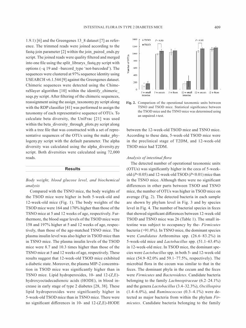
Analysis of intestinal floraThe detected number of operational taxonomic units
(OTUs) was significantly higher in the ceca of 5-week-old (P<0.05) and 12-week-old TSOD (P<0.01) mice than in the TSNO mice. Although there were no significant differences in other parts between TSOD and TSNO mice, the number of OTUs was higher in TSOD mice on average (Fig. 2). The detected bacteria in each sample are shown by phylum level in Fig. 3 and by species level in Fig. 4. The number of bacterial species in feces that showed significant differences between 12-week-old TSOD and TSNO mice was 26 (Table 1). The small in-testine was subject to occupancy by the- Firmicutes bacteria (>91.0%). In TSNO mice, the dominant species were Candidatus Arthromitus spp. (26.6–83.2%) in 5-week-old mice and Lactobacillus spp. (51.1–63.4%) in 12-week-old mice. In TSOD mice, the dominant spe-cies were Lactobacillus spp. in both 5- and 12-week-old mice (54.9–82.0% and 59.1–77.5%, respectively). The microbial flora in the cecum was similar to that in the feces. The dominant phyla in the cecum and the feces were Firmicutes and Bacteroidetes. Candidate bacteria belonging to the family Lachnospiraceae (6.2–24.1%) and the genera Lactobacillus (3.4–32.3%), Oscillospira (1.4–6.0%), and Ruminococcus (0.3–4.1%) were de-tected as major bacteria from within the phylum Fir-micutes. Candidate bacteria belonging to the family
Fig. 2. Comparison of the operational taxonomic units between TSNO and TSOD mice. Statistical significance between the TSOD mice and the TSNO mice was determined using an unpaired t-test.

M. Horie, ET AL.410
S24-7 (6.4–32.9%), family Rikenellaceae (0.7–11.0%), and genus Prevotella (0.1–3.1%) and Bacteroides acid-ifaciens (0.9–10.8%) were detected as major bacteria from within the phylum Bacteroidetes.
In order to compare the diversity among each bacte-rial flora, the UniFrac distance was calculated, and Prin-cipal Coordinate Analysis (PCoA) was performed (Fig. 5). As shown in Fig. 5, the small intestine content was positioned away from the other parts examined in the PCoA. According to the unweighted UniFrac analysis,
the detected species were different between the TSNO and TSOD mice. However, weighted UniFrac analysis showed that the interstrain difference in flora between TSOD and TSNO mice was small except in the small intestine content. Bifidobacteria were detected only in TSOD mice (Table 1). Significant interstrain differences were observed in 26 bacterial species in the feces of 12-week-old mice (Table 1). The percentage of Lacto-bacillus spp. against general intestinal flora was higher in TSod mice than in TSno mice. additionally, the
Fig. 3. Intestinal flora of individual mice at the phylum level. DNA was extracted from the small intestine content, cecum content, and feces, and the bacterial flora was analyzed. “Others” includes phyla that accounted for less than 1% in all samples.

INTESTINAL FLORA IN TYPE 2 DIABETES MICE 411
Fig. 4. Intestinal flora of individual mice at the species level. DNA was extracted from the small intestine content, cecum content, and feces, and the bacterial flora was analyzed. “Others” includes species that accounted for less than 1% in all samples.

M. Horie, ET AL.412
Table 1. Bacterial species that showed significant differences infeces between 12-weeks-old TSOD and TSNO mice
Phylum Class order Family genus Species TSno TSod P value (t-test)
Firmicutes Bacilli Turicibacterales Turicibacteraceae Turicibacter 0.000% 3.838% 0.003Firmicutes erysipelotrichi erysipelotrichales erysipelotrichaceae Coprobacillus 0.278% 0.023% 0.003Firmicutes Clostridia Clostridiales Peptostreptococcaceae Clostridium ruminantium 0.000% 0.004% 0.005Firmicutes Clostridia Clostridiales Clostridiaceae SMB53 0.000% 2.004% 0.005Firmicutes Bacilli Lactobacillales Lactobacillaceae Lactobacillus 8.997% 20.668% 0.006Firmicutes Clostridia Clostridiales Lachnospiraceae Coprococcus 1.681% 0.956% 0.009Firmicutes Clostridia Clostridiales Lachnospiraceae [Ruminococcus] 0.000% 0.031% 0.009Proteobacteria deltaproteobacteria desulfovibrionales desulfovibrionaceae Bilophila 0.000% 0.048% 0.009Firmicutes Clostridia Clostridiales Peptostreptococcaceae other other 0.000% 0.005% 0.011Firmicutes Clostridia Clostridiales Peptococcaceae 0.028% 0.012% 0.013Bacteroidetes Bacteroidia Bacteroidales S24-7 24.444% 14.887% 0.013Firmicutes erysipelotrichi erysipelotrichales erysipelotrichaceae 0.719% 0.215% 0.014Proteobacteria gammaproteobacteria enterobacteriales enterobacteriaceae escherichia coli 0.002% 0.023% 0.018actinobacteria actinobacteria Bifidobacteriales Bifidobacteriaceae Bifidobacterium pseudolongum 0.000% 0.153% 0.019Firmicutes Clostridia SHA-98 0.000% 0.011% 0.020Firmicutes Clostridia Clostridiales Lachnospiraceae 17.931% 10.719% 0.028Proteobacteria Betaproteobacteria Burkholderiales Alcaligenaceae Sutterella 0.338% 0.160% 0.029Firmicutes Clostridia Clostridiales ruminococcaceae anaerotruncus 0.704% 0.347% 0.031Firmicutes Clostridia Clostridiales Clostridiaceae Clostridium celatum 0.000% 1.128% 0.032Firmicutes Clostridia Clostridiales Lachnospiraceae Clostridium colinum 0.065% 0.016% 0.035Firmicutes Clostridia Clostridiales Clostridiaceae 0.000% 0.115% 0.037Firmicutes Clostridia Clostridiales ruminococcaceae oscillospira other 0.001% 0.000% 0.040Firmicutes Clostridia Clostridiales Lachnospiraceae Defluviitalea saccharophila 0.000% 0.001% 0.041Firmicutes Clostridia Clostridiales ruminococcaceae ruminococcus callidus 0.280% 1.081% 0.043Firmicutes Clostridia Clostridiales dehalobacteriaceae dehalobacterium 0.177% 0.131% 0.044Firmicutes Clostridia Clostridiales eubacteriaceae anaerofustis 0.004% 0.002% 0.045
Statistical significance between the TSOD mice and the TSNO mice was determined using an unpaired t-test.
Fig. 5. Principal Coordinate Analysis (PCoA) of the intestinal flora of each strain. The left side shows the results of unweighted mean PCoA by the genealogical relationship of the determined bacterial species. The right side shows the results of the weighted mean PCoA by genealogical relationship and read number of deter-mined bacterial species.

INTESTINAL FLORA IN TYPE 2 DIABETES MICE 413
percentages of Bacteroidales and Lachnospiraceae were higher in TSNO mice than in TSOD mice. Some species were observed only in TSOD mice, such as genera Tu-ricibacter and SMB53, and their percentages were 3.8% and 2.0%, respectively.
Discussion
Based on the analysis of intestinal flora in this study, the microbial flora in the cecum is similar to that in feces. This observation suggests that cecal flora can be deter-mined from fecal flora without dissection. Therefore, we can analyze consecutive changes in intestinal flora from an individual animal. In the present study, we collected and analyzed all feces for a 1-week habituation period, and the results of analysis of fecal flora showed vari-ability. Thus in order to analyze the intestinal flora, use of fresh feces collected within at least 24 h is recom-mended. Although the number of OTUs was signifi-cantly higher in the cecum of TSOD mice than in that of TSNO mice, weighted UniFrac analysis showed that the positions of the flora were similar in the cecum and feces of the TSOD and TSNO mice (Figs. 2 and 5). Therefore, the intestinal flora of TSOD mice was not remarkably different from that of TSNO mice. The intestinal flora of the TSOD mice was similar to that of the TSNO mice but not the same. Some bacterial species differed be-tween the intestinal flora of the TSOD mice and that of the TSNO mice. These mice were fed the same diet throughout the experimental period. Therefore, although the TSOD mice ate more than the TSNO mice, the effect of diet on the observed differences in intestinal flora was expected to be small. Ca. Arthromitus sp. was dominant in the small intestine content of 5-week-old TSNO mice. The dominant species in the small intestine contents of TSOD mice and 12-week-old TSNO mice were Lacto-bacillus spp. antimicrobial peptides such as alpha-de-fensin are secreted in the small intestine, and thus, the number of bacteria in the small intestine is small [22, 46]. Lactobacilli have bile and alpha-defensin tolerance [15, 23, 33]. Accordingly, they are one of the dominant species in the small intestine [24], and our findings cor-respond with this observation. It has been reported that lactobacilli in the small intestine are involved in gut immunity via the Peyer’s patch [11]. Although the rela-tive share of lactobacilli in the small intestine contents of the TSNO and TSOD mice were significantly differ-ent, the interaction between this difference and develop-
ment of T2dM is unclear. it is reported that intake of galactose improved hepatic insulin sensitivity in rats compared with intake of glucose and fructose [34] and that intake of an L.reuteri strain improved insulin sen-sitivity [25]. The metabolism of the lactobacilli in each mouse strain, such as sugar metabolism, may be impor-tant. However, we could not analyze this in the present study. additionally, Bifidobacterium was detected only in TSod mice. Some bacteria detected in TSod mice were not present in TSNO mice. The bacteria found only in TSod mice include Turicibacter and SMB53. The relative share of Turicibacter and SMB53 in the cecum of TSOD mice was more than 2%. Turicibacter is a Gram-positive, strictly anaerobic bacterium [4]. Analy-sis of the gut microbiome of the C57BL/6J mouse indi-cated a correlation between exercise and intestinal Tu-ricibacter. Turicibacter in the rectum and feces were decreased by voluntary wheel running (VWR) for 6 weeks [1]. The percentage of Turicibacter in the control group was 0.22% of the total representation. In contrast, the percentage of Turicibacter in the VWR group was 0%. Additionally, Turicibacter in the large intestine was increased by intake of a high resistant starch diet (raw potato starch) in pigs [35]. It has also been reported that Turicibacter was correlated with intestinal butyric acid [47]. However, the metabolism of Turicibacter and its interaction with the host in the intestine are still not clear.
in the present study, Turicibacter was not detected in 5-week-old mice. Turicibacter, however, was found in the cecum and feces of 12-week-old TSOD mice with advanced diabetes. Studies also show that the relative abundance of Turicibacter in obese mice as a result of a high-fat diet was lower than that of mice fed a normal diet [16]. It is possible that the relationship of intestinal flora with diabetes varies between mice with obesity induced by a high-calorie diet and mice with spontaneous odesity, such as the TSOD mouse. We observed differ-ences in the intestinal flora between TSNO and TSOD mice in this study. Particularly, characteristic species such as Turicibacter and SMB53 were observed in 12-week-old TSOD mice. In human studies, signifi-cantly higher counts were observed for Lactobacillus spp.in feces of Japanese T2dM patients than in control subjects [32], and obesity was associated with the intes-tinal flora at the phylum level, with the dominant phylum of the intestinal flora in obese people changed to Acti-nobacteria and Firmicutes and that in lean people changed to Bacteroidetes [37]. These observations were

M. Horie, ET AL.414
consistent with our data (Table 1). In the present study, the kinds of bacteria in the intestinal flora were different between the TSOD mice and the TSNO mice according to unweighted UniFrac analysis (Fig. 5). Diversity of the intestinal flora is associated with obesity and T2DM [20]. Further analysis of metabolites of bacteria in the intes-tinal flora is essential.
Twelve-week-old TSOD mice showed a typical clini-cal state of diabetes. However, observations of the in-testinal flora and diabetes are independent. Therefore, their interaction is still unclear. We have no grasp of cause and effect. Future studies on the metabolism of individual bacteria in the intestinal flora and on the cor-relation between the intestinal flora and diabetes are necessary. in the TSod mouse, T2dM symptoms de-velop into fatty liver and then hepatic disorder. Different from a conventional model, the T2dM symptoms of the TSOD mouse do not depend on intake of high-fat food. The T2dM symptoms of the TSod mouse are caused by genetic factors and overeating of a normal diet. If intestinal flora analysis is effective for determining the pathogenesis of T2DM, it may be a very valuable tool for prevention of T2dM.
Conflict of Interest
This study was funded by Eisai Food & Chemical Co., Ltd. (Tokyo, Japan).
References
1. Allen, J.M., Berg Miller, M.E., Pence, B.D., Whitlock, K., Nehra, V., Gaskins, H.R., White, B.A., Fryer, J.D., and Woods, J.A. 2015. Voluntary and forced exercise differen-tially alters the gut microbiome in C57BL/6J mice. J. Appl. Physiol. 118: 1059–1066. [Medline] [CrossRef]
2. Aronesty, E.2011. ea-utils: Command-line tools for process-ing biological sequencing data; Available online at: http://code.google.com/p/ea-utils.
3. Benno, Y. 1990. Effect of diets on human fecal microflora. Bifidus 4: 1–12.
4. Bosshard, P.P., Zbinden, R., and Altwegg, M. 2002. Tu-ricibacter sanguinis gen. nov., sp. nov., a novel anaerobic, gram-positive bacterium. Int. J. Syst. Evol. Microbiol. 52: 1263–1266. [Medline]
5. Burcelin, R., Serino, M., Chabo, C., Blasco-Baque, V., and Amar, J. 2011. Gut microbiota and diabetes: from pathogen-esis to therapeutic perspective. Acta Diabetol. 48: 257–273. [Medline] [CrossRef]
6. Caporaso, J.G., Kuczynski, J., Stombaugh, J., Bittinger, K., Bushman, F.D., Costello, E.K., Fierer, N., Peña, A.G.,
goodrich, J.k., gordon, J.i., Huttley, g.a., kelley, S.T., Knights, D., Koenig, J.E., Ley, R.E., Lozupone, C.A., Mc-Donald, D., Muegge, B.D., Pirrung, M., Reeder, J., Sevin-sky, J.R., Turnbaugh, P.J., Walters, W.A., Widmann, J., Yatsunenko, T., Zaneveld, J., and Knight, R. 2010. QIIME allows analysis of high-throughput community sequencing data. Nat. Methods 7: 335–336. [Medline] [CrossRef]
7. DeSantis, T.Z., Hugenholtz, P., Larsen, N., Rojas, M., Bro-die, E.L., Keller, K., Huber, T., Dalevi, D., Hu, P., and Ander-sen, G.L. 2006. Greengenes, a chimera-checked 16S rRNA gene database and workbench compatible with ARB. Appl. Environ. Microbiol. 72: 5069–5072. [Medline] [CrossRef]
8. Edberg, F., Andersson, A.F., and Holmström, S.J. 2012. Bac-terial community composition in the water column of a lake formed by a former uranium open pit mine. Microb. Ecol. 64: 870–880. [Medline] [CrossRef]
9. Edgar, R.C. 2010. Search and clustering orders of magnitude faster than BLaST. Bioinformatics 26: 2460–2461. [Med-line] [CrossRef]
10. Haas, B.J., Gevers, D., Earl, A.M., Feldgarden, M., Ward, D.V., Giannoukos, G., Ciulla, D., Tabbaa, D., Highlander, S.K., Sodergren, E., Methé, B., DeSantis, T.Z., Petrosino, J.F., Knight, R., and Birren, B.W. 2011. Chimeric 16S rRNA sequence formation and detection in Sanger and 454-pyrose-quenced PCR amplicons. Genome Res. 21: 494–504. [Med-line] [CrossRef]
11. Hachinura, S. 2007. immune-modulation by lactic acid bac-teria. Nihon Nyusankin Gakkaishi 18: 54–57.
12. Herlemann, D.P., Labrenz, M., Jürgens, K., Bertilsson, S., Waniek, J.J., and Andersson, A.F. 2011. Transitions in bacte-rial communities along the 2000 km salinity gradient of the Baltic Sea. ISME J. 5: 1571–1579. [Medline] [CrossRef]
13. Hirayama, I., Yi, Z., Izumi, S., Arai, I., Suzuki, W., Nagama-chi, Y., Kuwano, H., Takeuchi, T., and Izumi, T. 1999. Genet-ic analysis of obese diabetes in the TSod mouse. Diabetes 48: 1183–1191. [Medline] [CrossRef]
14. Honda, K. and Littman, D.R. 2016. The microbiota in adap-tive immune homeostasis and disease. Nature 535: 75–84. [Medline] [CrossRef]
15. Hsu, K.H., Pei, C., Yeh, J.Y., Shih, C.H., Chung, Y.C., Hung, L.T., and Ou, B.R. 2009. Production of bioactive human alpha-defensin 5 in Pichia pastoris. J. Gen. Appl. Microbiol. 55: 395–401. [Medline] [CrossRef]
16. Jung, M.J., Lee, J., Shin, N.R., Kim, M.S., Hyun, D.W., Yun, J.H., Kim, P.S., Whon, T.W., and Bae, J.W. 2016. Chronic Repression of mTOR Complex 2 Induces Changes in the Gut Microbiota of diet-induced obese Mice. Sci. Rep. 6: 30887. [Medline] [CrossRef]
17. Kataoka, K. 2016. The intestinal microbiota and its role in human health and disease. J. Med. Invest. 63: 27–37. [Med-line] [CrossRef]
18. Kimoto-Nira, H., Aoki, R., Sasaki, K., Suzuki, C., and Mizu-machi, K. 2012. Effect of oral intake of a Lactococcus lactis strain on skin properties of women -a Pilot Study-. Nihon Chikusan Gakkaiho 83: 307–313. [CrossRef]
19. Lau, E., Carvalho, D., Pina-Vaz, C., Barbosa, J.A., and Frei-tas, P. 2015. Beyond gut microbiota: understanding obesity and type 2 diabetes. Hormones (Athens) 14: 358–369. [Med-

INTESTINAL FLORA IN TYPE 2 DIABETES MICE 415
line] 20. Le Chatelier, E., Nielsen, T., Qin, J., Prifti, E., Hildebrand,
F., Falony, G., Almeida, M., Arumugam, M., Batto, J.M., Kennedy, S., Leonard, P., Li, J., Burgdorf, K., Grarup, N., Jørgensen, T., Brandslund, I., Nielsen, H.B., Juncker, A.S., Bertalan, M., Levenez, F., Pons, N., Rasmussen, S., Suna-gawa, S., Tap, J., Tims, S., Zoetendal, E.G., Brunak, S., Clé-ment, K., Doré, J., Kleerebezem, M., Kristiansen, K., Re-nault, P., Sicheritz-Ponten, T., de Vos, W.M., Zucker, J.D., Raes, J., Hansen, T., Bork, P., Wang, J., Ehrlich, S.D., and Pedersen, O. 2013. Richness of human gut microbiome cor-relates with metabolic markers. Nature 500: 541–546. [Med-line] [CrossRef]
21. Lozupone, C. and Knight, R. 2005. UniFrac: a new phylo-genetic method for comparing microbial communities. Appl. Environ. Microbiol. 71: 8228–8235. [Medline] [CrossRef]
22. Mathew, B. and Nagaraj, R. 2015. Antimicrobial activity of human α-defensin 5 and its linear analogs: N-terminal fatty acylation results in enhanced antimicrobial activity of the linear analogs. Peptides 71: 128–140. [Medline] [CrossRef]
23. Messaoudi, S., Madi, A., Prévost, H., Feuilloley, M., Manai, M., Dousset, X., and Connil, N. 2012. In vitro evaluation of the probiotic potential of Lactobacillus salivarius SMXD51. Anaerobe 18: 584–589. [Medline] [CrossRef]
24. Mitsuoka, T. 1982. Role of intestinal flora in human health. Eisei Kagaku 28: 43–59. [CrossRef]
25. Mobini, R., Tremaroli, V., Ståhlman, M., Karlsson, F., Levin, M., Ljungberg, M., Sohlin, M., Bertéus Forslund, H., Per-kins, R., Bäckhed, F., and Jansson, P.A. 2017. Metabolic ef-fects of Lactobacillus reuteri DSM 17938 in people with type 2 diabetes: A randomized controlled trial. Diabetes Obes. Metab. 19: 579–589. [Medline] [CrossRef]
26. Morishita, Y. 1998. Dietary components responsible for the stability and alteration of the intestinal microflora (1). Cho-nai Saikingaku Zasshi 12: 1–12.
27. Murotomi, K., Umeno, A., Yasunaga, M., Shichiri, M., Ishi-da, N., Abe, H., Yoshida, Y., and Nakajima, Y. 2014. Type 2 diabetes model TSOD mouse is exposed to oxidative stress at young age. J. Clin. Biochem. Nutr. 55: 216–220. [Medline] [CrossRef]
28. Murotomi, K., Umeno, A., Yasunaga, M., Shichiri, M., Ishi-da, N., Abe, H., Yoshida, Y., and Nakajima, Y. 2015. Switch-ing from singlet-oxygen-mediated oxidation to free-radical-mediated oxidation in the pathogenesis of type 2 diabetes in model mouse. Free Radic. Res. 49: 133–138. [Medline] [CrossRef]
29. Setchell, K.D., Brown, N.M., and Lydeking-Olsen, E. 2002. The clinical importance of the metabolite equol-a clue to the effectiveness of soy and its isoflavones. J. Nutr. 132: 3577–3584. [Medline]
30. Pedersen, H.K., Gudmundsdottir, V., Nielsen, H.B., Hyot-ylainen, T., nielsen, T., Jensen, B.a., Forslund, k., Hildeb-rand, F., Prifti, E., Falony, G., Le Chatelier, E., Levenez, F., Doré, J., Mattila, I., Plichta, D.R., Pöhö, P., Hellgren, L.I., Arumugam, M., Sunagawa, S., Vieira-Silva, S., Jørgensen, T., Holm, J.B., Trošt, K., Kristiansen, K., Brix, S., Raes, J., Wang, J., Hansen, T., Bork, P., Brunak, S., Oresic, M., Eh-rlich, S.D., and Pedersen, O. 2016. Human gut microbes im-
pact host serum metabolome and insulin sensitivity. Nature 535: 376–381. [Medline] [CrossRef]
31. Pope, J.L., Tomkovich, S., Yang, Y., and Jobin, C. 2017. Microbiota as a mediator of cancer progression and therapy. Transl. Res. 179: 139–154. [Medline]
32. Sato, J., Kanazawa, A., Ikeda, F., Yoshihara, T., Goto, H., Abe, H., Komiya, K., Kawaguchi, M., Shimizu, T., Ogihara, T., Tamura, Y., Sakurai, Y., Yamamoto, R., Mita, T., Fujitani, y., Fukuda, H., nomoto, k., Takahashi, T., asahara, T., Hi-rose, T., Nagata, S., Yamashiro, Y., and Watada, H. 2014. Gut dysbiosis and detection of “live gut bacteria” in blood of Japanese patients with type 2 diabetes. Diabetes Care 37: 2343–2350. [Medline] [CrossRef]
33. Schroeder, B.O., Ehmann, D., Precht, J.C., Castillo, P.A., Küchler, R., Berger, J., Schaller, M., Stange, E.F., and We-hkamp, J. 2015. Paneth cell α-defensin 6 (HD-6) is an anti-microbial peptide. Mucosal Immunol. 8: 661–671. [Medline] [CrossRef]
34. Stahel, P., Kim, J.J., Xiao, C., and Cant, J.P. 2017. Of the milk sugars, galactose, but not prebiotic galacto-oligosaccharide, improves insulin sensitivity in male Sprague-Dawley rats. PLoS One 12: e0172260. [Medline] [CrossRef]
35. Sun, Y., Su, Y., and Zhu, W. 2016. Microbiome-Metabolome Responses in the Cecum and Colon of Pig to a High Resis-tant Starch diet. Front. Microbiol. 7: 779. [Medline] [Cross-Ref]
36. Suzuki, W., Iizuka, S., Tabuchi, M., Funo, S., Yanagisawa, T., Kimura, M., Sato, T., Endo, T., and Kawamura, H. 1999. A new mouse model of spontaneous diabetes derived from ddy strain. Exp. Anim. 48: 181–189. [Medline] [CrossRef]
37. Turnbaugh, P.J., Hamady, M., Yatsunenko, T., Cantarel, B.L., Duncan, A., Ley, R.E., Sogin, M.L., Jones, W.J., Roe, B.A., Affourtit, J.P., Egholm, M., Henrissat, B., Heath, A.C., Knight, R., and Gordon, J.I. 2009. A core gut microbiome in obese and lean twins. Nature 457: 480–484. [Medline] [CrossRef]
38. umeno, a., Shichiri, M., ishida, n., Hashimoto, y., abe, k., Kataoka, M., Yoshino, K., Hagihara, Y., Aki, N., Funaki, M., Asada, Y., and Yoshida, Y. 2013. Singlet oxygen induced products of linoleates, 10- and 12-(Z,E)-hydroxyoctadecadi-enoic acids (HODE), can be potential biomarkers for early detection of type 2 diabetes. PLoS One 8: e63542. [Medline] [CrossRef]
39. Unoki, H., Takahashi, A., Kawaguchi, T., Hara, K., Horiko-shi, M., Andersen, G., Ng, D.P., Holmkvist, J., Borch-John-sen, K., Jørgensen, T., Sandbaek, A., Lauritzen, T., Hansen, T., nurbaya, S., Tsunoda, T., kubo, M., Babazono, T., Hi-rose, H., Hayashi, M., Iwamoto, Y., Kashiwagi, A., Kaku, K., Kawamori, R., Tai, E.S., Pedersen, O., Kamatani, N., Kadowaki, T., Kikkawa, R., Nakamura, Y., and Maeda, S. 2008. SNPs in KCNQ1 are associated with susceptibility to type 2 diabetes in east asian and european populations. Nat. Genet. 40: 1098–1102. [Medline] [CrossRef]
40. Upadhyaya, S. and Banerjee, G. 2015. Type 2 diabetes and gut microbiome: at the intersection of known and unknown. Gut Microbes 6: 85–92. [Medline] [CrossRef]
41. Wang, Q., Garrity, G.M., Tiedje, J.M., and Cole, J.R. 2007. Naive Bayesian classifier for rapid assignment of rRNA se-

M. Horie, ET AL.416
quences into the new bacterial taxonomy. Appl. Environ. Mi-crobiol. 73: 5261–5267. [Medline] [CrossRef]
42. World Health Organization2016. Global report on diabetes. WHO press.
43. Yassour, M., Lim, M.Y., Yun, H.S., Tickle, T.L., Sung, J., Song, Y.M., Lee, K., Franzosa, E.A., Morgan, X.C., Gevers, D., Lander, E.S., Xavier, R.J., Birren, B.W., Ko, G., and Hut-tenhower, C. 2016. Sub-clinical detection of gut microbial biomarkers of obesity and type 2 diabetes. Genome Med. 8: 17. [Medline] [CrossRef]
44. Yasuda, K., Miyake, K., Horikawa, Y., Hara, K., Osawa, H., Furuta, H., Hirota, y., Mori, H., Jonsson, a., Sato, y., Yamagata, K., Hinokio, Y., Wang, H.Y., Tanahashi, T., Na-kamura, N., Oka, Y., Iwasaki, N., Iwamoto, Y., Yamada, Y., Seino, Y., Maegawa, H., Kashiwagi, A., Takeda, J., Maeda, E., Shin, H.D., Cho, Y.M., Park, K.S., Lee, H.K., Ng, M.C., Ma, R.C., So, W.Y., Chan, J.C., Lyssenko, V., Tuomi, T., Nilsson, P., Groop, L., Kamatani, N., Sekine, A., Nakamura,
Y., Yamamoto, K., Yoshida, T., Tokunaga, K., Itakura, M., Makino, H., Nanjo, K., Kadowaki, T., and Kasuga, M. 2008. Variants in KCNQ1 are associated with susceptibility to type 2 diabetes mellitus. Nat. Genet. 40: 1092–1097. [Medline] [CrossRef]
45. Zapata, H.J. and Quagliarello, V.J. 2015. The microbiota and microbiome in aging: potential implications in health and age-related diseases. J. Am. Geriatr. Soc. 63: 776–781. [Medline] [CrossRef]
46. Zhao, C., Wang, I., and Lehrer, R.I. 1996. Widespread ex-pression of beta-defensin hBD-1 in human secretory glands and epithelial cells. FEBS Lett. 396: 319–322. [Medline] [CrossRef]
47. Zhong, Y., Nyman, M., and Fåk, F. 2015. Modulation of gut microbiota in rats fed high-fat diets by processing whole-grain barley to barley malt. Mol. Nutr. Food Res. 59: 2066–2076. [Medline] [CrossRef]