Embed Size (px)
Citation preview

Seediscussions,stats,andauthorprofilesforthispublicationat:https://www.researchgate.net/publication/304064482
Comparativeanalyticalmicrographsof“vassouras”(Baccharis,Asteraceae)
ArticleinRevistaBrasileiradeFarmacognosia·June2016
DOI:10.1016/j.bjp.2016.05.001
CITATIONS
0
READS
26
8authors,including:
Someoftheauthorsofthispublicationarealsoworkingontheserelatedprojects:
HerbariumECTatEmbrapaClimaTemperado,arepositoryforBraziliantemperateand
subtropicalfloraViewproject
SystematicsofBaccharis(Asteraceae:Astereae)Viewproject
GustavoHeiden
BrazilianAgriculturalResearchCorporation(…
76PUBLICATIONS152CITATIONS
SEEPROFILE
JosianePadilha
StateUniversityofPontaGrossa
30PUBLICATIONS120CITATIONS
SEEPROFILE
TomoeNakashima
UniversidadeFederaldoParaná
46PUBLICATIONS165CITATIONS
SEEPROFILE
JaneM.Budel
StateUniversityofPontaGrossa
71PUBLICATIONS346CITATIONS
SEEPROFILE
Allin-textreferencesunderlinedinbluearelinkedtopublicationsonResearchGate,
lettingyouaccessandreadthemimmediately.
Availablefrom:JaneM.Budel
Retrievedon:08November2016

O
C(
VJa
b
c
d
a
ARAA
KBBBBM
I
ra2bVSmsdMtie
aAsw
0c
Revista Brasileira de Farmacognosia 26 (2016) 665–672
ww w.elsev ier .com/ locate /b jp
riginal Article
omparative analytical micrographs of “vassouras”Baccharis, Asteraceae)
anessa B. Bobeka, Gustavo Heidenb, Camila Freitas de Oliveiraa, Valter Paes de Almeidac,osiane Padilha de Paulad, Paulo Vitor Faragod, Tomoe Nakashimaa, Jane Manfron Budeld,∗
Programa de Pós-graduac ão em Ciências Farmacêuticas, Universidade Federal do Paraná, Curitiba, PR, BrazilEmbrapa Clima Temperado, Pelotas, RS, BrazilCurso de Farmácia, Universidade Estadual de Ponta Grossa, Ponta Grossa, PR, BrazilPrograma de Pós-graduac ão em Ciências Farmacêuticas, Universidade Estadual de Ponta Grossa, Ponta Grossa, PR, Brazil
r t i c l e i n f o
rticle history:eceived 11 March 2016ccepted 3 May 2016vailable online 14 June 2016
a b s t r a c t
Baccharis belongs to the Asteraceae family and comprises a number of medicinal species. Baccharis bre-vifolia DC., B. microdonta DC., B. pauciflosculosa DC., and B. trilobata A.S. Oliveira & Marchiori, which arepopularly known in Brazil as “vassouras” (“broom”), are all found in Southern Brazil. The anatomicalfeatures of the leaf and stem were investigated by employing the usual light and scanning electron
eywords:accharis brevifoliaaccharis microdontaaccharis pauciflosculosaaccharis trilobata
microtechniques, as a means of differentiating the taxa. The following anatomical characteristics can beconsidered to be diagnostic: the occurrence and type of stomata, midrib, stem and crystal shapes, andthe presence of the petiole.
© 2016 Sociedade Brasileira de Farmacognosia. Published by Elsevier Editora Ltda. This is an openaccess article under the CC BY-NC-ND license (http://creativecommons.org/licenses/by-nc-nd/4.0/).
orpho-anatomy
ntroduction
Baccharis L., Asteraceae, includes about 354 species, whichange from the USA to Argentina (90% occur in South Americand about 178 species are found in Brazil) (Müller, 2013; BFG,015). However, further pharmacobotanical, genetic, chemical,iological, pharmacological and toxicological studies are required.arious ethnobotanical research studies have been carried out inouth American communities that use these plants for the treat-ent of several diseases. These communities mostly make use of
everal species of Baccharis as analgesic, diuretic, spasmolytic, anti-iabetic, anti-infective and stomachic medication (Zardini, 1984;entz et al., 1997; Abad and Bermejo, 2007). Many of these proper-
ies can be attributed to the presence of volatile oils that are foundn Baccharis species (Lago et al., 2008; Budel et al., 2012; Onofret al., 2013; Lage et al., 2015; Valarezo et al., 2015).
Previous data have revealed that Baccharis pauciflosculosa DC.nd B. microdonta DC. have antimicrobial activities (Perez and
nesini, 1993, 1994) and the volatile oil of Baccharis microdontahows a high concentration of oxygenated sesquiterpenes (49.91%),hich are mainly caryophyllene oxide (24.06%), �-cadinole∗ Corresponding author.E-mail: [email protected] (J.M. Budel).
http://dx.doi.org/10.1016/j.bjp.2016.05.001102-695X/© 2016 Sociedade Brasileira de Farmacognosia. Published by Elsevier Editorareativecommons.org/licenses/by-nc-nd/4.0/).
(8.44%) and viridiflorol (7.67%) (Lago et al., 2008). There is nopharmacobotanical, pharmacological or chemical characterizationavailable for B. brevifolia DC. and B. trilobata A.S. Oliveira &Marchiori.
Vegetable raw materials are a means of initially determining thequality of a medicinal plant or an herbal drug. The herbal indus-try experiences serious problems due to the substitution and/oradulteration of plant species by alternatives that are similar. Suchtampering impairs the effectiveness of herbal drugs and in somecases may cause intoxication (WHO, 2003). In the case of Baccharis,the problem is even worse because a lot of its species have a similarmorphology.
An additional problem is the inappropriate use of popularnames, which can cause mistakes in the identification of herbaldrugs. The same species often has several folk names and fur-thermore, a singular common name may designate several species(Upton et al., 2011). B. brevifolia, B. microdonta, B. pauciflosculosaand B. trilobata, popularly known in Brazil as “vassouras” (“broom”)occur in Southern Brazil and their morphology is similar. As a result,some confusion and/or mistakes can result from the popular use ofthis term.
For these reasons, the purpose of this paper was to studyanatomical data regarding the leaves and stems of B. brevifolia, B.microdonta, B. pauciflosculosa and B. trilobata as a means of provid-ing additional support for differentiating these species.
Ltda. This is an open access article under the CC BY-NC-ND license (http://

666 V.B. Bobek et al. / Revista Brasileira de Farmacognosia 26 (2016) 665–672
Fig. 1. (A, E, I, M) Baccharis brevifolia DC.; (B, F, J, N) Baccharis microdonta DC., (C, G, K, O) Baccharis pauciflosculosa DC., (D, H, L, P) Baccharis trilobata A.S. Oliveira & Marchiori;(A, B, C, D) vegetative and reproductive branch. (E, F, G, H) Abaxial side of leaf epidermis and (I, J, K, L) adaxial side of leaf epidermis, showing epidermal cell walls ands ct), noB
M
P
DOC2wn
tomatum (st). (M, N, O, P) View of the leaf surface, stomatum (st), striated cuticle (, C, D), 50 �m (E, F, G, H, I, J, K, L), 20 �m (M, N, O).
aterials and methods
lant material
Aerial parts of at least four specimens of Baccharis brevifoliaC., B. microdonta DC., B. pauciflosculosa DC. and B. trilobata A.S.liveira & Marchiori, Asteraceae, were collected in the region of
ampos Gerais, Ponta Grossa, Paraná, Southern Brazil (coordinates5◦ 08′ S and 50◦ 27′ W) during the summer of 2013. A voucheras identified by taxonomists and registered under the registrationumbers HUPG 20411 (B. brevifolia), HUPG 20406 (B. microdonta),n-glandular trichome (nt), and glandular trichome (gt) by SEM; Scale bar = 5 cm (A,
HUPG 20413 (B. trilobata) and HUPG 20409 (B. pauciflosculosa) inthe herbarium at the State University of Ponta Grossa.
Anatomical study
In order to determine the anatomy of the leaf and stem, leafand stem fragments were fixed in FAA 70 (Johansen, 1940) and
kept in 70% ethanol solution (Berlyn and Miksche, 1976). Trans-verse and longitudinal freehand sections were stained; either withtoluidine blue (O’Brien et al., 1964) or basic fuchsin combined withastra blue (Roeser, 1972). Slides were then mounted in 50% glycerin
V.B. Bobek et al. / Revista Brasileira de Farmacognosia 26 (2016) 665–672 667
Fig. 2. (A) Baccharis brevifolia DC., (B) Baccharis microdonta DC., (C) Baccharis pauciflosculosa DC., (D) Baccharis trilobata A.S. Oliveira & Marchiori. Cross-section of the leaves,s , paliss bar =
(aawws(r
tspwuFpt(
R
aiiGMP
howing collenchyma (co), cuticle (ct), epidermis (ep), endodermis (en), fibers (fi)tomatum (st), trichomes in cluster (tc), vascular bundle (vb), and xylem (xy). Scale
Berlyn and Miksche, 1976). These materials were also dehydratednd embedded in glycol methacrylate (Leica historesin®) (Federnd O’brien, 1968). After inclusion, the blocks that were obtainedere sectioned at 7–9 �m using a rotary microtome (Spencer 820)ith a type-C steel knife. Longitudinal sections were placed on
lides and stained with basic fuchsin combined with astra blueBrito and AlquinI, 1996). The slides were mounted in syntheticesin (Entellan®).
The diaphanization of the leaves was performed by following theechnique of Fuchs (1963), and that of Patel (1979) was used for thetomata classification. The results were illustrated with the aid ofhotos that were taken with an Olympus CX 31 optical microscope,hich was attached to a C7070 digital camera. In conducting theltra-structural analysis (Souza, 2007), the samples were fixed inAA 70, dehydrated in a graded ethanol series through the criticaloint procedure (Balzers CPD 030), coated with gold (Balzers Sput-ering SCD 030) and analyzed using a scanning electron microscopeJeol JSM 6360 LV).
esults and discussion
Morphological characteristics are often used to identify species,lthough when the morphology is similar mistakes can occur dur-ng classification. In this case, anatomical features can help in the
dentification, especially when they are powdered (Nodari anduerra, 2000). Similar morphological features tend to occur withikania spp. (Araújo et al., 2015), Passiflora spp. (Wosch et al., 2015),iper spp. (Gogosz et al., 2012), among other genera.
ade parenchyma (pp), phloem (ph), spongy parenchyma (sp), secretory duct (sd),100 �m (B), 50 �m (A, C, D).
In botanical products, adulterations involve the whole or par-tial replacement of one species for another, especially when theyare similar, belong to the same genus, or are used for the samepopular usage (Upton et al., 2011). In this context, B. brevifolia(Fig. 1A), B. microdonta (Fig. 1B), B. pauciflosculosa (Fig. 1C) andB. trilobata (Fig. 1D) showed a similar morphology to what hasbeen observed in relation to other Baccharis species (Barroso andBueno, 2002; Oliveira et al., 2011; Jasinski et al., 2014; Budel et al.,2015).
In several Baccharis species the anatomical shape of the anticli-nal epidermal cell walls of the leaves varies from straight to wavy(Souza et al., 2011; Oliveira et al., 2011; Bobek et al., 2015; Barretoet al., 2015). According to the pattern, all the species analyzed inthe present study show anticlinal epidermal cell walls that werestraight to slightly wavy and with relatively thin anticlinal walls onboth sides (Fig. 1E–L). The cuticle is striated (Fig. 1M) for the fourspecies, especially around the stomata (Fig. 1N–P).
The presence of stomata, as well as the amount and type ofstomata in leaf epidermis, are important features for characterizingand differentiating a species. Rodriguez et al. (2010) reported thatthe density of stomata can help to differentiate between B. articu-lata (Lam.) Pers. and B. trimera (Less.) DC. Both amphistomatic andhypostomatic leaves occur in Baccharis (Molares et al., 2009; Budelet al., 2013; Bobek et al., 2015; Barreto et al., 2015).
In the present study, only B. microdonta has hypostomatic leaves
(Fig. 1F and J), the others are amphistomatic (Fig. 1E and G–L).Van Cotthem (1970) stated that the type of stomatum does notonly have a diagnostic value but in many cases it can also beused as a marker of natural taxonomic affinity. Anomocytic and
668 V.B. Bobek et al. / Revista Brasileira de Farmacognosia 26 (2016) 665–672
Fig. 3. (A) Baccharis brevifolia DC., (B) Baccharis microdonta DC., (C) Baccharis pauciflosculosa DC., (D) Baccharis trilobata A.S. Oliveira & Marchiori. Cross-section of the midribs,showing cambium (ca), collenchyma (co), endodermis (en), fibers (fi), phloem (ph), secretory duct (sd), stomatum (st), vascular bundle (vb), and xylem (xy). Scale bar = 50 �m.
Fig. 4. Baccharis microdonta DC. Petiole in cross-section. (A) General appearance showing a vascular bundle (vb). (B) Detail of the vascular bundle, showing endodermis (en),fibers (fi), phloem (ph), secretory duct (sd), and xylem (xy). Scale bar = 200 �m (A), 50 �m (B).

V.B. Bobek et al. / Revista Brasileira de Farmacognosia 26 (2016) 665–672 669
F osculs
ac2se
attts
ctde2i
s2wflasfl
esp
wspt
ig. 5. (A) Baccharis brevifolia DC., (B) Baccharis microdonta DC., (C) Baccharis paucifltem in cross-section. Scale bar = 50 �m.
nisocytic stomata have been described for most species of Bac-haris (Budel and Duarte, 2008a,b; Souza et al., 2011; Oliveira et al.,011). However, other types of stomata have also been reported,uch as cyclocytic (Budel et al., 2013), staurocytic, tetracytic (Freiret al., 2007) and actinocytic (Pereira et al., 2014).
In the present study, B. brevifolia shows hexacytic (Fig. 1E)nd cyclocytic stomata (Fig. 1I), B. microdonta has signs of stauro-etracytic and tetracytic stomata (Fig. 1F), B. pauciflosculosa showsetracytic (Fig. 1G) and anomocytic stomata (Fig. 1K), and B.rilobata has stauro-tetracytic (Fig. 1H and L) and tetracytictomata.
A single-layer epidermis in a cross-section covered by a thinuticle, biseriate glandular trichome, flagelliform non-glandularrichome, isobilateral mesophyll, minor collateral vascular bun-les surrounded by an endodermis, and secretory ducts, have beenxtensively described in Baccharis (Molares et al., 2009; Budel et al.,012, 2015; Barreto et al., 2015; Bobek et al., 2015) and were found
n all the studied species (Figs. 1M, P and Figs. 2A–D).The midrib shape is a significant feature for differentiating
pecies (Oliveira et al., 2011; Gogosz et al., 2012; Barreto et al.,015; Wosch et al., 2015). In the present study this informationas also used to differentiate Baccharis species. B. brevifolia has aat-convex shape in the midrib (Fig. 2A), whereas B. microdonta has
concave–convex shape (Fig. 2B), B. pauciflosculosa has a biconvexhape (Fig. 2C) and in the case of B. trilobata the midrib is almostat on both sides (Fig. 2D).
In the case of all the studied species, beneath the uniseriatepidermis there are 2–5 layers of angular collenchyma on bothides and there is a single collateral vascular bundle in the groundarenchyma tissue (Figs. 3A–D).
In this study, only the B. microdonta leaf has a petiole (Fig. 4A),hich when viewed in cross-section, is concave-convex with three
light projections on the abaxial surface, the central part being morerominent. The epidermal coating has the same characteristics ashose observed in the leaf. The collenchyma is an angular type and
osa DC., (D) Baccharis trilobata A.S. Oliveira & Marchiori. General appearance of the
occurs in continuous strips of 1–2 sets of cells. There are three freecollateral vascular bundles surrounded by the endodermis and inopen arc organization (Fig. 4A) with secretory ducts in an externalphloem position (Fig. 4B).
In transection, the stem of B. brevifolia (Fig. 5A) and B. pauci-flosculosa (Fig. 5C) have an irregular shape with five conspicuousribs. B. microdonta (Fig. 5B) and B. trilobata (Fig. 5D) have an almosthexagonal shape with four conspicuous ribs.
In this study, the stem epidermis is very similar to the leaf epi-dermis for these taxa, since it possesses stomata, a striated cuticle,and non-glandular or glandular trichomes (Fig. 6A and D) whichcan either appear isolated or in clusters. Angular collenchymaalternates with chlorenchyma beneath the epidermis occurs in allstudied species (Fig. 6B–D) although a continuous stratum of col-lenchyma could be observed (Fig. 6A and B). This characteristic hasbeen reported for other Baccharis species, such as B. singularis (Vell.)G.M. Barroso (Souza et al., 2011). One to five layers of angular col-lenchyma can be found for the four studied species, particularly inthe ribs.
The endodermis bound the cortex internally with visible Cas-parian strips. Secretory ducts and perivascular fiber caps are foundnext to the phloem. Fiber is observed in the phloem. The xylemtracheary elements are arranged in rows in an orderly man-ner and separated by parenchyma cells and fibers (Fig. 6A–D).The pith consists of isodiametric thin-walled parenchymatic cells(Fig. 5A–D).
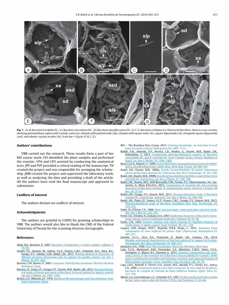
The type, presence or absence of crystals can be considered to betaxonomic characteristics (Meric, 2009). Calcium oxalate crystalsare often found in the perimedullary region of the pith in Baccharis(Budel and Duarte, 2008b; Souza et al., 2011; Oliveira et al., 2011;Jasinski et al., 2014; Barreto et al., 2015; Bobek et al., 2015).
In this study, B. brevifolia shows crystal sand, styloids with
pointed or square ends (Fig. 7A and B) and square dipyra-mids, B. microdonta has rare styloids with pointed ends (Fig. 7C),B. pauciflosculosa shows crystal sand and square dipyramids
670 V.B. Bobek et al. / Revista Brasileira de Farmacognosia 26 (2016) 665–672
F cifloscs mis (e(
(msli
mta
ig. 6. (A) Baccharis brevifolia DC., (B) Baccharis microdonta DC., (C) Baccharis pauhowing chlorenchyma (cl), collenchyma (co), cuticle (ct), epidermis (ep), endoderxy). Scale bar = 50 �m.
Fig. 7D), and B. trilobata has signs of crystal sand, square dipyra-ids, elongated square dipyramids, styloids with pointed and
quare ends (Fig. 7E and F), as well as tabular crystals in piles thatook like a tower (Fig. 7G). This type has not been mentioned beforen the genus.
Anatomical analysis is an inherent procedure for nearly all phar-
acopeias and is one of the main identification tests required forhe herbal industry. Although the individual structural elementsre relatively common within the same type of plant parts, the
Box 1: Anatomical characteristics of Baccharis brevifolia, B. m
Anatomical characteristics B. brevifolia B. m
Occurrence of stomata in the leaves Amphistomatic HypoTypes of stomata Ciclocytic and hexacytic Stau
and
Shape of midrib shape Flat-convex ConcPresence of petiole Absent PresShape of stem Irregular with six ribs Hexa
four
Types of crystals Crystal sand, styloids withpointed or square ends andsquare dipyramids
Rarepoin
ulosa DC., (D) Baccharis trilobata A.S. Oliveira & Marchiori. Stem in cross-section,n), fibers (fi), glandular trichome (gt), phloem (ph), secretory duct (sd), and xylem
manner in which the elements are set gives a vegetable speciesits characteristic fingerprint (Upton et al., 2011).
In that sense, even though most of the anatomical features of theleaf and stem of Baccharis spp. are quite similar, several character-istics observed in this study support the need for a differentiationof the four studied species, as confirmed in Box 1. The occurrence
and type of the stomata, midrib, stem and crystals shapes, and thepresence of the petiole are recommended as good markers for adiagnosis of the species.icrodonta, B. pauciflosculosa and B. trilobata.
icrodonta B. pauciflosculosa B. trilobata
stomatic Amphistomatic Amphistomaticro-tetracytictetracytic
Anomocytic andtetracytic
Stauro-tetracytic andtetracytic
ave-convex Biconvex Flat on both sidesent Absent Absentgonal withribs
Irregular with sixribs
Hexagonal with four ribs
styloids withted ends
Crystal sand andsquare dipyramids
Square dipyramids,elongated squaredipyramids, styloids withpointed and square ends,tabular crystals in piles
V.B. Bobek et al. / Revista Brasileira de Farmacognosia 26 (2016) 665–672 671
F ifloscus lp), st(
A
MttcsaAs
C
A
VU
R
A
A
B
B
B
ig. 7. (A, B) Baccharis brevifolia DC., (C) Baccharis microdonta DC., (D) Baccharis pauchowing perimedullary region with crystals, sand (sa), stiloyds with pointed ends (sesd), and tabular crystals in piles (tb). Scale bar = 10 �m (A, B, C, D).
uthors’ contributions
VBB carried out the research. These results form a part of herA course work. GH identified the plant samples and performed
he voucher. VPA and CFO assisted by conducting the anatomicalests. JPP and PVF provided a critical reading of the manuscript. TNreated the project and was responsible for arranging the scholar-hip. JMB created the project and supervised the laboratory work,s well as analyzing the data and providing a draft of the article.ll the authors have read the final manuscript and approved itsubmission.
onflicts of interest
The authors declare no conflicts of interest.
cknowledgments
The authors are grateful to CAPES for granting scholarships toBB. The authors would also like to thank the CME of the Federalniversity of Paraná for the scanning electron micrographs.
eferences
bad, M.J., Bermejo, P., 2007. Baccharis (Compositae): a review update. Caldasia 7,76–96.
raújo, F.F., Amorin, M., Santos, V.L.P., Franco, C.R.C., Folquitto, D.G., Silva, R.Z.,Farago, P.V., Takeda, I.J.M., Budel, J.M., 2015. Pharmacobotanical characters ofMikania micranta (Asteraceae) and its support for quality control. Lat. Am. J.Pharm. 34, 437–442.
arroso, G.M., Bueno, O., 2002. Composta: Subtribo Baccharidinae. Herbário BarbosaRodrigues, Itajaí.
arreto, I.F., Paula, J.P., Farago, P.V., Duarte, M.R., Budel, J.M., 2015. Pharmacobotan-
ical study of leaves and stems of Baccharis ochracea Spreng for quality control.Lat. Am. J. Pharm. 34, 1497–1502.erlyn, G.P., Miksche, J.P., 1976. Botanical Microtechnique and Cytochemistry. IowaState University, Ames.
losa DC., (E, F, G) Baccharis trilobata A.S. Oliveira & Marchiori. Stem in cross-section,iloyds with square ends (sls), square dipyramids (sd), elongated square dipyramids
BFG – The Brazilian Flora Group, 2015. Growing knowledge: an overview of seedplant diversity in Brazil. Rodriguésia 66, 1085–1113.
Bobek, V.B., Almeida, V.P., Pereira, C.B., Heiden, G., Duarte, M.R., Budel, J.M.,Nakashima, T., 2015. Comparative pharmacobotanical analysis of Bacchariscaprariifolia DC. and B. erioclada DC. from Campos Gerais, Paraná, Southern ofBrazil. Lat. Am. J. Pharm. 34, 1396–1402.
Brito, C.J.F.A., AlquinI., Y., 1996. A new method for staining botanical material embed-ded in glycolmethacrylate (GMA). Braz. Arch. Biol. Tecnol. 39, 949–951.
Budel, J.M., Duarte, M.R., 2008a. Estudo farmacobotânico de partes vegetativasaéreas de Baccharis anomala DC, Asteraceae. Rev. Bras. Farmacogn. 18, 761–768.
Budel, J.M., Duarte, M.R., 2008b. Estudo farmacobotânico de folha e caule de Baccharisuncinella DC, Asteraceae. Lat. Am. J. Pharm. 27, 740–746.
Budel, J.M., Duarte, M.R., Döll-Boscardin, P.M., Farago, P.V., Matzenbacher, N.I., Sar-toratto, A., Maia, B.H.L.N.S., 2012. Composition of essential oils and secretorystructures of Baccharis anomala, B. megapotamica and B. ochracea. J. Essent. OilRes. 24, 19–24.
Budel, J.M., Farago, P.V., Duarte, M.R., 2013. Pharmacobotanical study of Bacchariscognata DC. (Asteraceae: Astereae). Lat. Am. J. Pharm. 32, 540–544.
Budel, J.M., Paula, J.P., Santos, V.L.P., Franco, C.R.C., Farago, P.V., Duarte, M.R., 2015.Pharmacobotanical study of Baccharis pentaptera. Rev. Bras. Farmacogn. 25,314–319.
Feder, N., O’brien, T.P., 1968. Plant microtecnique: some principles and new meth-ods. Am. J. Bot. 55, 123–142.
Freire, S.E., Urtubey, E., Giuliano, D.A., 2007. Epidermal characters of Baccharis (Aster-aceae species used in traditional medicine). Caldasia 29, 23–38.
Fuchs, C.H., 1963. Fuchsin staining with NaOH clearing for lignified elements ofwhole plants or plants organs. Stain Technol. 38, 141–144.
Gogosz, A.M., Boeger, M.R.T., Negrelle, R.R.B., Bergo, C., 2012. Anatomia foliarcomparativa de nove espécies do gênero Piper (Piperaceae). Rodriguésia 63,405–417.
Jasinski, V.C.G., Silva, R.Z., Pontarolo, R., Budel, J.M., Campos, F.R., 2014.Morpho-anatomical characteristics of Baccharis glaziovii in support of its phar-macobotany. Rev. Bras. Farmacogn. 24, 506–515.
Johansen, D.A., 1940. Plant Microtechnique. MacGraw Hill Book, New York.Lage, T.C.A., Montanari, R.M., Fernandes, S.A., Monteiro, C.M.O., Senra, T.O.S.,
Zeringota, V., Matos, R.S., Daemon, E., 2015. Chemical composition and acari-cidal activity of the essential oil of Baccharis dracunculifolia De Candole (1836)and its constituents nerolidol and limonene on larvae and engorged females ofRhipicephalus microplus (Acari: Ixodidae). Exp. Parasitol. 148, 24–29.
Lago, J.H.G., Romoff, P., Fávero, O.A., Soares, M.G., Baraldi, P.T., Corrêa, A.G., Souza,F.O., 2008. Composic ão química dos óleos essenciais de seis espécies do gênero
Baccharis de Campos de Altitude da Mata Atlântica Paulista. Quim. Nova 31,727–730.Mentz, L.A., Lutzemberger, L.C., Schenkel, E.P., 1997. Da flora medicinal do Rio Grandedo Sul: notas sobre a obra de D’ávila (1910). Cad. Farmácia 13, 25–48.

6 de Fa
M
M
M
N
O
O
O
P
P
P
P
72 V.B. Bobek et al. / Revista Brasileira
eric, C., 2009. Calcium oxalate crystals in some species of the tribe inuleae (Aster-aceae). Acta Biol. Cracov. Ser. Bot. 51, 105–110.
olares, S., Gonzalez, S.B., Ladio, A., Agueda Castro, M., 2009. Etnobotánica, anatomíay caracterización físico-química del aceite esencial de Baccharis obovata Hook.et Arn. (Asteraceae: Astereae). Acta Bot. Bras. 23, 578–589.
üller, J., 2013. World checklist of Baccharis L. (Compositae–Astereae), Avail-able from: http://www.spezbot.uni-jena.de/wp-content/uploads/2013/09/World-checklist-of-Baccharis-L.pdf (accessed 20.04.16).
odari, R.O., Guerra, M.P., 2000. Aspectos genéticos e moleculares da produc ão vege-tal. In: Farmacognosia, da planta ao medicamento. UFSC/UFRGS, Porto Alegre.
’Brien, T.P., Feder, N., McCully, M.E., 1964. Polychromatic staining of plant cell wallsby toluidine blue O. Protoplasma 59, 368–373.
liveira, A.M.A., Santos, V.L.P., Franco, C.R.C., Farago, P.V., Duarte, M.R., Budel,J.M., 2011. Comparative morpho-anatomical study of Baccharis curitybensisHeering ex Malme and Baccharis spicata (Lam.). Baill. Lat. Am. J. Pharm. 30,1560–1566.
nofre, S.B., Canton, M., Pires, P.A., 2013. Action of essential oils obtained from Bac-charis coridifolia DC. (Asteraceae-Astereae) on the activity of antibiotics. Adv.Microb. 3, 166–170.
atel, J.D., 1979. New morphological classification of stomatal complexes. Phyto-morphology 29, 218–229.
ereira, C.B., Farago, P.V., Budel, J.M., Paula, J.P., Folquitto, D.G., Miguel, O.G., Miguel,M.D.A., 2014. New contribution to the pharmacognostic study of carquejas:
Baccharis milleflora DC, Asteraceae. Lat. Am. J. Pharm. 33, 841–847.erez, C., Anesini, C., 1993. Screening of plants used in Argentine folk medicine forantimicrobial activity. J. Ethnopharmacol. 39, 119–128.
erez, C., Anesini, C., 1994. Inhibition of Pseudomonas aeruginosa by argentineanmedicinal plants. Fitoterapia 65, 169–172.
rmacognosia 26 (2016) 665–672
Rodriguez, M.V., Martínez, M.L., Cortadi, A.A., Bandoni, A., Giuliano, D.A., Gattuso,S.J., Gattuso, M.A., 2010. Characterization of three sect. Caulopterae species(Baccharis-Asteraceae) inferred from morphoanatomy, polypeptide profiles andspectrophotometry data. Plant. Syst. Evol. 286, 175–190.
Roeser, K.R., 1972. Die Nadel der Schwarzkiefer-Massenprodukt und Kunstwerk derNatur. Mikrokosmos 61, 33–36.
Souza, W., 2007. Técnicas de microscopia eletrônica aplicadas às Ciências Biológicas.Sociedade Brasileira de Microscopia, Rio de Janeiro.
Souza, C.A., Farago, P.V., Duarte, M.R., Budel, J.M., 2011. Pharmacobotanical studyof Baccharis singularis (Vell.) G.M. Barroso, Asteraceae. Lat. Am. J. Pharm. 30,311–317.
Upton, R., Graff, A., Jolliffe, G., Länger, R., Williamson, E., 2011. American Herbal Phar-macopoeia Botanical Pharmacognosy: Microscopic Characterization of BotanicalMedicines. CRC Press.
Valarezo, E., Rosales, J., Morocho, V., Cartuche, L., Guaya, D., Ojeda-Riascos, S., Armi-jos, C., González, S., 2015. Chemical composition and biological activity of theessential oil of Baccharis obtusifolia Kunth from Loja, Ecuador. J. Essent. Oil Res.27, 212–216.
Van Cotthem, M.R.J., 1970. A classification of stomatal types. Bot. J. Linn. Soc. 63,235–246.
WHO, 2003. Guidelines on Good Agricultural and Collection Practices (GACP) forMedicinal Plants. World Health Organization, http://who.int (accessed May2014).
Wosch, L., Imig, D.C., Cervi, A.C., Moura, B.B., Budel, J.M., Santos, C.A.M., 2015. Com-parative study of Passiflora taxa leaves. I. A morpho-anatomical profile. Rev. Bras.Farmacogn. 25, 328–343.
Zardini, E.M., 1984. Etnobotánica de compuestas argentinas com especial referenciaa su uso farmacológico. Lat. Am. J. Pharm. 3, 77–99.


![PREFEITURA MUNICIPAL DE VASSOURAS SECRETARIA …€¦ · SSSSSS]S]SS]SSSSS]SSSS]SSS]SSSS]SSSSSSS S]SSSSSSSS]SSSSSS Estado do Rio de Janeiro Prefeitura Municipal de Vassouras PREFEITURA](https://img.pdfslide.net/doc/110x75/5f2081bf6d1ea8704657c558/prefeitura-municipal-de-vassouras-secretaria-ssssssssssssssssssssssssssssssss.jpg)