Embed Size (px)
Citation preview
Comparative anatomy of the mesosomal organs ofscorpions (Chelicerata, Scorpiones), with implicationsfor the phylogeny of the order
ERICH S. VOLSCHENK*†, CAMILO I. MATTONI‡ and LORENZO PRENDINI
Division of Invertebrate Zoology, American Museum of Natural History, Central Park West at79th Street, New York, NY 10024-5192, USA
Received 18 April 2007; accepted for publication 15 October 2007
We present a review and reassessment of anatomical variation in the ovariuterus (and associated follicles),digestive gland, and lateral lymphoid organs of scorpions, and discuss the contribution of these character systemsto the understanding of scorpion phylogeny. New data, obtained using light microscopy, are presented from anexamination of 55 scorpion species, representing most scorpion families, and are collated with observations fromthe literature. Six distinct types of ovariuterine anatomy are identified: five in the family Buthidae and one in theremaining (nonbuthid) families. The buthid genera Lychas C.L. Koch, 1845 and Rhopalurus Thorell, 1876 areexceptional in possessing multiple types of ovariuterine anatomy among the congeneric species studied. Thepresence or absence of lateral lymphoid organs appears to be phylogenetically informative: the organ is absent inbuthids, chaerilids and Pseudochactas Gromov, 1998. Embryo follicle morphology appears to be phylogeneticallyinformative within the superfamily Scorpionoidea Latreille, 1802, where it supports the controversial sister-grouprelationship between Urodacus Peters, 1861 and Heteroscorpion Birula, 1903. The mesosomal anatomy ofMicrocharmus Lourenço, 1995 (Microcharmidae Lourenço, 1996) is consistent with that of Buthidae C.L. Koch,1837, and we therefore propose the following new synonymy: Microcharmidae Lourenço, 1996 = Buthidae C.L.Koch, 1837. © 2008 The Linnean Society of London, Zoological Journal of the Linnean Society, 2008, 154,651–675.
ADDITIONAL KEYWORDS: digestive gland – follicle – lymphoid organs – ovariuterus – reproduction –systematics.
INTRODUCTION
Scorpion systematics is currently based almostentirely on characters of the external morphology(Sissom, 1990), despite an extensive body of literaturedocumenting phylogenetically informative charactersystems from the internal anatomy (particularly themesosoma) of scorpions. The mesosoma contains mostof the scorpion’s organ systems, including the intes-tine and glands of the digestive system, the reproduc-
tive system, the respiratory system, the circulatorysystem, and lymphoid organs (Hjelle, 1990). Phyloge-netically informative characters from the mesosomalanatomy were first reported a century ago (Laurie,1890, 1891, 1896a, b; Pavlovsky, 1913, 1915a, b, c, d,1917, 1924a, b, c, d, 1925, 1926), and, with thenotable exception of characters from the male repro-ductive system, the subject has since received littleattention from scorpion systematists. For example,the three family-level phylogenetic analyses of scor-pions presented to date included numerous charactersfrom the male reproductive system (23 in Stockwell,1989; 12 in Prendini, 2000; 8 in Soleglad & Fet, 2003),but only three characters from the female reproduc-tive system. A further problem concerns the limitednumber of taxa in which the mesosomal anatomy hasbeen studied in detail. Most observations in the earlyliterature were reported from a limited sample of
*Corresponding author. E-mail: [email protected]†Current address: Department of Terrestrial Invertebrates,Western Australian Museum, Locked Bag 49, Welshpool DC,WA 6986, Australia‡Current address: Cátedra de Diversidad Animal I,Facultad de Ciencias Exactas, Físicas y Naturales, Univer-sidad Nacional de Córdoba, Av. Vélez Sarsfield 299, 5000Córdoba, Argentina
Zoological Journal of the Linnean Society, 2008, 154, 651–675. With 7 figures
© 2008 The Linnean Society of London, Zoological Journal of the Linnean Society, 2008, 154, 651–675 651
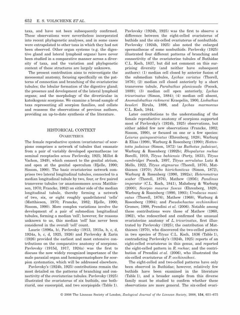
taxa, and have not been subsequently confirmed.These observations were nevertheless incorporatedinto recent phylogenetic analyses, and in some caseswere extrapolated to other taxa in which they had notbeen observed. Other organ systems (e.g. the diges-tive gland and lateral lymphoid organs) have neverbeen studied in a comparative manner across a diver-sity of taxa, and the variation and phylogeneticcontent of these structures are largely unknown.
The present contribution aims to reinvestigate themesosomal anatomy, focusing specifically on the pat-terns of connection and branching of the ovariuterinetubules; the lobular formation of the digestive gland;the presence and development of the lateral lymphoidorgans; and the morphology of the diverticulae inkatoikogenic scorpions. We examine a broad sample oftaxa representing all scorpion families, and collateand reassess the observations of previous authors,providing an up-to-date synthesis of the literature.
HISTORICAL CONTEXTOVARIUTERUS
The female reproductive system (ovariuterus) of scor-pions comprises a network of tubules that emanatefrom a pair of variably developed spermathecae (orseminal receptacles sensu Pavlovsky, 1925; Millot &Vachon, 1949), which connect to the genital atrium,and open at the genital operculum (Hjelle, 1990;Sissom, 1990). The basic ovariuterine network com-prises two lateral longitudinal tubules, connected to amedian longitudinal tubule by two, four, or five pairedtransverse tubules (or anastosomoses sensu Matthie-sen, 1970; Francke, 1982) on either side of the medianlongitudinal tubule, thereby forming a networkof two, six, or eight roughly quadrilateral ‘cells’(Matthiesen, 1970; Francke, 1982; Hjelle, 1990;Sissom, 1990). More complex variations involve thedevelopment of a pair of submedian longitudinaltubules, forming a median ‘cell’; however, for reasonsunknown to us, this median ‘cell’ has never beenconsidered in the overall ‘cell’ count.
Laurie (1896a, b), Pavlovsky (1913, 1915a, b, c, d,1924a, b, c, d, 1925, 1926) and Pavlovsky & Zarin(1926) provided the earliest and most extensive con-tributions on the comparative anatomy of scorpions.Pavlovsky (1915d, 1917, 1924a) was the first todiscuss the now widely recognized importance of themale paraxial organ and hemispermatophore for scor-pion systematics, which will be addressed elsewhere.
Pavlovsky’s (1924b, 1925) contributions remain themost detailed on the patterns of branching and con-nectivity of the overiuterine tubules. Pavlovsky (1925)illustrated the ovariuterus of six buthids, one both-riurid, one euscorpiid, and two scorpiopids (Table 1).
Pavlovsky (1924b, 1925) was the first to observe adifference between the eight-celled ovariuterus ofbuthids and the six-celled ovariuterus of nonbuthids.Pavlovsky (1924b, 1925) also noted the enlargedspermathecae of some nonbuthids. Pavlovsky (1925)illustrated four different patterns of branching andconnectivity of the ovariuterine tubules of ButhidaeC.L. Koch, 1837, but did not comment on this sur-prising diversity (and neither have subsequentauthors): (1) median cell closed by anterior fusion ofthe submedian tubules, Lychas variatus (Thorell,1876); (2) median cell closed anteriorly by a shorttransverse tubule, Parabuthus planicauda (Pocock,1889); (3) median cell open anteriorly, Lychastricarinatus (Simon, 1884); (4) median cell absent,Anomalobuthus rickmersi Kraepelin, 1900, Liobuthuskessleri Birula, 1898, and Lychas marmoreusC.L. Koch, 1844.
Later contributions to the understanding of thefemale reproductive anatomy of scorpions supportedmost of Pavlovsky’s (1924b, 1925) observations, buteither added few new observations (Francke, 1982;Sissom, 1990), or focused on one or a few species:Leiurus quinquestriatus (Ehrenberg, 1828), Warburg& Elias (1998), Warburg & Rosenberg (1990); Hotten-totta judaicus (Simon, 1872) (as Buthotus judaicus),Warburg & Rosenberg (1992b); Rhopalurus rochaeBorelli, 1910, Tityus bahiensis (Perty, 1833), Tityuscambridgei Pocock, 1897, Tityus serrulatus Lutz &Mello, 1922, Tityus stigmurus (Thorell, 1876), Mat-thiesen (1970); Nebo hierichonticus (Simon, 1872),Warburg & Rosenberg (1990, 1992a); Heterometrusscaber (Thorell, 1876), Mathew (1956); Pandinusimperator (C.L. Koch, 1841), Mahsberg & Warburg(2000); Scorpio maurus fuscus (Ehrenberg, 1829),Warburg & Rosenberg (1990, 1993); Urodacus mani-catus (Thorell, 1876), Mathew (1968), Warburg &Rosenberg (1994); and Pseudochactas ovchinnikoviGromov, 1998, Prendini et al. (2006). Notable amongthese contributions were those of Mathew (1960,1962), who redescribed and confirmed the unusualovariuterine anatomy of L. tricarinatus, first illus-trated by Pavlovsky (1925); the contribution of Mat-thiesen (1970), who discovered the two-celled patternin two species of Tityus C.L. Koch, 1836 (Table 1),contradicting Pavlovsky’s (1924b, 1925) reports of aneight-celled ovariuterus in this genus, and reportedthe eight-celled pattern in R. rochae; and the contri-bution of Prendini et al. (2006), who illustrated thesix-celled ovariuterus of P. ovchinnikovi.
The eight-celled and two-celled patterns have onlybeen observed in Buthidae; however, relatively fewbuthids have been examined in the literature(Table 1), and a broader sample from this diversefamily must be studied to confirm whether theseobservations are more general. The six-celled ovari-
652 E. S. VOLSCHENK ET AL.
© 2008 The Linnean Society of London, Zoological Journal of the Linnean Society, 2008, 154, 651–675
uterus, documented only in nonbuthid scorpions, isbased on equally few observations. Prendini et al.(2006: 238, table 7) provided the most recentsummary of published observations on variation inthe ovariuterine anatomy of scorpions, which weenlarge upon in the present contribution.
SPERMATHECAE
The spermathecae, which facilitate sperm storage andmaintenance after mating (Hjelle, 1990; Peretti &Battán-Horenstein, 2003; Peretti, 2003), are theswollen anterior extensions of the lateral longitudinaltubules. Although little is known about variation inthe spermathecal anatomy within the order Scorpi-ones, it is clear that considerable variation exists,ranging from only a slight anterior swelling of theovariuterine tubules in some buthids, to the forma-tion of large sac-like structures in some bothriurids(Pavlovsky, 1925).
LOCATION OF EMBRYONIC DEVELOPMENT
Differences in the type of embryonic development(katoikogenic vs. apoikogenic development) have beenstudied in many scorpion species, and were first sum-marized by Polis & Sissom (1990: 184–187, table 4.2);however, few attempts have been made to analyseand summarize the data comparatively (Laurie, 1890,1891, 1896a, b; Francke, 1982; Polis & Sissom, 1990;Farley, 2001). Laurie (1896a) first recognized twodistinct types of embryonic development in scorpions:apoikogenic, in which embryonic development occursinside the ovariuterine tubules, and the embryos arenourished by yolk; and katoikogenic, in which devel-opment occurs inside diverticulae of the ovariuterus,and the embryos are nourished via a placenta-likeorgan. Apoikogenic and katoikogenic development areamong the few characters from the female reproduc-tive system to have been included in phylogeneticanalyses of the higher phylogeny of Scorpiones(Stockwell, 1989; Prendini, 2000; Soleglad and Fet,2003).
Francke (1982) clarified several misconceptionsconcerning the development of scorpions, and con-cluded that all are viviparous, but with some impor-tant differences in the morphology and developmentof the ovarian follicles and embryos, first noted byLaurie (1896a, b), and discussed further below.Warburg (2001) described a putative third type ofdevelopment in Vaejovis spinigerus (Wood, 1863) andCompsobuthus werneri schmiedechnechti Vachon,1949 (as C. werneri judaicus Levy et al., 1973), inwhich the oocytes apparently mature inside theovarian tubes, rather than inside follicles on the outersurface.
FOLLICLES
The oocytes are located in follicles that are variablysituated on the exterior of the ovariuterine tubules.Three types of follicles have been recognized(Francke, 1982; Sissom, 1990; Lourenço, 2002):(1) sessile and in direct contact with the ovariuterus(Bothriuridae Simon, 1880; Buthidae, ChactidaePocock, 1893; Chaerilidae Pocock, 1893); (2) con-nected to the ovariuterus by a short stalk or pedicel(Iuridae Thorell, 1876; Vaejovidae Thorell, 1876);(3) oocytes located within diverticulae that arise fromthe ovariuterine tubules (Diplocentridae Karsch,1880; Hemiscorpiidae Pocock, 1893; Liochelidae Fetand Bechly, 2001; Scorpionidae Latreille, 1802; Uro-dacidae Pocock, 1893). The ovariuterus of Smeringu-rus mesaensis (Stahnke, 1957) is unusual in that thefollicles are initially sessile and in direct contact withthe ovariuterus, but, during early embryology, atrophic layer of cells develops and completely coversthe maturing follicles (Farley, 1998, 2001).
The size and shape of the follicles are directlyrelated to the type of embryonic development. Sessileand stalked follicles are characteristic of scorpionswith apoikogenic development (Laurie, 1896a;Francke, 1982; Polis & Sissom, 1990; Farley, 2001).Follicles of apoikogenic scorpions are oval or rounded,whereas those of katoikogenic scorpions are moreelongated (Laurie, 1896a, b; Francke, 1982; Polis &Sissom, 1990; Farley, 2001), which may be related tothe development of the follicle into the diverticulumin katoikogenic scorpions.
Stalked follicles (with a pedicel) were once thoughtto be uniquely present in and potentially synapo-morphic for Iuridae and Vaejovidae (Laurie, 1896a;Francke, 1982; Stockwell, 1989; Sissom, 1990);however, stalked follicles have also been reported inthe buthids Lychas tricarinatus (Mathew, 1962: 348,fig. 1), Tityus bahiensis and T. serrulatus (Matthiesen,1970: 628, figs. 3, 5), Hottentotta judaicus (Warburg &Rosenberg, 1992b: 34, figs. 3, 4, 7), and Leiurus quin-questriatus (Warburg, Elias & Rosenberg, 1995), andin the euscorpiids, Euscorpius italicus (Herbst, 1800)(Laurie, 1890) and Euscorpius flavicaudis (DeGeer,1778) (Lourenço, 2002: 73, fig. 3b).
DIGESTIVE GLAND
The digestive gland is the largest organ in the scor-pion’s body, comprising most of the contents of themesosoma. The organ is formed by six pairs of glandsconnected to the intestine by means of fine ducts. Thefirst pair of diverticulae are situated in the prosoma;the remaining five are in each of the first five meso-somal segments. The digestive gland contains cellsthat produce enzymes for degrading ingested materi-als, and cells that absorb and store digested materials
MESOSOMAL ANATOMY OF SCORPIONES (CHELICERATA) 653
© 2008 The Linnean Society of London, Zoological Journal of the Linnean Society, 2008, 154, 651–675
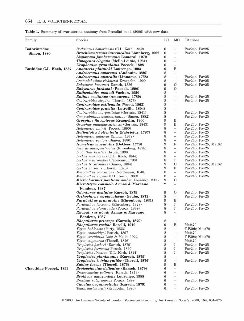
Table 1. Summary of ovariuterine anatomy from Prendini et al. (2006) with new data
Family Species LC MC Citations
BothriuridaeSimon, 1880
Bothriurus bonariensis (C.L. Koch, 1842) 6 – Pav24b, Pav25Brachistosternus intermedius Lönnberg, 1902 6 – Pav24b, Pav25Lisposoma josehermana Lamoral, 1979 6 –Timogenes elegans (Mello-Leitão, 1931) 6 –Urophonius granulatus Pocock, 1898 6 –
Buthidae C.L. Koch, 1837 Ananteris platnicki Lourenço, 1993 9 BAndroctonus amoreuxi (Audouin, 1826) 8 –Androctonus australis (Linnaeus, 1758) 8 – Pav24b, Pav25Anomalobuthus rickmersi Kraepelin, 1900 8 – Pav24b, Pav25Babycurus buettneri Karsch, 1886 9 O Pav24b, Pav25Babycurus jacksoni (Pocock, 1890) 9 OButheoloides monodi Vachon, 1950 8 –Buthus occitanus (Amoureux, 1789) 8 – Pav24b, Pav25Centruroides elegans (Thorell, 1876) 8 – Pav24b, Pav25Centruroides exilicauda (Wood, 1863) 8 –Centruroides gracilis (Latreille, 1804) 8 –Centruroides margaritatus (Gervais, 1841) 8 – Pav24b, Pav25Compsobuthus acutecarinatus (Simon, 1882) 8 – Pav24b, Pav25Grosphus flavopiceus Kraepelin, 1900 9 BGrosphus madagascariensis (Gervais, 1843) 9 B Pav24b, Pav25Hottentotta eminii (Pocock, 1890) 8 – Pav24b, Pav25Hottentotta hottentotta (Fabricius, 1787) 8 – Pav24b, Pav25Hottentotta judaicus (Simon, 1872) 8 – Pav24b, Pav25Hottentotta saulcyi (Simon, 1880) 8 – Pav24b, Pav25Isometrus maculatus (DeGeer, 1778) 9 F Pav24b, Pav25; Mat62Leiurus quinquestriatus (Ehrenberg, 1828) 8 – Pav24b, Pav25Liobuthus kessleri Birula, 1898 8 – Pav24b, Pav25Lychas marmoreus (C.L. Koch, 1844) 8 – Pav24b, Pav25Lychas mucronatus (Fabricius, 1798) 8 ? Pav24b, Pav25Lychas tricarinatus (Simon, 1884) 9 O Pav24b, Pav25; Mat62Lychas variatus (Thorell, 1876) 9 F Pav24b, Pav25Mesobuthus caucasicus (Nordmann, 1840) 8 – Pav24b, Pav25Mesobuthus eupeus (C.L. Koch, 1839) 8 – Pav24b, Pav25Microcharmus pauliani amber Lourenço, 2006 9 OMicrotityus consuelo Armas & Marcano
Fondeur, 19872 –
Odonturus dentatus Karsch, 1879 9 O Pav24b, Pav25Orthochirus scrobiculosus (Grube, 1873) 8 – Pav24b, Pav25Parabuthus granulatus (Ehrenberg, 1831) 9 BParabuthus leiosoma (Ehrenberg, 1828) 8 ? Pav24b, Pav25Parabuthus planicauda (Pocock, 1889) 8 ? Pav24b, Pav25Rhopalurus abudi Armas & Marcano
Fondeur, 19878 –
Rhopalurus princeps (Karsch, 1879) 8 –Rhopalurus rochae Borelli, 1910 9 B Matt70Tityus bahiensis (Perty, 1833) 2 – T-P39b; Matt70Tityus cambridgei Pocock, 1897 2 – Matt70Tityus serrulatus Lutz & Mello, 1922 2 – T-P39a; Matt70Tityus stigmurus (Thorell, 1876) 2 – Matt70Uroplectes fischeri (Karsch, 1879) 8 ? Pav24b, Pav25Uroplectes formosus Pocock, 1890 8 ? Pav24b, Pav25Uroplectes lineatus (C.L. Koch, 1844) 8 ? Pav24b, Pav25Uroplectes planimanus (Karsch, 1879) 8 –Uroplectes t. triangulifer (Thorell, 1876) 8 – Pav24b, Pav25Zabius fuscus (Thorell, 1876) 9 B
Chactidae Pocock, 1893 Broteochactas delicatus (Karsch, 1879) 6 –Broteochactas gollmeri (Karsch, 1879) 6 – Pav24b, Pav25Brotheas amazonicus Lourenço, 1988 6 –Brotheas subgranosus Pocock, 1898 6 – Pav24b, Pav25Chactas aequinoctialis (Karsch, 1879) 6 –Teuthraustes witti (Kraepelin, 1896) 6 – Pav24b, Pav25
654 E. S. VOLSCHENK ET AL.
© 2008 The Linnean Society of London, Zoological Journal of the Linnean Society, 2008, 154, 651–675
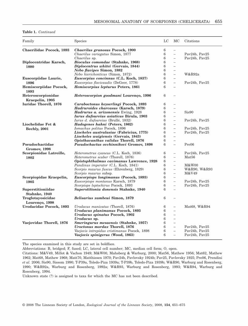
Table 1. Continued
Family Species LC MC Citations
Chaerilidae Pocock, 1893 Chaerilus granosus Pocock, 1900 6 –Chaerilus variegatus Simon, 1877 6 – Pav24b, Pav25Chaerilus sp. 6 – Pav24b, Pav25
Diplocentridae Karsch,1880
Bioculus comondae (Stahnke, 1968) 6 –Diplocentrus whitei (Gervais, 1844) 6 –Nebo flavipes Simon, 1882 6 –Nebo hierichonticus (Simon, 1872) 6 – W&R92a
Euscorpiidae Laurie,1896
Euscorpius concinnus (C.L. Koch, 1837) 6 –Euscorpius flavicaudis (DeGeer, 1778) 6 – Pav24b, Pav25
Hemiscorpiidae Pocock,1893
Hemiscorpius lepturus Peters, 1861 6 –
HeteroscorpionidaeKraepelin, 1905
Heteroscorpion goodmani Lourenço, 1996 6 –
Iuridae Thorell, 1876 Caraboctonus keyserlingi Pocock, 1893 6 –Hadruroides charcasus (Karsch, 1879) 6 –Hadrurus a. arizonensis Ewing, 1928 6 – Sis90Iurus dufoureius asiaticus Birula, 1903 6 –Iurus d. dufoureius (Brullé, 1832) 6 – Pav24b, Pav25
Liochelidae Fet &Bechly, 2001
Hadogenes hahni (Peters, 1862) 6 –Iomachus politus Pocock, 1896 6 – Pav24b, Pav25Liocheles australasiae (Fabricius, 1775) 6 – Pav24b, Pav25Liocheles waigiensis (Gervais, 1843) 6 –Opisthacanthus validus Thorell, 1876 6 –
PseudochactidaeGromov, 1998
Pseudochactas ovchinnikovi Gromov, 1898 6 – Pre06
Scorpionidae Latreille,1802
Heterometrus cyaneus (C.L. Koch, 1836) 6 – Pav24b, Pav25Heterometrus scaber (Thorell, 1876) 6 – Mat56Opistophthalmus cavimanus Lawrence, 1928 6 –Pandinus imperator (C.L. Koch, 1841) 6 – M&W00Scorpio maurus fuscus (Ehrenberg, 1829) 6 – W&R90, W&R93Scorpio maurus subsp. 6 – M&V49
Scorpiopidae Kraepelin,1905
Euscorpiops longimanus (Pocock, 1893) 6 –Euscorpiops montanus Karsch, 1879 6 – Pav24b, Pav25Scorpiops leptochirus Pocock, 1893 6 – Pav24b, Pav25
SuperstitioniidaeStahnke, 1940
Superstitionia donensis Stahnke, 1940 6 –
TroglotayosicidaeLourenço, 1998
Belisarius xambeui Simon, 1879 6 –
Urodacidae Pocock, 1893 Urodacus manicatus (Thorell, 1876) 6 – Mat68, W&R94Urodacus planimanus Pocock, 1893 6 –Urodacus spinatus Pocock, 1902 6 –Urodacus sp. 6 –
Vaejovidae Thorell, 1876 Smeringurus mesaensis (Stahnke, 1957) 6 –Uroctonus mordax Thorell, 1876 6 – Pav24b, Pav25Vaejovis intrepidus cristimanus Pocock, 1898 6 – Pav24b, Pav25Vaejovis spinigerus (Wood, 1863) 6 – Pav24b, Pav25
The species examined in this study are set in boldface.Abbreviations: B, bridged; F, fused; LC, lateral cell number; MC, median cell form; O, open.Citations: M&V49, Millot & Vachon 1949; M&W00, Mahsberg & Warburg, 2000; Mat56, Mathew 1956; Mat62, Mathew1962; Mat68, Mathew 1968; Matt70, Matthiesen 1970; Pav24b, Pavlovsky 1924b; Pav25, Pavlovsky 1925; Pre06, Prendiniet al. 2006; Sis90, Sissom 1990; T-P39a, Toledo-Piza 1939a; T-P39b, Toledo-Piza 1939b; W&R90, Warburg and Rosenberg,1990; W&R92a, Warburg and Rosenberg, 1992a; W&R93, Warburg and Rosenberg, 1993; W&R94, Warburg andRosenberg, 1994.Unknown state (?) is assigned to taxa for which the MC has not been described.
MESOSOMAL ANATOMY OF SCORPIONES (CHELICERATA) 655
© 2008 The Linnean Society of London, Zoological Journal of the Linnean Society, 2008, 154, 651–675
(Pavlovsky & Zarin, 1926; Snodgrass, 1952; Hjelle,1990). Anatomical variation in the digestive glandwas first observed by Pavlovsky (1925: fig. 1, platesVII and VIII), who illustrated the compact digestivegland of Centruroides margaritatus (Gervais, 1841),and the markedly lobate digestive gland of Scorpiomaurus Linnaeus, 1758 in dorsal aspect. Pavlovsky& Zarin (1926) subsequently reported a less lobatedigestive gland in Chactidae and Vaejovidae, butunfortunately provided no identification of the speciesexamined.
LATERAL LYMPHOID ORGANS
These organs consist of a pair of large, two-cell-thicktubular structures connected anteriorly to the dia-phragm dividing the prosoma from the mesosoma.Laurie (1896b) first noticed the variation of the tubes,and considered them to be associated with the coxalglands, part of the excretory system of scorpions,located in the prosoma (Hjelle, 1990). Laurie (1896b)also noted the absence of these tubes in the buthidshe examined. Pavlovsky (1924c) provided a moredetailed description of these structures and namedthem ‘lymphoid organs’, believing them to be associ-ated with the lymphatic system. Additional compo-nents of the scorpion lymphatic system comprisestrand-like lymphatic glands, extending along thedorsal surface of the ventral nerve cord in the meso-soma, that are referred to as the supraneural glands(Pavlovsky, 1924c; Millot & Vachon, 1949); Farley,1984, 1999; Hjelle, 1990).
Pavlovsky (1924c) described two kinds of lymphaticsystem in scorpions: (1) simple lymphatic system,without lateral lymphoid organs; (2) complex lym-phatic system, with lateral lymphoid organs. Pav-lovsky (1924c) observed the simple system inButhidae and the complex system in other families(Bothriuridae, Chactidae, Euscorpiidae Laurie, 1893,Iuridae, Liochelidae, Scorpionidae, ScorpiopidaeKraepelin, 1905, Urodacidae, and Vaejovidae), andnoted that the form and size of the tubes varied, fromshort ovoid to long tubes. Unfortunately, Pavlovsky(1924c) provided no further details about the differ-ences observed.
The function of these organs was largely unkownuntil recently (Farley, 1984; Hjelle, 1990). Previousresearchers reported the presence of phagocytic cells,which eliminate foreign substances (Pavlovsky, 1924c;Millot & Vachon, 1949), but Nayar (1966) suggestedthat they may serve an endocrine function. Farley(1984, 1999) demonstrated the hematocytopoieticfunction of the lateral lymphoid organs by means ofultrastructural studies.
The lateral lymphoid organs have been identifiedin the following families: Bothriuridae (Pavlovsky,
1924c); Chactidae (Pavlovsky, 1924c); Euscorpiidae(Pavlovsky, 1924c, as Chactidae); Iuridae (Pavlovsky,1924c, as Vaejovidae and Chaerilidae); Liochelidae(Laurie, 1896b, as Ischnuridae Simon, 1879; Pav-lovsky, 1924c, as Scorpionidae); Scorpionidae (Laurie,1896b; Pavlovsky, 1924c); Scorpiopidae (Pavlovsky,1924c, as Chactidae); Urodacidae (Pavlovsky, 1924c,as Scorpionidae); and Vaejovidae (Pavlovsky, 1924c).The absence of these organs has only been confirmedin Buthidae (Laurie, 1896b; Pavlovsky, 1924c),whereas the presence or form of these organs has notbeen reported in the remaining scorpion families,including Chaerilidae and Pseudochactidae Gromov,1998.
MATERIAL AND METHODSMATERIAL EXAMINED
Taxon sampling for this study aimed primarily atsampling exemplar species of most families currentlyrecognized in Scorpiones (classification followsPrendini & Wheeler, 2005), and secondarily at cor-roborating previous observations, particularly thoseof Pavlovsky (1924b, 1925). Owing to the destructiveprocedures required to obtain observations on themesosomal anatomy, we selected specimens with nolocality data, which were thus of limited systematicutility, or specimens from large series, whereverpossible.
The broad scope of taxon sampling (representa-tives of almost every family and a large sample ofthe family Buthidae), from an equally broad distri-bution of habitats around the world, necessitatedthe use of museum specimens. All specimens werepreserved in 75% ethanol. Whereas most of thespecimens are known to have been fixed in ethanol,some specimens were originally fixed in formalin,causing the digestive gland and muscles to be verybrittle. The material examined is listed in theAppendix.
DISSECTIONS
In order to examine the mesosomal organs, particu-larly the ovariuterus, a careful dissection is required.Dissections were conducted using fine-tipped forceps,dissection needles, iris scissors (Miltex 18-1620) andNikon SMZ-1500 dissection stereomicroscopes. Dis-sections were conducted in glass or plastic Petridishes, and specimens were either immersed in 75%ethanol or placed in a plastic Petri dish withoutethanol.
Using the iris scissors, a shallow incision was madethrough the pleural membrane, between the tergitesand sternites of the mesosoma, starting beneath terg-ite VI and cutting anteriorly around the lateral and
656 E. S. VOLSCHENK ET AL.
© 2008 The Linnean Society of London, Zoological Journal of the Linnean Society, 2008, 154, 651–675
anterior carapace margins of the prosoma, then pos-teriorly between tergite VII and sternite VII, andfinally through the membrane connecting tergite VIIand metasomal segment I. The opisthosomal tergawere then carefully removed by cutting through thedorsoventral muscles that restrain the dorsal scleritesand carapace. Once the tergites were removed, thedorsal aspect of the digestive gland was completelyexposed for examination, prior to being carefully dis-sected away. Dissections usually commenced anteri-orly, in the area covered by tergites I and II, andproceeded posteriorly after the anterior branches ofthe ovariuterus were located. The ovariuterinetubules run through the digestive gland, the removalof which must be undertaken carefully to avoid dam-aging the ovariuterus.
The preservation of specimens available for dissec-tions was variable. Near complete exposure of theovariuterus was obtained by careful dissection ofsofter and fresher specimens. Brittle specimens weresoaked in 20% ethanol or distilled water for 2–5 daysto soften the digestive gland, and increase the elas-ticity of the ovariuterine tubules, thus significantlyincreasing the ease of dissection and reducing thedamage to the ovariuterus. In a few cases, soakingonly slightly improved the specimens, and dissectionsremoved just enough of the digestive gland to confi-dently make observations. Specimens satisfactorilydissected were returned to 75% ethanol, and werelater photographed using a Microptics™ ML1000digital imaging system, or a Nikon Coolpix 4500digital camera attached to a Nikon SMZ-1500 micro-scope. Suitable images were then used to render linedrawings of the ovariuterus.
Measurements were taken directly from the digitalphotographs using the software UTHSCSA IMAGETOOL 3.00 (1996–2002), which was developed at theUniversity of Texas Health Science Center at SanAntonio, TX, USA, and is available from http://ddsdx.uthscsa.edu/dig/itdesc.html. The body widthwas measured at diaphragm level. The length andwidth of the lymphoid organ represent the maximumdistances observed on the specimen.
TERMINOLOGY
This study is concerned with anatomy, the ‘science ofinternal morphology, as revealed by dissection’ (Torre-Bueno et al., 1989: 38). Anatomical terminologymostly follows Hjelle (1990); however, the followingterms were developed based on our observations.
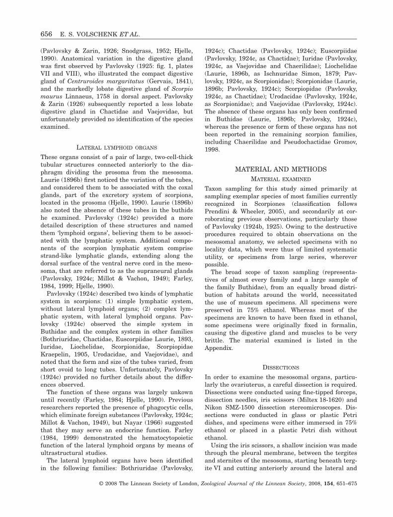
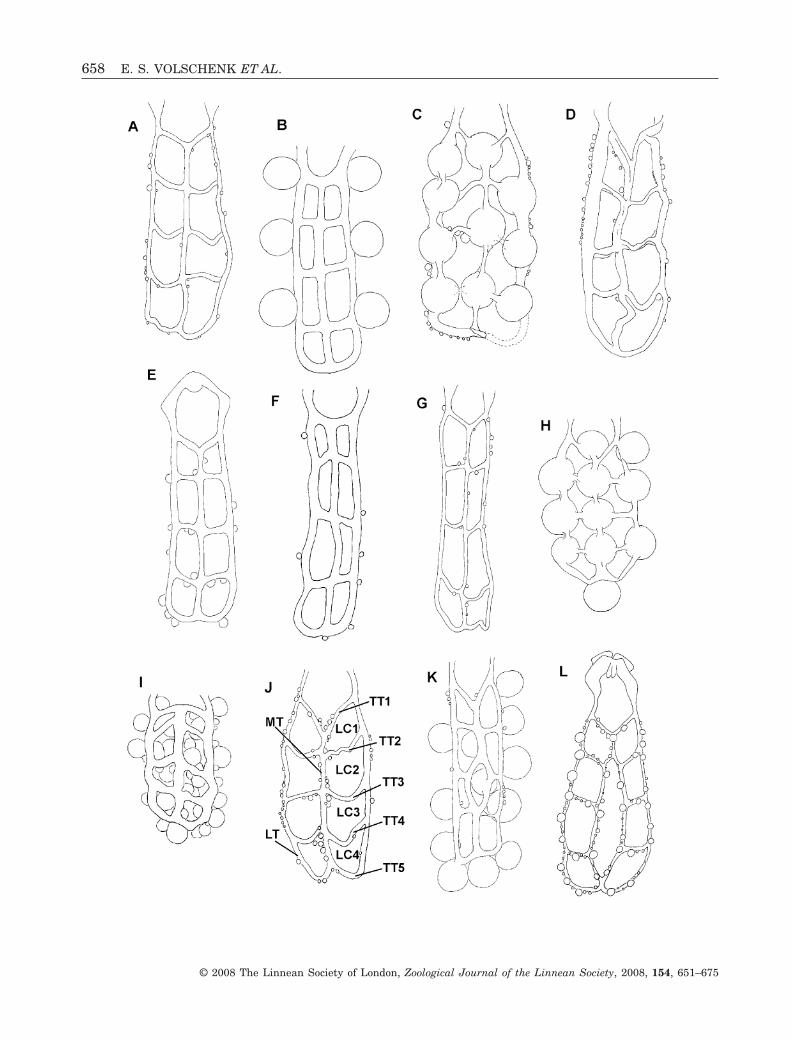
The lateral longitudinal tubules (LT; Figs 1J, 3E,6B) are the primary longitudinal tubules, leadingdirectly from the spermathecae and extending poste-riorly to mesosomal segment VI, where they curveinwards towards each other, and in so doing become
the fifth pair of transverse tubules. The lateral lon-gitudinal tubules are usually situated slightly abovethe intestine in the body cavity.
The median longitudinal tubule (MT; Figs 1J, 3B) islocated below the intestine, and is usually lower inthe body cavity than the LTs. The MT divides ante-riorly, forming the anteriormost pair of transversetubules. All nonbuthids and some buthids exhibitcomplete development of the MT. Francke (1982) andHjelle (1990) considered this tubule to be the result offusion of the submedian longitudinal tubules.
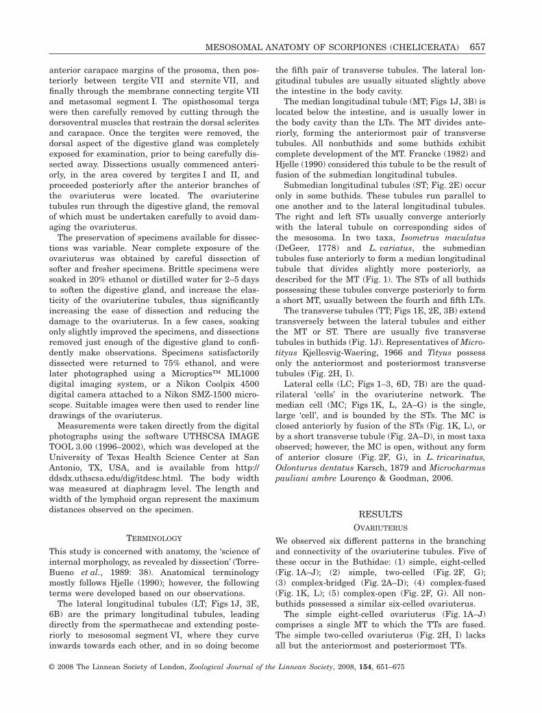
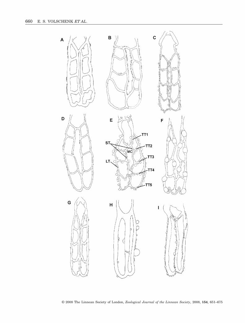
Submedian longitudinal tubules (ST; Fig. 2E) occuronly in some buthids. These tubules run parallel toone another and to the lateral longitudinal tubules.The right and left STs usually converge anteriorlywith the lateral tubule on corresponding sides ofthe mesosoma. In two taxa, Isometrus maculatus(DeGeer, 1778) and L. variatus, the submediantubules fuse anteriorly to form a median longitudinaltubule that divides slightly more posteriorly, asdescribed for the MT (Fig. 1). The STs of all buthidspossessing these tubules converge posteriorly to forma short MT, usually between the fourth and fifth LTs.
The transverse tubules (TT; Figs 1E, 2E, 3B) extendtransversely between the lateral tubules and eitherthe MT or ST. There are usually five transversetubules in buthids (Fig. 1J). Representatives of Micro-tityus Kjellesvig-Waering, 1966 and Tityus possessonly the anteriormost and posteriormost transversetubules (Fig. 2H, I).
Lateral cells (LC; Figs 1–3, 6D, 7B) are the quad-rilateral ‘cells’ in the ovariuterine network. Themedian cell (MC; Figs 1K, L, 2A–G) is the single,large ‘cell’, and is bounded by the STs. The MC isclosed anteriorly by fusion of the STs (Fig. 1K, L), orby a short transverse tubule (Fig. 2A–D), in most taxaobserved; however, the MC is open, without any formof anterior closure (Fig. 2F, G), in L. tricarinatus,Odonturus dentatus Karsch, 1879 and Microcharmuspauliani ambre Lourenço & Goodman, 2006.
RESULTSOVARIUTERUS
We observed six different patterns in the branchingand connectivity of the ovariuterine tubules. Five ofthese occur in the Buthidae: (1) simple, eight-celled(Fig. 1A–J); (2) simple, two-celled (Fig. 2F, G);(3) complex-bridged (Fig. 2A–D); (4) complex-fused(Fig. 1K, L); (5) complex-open (Fig. 2F, G). All non-buthids possessed a similar six-celled ovariuterus.
The simple eight-celled ovariuterus (Fig. 1A–J)comprises a single MT to which the TTs are fused.The simple two-celled ovariuterus (Fig. 2H, I) lacksall but the anteriormost and posteriormost TTs.
MESOSOMAL ANATOMY OF SCORPIONES (CHELICERATA) 657
© 2008 The Linnean Society of London, Zoological Journal of the Linnean Society, 2008, 154, 651–675
658 E. S. VOLSCHENK ET AL.
© 2008 The Linnean Society of London, Zoological Journal of the Linnean Society, 2008, 154, 651–675

The complex ovariuterus is defined by the forma-tion of an MC or mesial branches, according toFrancke (1982). In our opinion, the MC should beadded to the ‘cell’ count, and these ovariuterine pat-terns are in fact ‘nine-celled’, not ‘eight-celled’ as isusually reported in the literature (Table 1). Thecomplex ovariuterus may be subdivided into threetypes, depending on whether or not the MC is closed,and on the manner of the closure. All types of complexovariuterus examined are eight-celled. The complex-bridged ovariuterus (Fig. 2A–E) possesses a closedMC formed by a short TT bridging the STs.
The complex-fused ovariuterus (Fig. 1K, L) pos-sesses a closed MC, formed by anterior fusion of theSTs. The complex-open ovariuterus (Fig. 2F, G) ismost similar to the complex-bridged form, but lacksthe anterior bridging tubule, leaving the MC incom-plete, or open.
The simple six-celled ovariuterus (Fig. 3) lacks theanteriormost TTs, and may be planar in cross section,the TTs situated almost level with the MT (in katoiko-genic taxa), or W-shaped, the TTs forming two ven-trally directed arcs between the MT and the LTs (inapoikogenic taxa) (Table 2). The planar ovariuterus ofkatoikogenic taxa is situated between the dorsal andventral sections of the digestive gland, and does notextend ventrally as in apoikogenic taxa (Fig. 6D). Thediverticulae mostly emanate from the lateral andventral surfaces of the ovariuterine tubules, inbetween the lobes of the digestive gland, with theappendices (when present) directed randomly out-wards. The tubules of apoikogenic taxa are usuallyrounded in cross section, whereas those of katoiko-genic taxa are dorsoventrally compressed.
SPERMATHECAE
The spermathecae of most Buthidae (O. dentatusappears to be an exception), Pseudochactidae, Vaejo-vidae, Liochelidae, Hemiscorpiidae, and Urodacidaeare weakly developed, and are usually visible as aslight expansion of the anteriormost part of the LTsof the ovariuterus. The spermathecae are enlargedand sac-like in Bothriuridae, Chactidae, O. dentatus
(Buthidae), Caraboctonus keyserlingi Pocock, 1893and Hadrurus a. arizonensis Ewing, 1928 (Iuridae).The LTs of the ovariuterus attach medially to theinternal side of the spermathecae (Fig. 3G) in allbothriurids except Lisposoma josehermana Lamoral,1979, and at the posterior end of the spermathecae(Fig. 3E) in all other taxa examined (including L.josehermana). The spermathecae of H. a. arizonensis(Iuridae) are extremely large and attached almostposteriorly, but the first section of the ovariuterinetubule (oviduct, according to Hjelle, 1990) forms ananteriorly-directed ‘handle’ close to the spermathecalwall, creating the impression that it is attached medi-ally (as in Sissom, 1990: 80, fig. 3.13D).
FOLLICLES AND LOCATION OF
EMBRYONIC DEVELOPMENT
The shape of the follicle is related to the type ofembryonic development. Rounded or oval follicles arecharacteristic of apoikogenic taxa, whereas elongatedfollicles are characteristic of katoikogenic taxa. Themature follicles of chactids, euscorpiids, scorpiopids,and most iurids are oval (as in Fig. 5A), whereas thefollicles of the remaining apoikogenic families arerounded (as in Fig. 5B).
The follicles of all taxa examined arise predomi-nantly from the ventral and lateral surfaces of theovariuterine tubules. All apoikogenic taxa examined,including V. spinigerus, display small, rounded, oroval follicles, with a well-developed stalk or pedicel(Fig. 5A, B). The only exception was observed inS. mesaensis (Vaejovidae), in which the follicles aresurrounded by a trophic layer (Figs 3F, 5C). We did notobserve stalked follicles in this species, although theywere illustrated by Sissom (1990: 80, fig. 3.13E). Thefollicles of katoikogenic scorpions are more elongated,forming broad-based, thumb-like processes (Fig. 3H, I;Fig 5D).
The appendix of the diverticula was observed in allkatoikogenic taxa except for Urodacus Peters, 1861(Urodacidae) and Heteroscorpion goodmani Lourenço,1996 (Heteroscorpionidae Kraepelin, 1905), in whichthe diverticula possess only a rounded distal end
Figure 1. Eight-celled (A–J) and nine-celled (K, L) ovariuteri of selected buthid scorpion species, depicting variation inthe simple ovariuterus (A–J) and the complex-fused ovariuterus (K–L). Ovariuteri display embryos developing withintubules (C, H) or follicles containing mature, or nearly mature, oocytes on ovariuterine walls (B, I, K). A, Hottentottahottentotta Fabricius, 1787. B, Rhopalurus princeps Karsch, 1879. C, Centruroides gracilis (Latreille, 1804). D, Rhopalu-rus abudi Armas & Marcano Fondeur, 1987, E, Lychas marmoreus (C.L. Koch, 1844) (after Pavlovsky, 1925: plate VI, fig.8). F, Butheoloides monodi Vachon, 1950. G, Uroplectes planimanus (Karsch, 1879). H, Orthochirus scrobiculosus (Grube,1873). I. Anomalobuthus rickmersi Kraepelin, 1990 (after Pavlovsky, 1925: plate VI, fig. 12). J, Androctonus amoreuxi(Audouin, 1826). K, Isometrus maculatus DeGeer, 1778. L, Lychas variatus (Thorell, 1876) (after Pavlovsky, 1925: plateVII, fig. 9) Abbreviations: LC, lateral cell; LT, lateral longitudinal tubule; MT, median longitudinal tubule; TT, transversetubule.�
MESOSOMAL ANATOMY OF SCORPIONES (CHELICERATA) 659
© 2008 The Linnean Society of London, Zoological Journal of the Linnean Society, 2008, 154, 651–675
660 E. S. VOLSCHENK ET AL.
© 2008 The Linnean Society of London, Zoological Journal of the Linnean Society, 2008, 154, 651–675
(Fig. 4A). Considerable variation was observed in theshape of the appendix, from the long, narrow tubeof Opistophthalmus cavimanus Lawrence, 1928(Fig. 4D), to the bottle-shaped distal end (Fig. 4B, C)of Hemiscorpiidae and most Liochelidae (except forOpisthacanthus validus Thorell, 1876, in which it isstraight, and ends helicoidally), or the distal ‘button’of Diplocentridae (Fig. 4B).
DIGESTIVE GLAND
We identified three types of digestive glands in thisstudy: (1) compact (Fig. 6A); (2) digitiform (Fig. 6B);(3) a new, perhaps intermediate, hemidigitiformdigestive gland (Fig. 6C, D). All apoikogenic scorpionsexamined possess a compact digestive gland (Table 2).Although dorsal and ventral divisions are evident inthe compact digestive gland, longitudinal divisionsare difficult or impossible to identify. The lateraltubules of the ovariuterus are situated between thedorsal and ventral portions, and the transversetubules are situated in between the ventral lobes ofeach segment.
The digitiform digestive gland was observed in allkatoikogenic taxa, excepting Urodacus, which possessa hemidigitiform digestive gland. The digitiformdigestive gland displays well-developed lobes on boththe dorsal and ventral portions; however, thereappears to be no obvious association between the‘digits’ and particular mesosomal segments (Fig. 6B).The dorsal portion of the hemidigitiform digestivegland is extremely compact, and is slightly narrowerthan the ventral portion, the lateral sides of whichare visible in dorsal aspect (Fig. 6C). The ventralportion of the hemidigitiform digestive gland hasthickened lobes (Fig. 6D).
LATERAL LYMPHOID ORGANS
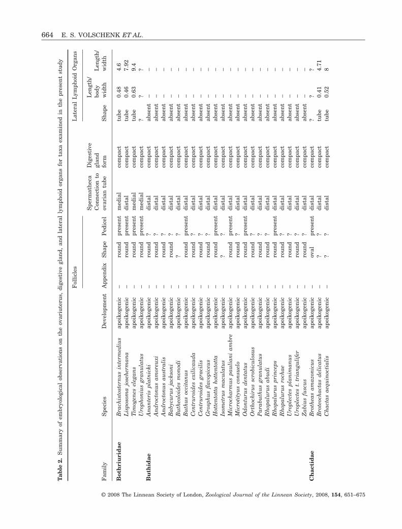
These organs were absent in all buthids and chaeril-ids examined, as well as in P. ovchinnikovi andM. pauliani ambre. All remaining species examinedpossess lateral lymphoid organs. We observed con-siderable variation in the shape and size of theseorgans. Some taxa, e.g. C. keyserlingi and Hadruroi-
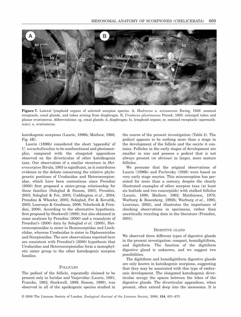
des charcasus Pocock, 1900 (Iuridae), Superstitioniadonensis Stahnke, 1940 (Superstitioniidae Stahnke,1940), and the Diplocentridae, exhibit small, sac-likestructures that do not extend beyond mesosomal seg-ment II, with a length/width ratio of less than 4.5(Table 2). Others, e.g. Urodacus, exhibit narrowtubes, which may extend as far as segment IV, witha length/width ratio up to 16.67 (Table 2, Fig. 7A, B).The lateral lymphoid organs are straight in mostcases, extending slightly to the ventral surface, andlie between the ovariuterine LTs. The lateral lym-phoid organs of Urodacus planimanus Pocock, 1893are very long (organ length/body width ratio of 1.22),and become tortuous distally (Table 2, Fig. 7B).Superstitionia donensis has the smallest lymphoidorgan observed, with an organ length/body widthratio of 0.15 (Table 2).
DISCUSSIONOVARIUTERUS
All nonbuthid scorpions described in the literature,and examined in the present study, possess a six-celled ovariuterus. Two distinct types of six-celledovariuterus were identified, conforming to taxa withapoikogenic and katoikogenic development. Apoiko-genic taxa possess a generally broader, oftenW-shaped arrangement of tubules, in which the TTsform two ventrally directed arcs between the MT andthe LTs (Fig. 3A–G). Katoikogenic scorpions possess aplanar, more elongated arrangement, in which theTTs are almost level with the MT in cross-section(Fig. 3H–I). As discussed further in the section onembryonic development, katoikogeny is restricted tothe scorpionoid families, excluding Bothriuridae, andis apomorphic relative to apoikogeny (Stockwell,1989; Prendini, 2000, 2003).
Pseudochactas ovchinnikovi also possesses a six-celled ovariuterus, yet this enigmatic scorpion alsoexhibits numerous buthid-like characters (Prendiniet al., 2006). Although its phylogenetic positionremains to be rigorously tested, the evidence suggeststhat Pseudochactas Gromov, 1998 is the sister groupof Buthidae (Prendini et al., 2006). If this is the case,
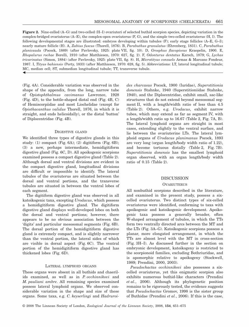
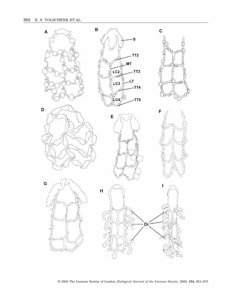
Figure 2. Nine-celled (A–G) and two-celled (H–I) ovariuteri of selected buthid scorpion species, depicting variation in thecomplex-bridged ovariuterus (A–E), the complex-open ovariuterus (F, G), and the simple two-celled ovariuterus (H, I). Thefollowing developmental stages are illustrated: embryos developing within tubules (F); early stage follicles (A–E, G–I);nearly mature follicle (H). A, Zabius fuscus (Thorell, 1876). B, Parabuthus granulatus (Ehrenberg, 1831). C, Parabuthusplanicauda (Pocock, 1889) (after Pavlovsky, 1925: plate VII, fig. 10). D, Grosphus flavopiceus Kraepelin, 1900. E,Rhopalurus rochae Borelli, 1910 (after Matthiesen, 1970: 627, fig. 2). F, Odonturus dentatus Karsch, 1879; G, Lychastricarinatus (Simon, 1884) (after Pavlovsky, 1925: plate VII, fig. 8). H, Microtityus consuelo Armas & Marcano Fondeur,1987. I, Tityus bahiensis (Perty, 1833) (after Matthiesen, 1970: 628, fig. 5). Abbreviations: LT, lateral longitudinal tubule;MC, median cell; ST, submedian longitudinal tubule; TT, transverse tubule.�
MESOSOMAL ANATOMY OF SCORPIONES (CHELICERATA) 661
© 2008 The Linnean Society of London, Zoological Journal of the Linnean Society, 2008, 154, 651–675
662 E. S. VOLSCHENK ET AL.
© 2008 The Linnean Society of London, Zoological Journal of the Linnean Society, 2008, 154, 651–675
and likewise if Pseudochactas is the sister group of allother Recent scorpions, as proposed by Soleglad & Fet(2003), the eight-celled ovariuterus found in mostButhidae must be apomorphic.
Among the buthid genera examined during thepresent investigation, Androctonus Ehrenberg, 1828,Anomalobuthus Kraepelin, 1900, Hottentotta Birula,1908, Liobuthus Birula, 1898, and OrthochirusKarsch, 1891, representing the Old-World Palaearcticbuthid clade (Fet, Soleglad & Lowe, 2003, 2005;Coddington et al., 2004), all possess the simpleeight-celled ovariuterus, suggesting that it may besynapomorphic for this clade.
The complex eight-celled ovariuterus has appar-ently evolved several times independently from thesimple condition. All buthids in which the complexeight-celled ovariuterus was observed, originate fromparts of the former Gondwanaland (Australia, India,Madagascar, southern Africa, and South America). Inthe present study, Uroplectes planimanus Karsch,1879 and Uroplectes t. triangulifer Thorell, 1876 werefound to possess the simple eight-celled ovariuterus,whereas representatives of Grosphus Simon, 1880and Parabuthus Pocock, 1890 were found to possess acomplex-bridged eight-celled ovariuterus. Prendini(2004) proposed that Uroplectes Peters, 1861 is thesister group of Parabuthus, and that the two Africangenera form a monophyletic sister group of the Mala-gasy Grosphus. Prendini’s (2004) hypothesis impliesthat the simple eight-celled ovariuterus was indepen-dently derived in Uroplectes and the Palaearcticbuthids, which are not closely related (Fet et al.,2003).
Two notable cases of variation in ovariuterineanatomy were observed among congeners. Rhopalu-rus princeps (Karsch, 1879) and R. abudi fromthe Dominican Republic both exhibit the simpleeight-celled ovariuterus. In contrast, the BrazilianR. rochae displays the complex-bridged ovariuterus,first observed by Matthiesen (1970), and confirmed inthe present study. Rhopalurus Thorell, 1876 is dis-continuously distributed in the Caribbean and Brazil(Lourenço, 1986, 2000a; Lourenço & Pinto-da-Rocha,1997; Armas, 1999; Armas, Ottenwalder & Guerrero,
1999). The observed differences in ovariuterineanatomy suggest that it may contain informativecharacters for the systematics of the genus. The phy-logenetic relationships of Rhopalurus are currentlyunknown, but it is possible that the genus may beparaphyletic with respect to Centruroides Marx, 1890,with which its component species share several syna-pomorphies (Sissom, 1990; Fet & Lowe, 2000). Weexamined Centruroides exilicauda (Wood, 1863) andCentruroides gracilis (Latreille, 1804) in the presentstudy, both of which possess the simple eight-celledovariuterus.
Pavlovsky (1925) illustrated three different typesof ovariuterine anatomy in the buthid genus LychasC.L. Koch, 1845. Lychas marmoreus exhibits thesimple eight-celled ovariuterus (Fig. 1E), L. variatusexhibits the complex-fused ovariuterus (Fig. 1L), andL. tricarinatus exhibits the complex-open ovariuterus(Fig. 2G). If Pavlovsky’s (1925) illustrations correctlydepict the anatomy of these species, then theseobservations suggest that this widespread genus(distributed in Africa, Australasia, and India) mayalso be paraphyletic. The ovariuterine anatomyof L. tricarinatus was independently confirmed byMathew (1960, 1962), and the anatomy of the otherLychas species illustrated by Pavlovsky (1925) wasverified during the present study. The taxonomy ofthe Australian Lychas (particularly L. marmoreus,L. variatus, and their respective synonyms) ischaotic (Kraepelin, 1916; Glauert, 1925; Koch, 1977;Kovarík, 1997). Additional species of Lychas shouldbe examined to illuminate the patterns observed byPavlovsky (1925). All other buthid genera examinedduring the present investigation, except Rhopalurus,possess the same ovariuterine anatomy amongcongeners.
Our observations on the ovariuterine anatomy alsosupport mounting evidence that the genus Micro-charmus Lourenço, 1996, currently placed in a uniquefamily, Microcharmidae Lourenço, 1996, is a buthid(Coddington et al., 2004). We observed the complexopen form of the eight-celled ovariuterus inM. pauliani ambre and two buthids, Babycurus jack-soni (Pocock, 1890) and L. tricarinatus. Microcharmus
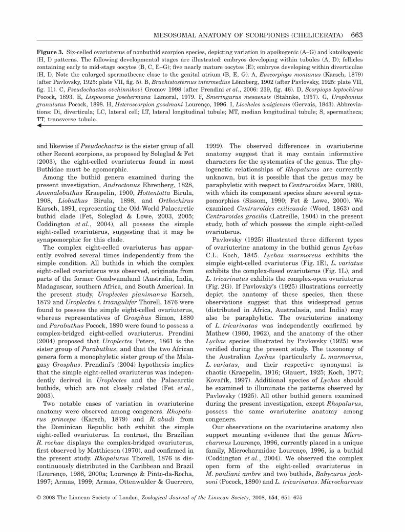
Figure 3. Six-celled ovariuterus of nonbuthid scorpion species, depicting variation in apoikogenic (A–G) and katoikogenic(H, I) patterns. The following developmental stages are illustrated: embryos developing within tubules (A, D); folliclescontaining early to mid-stage oocytes (B, C, E–G); five nearly mature oocytes (E); embryos developing within diverticulae(H, I). Note the enlarged spermathecae close to the genital atrium (B, E, G). A, Euscorpiops montanus (Karsch, 1879)(after Pavlovsky, 1925: plate VII, fig. 5). B, Brachistosternus intermedius Lönnberg, 1902 (after Pavlovsky, 1925: plate VII,fig. 11). C, Pseudochactas ovchinnikovi Gromov 1998 (after Prendini et al., 2006: 239, fig. 46). D, Scorpiops leptochirusPocock, 1893. E, Lisposoma josehermana Lamoral, 1979. F, Smeringurus mesaensis (Stahnke, 1957). G, Urophoniusgranulatus Pocock, 1898. H, Heteroscorpion goodmani Lourenço, 1996. I, Liocheles waigiensis (Gervais, 1843). Abbrevia-tions: Di, diverticula; LC, lateral cell; LT, lateral longitudinal tubule; MT, median longitudinal tubule; S, spermatheca;TT, transverse tubule.�
MESOSOMAL ANATOMY OF SCORPIONES (CHELICERATA) 663
© 2008 The Linnean Society of London, Zoological Journal of the Linnean Society, 2008, 154, 651–675
Tab
le2.
Su
mm
ary
ofem
bryo
logi
cal
obse
rvat
ion
son
the
ovar
iute
rus,
dige
stiv
egl
and,
and
late
ral
lym
phoi
dor
gan
sfo
rta
xaex
amin
edin
the
pres
ent
stu
dy
Fam
ily
Spe
cies
Dev
elop
men
t
Fol
licl
es
Spe
rmat
hec
aC
onn
ecti
onto
ovar
ian
tube
Dig
esti
vegl
and
form
Lat
eral
Lym
phoi
dO
rgan
s
App
endi
xS
hap
eP
edic
elS
hap
e
Len
gth
/bo
dyw
idth
Len
gth
/w
idth
Bot
hri
uri
dae
Bra
chis
tost
ern
us
inte
rmed
ius
apoi
koge
nic
–ro
un
dpr
esen
tm
edia
lco
mpa
cttu
be0.
484.
6L
ispo
som
ajo
seh
erm
ana
apoi
koge
nic
–ro
un
dpr
esen
tdi
stal
com
pact
tube
0.46
7.92
Tim
ogen
esel
egan
sap
oiko
gen
ic–
rou
nd
pres
ent
med
ial
com
pact
tube
0.63
9.4
Uro
phon
ius
gran
ula
tus
apoi
koge
nic
–ro
un
dpr
esen
tm
edia
lco
mpa
ct?
??
Bu
thid
aeA
nan
teri
spl
atn
icki
apoi
koge
nic
–ro
un
d?
dist
alco
mpa
ctab
sen
t–
–A
nd
roct
onu
sam
oreu
xiap
oiko
gen
ic–
rou
nd
?di
stal
com
pact
abse
nt
––
An
dro
cton
us
aust
rali
sap
oiko
gen
ic–
rou
nd
?di
stal
com
pact
abse
nt
––
Bab
ycu
rus
jack
son
iap
oiko
gen
ic–
rou
nd
?di
stal
com
pact
abse
nt
––
Bu
theo
loid
esm
onod
iap
oiko
gen
ic–
??
dist
alco
mpa
ctab
sen
t–
–B
uth
us
occi
tan
us
apoi
koge
nic
–ro
un
dpr
esen
tdi
stal
com
pact
abse
nt
––
Cen
tru
roid
esex
ilic
aud
aap
oiko
gen
ic–
rou
nd
?di
stal
com
pact
abse
nt
––
Cen
tru
roid
esgr
acil
isap
oiko
gen
ic–
rou
nd
?di
stal
com
pact
abse
nt
––
Gro
sph
us
flav
opic
eus
apoi
koge
nic
–ro
un
d?
dist
alco
mpa
ctab
sen
t–
–H
otte
nto
tta
hot
ten
tott
aap
oiko
gen
ic–
rou
nd
pres
ent
dist
alco
mpa
ctab
sen
t–
–Is
omet
rus
mac
ula
tus
apoi
koge
nic
–?
?di
stal
com
pact
abse
nt
––
Mic
roch
arm
us
pau
lian
iam
bre
apoi
koge
nic
–ro
un
dpr
esen
tdi
stal
com
pact
abse
nt
––
Mic
roti
tyu
sco
nsu
elo
apoi
koge
nic
–ro
un
d?
dist
alco
mpa
ctab
sen
t–
–O
don
turu
sd
enta
tus
apoi
koge
nic
–ro
un
dpr
esen
tdi
stal
com
pact
abse
nt
––
Ort
hoc
hir
us
scro
bicu
losu
sap
oiko
gen
ic–
rou
nd
?di
stal
com
pact
abse
nt
––
Par
abu
thu
sgr
anu
latu
sap
oiko
gen
ic–
rou
nd
?di
stal
com
pact
abse
nt
––
Rh
opal
uru
sab
ud
iap
oiko
gen
ic–
rou
nd
?di
stal
com
pact
abse
nt
––
Rh
opal
uru
spr
ince
psap
oiko
gen
ic–
rou
nd
pres
ent
dist
alco
mpa
ctab
sen
t–
–R
hop
alu
rus
roch
aeap
oiko
gen
ic–
rou
nd
?di
stal
com
pact
abse
nt
––
Uro
plec
tes
plan
iman
us
apoi
koge
nic
–ro
un
d?
dist
alco
mpa
ctab
sen
t–
–U
ropl
ecte
st.
tria
ngu
life
rap
oiko
gen
ic–
rou
nd
?di
stal
com
pact
abse
nt
––
Zab
ius
fusc
us
apoi
koge
nic
–ro
un
d?
dist
alco
mpa
ctab
sen
t–
–C
hac
tid
aeB
roth
eas
amaz
onic
us
apoi
koge
nic
–ov
alpr
esen
tdi
stal
com
pact
??
?B
rote
och
acta
sd
elic
atu
sap
oiko
gen
ic–
??
dist
alco
mpa
cttu
be0.
414.
71C
hac
tas
aequ
inoc
tial
isap
oiko
gen
ic–
??
dist
alco
mpa
cttu
be0.
528
664 E. S. VOLSCHENK ET AL.
© 2008 The Linnean Society of London, Zoological Journal of the Linnean Society, 2008, 154, 651–675
Ch
aeri
lid
aeC
hae
rilu
sgr
anos
us
apoi
koge
nic
–ro
un
dpr
esen
tdi
stal
com
pact
abse
nt
––
Dip
loce
ntr
idae
Bio
culu
sco
mon
dae
kato
ikog
enic
pres
ent
thu
mb
abse
nt
dist
aldi
giti
form
sac
0.42
2.88
Dip
loce
ntr
us
wh
itei
kato
ikog
enic
pres
ent
??
dist
aldi
giti
form
sac
0.24
2.3
Neb
ofl
avip
eska
toik
ogen
icpr
esen
t?
?di
stal
??
??
Eu
scor
pii
dae
Eu
scor
piu
sco
nci
nn
us
apoi
koge
nic
–ov
alpr
esen
tdi
stal
com
pact
tube
0.47
8.66
Hem
isco
rpii
dae
Hem
isco
rpiu
sle
ptu
rus
kato
ikog
enic
pres
ent
thu
mb
abse
nt
dist
aldi
giti
form
tube
0.44
4.65
Het
eros
corp
ion
idae
Het
eros
corp
ion
good
man
ika
toik
ogen
icab
sen
tco
ne
abse
nt
dist
aldi
giti
form
??
?Iu
rid
aeC
arab
octo
nu
ske
yser
lin
giap
oiko
gen
ic–
oval
pres
ent
subd
ista
lco
mpa
ctsa
c0.
444.
43H
adru
roid
esch
arca
sus
apoi
koge
nic
–ov
alpr
esen
tdi
stal
com
pact
sac
0.34
3.1
Had
ruru
sa.
ariz
onen
sis
apoi
koge
nic
–ro
un
dpr
esen
tsu
bdis
tal*
com
pact
tube
0.31
8.05
Iuru
sd
ufo
ure
ius
asia
ticu
sap
oiko
gen
ic–
oval
pres
ent
dist
alco
mpa
cttu
be0.
354.
55L
ioch
elid
aeH
adog
enes
hah
ni
kato
ikog
enic
pres
ent
thu
mb
abse
nt
dist
aldi
giti
form
tube
0.35
6.36
Lio
chel
esau
stra
lasi
aeka
toik
ogen
icpr
esen
tth
um
bab
sen
tdi
stal
digi
tifo
rm?
??
Lio
chel
esw
aigi
ensi
ska
toik
ogen
icpr
esen
t?
abse
nt
dist
aldi
giti
form
tube
0.46
13.7
5O
pist
hac
anth
us
vali
du
ska
toik
ogen
icpr
esen
tth
um
bab
sen
tdi
stal
digi
tifo
rmtu
be0.
475.
63P
seu
doc
hac
tid
aeP
seu
doc
hac
tas
ovch
inn
ikov
iap
oiko
gen
ic–
rou
nd
pres
ent
dist
alco
mpa
ctab
sen
t–
–S
corp
ion
idae
Opi
stop
hth
alm
us
cavi
man
us
kato
ikog
enic
pres
ent
thu
mb
abse
nt
dist
aldi
giti
form
??
?S
corp
iop
idae
Eu
scor
piop
slo
ngi
man
us
apoi
koge
nic
–ov
alpr
esen
tdi
stal
com
pact
tube
0.33
8.25
Su
per
stit
ion
iid
aeS
upe
rsti
tion
iad
onen
sis
apoi
koge
nic
–ro
un
dpr
esen
tdi
stal
com
pact
sac
0.15
1.5
Tro
glot
ayos
icid
aeB
elis
ariu
sxa
mbe
ui
apoi
koge
nic
–?
?di
stal
com
pact
tube
??
Uro
dac
idae
Uro
dac
us
plan
iman
us
kato
ikog
enic
abse
nt
thu
mb
abse
nt
dist
alh
emid
igit
ifor
mtu
be1.
2215
.28
Uro
dac
us
spin
atu
ska
toik
ogen
icab
sen
tth
um
bab
sen
tdi
stal
hem
idig
itif
orm
tube
??
Uro
dac
us
sp.
kato
ikog
enic
abse
nt
thu
mb
abse
nt
dist
alh
emid
igit
ifor
mtu
be0.
7716
.67
Vae
jovi
dae
Sm
erin
guru
sm
esae
nsi
sap
oiko
gen
ic–
–ab
sen
tdi
stal
com
pact
tube
0.38
6.71
Uro
cton
us
mor
dax
apoi
koge
nic
–ro
un
dpr
esen
tdi
stal
com
pact
tube
0.68
9.5
Vaej
ovis
spin
iger
us
apoi
koge
nic
–ro
un
dpr
esen
tdi
stal
com
pact
tube
0.41
6.5
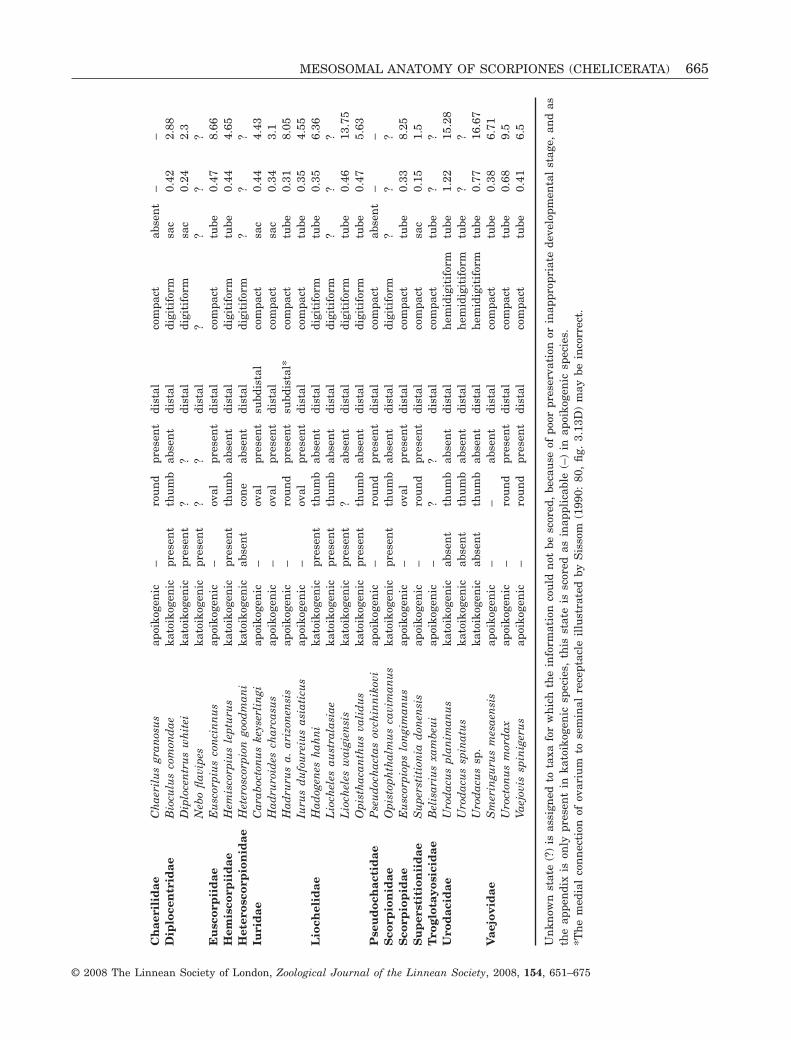
Un
know
nst
ate
(?)
isas
sign
edto
taxa
for
wh
ich
the
info
rmat
ion
cou
ldn
otbe
scor
ed,
beca
use
ofpo
orpr
eser
vati
onor
inap
prop
riat
ede
velo
pmen
tal
stag
e,an
das
the
appe
ndi
xis
only
pres
ent
inka
toik
ogen
icsp
ecie
s,th
isst
ate
issc
ored
asin
appl
icab
le(–
)in
apoi
koge
nic
spec
ies.
*Th
em
edia
lco
nn
ecti
onof
ovar
ium
tose
min
alre
cept
acle
illu
stra
ted
byS
isso
m(1
990:
80,
fig.
3.13
D)
may
bein
corr
ect.
MESOSOMAL ANATOMY OF SCORPIONES (CHELICERATA) 665
© 2008 The Linnean Society of London, Zoological Journal of the Linnean Society, 2008, 154, 651–675
also lacks lateral lymphoid organs, which is anotherbuthid characteristic. These anatomical characterssupport numerous external morphological characters(e.g. the presence of the type-A trichobothrial patternon the pedipalps) otherwise unique to Buthidae, fromwhich Microcharmidae is separated principally on thebasis of size and ecology (Lourenço, 2000b). Thebalance of evidence does not, in our opinion, warrantcontinued recognition of Microcharmidae, whichrenders Buthidae paraphyletic (E. S. Volschenk & L.Prendini, unpubl. data). We therefore propose thefollowing new synonymy: Microcharmidae Lourenço,1996 = Buthidae C.L. Koch, 1837.
SPERMATHECAE
The spermatheca is an elastic structure, which isprobably subject to slight changes in size depending
on the quantity of sperm contained within; however,several characters of this organ may be phylogeneti-cally informative. Knowledge of the extent of varia-tion in the spermathecae is limited, as spermathecalsize is probably dependent on time elapsed since thelast mating, as well as on the stage of the reproduc-tive cycle. Three independent studies observedenlarged spermathecae in the iurid, H. a. arizonensis(Sissom, 1990; Farley, 2001; this study). Spermathe-cal size may also be phylogenetically informative inthe other iurids, bothriurids, and chactids, in whichenlarged spermathecae have been observed. Theabsence or small size of the spermathecae of mostother scorpions, including Buthidae and Pseudo-chactas, suggests that enlarged spermathecae areapomorphic.
The extremely enlarged spermathecae observed insome taxa (e.g. H. a. arizonensis and Urophonius
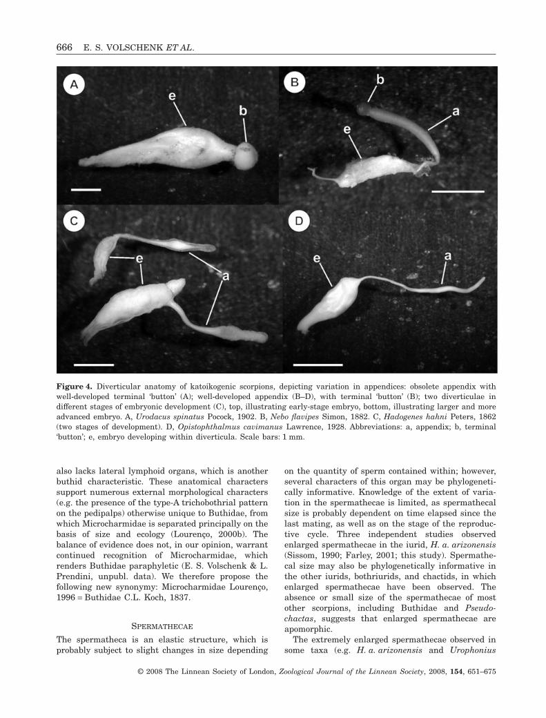
Figure 4. Diverticular anatomy of katoikogenic scorpions, depicting variation in appendices: obsolete appendix withwell-developed terminal ‘button’ (A); well-developed appendix (B–D), with terminal ‘button’ (B); two diverticulae indifferent stages of embryonic development (C), top, illustrating early-stage embryo, bottom, illustrating larger and moreadvanced embryo. A, Urodacus spinatus Pocock, 1902. B, Nebo flavipes Simon, 1882. C, Hadogenes hahni Peters, 1862(two stages of development). D, Opistophthalmus cavimanus Lawrence, 1928. Abbreviations: a, appendix; b, terminal‘button’; e, embryo developing within diverticula. Scale bars: 1 mm.
666 E. S. VOLSCHENK ET AL.
© 2008 The Linnean Society of London, Zoological Journal of the Linnean Society, 2008, 154, 651–675
granulatus Pocock, 1899) probably contain sperm frommultiple inseminations, because the quantity of spermcontained in a single spermatophore from these speciesis a small fraction of the volume of the spermatheca(A.V. Peretti, pers. comm.). The purpose of this accu-mulation of sperm is unknown, but is presumed tofacilitate the production of multiple broods.
The point of attachment of the ovariuterus to thespermathecae also varies. The ovariuterus of mostscorpions possessing spermathecae is attached poste-riorly, and it is likely that, during parturition, anysperm retained in the spermathecae will be expelledas the first of the brood pass through to the exterior.The ovariuterus of all bothriurids (except L. joseher-mana) examined in the present study attaches inter-nolaterally to the spermathecae. Lateral attachmentto the spermathecae creates a broad pocket that mayfacilitate the storage of large quantities of sperm. Theextreme size of the spermathecae observed in iurids
and bothriurids may thus serve not only to store alarge mass of sperm, but also to prevent its expulsionduring parturition.
LOCATION OF EMBRYONIC DEVELOPMENT
Several studies (e.g. Stockwell, 1989; Prendini, 2000,2003) demonstrated that apoikogenic developmentis plesiomorphic, and katoikogenic developmentapomorphic, in scorpions. Katoikogenic scorpionsinclude all Scorpionoidea Latreille, 1802 (sensuPrendini, 2000) except Bothriuridae, which areapoikogenic. Katoikogeny appears to have evolvedonly once (Prendini, 2000, 2003).
Lourenço (2002) speculated that Lisposoma wouldpossess well-developed diverticulae like other scorpi-onoid taxa, contrary to Stockwell (1989) and Prendini(2000). We confirmed the presence of a typical,
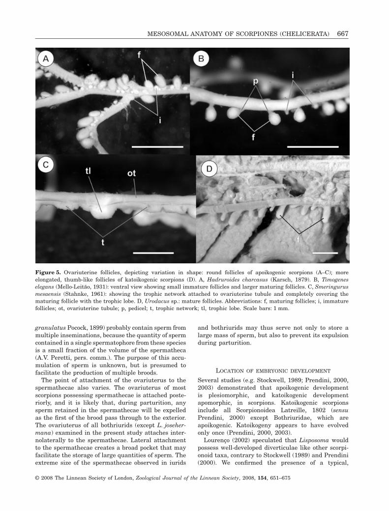
Figure 5. Ovariuterine follicles, depicting variation in shape: round follicles of apoikogenic scorpions (A–C); moreelongated, thumb-like follicles of katoikogenic scorpions (D). A, Hadruroides charcasus (Karsch, 1879). B, Timogeneselegans (Mello-Leitão, 1931): ventral view showing small immature follicles and larger maturing follicles. C, Smeringurusmesaensis (Stahnke, 1961): showing the trophic network attached to ovariuterine tubule and completely covering thematuring follicle with the trophic lobe. D, Urodacus sp.: mature follicles. Abbreviations: f, maturing follicles; i, immaturefollicles; ot, ovariuterine tubule; p, pedicel; t, trophic network; tl, trophic lobe. Scale bars: 1 mm.
MESOSOMAL ANATOMY OF SCORPIONES (CHELICERATA) 667
© 2008 The Linnean Society of London, Zoological Journal of the Linnean Society, 2008, 154, 651–675
apoikogenic ovariuterus in L. josehermana (Fig. 4E),as reported by Stockwell (1989) and Prendini (2000,2003).
The alleged ‘third type’ of embryonic developmentreported for V. spinigerus and C. werneri judaicus(Warburg & Rosenberg, 1996; Warburg, 2001) appearsto be a misinterpretation of an advanced stage in thedevelopment of the embryos inside the ovariuterus.We observed regular follicles in V. spinigerus, con-forming to typical apoikogenic development.
The typical, elongated diverticular appendix is notpresent in all katoikogenic taxa (Laurie, 1896b). Wedid not observe elongated appendices on the diver-ticulae of H. goodmani (Heteroscorpionidae) or Uro-dacus spinatus Pocock, 1902 (Urodacidae), which
was the only urodacid with developing embryos thatwe examined in the course of this study. We insteadobserved a thickened structure, on a short pedicel(perhaps a rudimentary appendix), situated distallyon the embryos (Fig. 4A) of these taxa. Laurie (1896b)and Mathew (1968) documented similar structuresin the early embryos of Urodacus novaehollandiaePeters, 1861 and U. manicatus, respectively. Mathew(1968) conducted a detailed investigation of the inter-nal structure of the follicles of U. manicatus, anddemonstrated that the thick, button-like structure isan accumulation of gland cells, which may secretenourishment for the developing embryo. This secre-tory structure is located at the distalmost part of theelongated appendices that are characteristic of other
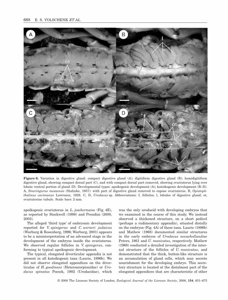
Figure 6. Variation in digestive gland: compact digestive gland (A); digitiform digestive gland (B); hemidigitiformdigestive gland, showing compact dorsal part (C), and with compact dorsal part removed, showing ovariuterus lying overlobate ventral portion of gland (D). Developmental types: apoikogenic development (A); katoikogenic development (B–D).A, Smeringurus mesaensis (Stahnke, 1957): with part of digestive gland removed to expose ovariuterus. B, Opistoph-thalmus cavimanus Lawrence, 1928. C, D, Urodacus sp. Abbreviations: f, follicles; l, lobules of digestive gland; ot,ovariuterine tubule. Scale bars: 2 mm.
668 E. S. VOLSCHENK ET AL.
© 2008 The Linnean Society of London, Zoological Journal of the Linnean Society, 2008, 154, 651–675
katoikogenic scorpions (Laurie, 1896b; Mathew, 1968;Fig. 4B).
Laurie (1896b) considered the short ‘appendix’ ofU. novaehollandiae to be nonfunctional and plesiomor-phic, compared with the elongated appendicesobserved on the diverticulae of other katoikogenictaxa. Our observation of a similar structure in Het-eroscorpion Birula, 1903 is significant, as it contributesevidence to the debate concerning the relative phylo-genetic positions of Urodacidae and Heteroscorpion-idae, which have been contentious since Prendini(2000) first proposed a sister-group relationship forthese families (Soleglad & Sissom, 2001; Prendini,2003; Soleglad & Fet, 2003; Coddington et al., 2004;Prendini & Wheeler, 2005; Soleglad, Fet & Kovarík,2005; Lourenço & Goodman, 2006; Volschenk & Pren-dini, 2008). According to the alternative hypothesis,first proposed by Stockwell (1989), but also obtained insome analyses by Prendini (2000) and a reanalysis ofPrendini’s (2000) data by Soleglad et al. (2005), Het-eroscorpionidae is sister to Hemiscorpiidae and Lioch-elidae, whereas Urodacidae is sister to Diplocentridaeand Scorpionidae. The new observations reported hereare consistent with Prendini’s (2000) hypothesis thatUrodacidae and Heteroscorpionidae form a monophyl-etic sister group to the other katoikogenic scorpionfamilies.
FOLLICLES
The pedicel of the follicle, repeatedly claimed to bepresent only in Iuridae and Vaejovidae (Laurie, 1891;Francke, 1982; Stockwell, 1989; Sissom, 1990), wasobserved in all of the apoikogenic species studied in
the course of the present investigation (Table 2). Thepedicel appears to be nothing more than a stage inthe development of the follicle and the oocyte it con-tains. Follicles in the early stages of development aresmaller in size and possess a pedicel that is notalways present (or obvious) in larger, more maturefollicles.
We presume that the original observations ofLaurie (1896b) and Pavlovsky (1926) were based onvery early stage oocytes. This misconception has per-sisted for more than a century, despite the clearlyillustrated examples of other scorpion taxa (at leastsix buthids and two euscorpiids) with stalked follicles(Laurie, 1890; Mathew, 1962; Matthiesen, 1970;Warburg & Rosenberg, 1992b; Warburg et al., 1995;Lourenço, 2002), and illustrates the importance ofchecking observations in specimens, rather thanuncritically recycling data in the literature (Prendini,2001).
DIGESTIVE GLAND
We observed three different types of digestive glandsin the present investigation: compact, hemidigitiform,and digitiform. The function of the digitiformdigestive gland is unknown, and we suggest twopossibilities.
The digitiform and hemidigitiform digestive glandsare only known in katoikogenic scorpions, suggestingthat they may be associated with this type of embry-onic development. The elongated katoikogenic diver-ticulae occupy the spaces between the lobes of thedigestive glands. The diverticular appendices, whenpresent, often extend deep into the mesosoma. It is
Figure 7. Lateral lymphoid organs of selected scorpion species. A, Hadrurus a. arizonensis Ewing, 1928: seminalreceptacle, coxal glands, and tubes arising from diaphragm. B, Urodacus planimanus Pocock, 1893: enlarged tubes andplanar ovariuterus. Abbreviations: cg, coxal glands; d, diaphragm; lo, lymphoid organs; sr, seminal receptacle (spermath-ecae); o, ovariuterus.
MESOSOMAL ANATOMY OF SCORPIONES (CHELICERATA) 669
© 2008 The Linnean Society of London, Zoological Journal of the Linnean Society, 2008, 154, 651–675
thus possible that the diverticulae may requireadditional space in the mesosoma for growth anddevelopment.
The digitiform and hemidigitiform digestive glandsmay also increase the surface area that is exposed tothe haemolymph. Although the appendix of the diver-ticula is known to channel nutrients to the embryo atits base (Mathew, 1968), the mechanism by whichthese nutrients enter the appendix is unknown. Onthe assumption that this process occurs by diffusionfrom the haemolymph, the increased surface area ofthe digitiform and hemidigitiform digestive glandswould increase the rate at which nutrients coulddiffuse from the glands into the haemolymph.
Contrary to Pavlovsky & Zarin (1926), we did notobserve a digitiform digestive gland in the chactidsand vaejovids we examined, all of which possessed acompact digestive gland.
The digestive gland does not appear to be sexuallydimorphic. The same anatomy has been observed inmales and females in all species possessing a compactdigestive gland. Similarly, the digitiform digestivegland was observed in both sexes in at least twokatoikogenic scorpions (Bioculus comondae Stahnke,1968 and O. validus).
LATERAL LYMPHOID ORGANS
The absence of lateral lymphoid organs in P. ovchin-nikovi, Buthidae, and Chaerilidae is congruent withall hypotheses concerning the possible placement ofPseudochactidae in the phylogeny of scorpions: sistergroup of all recent scorpions, sister group of Buthidae,or sister group of Chaerilidae (Prendini et al., 2006).Considering the most recent hypothesis of scorpionphylogeny (Coddington et al., 2004), the presence oflateral lymphoid organs may be apomorphic in theorder. The morphology and degree of development ofthese organs is quite variable (Table 2), but is con-sistent within some taxa (e.g. Urodacus), and maycarry phylogenetic information. Further physiologicalresearch is needed to completely clarify the functionof these organs (Farley, 1999).
CONCLUSIONS
Phylogenetically informative characters from themesosomal anatomy of scorpions were first reported acentury ago (Laurie, 1896a, b; Pavlovsky, 1913,1915a, b, c, d, 1917, 1924a, b, c, d, 1925, 1926;Pavlovsky & Zarin, 1926), and have received littleattention since. Many of the observations in theseearly studies were reported from a limited sample oftaxa, and few were subsequently confirmed. Theseobservations were nevertheless incorporated into theanatomical literature and into recent phylogenetic
analyses, and, in some cases, were extrapolated toother taxa in which they had not been observed. Someorgan systems (e.g. the digestive gland and laterallymphoid organs) have never been studied compara-tively across diverse taxa, or from a systematicperspective, and their reference in the literature islimited to their original discovery.
Here, we summarized existing data on theseorgans, reassessed the original observations as far aspossible, augmented the set of observations across asample of taxa representing most scorpion families,and revised the terminology to accommodate therange of variation observed. In so doing, we confirmedmost of the original observations of previous workersand also identified a few misconceptions. In spite ofthese advances, the mesosomal anatomy of scorpionsremains poorly known. Further study is needed toassess the extent of intraspecific variation in theseorgans, and to further enlarge the taxon sample atthe generic level. Just as male hemispermatophoresare increasingly included in taxonomic descriptionsand phylogenetic analyses on scorpions, we hope thatthis contribution will inspire the inclusion of morecharacters from the internal anatomy of scorpions.
ACKNOWLEDGEMENTS
ESV and CIM were each supported for two years byan Ambrose Monell Postdoctoral Fellowship from theAMNH; ESV received support for a third year from agrant to LP, from the Richard Lounsbery Foundation.Fieldwork, during which some of the specimens usedin the present study were collected, was supported bythe American Museum of Natural History (AMNH)and by grants to LP from the following organizations:US National Science Foundation (BIO-DEB 0413453,EAR 0228699, EAR 0313698); Richard LounsberyFoundation; Stavros Niarchos Foundation. We thankthe following for loans of material and permission todissect specimens: Alexander Gromov (Alexander V.Gromov Private Collection, Almaty, Kazakhstan);Charles Griswold and Darrel Ubick (CaliforniaAcademy of Sciences, San Francisco, CA, USA);Gonzalo Giribet and Laura Liebensperger (Museumof Comparative Zoology, Harvard University, Cam-bridge, MA, USA); Robert Raven and Owen Seaman(Queensland Museum, Brisbane, Australia); JonathanCoddington and Dana De Roche (US NationalMuseum of Natural History, Smithsonian Institution,Washington, DC, USA); Mark Harvey and JulianneWaldock (Western Australian Museum, Perth,Australia); Jason Dunlop and Shahin Nawai(Museum für Naturkunde der Humboldt-Universität,Berlin, Germany). We also thank Andrés OjangurenAffilastro (Museo Argentino de Ciencias Naturales‘Bernardino Rivadavia’, Argentina) for the gift of the
670 E. S. VOLSCHENK ET AL.
© 2008 The Linnean Society of London, Zoological Journal of the Linnean Society, 2008, 154, 651–675
Zabius fuscus (Thorell, 1876) specimens used in thisstudy. Finally, we thank Lauren Esposito, EdmundoGonzález, Lionel Monod (all at the AMNH) for dis-cussions on the topic, and Roger Farley and AlfredoPeretti for constructive comments on previous ver-sions of the manuscript.
REFERENCES
de Armas LF. 1999. Quince nuevos alacranes de La Españolay Navassa, Antillas Mayores (Arachnida: Scorpiones). Avi-cennia 10/11: 101–136.
de Armas LF, Ottenwalder JA, Guerrero KA. 1999. Escor-piones de las Islas Saona, Beata y Catalina, RepublicaDominicana (Arachnida: Scorpiones). Cocuyo 8: 30–32.
Coddington JA, Giribet G, Harvey MS, Prendini L,Walter DE. 2004. Arachnida. In: Cracraft J, Donoghue M,eds. Assembling the tree of life. Oxford: Oxford UniversityPress, 296–318.
Farley RD. 1984. The ultrastructure of hematocytopoieticorgans in the desert scorpion Paruroctonus. Tissue and Cell16: 577–588.
Farley RD. 1998. Matrotrophic adaptations and early stagesof embryogenesis in the desert scorpion Paruroctonusmesaensis (Vaejovidae). Journal of Morphology 237: 187–211.
Farley RD. 1999. Scorpiones. In: Harrison FW, Foelix RF,eds. Microscopic anatomy of invertebrates. Cheliceratearthropoda. Vol. 8A. New York: Wiley-Liss, 117–222.
Farley RD. 2001. Structure, reproduction, and development.In: Brownell P, Polis GA, eds. Scorpion biology andresearch. Oxford: Oxford University Press, 13–78.
Fet V, Gantenbein B, Gromov AV, Lowe G. 2003. The firstmolecular phylogeny of Buthidae (Scorpiones). Euscorpius4: 1–10.
Fet V, Lowe G. 2000. Family Buthidae C.L. Koch, 1837. In:Fet V, Sissom WD, Lowe G, Braunwalder ME, eds. Catalogof the Scorpions of the World (1758–1998). New York: NewYork Entomological Society, 54–286.
Fet V, Soleglad ME, Lowe G. 2005. A new trichobothrialcharacter for high-level systematics of Buthidae (Scorpi-ones: Buthidae). Euscorpius 23: 1–40.
Francke OF. 1982. Parturition in scorpions (Arachnida,Scorpiones): a review of the ideas. Revue Arachnologique4: 27–37.
Glauert L. 1925. Australian Scorpionidea. Part 1. Journal ofthe Royal Society of Western Australia 11: 89–118.
Hjelle JT. 1990. Anatomy and morphology. In: Polis GA, ed.The biology of scorpions. Stanford: Stanford UniversityPress, 9–63.
Koch LE. 1977. The taxonomy, geographic distributionand evolutionary radiation of Australo-Papuan scorpions.Records of the Western Australian Museum 5: 83–367.
Kovarík F. 1997. Revision of the genera Lychas and Hemily-chas, with descriptions of six new species (Scorpiones:Buthidae). Acta Societatis Zoologicae Bohemoslovacae 61:311–371.
Kraepelin K. 1916. Results of Dr Mjöberg’s Swedish Scien-tific Expeditions to Australia 1910–1913. 4. Scolopendridenund Scorpione. Arkiv für Zoologie 10: 18–43.
Laurie M. 1890. The embryology of the scorpion (Euscorpiusitalicus). Quarterly Journal of Microscopical Science 31:105–141.
Laurie M. 1891. Some points in the development of Scorpiofulvipes. Quarterly Journal of Microscopical Science 32:587–597.
Laurie M. 1896a. Further notes on the anatomy and devel-opment of scorpions, and their bearing on the classificationof the order. Annals and Magazine of Natural History (6) 18:121–133.
Laurie M. 1896b. Notes on the anatomy of some scorpions,and its bearing on the classification of the order. Annals andMagazine of Natural History (6) 17: 185–184.
Lourenço WR. 1986. Biogéographie et phylogénie des scor-pions du genre Rhopalurus Thorell, 1876 (Scorpiones,Buthidae). Mémoires de la Société Royale Belged’Entomologie 33: 129–137.
Lourenço WR. 2000a. Panbiogeographie, les familles desscorpions et leur repartition geographique. Biogeographica76: 21–39.
Lourenço WR. 2000b. Un nouveau genre de scorpion mal-gache, maillon possible entre les Microcharmidae et lesButhidae. Comptes Rendus de l’Academie des Sciences. SerieIII. Sciences de la Vie 323: 877–881.
Lourenço WR. 2002. Reproduction in scorpions, with specialreference to parthenogenesis. In: Toft S, Scharff N, eds.European arachnology 2000. Aarhus: Aarus UniversityPress, 71–85.
Lourenço WR, Goodman SM. 2006. Description of a newspecies of Heteroscorpion Birula, 1903 (Scorpiones, Het-eroscorpionidae) from the Montagne des Français inextreme northern Madagascar. Zootaxa 1269: 31–41.
Lourenço WR, Pinto-da-Rocha R. 1997. A reappraisal ofthe geographic distribution of the genus Rhopalurus Thorell(Scorpiones, Buthidae) and description of two new species.Biogeographica (Paris) 73: 181-191.
Mahsberg D, Warburg MR. 2000. Ovariuterus of Pandinusimperator, Koch (Scorpiones; Scorpionidae): comparison ofvirgin female with mother. Annals of Anatomy 182: 171–174.
Mathew AP. 1956. The ovarian diverticule of Heterometrus(Palamnaeus) scaber. Proceedings of the Indian Science Con-gress Association 4: 28.
Mathew AP. 1960. Embryonic nutrition in Lychas tricarina-tus. Journal of the Zoological Society of India 12: 220–228.
Mathew AP. 1962. Reproductive biology of Lychas tricarina-tus (Simon). Biological Bulletin 123: 344–350.
Mathew AP. 1968. Embryonic nutrition in Urodacus abrup-tus, Pocock. Journal of Animal Morphology and Physiology15: 162–167.
Matthiesen FA. 1970. Reproductive system and embryos ofBrazilian scorpions. Anais da Academia Brasileira de Cien-cias 42: 627–632.
Millot J, Vachon M. 1949. Ordre des Scorpiones. In: GrasséP-P, ed. Traité de Zoologie. Paris: Masson et Cie, 386–436.
MESOSOMAL ANATOMY OF SCORPIONES (CHELICERATA) 671
© 2008 The Linnean Society of London, Zoological Journal of the Linnean Society, 2008, 154, 651–675
Nayar KK. 1966. Endocrine nature of the lymphoid organs ofthe scorpion Heterometrus scaber Thorell. Proceedings of the2nd All-India Congress of Zoology 2: 173–174.
Pavlovsky EN. 1913. Scorpiotomische Mitteilungen. I. EinBeitrag zur Morphologie der Giftdrüsen der Skorpione.Zeitschrift für Wissenschaftliche Zoologie 105: 157–177.
Pavlovsky EN. 1915a. Contribution à la connaissance de lastructure et du développement post-embryonnaire desorganes génitaux du Buthus australis L. Entomologicheskoeobozrenie 15: 61–63.
Pavlovsky EN. 1915b. Sur la structure des organes phago-cytaires chez Scorpio maurus L. Comptes Rendus desSéances de la Société de Biologie Paris 78: 746–748.
Pavlovsky EN. 1915c. Sur la phagocytose chez Scorpiomaurus L. Comptes Rendus des Séances de la Société deBiologie Paris 78: 748–750.
Pavlovsky EN. 1915d. Sur la structures et sur le développe-ment post-embyonnaire des organes génitaux mâles duButhus australis L. Comptes Rendus de la Société de Biolo-gie Paris 80: 61–63.
Pavlovsky EN. 1917. Opuscula Scorpiotomica. 1. Surl’appareil génital male et sur un cas d’anomalie de cetappareil chez Isometrus maculatus (Scorpionides, fam.Buthidae). Comptes Rendus des Séances de la Société deBiologie Paris 80: 502–505.
Pavlovsky EN. 1924a. [On the morphology of the malegenital apparatus in scorpions]. Trudy LeningradskogoObshchetsva Lyubitelei Prirody [Transactions of the Lenin-grad Society of Naturalists] 53: 17–86.
Pavlovsky EN. 1924b. Scorpiotomische Mitteilungen. III.Zur äusseren Morphologie der Scorpione. IV. Zur Morpholo-gie der weiblichen Genitalorgane der Scorpione. ZoologischeJahrbücher, Anatomie und Ontogenie 46: 473–508.
Pavlovsky EN. 1924c. Studies on the organisation and devel-opment of Scorpions. [1. On the external morphology of thescorpions. 2: The integument and its processes. 3: Thecirculatory system. 4: The system of lymphatic/phagocyticorgans]. Quarterly Journal of Microscopical Science 68:615–640.
Pavlovsky EN. 1924d. Zur äusseren Morphologie der Scor-pione. Annuaire du Musée Zoologique de l’Academie desSciences (Leningrad) 25: 125–142.
Pavlovsky EN. 1925. Zur morphologie der weiblichen Geni-talapparates und zur embryologie der Skorpione. Annuairedu Musée Zoologique de l’Academie des Sciences (Leningrad)26: 137–205.
Pavlovsky EN. 1926. Studies on the organization and devel-opment of scorpions. 5. The Lungs. Quarterly Journal ofMicroscopical Science 70: 135–146.
Pavlovsky EN, Zarin EJ. 1926. On the structure and fer-ments of the digestive organs of scorpions. QuarterlyJournal of Microscopical Science 70: 221–261.
Peretti AV. 2003. Spermatophores and female genitalia ofsome scorpions (Bothriuridae, Buthidae): genitalic copula-tory courtship, coercion and other mechanisms. Journal ofZoology 261: 135–153.
Peretti AV, Battán-Horenstein M. 2003. Comparativeanalysis of the male reproductive system in Bothriuridae
scorpions (Arachnida, Scorpiones): structures associatedwith the paraxial organ and sperm packages. ZoologischerAnzeiger 241: 21–31.
Polis GA, Sissom WD. 1990. Life history. In: Polis GA, ed.The biology of scorpions. Stanford: Stanford UniversityPress, 161–223.
Prendini L. 2000. Phylogeny and classification of the super-family Scorpionoidea Latreille 1802 (Chelicerata, Scorpi-ones): an exemplar approach. Cladistics 16: 1–78.
Prendini L. 2001. Species or supraspecific taxa as terminalsin cladistic analysis? Groundplans versus exemplars revis-ited. Systematic Biology 50: 290–300.
Prendini L. 2003. A new genus and species of bothriuridscorpion from the Brandberg Massif, Namibia, with areanalysis of bothriurid phylogeny and a discussion of thephylogenetic position of Lisposoma Lawrence. SystematicEntomology 28: 149–172.
Prendini L. 2004. Systematics of the genus PseudolychasKraepelin (Scorpiones: Buthidae). Annals of the Entomologi-cal Society of America 97: 37–63.
Prendini L, Volschenk ES, Maaliki S, Gromov AV. 2006.A ‘living fossil’ from Central Asia: the morphology ofPseudochactas ovchinnikovi Gromov, 1998 (Scorpiones:Pseudochactidae), with comments on its phylogenetic posi-tion. Zoologischer Anzeiger 245: 211–248.
Prendini L, Wheeler WC. 2005. Scorpion higher phylogenyand classification, taxonomic anarchy, and standards forpeer review in online publishing. Cladistics 21: 446–494.
Sissom WD. 1990. Systematics, biogeography, and paleontol-ogy. In: Polis GA, ed. The biology of scorpions. Stanford:Stanford University Press, 64–160.
Snodgrass RE. 1952. A textbook of arthropod anatomy.Ithaca (NY): Comstock Publishing Associates.
Soleglad ME, Fet V. 2003. High-level systematics and phy-logeny of the extant scorpions (Scorpiones: Orthosterni).Euscorpius 11: 1–175.
Soleglad ME, Fet V, Kovarík F. 2005. The systematicposition of the scorpion genera Heteroscorpion Birula, 1903and Urodacus Peters, 1861 (Scorpiones Scorpionoidea).Revista Ibérica Aracnología 20: 1–38.
Soleglad ME, Sissom DW. 2001. Phylogeny of the familyEuscorpiidae Laurie, 1896: A major revision. In: Fet V,Selden PA, eds. Scorpions 2001. In memoriam Gary A Polis.Burnham Beeches, Bucks: British Arachnological Society,25–111.
Stockwell SA. 1989. Revision of the Phylogeny and HigherClassification of Scorpiones (Chelicerata). Unpublished PhDDissertation, University of California, Berkeley.
de Toledo Piza S Jr. 1939a. Estudos anatomicos em escor-pioes brasileiros. I. O aparelho reproductor feminino eoinicio do desenvolvimento do embriào de Tityus serrulatus,compativamente ao que foi observado em Tityus bahiensis.Jornal de Agronomia 2: 273–276.
de Toledo Piza S Jr. 1939b. Estudos anatomicos em escor-pioes brasileiros. II. Sobre os osgaos copuladores dos escor-pioes. Jornal de Agronomia 2: 285–290.
Torre-Bueno JR, Tulloch GT, Nichols SW, Schuh RT.
672 E. S. VOLSCHENK ET AL.
© 2008 The Linnean Society of London, Zoological Journal of the Linnean Society, 2008, 154, 651–675
1989. The Torre-Bueno glossary of entomology. New York:New York Entomological Society.
Volschenk ES, Prendini L. 2008. Aops oncodactylus gen. etsp. nov., the first troglobitic urodacid (Urodacidae: Scorpi-ones), with a re-assessment of cavernicolous, troglobitic andtroglomorphic scorpions. Invertebrate Systematics 22: 235–257.
Warburg MR. 2001. Scorpion reproductive strategies, poten-tial and longevity: an ecomorphologist’s interpretation. In:Fet V, Selden PA, eds. Scorpions 2001. In memoriam GaryA Polis. Burnham Beeches, Bucks: British ArachnologicalSociety, 349–358.
Warburg MR, Elias R. 1998. Differences in the scorpionfemale reproductive system of Leiurus quinquestriatus Hand E (Buthidae), in two populations inhabiting differentzoogeographical regions in Israel. Journal of Arid Environ-ments 40: 91–95.
Warburg MR, Elias R, Rosenberg M. 1995. Ovariuterusand oocyte dimensions in the female buthid scorpion,Leiurus quinquestriatus, H. and E. (Scorpiones: Buthidae),and the effect of higher temperature. Invertebrate Repro-duction and Development 27: 21–28.
Warburg MR, Rosenberg M. 1990. The morphology of thefemale reproductive system in three scorpion species. ActaZoologica Fennica 190: 393–396.
Warburg MR, Rosenberg M. 1992a. The reproductivesystem of a scorpion, Nebo hierichonticus (Simon) (Scorpi-ones: Diplocentridae). International Journal of Insect Mor-phology and Embryology 21: 365–368.
Warburg MR, Rosenberg M. 1992b. The reproductivesystem of female Buthotus judaicus (Scorpiones: Buthidae).Biological Structures and Morphogenesis 4: 33–37.
Warburg MR, Rosenberg M. 1993. The female reproductivesystem in Scorpio maurus fuscus (Scorpiones; Scorpionidae).Israel Journal of Zoology 39: 23–27.
Warburg MR, Rosenberg M. 1994. The female reproductivesystem of the eastern Australian scorpion. Tissue and Cell26: 779–783.
Warburg MR, Rosenberg M. 1996. A different oogeneticpattern in a US apoikogenous scorpion, Vaejovis spinigerus(Scorpiones, Vaejovidae). Tissue and Cell 28: 751–757.
APPENDIX: MATERIAL EXAMINED
Where specimens possess a museum catalognumber, that number is documented. For specimenswithout institutional catalog numbers, an Erich S.Volschenk registration (ESV) number, correspondingto the first author’s database of material examined,is provided in parentheses following the institu-tional abbreviation. ESV numbers without parenthe-ses are from specimens in the private collection ofESV. All specimens given ESV numbers possess thesame number printed on a small card with thespecimens.
The museum abbreviations used are as follows:AMNH, American Museum of Natural History, New
York, NY, USA; AVG, Alexander V. Gromov PrivateCollection, Almaty, Kazakhstan; CAS, CaliforniaAcademy of Sciences, San Francisco, CA, USA; ESV,Erich S. Volschenk Private Collection, Brisbane,Australia; MCZ, Museum of Comparative Zoology,Harvard University, Cambridge, MA, USA; QM,Queensland Museum, Brisbane, Australia; USNM,US National Museum of Natural History, Smithso-nian Institution, Washington, DC, USA; WAM,Western Australian Museum, Perth, Australia; ZMB,Museum für Naturkunde der Humboldt-Universität,Berlin, Germany.
BOTHRIURIDAE SIMON, 1880
Brachistosternus intermedius Lönnberg, 1902: 1 �AMNH (ESV7135), 19 km N Humahuaca, Jujuy Prov-ince, Argentina, 23°02′15.4″S 65°22′53.7″W, 3150–3500 m a.s.l., 5.i.2005, C. Mattoni & A. Ojanguren.
Lisposoma josehermana Lamoral, 1979: 1 � AMNH(ESV7628), Farm Varianto on Elandshoek 771,Tsumeb District, Oshikoto Region, Namibia,19°22.773′S 17°44.456′E, 1500 m a.s.l., 4.i.2004, L.Prendini, E. Scott, T. & C. Bird, Q. & N. Martins.
Timogenes elegans (Mello-Leitão, 1931): 1 �AMNH, Camping Oasis, Las Grutas, Río NegroProvínce, Argentina, 40°46′8.9″S 65°2′21.4″W,14 m a.s.l., 11.i.2005, M. Magnanelli & E.G. Lopez.
Urophonius granulatus Pocock, 1898: 1 � AMNH(ESV7338), Destacamento Policial Las Sierras,SW Pico Truncado, Santa Cruz Province, Argentina,47°18′14.2″S 68°31′57.7″W, 232 m a.s.l., 16.i.2005,M. Magnanelli & E.G. Lopez.
BUTHIDAE C.L. KOCH, 1837
Ananteris platnicki Lourenço, 1993: 1 � AMNH(LP 6100), Reserva Biológica ‘Hitoy Carere’, Limón,Cahuita, Limón Provincia, Costa Rica, 09°40′18″N83°01′30″W, 95 m a.s.l., 17–18.v.2006, V. Vignoli, C.Viquez & H. Ajuria.
Androctonus amoreuxi (Audouin, 1826): 1 � AMNH(ESV7321), 22 km N Ourosogui, Senegal,15°47′59.3″N 13°26′30.6″W, 52 m a.s.l., 6.vii.2005, J.Huff & V. Vignoli.
Androctonus australis (Linnaeus, 1758): 1 � AMNH(ESV7137), Medira, Algeria, Spring 1969.
Babycurus jacksoni (Pocock, 1890): 1 � AMNH(ESV7437), East Usambara Mt., Amani, Tanzania,xi.2006, J. Beraducci.
Butheoloides monodi Vachon, 1950: 1 � AMNH(ESV7944), Riverzoo Farm, Bambadinca, Guinea-Bissau, 12°00′09″N 14°53′25.9″W, 28 m a.s.l., 29.vi–2.vii.2005, J. Huff & V. Vignoli.
Buthus occitanus (Amoreux, 1789): 1 �, 1 � AMNH(ESV7139), 22 km SW Kidira, Senegal, 14°32′22.9″N12°21′90.6″W, 61 m a.s.l., 5.vii.2005, J. Huff &V. Vignoli.
MESOSOMAL ANATOMY OF SCORPIONES (CHELICERATA) 673
© 2008 The Linnean Society of London, Zoological Journal of the Linnean Society, 2008, 154, 651–675
Centruroides exilicauda (Wood, 1863): 1 � AMNH(ESV7025), 15 mi. W San Ignacio, Baja CaliforniaSur, Mexico, 200 ft, 28.vi.1974, T. Lutz & H.L.Stahnke; 1 �, 2 � AMNH (ESV7526), Middle MarchCanyon Road, 2.1 mi. from turnoff to Pearce,towards China Peak, Middle March Canyon,Dragoon Mountains, Coronado National Forest,Cochise County, Arizona, USA, 31°53.644′N109°58.769′W, 1976 m a.s.l., 23.vi.2006, R. Mercurio& W.E. Savary.
Centruroides gracilis (Latreille, 1804): 1 � AMNH(ESV7324), vicinity Cuevos de los Savinos, nearValles, San Luis Potosí, Mexico, 8.iii–4.iv.1946,B.J. Dontzin & E. Runda; 1 � AMNH (ESV7005),2 mi. NE Comalcalco, Tabasco, Mexico, ii–v.1956,G. Ekholm.
Grosphus flavopiceus (Kraepelin, 1900): 1 � AMNH(ESV7339), Nosy Lava, Madagascar.
Hottentotta hottentotta Fabricius, 1787: 1 � AMNH(ESV7304), 21 km W Thies, Senegal, 14°46′35.9″N16°59′16.9″W, 90 m a.s.l., 26.vi.2005, J. Huff &V. Vignoli.
Isometrus maculatus (DeGeer, 1778): 1 � CAS(ESV5110), Maui, Hawaii, 12.viii.1972, L. Gomez.
Microcharmus pauliani ambre Lourenço, 2006:1 �, 1 � AMNH (ESV7419), Réserve Spécialed’Ambre, 3.5 km 235° SW Sakaram, AntsirananaProvince, Madagascar, 12°28′08″S 49°14′32″E,26–31.i.2001, B.L. Fisher, C.E. Griswold, et al.
Microtityus consuelo Armas & Marcano Fondeur,1987: 1 �, 1 � AMNH (ESV6087), Parque NacionalDel Este, Track between Ranger Station at Bayahibeand La Tortuga, La Altagracia Province, DominicanRepublic, 8.5 m, 18°19′41″N 68°48′10.4″W,13.vii.2004, E.S. Volschenk & J. Huff.
Odonturus dentatus Karsch, 1879: 1 � CAS(ESV5054), 11 mi. N Kajiado, Kenya, 1550 m a.s.l.,20.x.1957, E.S. Ross & R.E. Leech.
Orthochirus scrobiculosus (Grube, 1873): 1 �AMNH (ESV7305), c. 10 km E Lebap (Turkmenistan),c. 19 km N Turpakkala, Uchizak Hills, KyzylkumDesert, Hazorasp District, Uzbekistan, 41°01.673′N62°00.361′E, 227 m a.s.l., 31.v.2003, L. Prendini &A.V. Gromov.
Parabuthus granulatus (Ehrenberg, 1831): 1 �,1 � AMNH (ESV7323), Marienfluss, Opuwo District,Kunene Region, Namibia, 512 m a.s.l., 17°34.637′S12°33.900′E, 10.i.2004, L. Prendini, E. Scott, T. & C.Bird, Q. & N. Martins.
Rhopalurus abudi Armas & Marcano Fondeur,1987: 1 � AMNH (ESV6039), 1 � AMNH (ESV6010),Parque Nacional Del Este, Track between RangerStation at Boca de Yuma and Punta Faustino, LaAltagracia Province, Dominican Republic,18°21′17.2″N 68.36′52.3″W, 3.25 m a.s.l., 15.vii.2004,E.S. Volschenk & J. Huff.
Rhopalurus princeps (Karsch, 1879): 1 � AMNH(ESV6033), Parque Nacional Sierra de Baoruco onroad between Rabo de Gato and Duverge, Indepen-dencia Province, Dominican Republic, 18°19′37.8″N71°33′55.4″W, 447 m a.s.l., 7.vii.2004, E.S. Volschenk& J. Huff.
Rhopalurus rochae Borelli, 1910: 1 � AMNH(ESV7210), 18 km Exu, Pernambuco, Brazil,5.iii.1977, L.J. Vitt.
Uroplectes planimanus (Karsch, 1879): 1 �, 1 �AMNH (ESV7147), Munutum riverbed, at junctionwith road Orange Drum–Orupembe, Opuwo District,Kunene Region, Namibia, 18°10.882′S 12°24.186′E,576 m a.s.l., 16.i.2004, L. Prendini, E. Scott, T. & C.Bird, Q. & N. Martins.
Uroplectes triangulifer triangulifer (Thorell, 1876):1 � AMNH (ESV7345), South Africa.
Zabius fuscus (Thorell, 1876): 1 � ESV7915, LaGranja, Alta Gracia, Córdoba, Argentina,15–30.i.1924.
CHACTIDAE POCOCK, 1893
Broteochactas delicatus (Karsch, 1879): 1 � AMNH(ESV7123), Guyana, i.1932, Kamakusa; 1 � AMNH(ESV7641), Manaus, Brazil, 1943, T. Gilliard.
Brotheas amazonicus Lourenço, 1988: 1 � AMNH(ESV7136), Manaus, Brazil, ii.1943.
Chactas aequinoctialis (Karsch, 1879): 1 � AMNH(ESV7131), Laguna Herrera, Dabana de Bogota,Colombia, 2650 m a.s.l., iv.1958, F. Medem.
CHAERILIDAE POCOCK, 1893
Chaerilus granosus (Pocock, 1900): 2 � MCZ(ESV7048), Kooloo, Himalaya Mts., India, Carleton.
DIPLOCENTRIDAE KARSCH, 1880
Bioculus comondae Stahnke, 1968: 1 �, 1 � AMNH(ESV7335), c. 10 km SE La Paz on BCS 286 to SanJuan de Los Planes, Baja California Sur, Mexico,24°08.433′N 110°15.333′W, 106 m a.s.l., 9.vii.2005, L.Prendini & R. Mercurio.
Diplocentrus whitei (Gervais 1844): 2 � AMNH(ESV7334), Dry Lake Bed, 2.2 mi. NE Punta SanMarcos, 5 mi. SW Cuatro Cienegas, Coahuila, Mexico,13.viii.1968, M.A. Cazier et al.
Nebo flavipes Simon, 1882: 1 � AMNH (ESV7212),Wadi Quatam, Jabal Harra, Hatim al Khalidi, SaudiArabia, 18°12′N 44°05′E, 1500 m a.s.l., 1982, J.Gasparetti.
EUSCORPIIDAE LAURIE 1896
Euscorpius concinnus (C.L. Koch, 1837): 1 � AMNH(ESV7307), Italy, J.D. Nopp.
674 E. S. VOLSCHENK ET AL.
© 2008 The Linnean Society of London, Zoological Journal of the Linnean Society, 2008, 154, 651–675
HEMISCORPIIDAE POCOCK, 1893
Hemiscorpius lepturus Peters, 1861: 1 � USNM2035782, 35 km E Gach Saran, Khuzistan Province,Iran, 6.ii.1964, J. Neal.
HETEROSCORPIONIDAE KRAEPELIN, 1905
Heteroscorpion goodmani (Kraepelin, 1896): 1 �AMNH (ESV7211), S.E. Madagascar, ix.1932.
IURIDAE THORELL, 1776
Caraboctonus keyserlingi (Pocock, 1893): 1 � AMNH(ESV7947), Quebrada Huaquen, 2 km from Huaquen,Provincia de Petorca, Region V (Valparaiso), Chile,32°19.758′S 71°24.954′W, 21 m a.s.l., 11.xi.2003,C. Mattoni, L. Prendini & J. Ochoa.
Hadruroides charcasus (Karsch, 1879): 1 � AMNH(ESV7942), Top of Quebrada Honda, c. 1/2 mi. aboveand below Oky Well, near Talara, Peru, 10.v.1981, J.Boos.
Hadrurus arizonensis arizonensis Ewing, 1928: 1 �AMNH (ESV7231), Crater Mts., 20 mi. NW Ajo, YumaCounty, Arizona, USA.
Iurus dufoureius asiaticus (Brullé, 1832): 1 � ZMB(ESV7928), Turkey.
LIOCHELIDAE FET AND BECHLY, 2001
Hadogenes hahni (Peters, 1862): 1 � AMNH(ESV7240), Farm Uisib 427, 15 km NW Otavi, Groot-fontein District, Otjozondjupa Region, Namibia,19°33.132′S 17°14.124′E, 1293 m a.s.l., 2.ii.2004, L.Prendini, E. Scott, T. & C. Bird, Q. & N. Martins.
Liocheles australasiae (Fabricius, 1775): 1 �AMNH (ESV7810), Wandumi, Wau Valley, MorobeDistrict, NE New Guinea, 15.xii.1968, R.C.A. Rice.
Liocheles waigiensis (Gervais 1843): 1 � AMNH(ESV7337), New Caledonia, iii–iv.1939, L. Macmillan.
Opisthacanthus validus Thorell, 1876: 1 � AMNH(ESV5713), 1 � AMNH (ESV7922), South Africa.
PSEUDOCHACTIDAE GROMOV, 1998
Pseudochactas ovchinnikovi Gromov, 1998: 3 �AMNH (ESV7331, ESV7919, and ESV7608), DikhanaCanyon, foothills of E slope of Babatag Mountainrange, c. 5 km WSW Ochmachit (Akmechet) Village,Uzun District, Surkhandarya Area, Uzbekistan,38°01.638′N 68°15.198′E, 722 m a.s.l., 20–24.v.2003,L. Prendini & A.V. Gromov; 2 subad. � AVG(ESV7601), Dikhana Canyon, foothills of E slope ofBabatag Mountain range, 5–6 km WSW AkmechetVillage, Uzun District, Surkhandarya Area, Uzbeki-stan, 38°02′01″N 68°14′03″E, 730–870 m a.s.l.,4.v.2002, A.V. Gromov.
SCORPIONIDAE LATREILLE, 1802
Opistophthalmus cavimanus Lawrence, 1928: 1 �AMNH (ESV7333), 4 km W Rooidrum, OpuwoDistrict, Kunene Region, Namibia, 17°48.131′S12°30.584′E, 896 m a.s.l., 11.i.2004, L. Prendini, E.Scott, T. & C. Bird, Q. & N. Martins.
SCORPIOPIDAE KRAEPELIN, 1905
Euscorpiops longimanus Pocock, 1893: 1 � AMNH,Thailand, B. & A. Steele.
SUPERSTITIONIIDAE STAHNKE, 1940
Superstitionia donensis Stahnke, 1940: 1 � AMNH(ESV7328), Upper Campground, Molino BasinStation, Catalina Mts., 4400 ft, Pima County, Arizona,USA, 29–30.iii.1975, E. Minch.
TROGLOTAYOSICIDAE LOURENÇO, 1998
Belisarius xambeui Simon, 1879: 1 � AMNH(ESV6007), Haut Vallespir, D54 to Montferrer,Klein Geröllhalde, 24°27.120′N 2°36.191′E, 1305 ft,Pyrenées-Orientales, France, 5.vii.2003, S. Huber.
URODACIDAE POCOCK, 1893
Urodacus planimanus Pocock, 1893: 1 � ESV7920,Jarrahdale, near Alcoa mine site, Western Australia,Australia, ix.1999, E.S. Volschenk & K.E.C. Brannan.
Urodacus spinatus Pocock, 1902: 1 � ESV7946,James Cook University Campus, Townsville, Queen-sland, Australia, 5.v.1996, E.S. Volschenk.
Urodacus sp.: 1 � QM S48432, Oakwells, W ofInjune, Queensland, Australia, 21.ix.2002, C. Eddie &B. Cosh.
VAEJOVIDAE THORELL, 1876
Smeringurus mesaensis (Stahnke, 1957): 1 � AMNH(ESV7925), Algodones Dunes, Junction of Ogilby Rd(S34) and Interstate Hwy 8, Imperial County,California, USA, 32°45.454′N 114°50.203′W,50 m a.s.l., 31.viii.2005, R. Mercurio & L. Prendini;1 � AMNH (ESV7145), City Dump, 2.5 mi N Sumer-ton, Yuma County, Arizona, 14.viii.1969, J. Bigelow.
Uroctonus mordax Thorell, 1836: 1 � AMNH(ESV7325), McAllister Soda Spring, Jackson County,Oregon, 3000 ft, 4.vii.1946, B. Malkin & V.E.Thatcher; 1 � AMNH (ESV 7442), Ashland, Oregon,17.vii.1943, H.A. Scullen.
Vaejovis spinigerus (Wood, 1863): 1 � AMNH(ESV7207), Canyons, Sonoita Ranch, 4 mi. S Patago-nia, Santa Cruz County, Canyon, Arizona, USA,24.iii.1970, 4000 ft, O.F. Francke, M.A. Cazier &J. Bigelow.
MESOSOMAL ANATOMY OF SCORPIONES (CHELICERATA) 675
© 2008 The Linnean Society of London, Zoological Journal of the Linnean Society, 2008, 154, 651–675