Embed Size (px)
Citation preview

STUDY PROTOCOL Open Access
Comparative effectiveness of Biobrane®,RECELL® Autologous skin Cell suspensionand Silver dressings in partial thicknesspaediatric burns: BRACS randomised trialprotocolAnjana Bairagi1,2,3* , Bronwyn Griffin1,2,3, Zephanie Tyack1,4,6, Dimitrios Vagenas5, Steven M. McPhail4,7 andRoy Kimble1,2,3,6
Abstract
Background: Mixed partial thickness burns are the most common depth of burn injury managed at a largeAustralian paediatric hospital specialty burns unit. Prolonged time until re-epithelialisation is associated withincreased burn depth and scar formation. Whilst current wound management approaches have benefits such asanti-microbial cover, these are not without inherent limitations including multiple dressing changes. The Biobrane®RECELL® Autologous skin Cell suspension and Silver dressings (BRACS) trial aims to identify the most effectivewound management approach for mixed partial thickness injuries in children.
Methods: All children presenting with an acute burn injury to the study site will be screened for eligibility. This is asingle-centre, three-arm, parallel group, randomised trial. Children younger than 16 years, with burns ≥ 5% totalbody surface area involving any anatomical location, up to 48 h after the burn injury, and of a superficial partial tomid-dermal depth, will be included. A sample size of 84 participants will be randomised to standard silver dressingor a Regenerative Epithelial Suspension (RES™) with Biobrane® or Biobrane® alone. The first dressing will be appliedunder general anaesthesia and subsequent dressings will be changed every 3 to 5 days until the wound is ≥ 95%re-epithelialised, with re-epithelialisation time the primary outcome. Secondary outcomes of acute pain, acute itch,scar severity, health-related quality of life, treatment satisfaction, dressing application ease and healthcare resourceuse will be assessed at each dressing change and 3, 6 and 12 months post-burn injury.
Discussion: The findings of this study can potentially change the wound management approach for superficialpartial to mid-dermal burns in children locally and worldwide.
Trial registration: The Australian New Zealand Clinical Trials Registry (ACTRN12618000245291) approvedprospective registration on 15 February 2018. Registration details can be viewed at https://www.anzctr.org.au/Trial/Registration/TrialReview.aspx?id=374272&isReview=true.
Keywords: Partial thickness burns, Children, Regenerative epidermal suspension, Re-epithelialisation, Wound healing
© The Author(s). 2019 Open Access This article is distributed under the terms of the Creative Commons Attribution 4.0International License (http://creativecommons.org/licenses/by/4.0/), which permits unrestricted use, distribution, andreproduction in any medium, provided you give appropriate credit to the original author(s) and the source, provide a link tothe Creative Commons license, and indicate if changes were made. The Creative Commons Public Domain Dedication waiver(http://creativecommons.org/publicdomain/zero/1.0/) applies to the data made available in this article, unless otherwise stated.
* Correspondence: [email protected] for Children’s Burns and Trauma Research, Centre for Children’sHealth Research, Brisbane, Queensland, Australia2Pegg Leditschke Children’s Burns Centre, Queensland Children’s Hospital,Brisbane, Queensland, AustraliaFull list of author information is available at the end of the article
Bairagi et al. Burns & Trauma (2019) 7:33 https://doi.org/10.1186/s41038-019-0165-0

BackgroundBurns are the fifth most common cause of non-fatal injur-ies in children younger than 16 years [1]. Paediatric burninjuries are associated with the burden of both functionaland psychological challenges to the child [2–4] as well asthe considerable expense incurred to health service pro-viders who manage the burn care [5]. In 2017, three-fourths of acute burns treated at the Queensland Chil-dren’s Hospital in Brisbane and approximately half of re-corded burn cases across Australia and New Zealand [6]were partial thickness burn injuries. The available woundmanagement options are not without inherent disadvan-tages including but not limited to delayed wound healing[7–12]. Thus, current practice is driven to facilitate fasterwound re-epithelialisation to improve scar outcomes [13]and reduce healthcare costs [10, 14].Partial thickness burn injuries have a varied time to re-
epithelialisation. Superficial partial thickness burns ideallyheal within 14 days without requiring surgical interven-tion. Typically, deep partial thickness wounds are antici-pated to have a prolonged time to re-epithelialisation(TTRE), often requiring a skin graft. Mid-dermal thicknesswounds can potentially re-epithelialise spontaneously,within 14 days and without surgical intervention [15].However, this is not always the status quo and manage-ment of intermediate depth burns remains contentious.Some surgeons prefer early skin grafting, whereas othersadvocate for conservative non-surgical management.One third of paediatric burns will develop hyper-
trophic scar, if wound re-epithelialisation occurs be-tween 14 and 21 days [15–17]. It is presumed thatchildren have an increased collagen production ratehence the higher incidence of hypertrophic scar forma-tion as compared with adults [18]. In a systematic re-view, Vloemans et al. found that membranous dressingswere better than topical standard of care options for thetreatment of partial thickness scald burns in children[19]. However, there was no clear recommendation re-garding the best membranous dressing for treatment ofpartial thickness paediatric burns [16]. Burn depth,TTRE and hypertrophic scar have an established associ-ation [13, 15–17]. Yet, despite advances in wound man-agement approaches, the ideal dressing to managepartial thickness burns in children remains undefined.The three wound management approaches to be investi-gated in this trial are a topical anti-microbial dressing,epidermal replacement using an autologous skin cellsuspension and dermal salvage using a biosynthetic skinsubstitute.Topical silver impregnated dressings used as the stand-
ard of care at the study site are (i) Acticoat® (Smith andNephew, Hull, UK) with Mepitel® (Mölnlycke, Göteborg,Sweden) or (ii) Mepilex Ag® (Mölnlycke, Göteborg,Sweden) alone [9]. Acticoat®, is a multi-layered
polyethylene nanocrystalline silver pad that is attached toa soaking coat of polyester [20] and has been in clinicaluse since 1993. When compared to silver sulphadiazineointment, Acticoat® is safe, cost-effective and reduces theTTRE, requirement for grafting and long term scar man-agement in paediatric burn injuries [21–23] . Mepitel® is asilicone-coated, nylon dressing with a silicone woundinterface layer. Acticoat® combined with Mepitel deliverantimicrobial properties of silver nanocrystalline particlesand an atraumatic pain-free wound contact layer. Thispromotes faster re-epithelialisation by minimising wound-related trauma and pain [24].Mepilex Ag® is a silver sulfate soft silicone foam dressing
that allows for continuous silver delivery to the wound, goodexudate management, provides a moist environment andthermal insulation making this dressing ideal for partialthickness burns [25]. In a randomised trial of children (n =96) with partial thickness burns, silver-impregnated foamdressings were effective in shortening the TTRE and re-duced pain during dressing changes [9]. Despite this, silverimpregnated dressings are more expensive per dressingchange when compared to Biobrane® and a RegenerativeEpithelial Suspension (RES™) [10], require frequent dressingchanges due to non-transparent design and are associatewith adverse effects such as cytotoxicity and argyria. Overthe last 30 years, epithelial suspension preparation for burnwound management has undergone a multitude of transfor-mations including the number of enzymatic degradationagents used, co-delivery systems with various cell carriersand a range of preparation times (few weeks to under anhour) [7, 26]. The RECELL® Autologous Cell Harvesting(ACH) device (Avita Medical, California, USA) was first in-troduced almost two decades ago [27]. In approximately 30min, a Regenerative Epithelial Suspension (RES™) containingbasal keratinocytes, melanocytes, fibroblasts, melanocytes,Langerhans cells and epidermal basal cells [28] is obtainedfrom a donor skin sample as small as 1 cm2. The fasterpreparation allows for a readily available suspension com-pared to the cultured keratinocytes, where wait times can bea few weeks.Few studies have comprehensively assessed the effect-
iveness of the RES™ in the management of childhoodburn injuries. Most of the published literature is basedon the adult cohort and there is a paucity of data basedon the paediatric population. When compared to stand-ard care, wounds treated with RES™ had longer operativetimes [29], more wound infection [10] and once appliedthe RES™ requires a retention dressing such as Biobrane®,to keep in situ [10]. However, in the other studies, appli-cation of RES™ was associated with less post-operativepain [10, 29], smaller donor site area [29, 30], shorterTTRE [10, 29, 30], fewer dressing changes and cost-ef-fectiveness [10, 31]. Autologous skin cell suspensionsshow promising potential to change current burn wound
Bairagi et al. Burns & Trauma (2019) 7:33 Page 2 of 12

management approaches from dressing based to cell-based therapy.First introduced in 1979 [11], Biobrane® (Smith and
Nephew, Hull, UK) is a composite biosynthetic skin sub-stitute composed a matrix of short porcine collagen pep-tides bonded to a layer of nylon that is enveloped insilicone [8]. The benefits of using Biobrane® includeshorter TTRE, improved mobility, shorter hospital stay,ease of application, ability to visualise the wound [32]and reduced pain [10–12, 33, 34]. For clean wounds, thistransparent dressing is able to stay in situ for up to 14days, thus supporting the cost-effective treatment ofburn injuries [35]. Failure of Biobrane® to adhere to thewound bed occurs once counts exceed 105 organisms/gram of tissue [36]. Reported infection rates associatedwith Biobrane® range from 5 to 22% [12, 37, 38]. How-ever, Lal. et al. demonstrated that infection rate uponapplication of Biobrane® to superficial partial thicknessscald burns in children with 48 h of injury did not differwhen compared to a topical silver dressing [39]. Bio-brane® recreates the barrier function of the skin and hasbeen used as a dermal salvage option for burn woundsover the last four decades.
AimsThe Biobrane®, RECELL® Autologous skin Cell suspen-sion and Silver dressings Trial (BRACS Trial), will evalu-ate partial thickness, paediatric and burn injuries withthe following aims:
PrimaryTo evaluate the effectiveness of three wound manage-ment approaches (standard care silver dressings (Acti-coat® and Mepitel® or Mepilex Ag®)) or an autologousskin cell suspension (ASCS, harvested with the RECELL®ACH device) with Biobrane® or Biobrane® alone, in redu-cing the re-epithelialisation time of superficial partial tomid-dermal thickness burn injuries in children.
SecondaryTo examine the effectiveness of three wound manage-ment approaches in the same group of children for redu-cing pain, itch, scar severity and healthcare resource useas well as, improving health-related quality of life, treat-ment satisfaction and dressing application ease.
Study designThe BRACS Trial is a parallel group, single-centre andrandomised trial. The null hypothesis is that there is nodifference between the three intervention groups. Thealternative hypothesis is that there is a statistically sig-nificant difference between the interventions. If null hy-pothesis is not rejected, this is still important, as treatingclinicians will be able to select an intervention knowing
that patient well-being will not be compromised. An ac-tive treatment control group (group A: standard silverdressings) was included as it was considered unethical touse a placebo group for this study.
MethodsStudy settingThis single-centre study will be conducted at a majorpaediatric burn centre in Brisbane, Queensland,Australia. This large state hospital has a catchment areaspanning more than 1.73 million km2 and with a popula-tion of approximately 5 million inhabitants [40]. Thecentre treats over 1200 new burns patients annually.
Eligibility criteriaAll new patients attending the study site will be evalu-ated for eligibility. Children younger than 16 years, whohave sustained a burn of superficial partial thickness tomid-dermal depth, within 48 h of injury and with a totalbody surface area burned (TBSAB) ≥ 5% as assessed bythe attending burns surgeon, will be included. To allowfor patients who travel from regional referring centres tobe included, application of silver-impregnated dressingsonto wounds prior transfer to the study setting will beincluded. Patients with superficial burns, deep dermal tofull thickness depth wounds and injuries deemed notcompatible with life by the attending burns surgeon, willbe excluded from participation in the study, see Fig. 1.
RecruitmentTreating clinicians of all children presenting to the studysetting will determine eligibility for enrolment in thestudy based on the set inclusion and exclusion criteria.Participant (where applicable) ‘assent’ is sought at the re-cruitment of each potential participant. Once guardianpermission is confirmed, informed consent for participa-tion will be obtained by an investigator aligned with thestudy. A translator will be present for all participants ofnon-English speaking background throughout the study.All participants and their guardians will be given an op-portunity to discuss the study with the investigator priorto signed consent. Participants, who decline enrolmentinto the study, will be assigned standard care and onlytheir de-identified data will be collected.
InterventionsEligible participants will be randomised to one of thethree interventions:
Group A: standard silver dressings. Silver dressings willbe applied ((i) Acticoat® with Mepitel® or (ii) MepilexAg®) as per standard protocol at the study site.Group B: Biobrane® combined with RES™Group C: Biobrane® only.
Bairagi et al. Burns & Trauma (2019) 7:33 Page 3 of 12

Operative proceduresThe initial dressing application will be completed undera general anaesthesia for all consented participantsunder aseptic conditions. The operative timeline is illus-trated in Table 1. For eligibility to enrol, the attendingburns surgeon will determine TBSAB and this will bethe primary approach for TBSAB assessment. The in-accuracy of human TBSAB assessment arises from vari-ability associated with children, body mass index andgender [41]. Consequently, the secondary measure to es-timate TBSAB will be the NSW Trauma Applicationand E-Burn Application. Available software programshave found some application but not much validation[42–44] and stems from the idea of rapid accurateTBSAB calculation to guide subsequent management.These mobile applications facilitate instant TBSAB cal-culation and both are used at the study centre andworldwide.Objective burn depth will be measured with the Moor
LDLS-BI Laser Doppler Imager® (Moor InstrumentsLtd., Axminster, and Devon, UK). The reported sensitiv-ity and specificity of Laser Doppler Imaging (LDI) inpredicting outcome of paediatric injuries when com-pared with clinician assessment ranges from 80.6–97%and 76.9–100% [45–47], respectively. The assessment of
burn depth by the attending burns surgeon will be takenas the primary approach. Burn depth assessed with LDIwill be the secondary approach and measured at the ini-tial dressing application after randomisation. Burn depthassessment using LDI in the first 48 h post-burn injuryhas been showed to be accurate [48–51] and is used torecord burn depth progression and not guide clinicalpractice at the study site.A three-part process will be required for the initial
dressing application of participants in group B thatincludes harvesting of donor skin, preparation and appli-cation of the ASCS to the wound followed by Biobrane®and secondary dressings. A suitable donor site, wherepossible, will be identified adjacent to the wound. Thesize of skin sample will be based on the TBSAB as deter-mined by the RES™ preparation guidelines. A donor sam-ple of healthy skin will be obtained at a depth of 0.15mm (0.006 in.) in depth using a pneumatic dermatome(Zimmer, Dover, Ohio, USA). The correct thickness ofdonor is evidenced by an almost translucent sample thatleaves behind pinpoint bleeding at the donor site, theedges of the sample do not curl and there is an absenceof the white dermis surface. Any excess RES™ if presentwill be applied to the donor site then dressed with Cuti-cerin® (Smith and Nephew Medical Ltd., Hull, UK). The
Fig. 1 Biobrane®, RECELL® Autologous skin Cell suspension and Silver dressings (BRACS) Trial flow diagram. SPT superficial partial thickness, MDmid-dermal, TBSAB total body surface area burned, RES™ Regenerative Epithelial Suspension, HRU Healthcare Resource Use, HRQOL Health-relatedquality of life, OPD outpatient department
Bairagi et al. Burns & Trauma (2019) 7:33 Page 4 of 12

sequence of secondary dressings used in groups B and Cwill maintain moisture, allow for absorption of exudateand a degree of compression to minimise disturbance ofthe fragile epidermal surface. Application of these dress-ing changes is not required under general anaesthesiaunless otherwise indicated. The attending burns surgeonor paediatric surgery registrar assigned to the care of thepatient will apply the RES™ and Biobrane®. Both doctorsand nurses will apply the standard silver dressings.
Post-operative carePost-operatively, all in-patient participants will receivemulti-disciplinary support as part of rehabilitation in-cluding allied health, multi-modal analgesia, and criticalcare for those hospitalised in the intensive care unit.Participants deemed fit to be discharged from the hos-pital in between the dressing changes prior to full re-epithelialisation; will be given a standardised patient in-formation card with emergency numbers and basic
Table 1 Initial dressing application procedure
RESTM Regenerative Epidermal Suspension, ACH Autologous cell farvesting, FR French gauge
Bairagi et al. Burns & Trauma (2019) 7:33 Page 5 of 12

Table 2 Data collection and assessment timeline for the trial
COD change of dressing, DOI date of injury, TBSAB total body surface area burned, 3D three-dimensional, 2D two-dimensional, FLACC Face, Legs, Activity, Cry,Consolability, FPS-R Faces Pain Scale-Revised, NRS-P Numeric Rating Scale–Pain, NRS-P Proxy Numeric Rating Scale–Pain Proxy, NRS-I Numeric Rating Scale–Itch,NRS-I Proxy Numeric Rating Scale–Itch Proxy, POSAS Patient and Observer Scar Assessment Scale, BBSIP Brisbane Burn Scar Impact Profile, CHU9D Child HealthUtility 9D, RES™ Regenerative Epithelial Suspension
Bairagi et al. Burns & Trauma (2019) 7:33 Page 6 of 12
dressing advice. In addition, participants will be advisedto report any concerns regarding the dressing to staff atthe study setting especially should fever or erythema de-velop at home. A schematic representation of the assess-ment timeline is illustrated in Table 2.
MonitoringMinimal adverse events are expected from the proposedinterventions. At the study centre, known potential ad-verse events (e.g. infection, haematoma, intensive careadmissions) have a standardised management protocol.Adverse events related to the interventions will be moni-tored through review of patient medical records, byguardian and participant (where applicable) self-reportand by the treating clinicians. All adverse events will becommunicated to the clinical health service and HumanResearch Ethics Committee (HREC). Discontinuation oralteration of treatment will be at the discretion of thetreating clinical team. An independent Safety Monitor-ing Group will also monitor all aspects of patient safetythroughout the study on a regular basis. An interim ana-lysis will be completed by an independent statistician,who will be blinded to the treatment allocation after aminimum of 30 participants have been recruited.
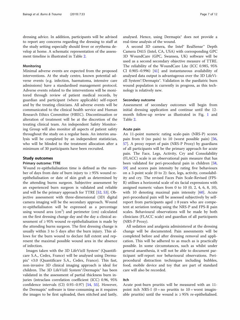
Study outcomesPrimary outcome: TTREWound re-epithelialisation time is defined as the num-ber of days from date of burn injury to ≥ 95% wound re-epithelialisation or date of skin graft as determined bythe attending burns surgeon. Subjective assessment byan experienced burn surgeon is validated and reliableand will be the primary approach for TTRE [52, 53]. Ob-jective assessment with three-dimensional (3D) digitalcamera imaging will be the secondary approach. Woundre-epithelialisaton will be expressed as a percentageusing wound area (cm2) and perimeter (cm) calculatedon the first dressing change day and the day a clinical as-sessment of ≥ 95% wound re-epithelialisation is made bythe attending burns surgeon. The first dressing change isusually within 3 to 5 days after the burn injury. This al-lows for the burn wound to declare full extent and rep-resent the maximal possible wound area in the absenceof infection.Images taken with the 3D LifeVizII System® (Quantifi-
care S.A., Cedex, France) will be analysed using Derma-pix® v3.0 (Quantificare S.A., Cedex, France). This fast,non-invasive 3D clinical imaging approach is ideal forchildren. The 3D LifeVizII System®/Dermapix® has beenvalidated in the assessment of partial thickness burn in-juries (intraclass correlation coefficient (ICC) 0.96, 95%confidence intervals (CI) 0.93–0.97) [54, 55]. However,the Dermapix® software is time-consuming as it requiresthe images to be first uploaded, then stitched and lastly,
analysed. Hence, using Dermapix® does not provide areal-time analysis of the wound.A second 3D camera, the Intel® RealSense™ Depth
Camera D415 (Intel, CA, USA) with corresponding GPC3D WoundCare (GPC, Swansea, UK) software will beused as a second secondary objective measure of TTRE.The reliability of the WoundCare Lite (ICC 0.985, 95%CI 0.905–0.996) [41] and instantaneous availability ofanalysed data output is advantageous over the 3D LifeVi-zII System®/Dermapix®. Validation in the paediatric burnwound population is currently in progress, as this tech-nology is relatively new.
Secondary outcomeAssessment of secondary outcomes will begin frominitial dressing application and continue until the 12-month follow-up review as illustrated in Fig. 1 andTable 2.
Acute painAn 11-point numeric rating scale-pain (NRS-P) scorespain from 0 (no pain) to 10 (worst possible pain) [56,57]. A proxy report of pain (NRS-P Proxy) by guardiansof all participants will be the primary approach for acutepain. The Face, Legs, Activity, Cry and Consolability(FLACC) scale is an observational pain measure that hasbeen validated for peri-procedural pain in children [58,59] and scores pain intensity by rating five behaviourson a 3-point scale (0 to 2): face, legs, activity, consolabil-ity and cry. The revised Faces Pain Scale-Revised (FPS-R) utilises a horizontal scale of six facial expressions withassigned numeric values from 0 to 10 (0, 2, 4, 6, 8, 10),with 10 denoting maximal pain intensity [60]. Acuteperi-procedural pain will be assessed subjectively by self-report from participants aged ≥ 8 years who are compe-tent at seriation testing using the NRS-P and FPS-R painscales. Behavioural observations will be made by bothclinicians (FLACC scale) and guardian of all participants(NRS-P Proxy).All sedation and analgesia administered at the dressing
change will be documented. Pain assessments will becompleted before and after dressing removal and appli-cation. This will be adhered to as much as is practicallypossible. In some circumstances, such as whilst undergeneral anaesthesia, it will not be able to document par-ticipant self-report nor behavioural observations. Peri-procedural distraction techniques including bubbles,food, mobile device and toy that are part of standardcare will also be recorded.
ItchAcute post-burn pruritis will be measured with an 11-point itch NRS-I (0 = no pruritis to 10 = worst imagin-able pruritis) until the wound is ≥ 95% re-epithelialised.
Bairagi et al. Burns & Trauma (2019) 7:33 Page 7 of 12

Participants ≥ 8 years will self-report (NRS-I). A proxyreport of participant itch (NRS-I proxy) will be takenfrom the accompanying guardian for all participants andwill the primary approach for itch. The NRS-I has showngood correlation when compared with visual analoguescale and Itch Man Scale and is easier to use in theevaluation of itch intensity [61, 62]. The itch item of thePatient and Observer Scar Assessment Scale (POSAS)will be used to assess chronic pruritis and will be evalu-ated by all participants 8 years or older as well as by allguardians (POSAS Patient) and by an investigator(POSAS Observer) [63].
Scar severityThe re-epithelialised wound will be evaluated for the se-verity of scars using objective (ultrasonography and col-orimetry) and subjective (POSAS and Brisbane BurnScar Impact Profile (BBSIP)) measures [63–66]. The ob-jective measures are the principal approach based onevidence of the ability to detect change. Scar thicknesswill be measured using the portable Venue40 MSK®Ultrasound machine (GE Healthcare, Fairfield, CT,USA). Ultrasound test-retest and interrater reproduci-bility of scar thickness is acceptable for paediatricscars [67].Scar colorimetry will be evaluated with the DSM II Col-
orMeter® (Cortex Technology, Hadsund, Denmark). Thisdevice uses tristimulus reflectance, colorimetry and nar-row-band photometry to assess scar lightness (L*), ery-thema (a*) and pigmentation (b*) [68]. An average of threemeasurements of L*, a*, b* from both the scar and healthyskin will be recorded. The primary measure of scar pig-mentation and scar erythema will be L* and a* respectively.Both the investigator and guardian of all participants
will complete the POSAS. This survey assesses scarthickness, vascularity, pliability, pigmentation and relief,as well as patient scale items of itch, pain, colour, stiff-ness, thickness and irregularity [63]. The BBSIP physicalscar subscales and BBSIP sensory subscale will alsomeasure scar severity [64–66]. An investigator havingcompleted scar assessment training to the satisfaction ofthe occupational therapist at the study site will completethe scar assessments of all participants.
Health-related quality of life (HRQOL)The BBSIP measures HRQOL and patient-reported scarcharacteristics for individuals with burn scars [64–66].The Child Health Utility 9D (CHU9D) is a preference-based measure that assesses both participant HQROLand resource utilisation of the interventions [69–72].Participant guardians will complete both HRQOL assess-ments for all participants.
Treatment satisfactionTreatment satisfaction is a patient-reported outcome[73] that guides quality of service delivery by a treatinghealth service provider. In a study of parents (n = 62) ofchildren receiving burn treatment, the perceived supportand respectful communication by clinicians was stronglyassociated with the quality of received care [74]. Bothclinicians and participant guardian will complete an 11-point NRS (0 = not at all satisfied to 10 = extremely satis-fied) to measure treatment satisfaction.
Ease of dressing applicationThe ease of dressing application will be assessed using aquestionnaire regarding the dressings (application ease,dressing conformability, dressing application duration)including additional space for comments by treatingclinicians.
Healthcare resource useHealthcare resource use related to the management ofburn injuries will be collected for each patient from theperspective of the healthcare provider with a 12-monthtime-horizon. Healthcare resource use to be collectedfor each participant will include trial interventions andother wound management-related resource use. Inaddition, product(s) used in wound and scar manage-ment as well as associated clinician labour time (e.g. forassessments and treatments during outpatient clinic ap-pointments). Resource use for burn-related hospitalisa-tions (e.g. intensive care admission) will also becollected. Resource use will be costed at market rates(e.g. industrial award rates for clinician labour time,standing offer arrangement rates for products supplied,values consistent with the (Australian) IndependentHospital Pricing Authority for hospitalisation costs).
Data managementSample size estimateThe sample size estimate conducted for this trial isbased on the primary outcome of TTRE. Using thePower Analysis and Sample Size (PASS) software (ver-sion 11.0.7; PASS, NCSS, LLC), a two-sided logrank testprocedure was used. A total sample size of 84 subjects(28 participants per group) that accounted for a 10% at-trition rate would achieve 80% power at a significancelevel of 0.05 [10, 75, 76]. A clinically important differ-ence of 4 days for re-epithelialisation was used for thesample size estimation.
Randomisation and allocationTo reduce the chance of prediction of treatment alloca-tion, a procedure which randomly allocates participantsin groups using a random step size will be used [77].The randomisation sequence will then be uploaded into
Bairagi et al. Burns & Trauma (2019) 7:33 Page 8 of 12

Research Electronic Data Capture (REDCap) randomisa-tion module [78] by a third party person not associatedwith the study. Upon obtaining informed consent for theparticipation, baseline data (participant demographicsincluding Fitzpatrick Skin Type, burn injury time, depth,TBSAB, injury mechanism and history of first aid priorto enrollment) will be collected. An investigator alignedwith the study will complete the randomisation usingREDCap and inform the treating clinicians of the allo-cated intervention group.
BlindingOnce data collection is complete, a panel of burn woundspecialists inclusive of preselected burn surgeons andnurses will conduct a blinded review of wound and scarimaging. Any possible identifying material that could in-dicate to the blinded assessors, which group the partici-pant, was allocated to, will be removed from images.The participants and their care providers will be blindedto their intervention assignment. Although it will be un-avoidable to detect remnants of silver dressing in somecases, it is anticipated that it will be almost impossible todifferentiate between the group B and group C. An expe-rienced radiographer from the study centre willcomplete blinded assessments of scar thickness.
Statistical analysisIn the exploratory stage, data will be graphed, andsummary statistics will be calculated for all outcomes.Data will be analysed using the ‘Intention to Treat’principle. The primary outcome data will use the sur-vival analysis model (both Kaplan-Meier analysis andCox proportional hazards regression), with the time tohealing as the main outcome and dressing group as theexplanatory variable. All other data will be analysedusing appropriate methods for longitudinal data such asmixed model’s regression analysis or generalised estimat-ing equations (GEE). A GEE is better suited for estimat-ing population average effects whereas mixed models arebetter suited for understanding the source of correlationand its structure. Thus, they are complementary ways ofanalysing the data rather than opposing. It should alsobe noted that for symmetrical outcome variable distribu-tions (such as normally distributed) both methodsshould give similar results. Statistical significance will beset at p < 0.05. The data set will be analysed with SPSS(IBM Corporation, Armonk, NY, USA) and Stata(StataCorp, College Station, TX, USA) software whereappropriate.
Data collection and storageData collection will be in the form of completion ofquestionnaires and clinical imaging (2D and 3Dphotographs, laser Doppler imaging and sonography),
colorimetry, assessments of pain, itch intensity, treat-ment satisfaction, ease of dressing application,HRQOL, wound intervention fidelity, healthcare re-source utilisation and scar severity as well as partici-pant demographics, socioeconomic status and clinicalcharacteristics. Data collection and management willbe completed by an investigator and entered into theREDCap software [78] system for managing the data.At the study site, the de-identified data will be keptin a locked filing cabinet and backed up onto theQueensland University of Technology (QUT), Re-search Data Storage Service. This data will be storedfor 15 years after the completion of the trial in ac-cordance with National Health and Medical ResearchCouncil guidelines. Results of this study are to bepublished in peer-reviewed journals and presented atnational and international burns conferences. Thisprotocol was completed in accordance with theSPIRIT 2013 guidelines [79, 80].
DiscussionThe overall reported rate of hypertrophic scar formationin children ranges from 17 to > 50% in children [17]. Toavoid the costly burden of managing burn scars and theimpact on participants and families, current woundmanagement approaches focus on shortening re-epithe-lialisation time. An ideal skin substitute provides effect-ive and scar free healing. However, current woundmanagement approaches incorporate some but not allproperties of a functioning integumentary system thusthe debate continues regarding how best to treat theseburn wounds. For children with larger TBSAB, fasterTTRE assists these burn survivors to begin the processof rehabilitation earlier, enables earlier return to routinedaily activities and reduces scarring.Effective pain management is of paramount import-
ance, from both a psychological and clinical perspectivein paediatric burn patients. Sub-optimal peri-proceduralanalgesia was significantly associated with a 2.2% delayin re-epithelialisation, for every point increase in painmeasured using the FPS-R [81]. In addition, reducingthe number of dressing changes may reduce the poten-tial pain and distress associated with dressing changes.Post-burn pruritis causes much distress in children andhas a moderate association with mental health [82]. Un-controlled scratching hinders the re-epithelialisationprocess and potentially disrupts the fragile newly regen-erated epithelium. In addition, the potential introductionof infective pathogens would also delay TTRE. Inevit-ably, a proportion of children post mixed partial thick-ness burn will develop a scar. A multi-disciplinaryapproach that incorporates both operative (contracturerelease, medical needling) and non-operative treatments
Bairagi et al. Burns & Trauma (2019) 7:33 Page 9 of 12

(topical silicone, pressure garments) is vital to successfulscar management. Efficient health resource utilisationimproves the quality of health care provided to currentand future children with mixed partial thickness burninjuries and most importantly enables clinicians to makeinformed decisions on best practice.Based on past trials and studies at the study site, a
limitation of this study is the anticipated large dropoutrate. Potential attrition bias will be addressed by examin-ing whether the extent and reasons for dropout are bal-anced across the groups [83]. Although differentdropout rates across the three groups are not antici-pated, a sensitivity analysis will be run to check for anydifferences. It is expected that random drop out will re-sult in larger confidence intervals but not bias. Inaddition, if there is no imbalance, then attrition bias isnot likely to be a problem [83]. Limitations associatedwith burn depth assessment and wound infection diag-nosis will be adjusted for during statistical analysis. TheBRACS randomised trial will add new knowledge re-garding the comparative effectiveness of autologous skincell suspension for the management of partial thicknessand paediatric burn injuries.
Trial statusRecruitment started in May 2018 and will proceed for aperiod of 18 months. Follow-up consultations will be inthe outpatient setting at the 3, 6 and 12-month post-dateof burn injury. Intended completion of the study is De-cember 2020.
Abbreviations2D: Two-dimensional; 3D: Three-dimensional; ACH: Autologous cellharvesting; ASCS: Autologous skin cell suspension; BBSIP: Brisbane Burn ScarImpact Profile; BRACS: Biobrane®, RECELL® Autologous skin Cell suspensionand Silver dressings; CHU9D: Child Health Utility 9D; COD: Change ofdressing; DOI: Date of injury; FLACC: Face, Legs, Activity, Cry, Consolability;FPS-R: Faces Pain Scale-Revised; GEE: Generalised Estimating Equations;HREC: Human Research Ethics Committee; HRQOL: Health-related quality oflife; ICC: Intraclass correlation coefficient; LDI: Laser Doppler Imaging; NRS-IProxy: Numeric Rating Scale–Itch Proxy; NRS-I: Numeric Rating Scale–Itch;NRS-P Proxy: Numeric Rating Scale–Pain Proxy; NRS-P: Numeric Rating Scale–Pain; OPD: Outpatient department; POSAS: Patient and Observer ScarAssessment Scale; QLD: Queensland; QUT: Queensland University ofTechnology; REDCap: Research electronic data capture; RES™: RegenerativeEpithelial Suspension; SPIRIT: Standard Protocol Items Recommendations forInterventional Trials; SSA: Site-specific approval; TBSAB: Total body surfacearea burned; TTRE: Time to re-epithelialisation
AcknowledgementsThe investigators would like to thank the participants and their families aswell as the staff of the study site for their continued support throughout thetrial.
Authors’ contributionsAll authors contributed towards the study design. AB drafted the manuscript.Critical review and editing of the final manuscript draft were done by allauthors. The final version of this manuscript was read and approved by allthe authors.
FundingAvita Medical has made available an industry research grant to beadministrated by QUT. However, Avita Medical will not have any input intoprotocol design nor the conduct of the trial. The principal investigator is aPhD candidate of QUT and has no financial interest in the products used inthis study. SMM is supported by an Australian National Health and MedicalResearch Council administered fellowship(#1161138).
Availability of data and materialsNot applicable
Ethics approval and consent to participateEthics was obtained from the study site (HREC/17/QRCH/278, SSA/18/QRCH/41 QUT HREC 80227). The trial is prospectively registered with the AustralianNew Zealand Clinical Trials Registry (ACTRN12618000245291). Consent forparticipation will be obtained from all eligible participants. Protocolamendments will be communicated with both the study centre HREC andthe Australian New Zealand Clinical Trials Registry.
Consent for publicationNot applicable
Competing interestsA co-investigator of the study is a paediatric burns surgeon (RK) treating par-ticipants at the study site; however, this surgeon will not have any role inthe participant recruitment or allocation to groups. All other authors declarethat they have no competing interests.
Author details1Centre for Children’s Burns and Trauma Research, Centre for Children’sHealth Research, Brisbane, Queensland, Australia. 2Pegg Leditschke Children’sBurns Centre, Queensland Children’s Hospital, Brisbane, Queensland,Australia. 3School of Nursing, Institute of Health and Biomedical Innovation,Queensland University of Technology, Brisbane, Queensland, Australia.4Australian Centre for Health Services Innovation, Institute of Health andBiomedical Innovation, School of Public Health and Social Work, QueenslandUniversity of Technology, Brisbane, Queensland, Australia. 5Research MethodsGroup, Institute of Health and Biomedical Innovation, Queensland Universityof Technology, Brisbane, Queensland, Australia. 6The University ofQueensland, Brisbane, Queensland, Australia. 7Centre for Functioning andHealth Research, Metro South Hospital and Health Service, Brisbane,Queensland, Australia.
Received: 2 December 2018 Accepted: 26 July 2019
References1. Peck MD. Epidemiology and prevention of burns throughout the world.
Jeschke MG, Kamolz L-P, Sjöberg F, Wolf SE, editors. Vienna: SpringerVienna; 2012.
2. Simons M, Price N, Kimble R, Tyack Z. Patient experiences of burn scars inadults and children and development of a health-related quality of lifeconceptual model: a qualitative study. Burns. 2016;42(3):620–32.
3. De Young AC, Kenardy JA, Cobham VE, Kimble R. Prevalence, comorbidityand course of trauma reactions in young burn-injured children. J ChildPsychol Psychiatry. 2012;53(1):56–63.
4. Maskell J, Newcombe P, Martin G, Kimble R. Psychosocial functioningdifferences in pediatric burn survivors compared with healthy norms. J BurnCare Res. 2013;34(4):465–76.
5. Hop MJ, Polinder S, Vlies CH, Middelkoop E, Baar ME. Costs of burn care: asystematic review. Wound Repair Regen. 2014;22:436.
6. Tracy L, McInnes J, Gong J, Gabbe B, Thomas T. BRANZ 8th Annual Report_Jul 16 - Jun 17. Burns registry of Australia and New Zealand; 2017.
7. Ter Horst B, Chouhan G, Moiemen NS, Grover LM. Advances in keratinocytedelivery in burn wound care. Adv Drug Deliv Rev. 2018;123:18–32.
8. Watt SM, Pleat JM. Stem cells, niches and scaffolds: applications to burnsand wound care. Adv Drug Deliv Rev. 2018;123:82–106.
9. Gee Kee EL, Kimble RM, Cuttle L, Khan A, Stockton KA. Randomizedcontrolled trial of three burns dressings for partial thickness burns inchildren. Burns. 2015;41(5):946–55.
Bairagi et al. Burns & Trauma (2019) 7:33 Page 10 of 12

10. Wood F, Martin L, Lewis D, Rawlins J, McWilliams T, Burrows S, et al. Aprospective randomised clinical pilot study to compare the effectiveness ofBiobrane(R) synthetic wound dressing, with or without autologous cellsuspension, to the local standard treatment regimen in paediatric scaldinjuries. Burns. 2012;38(6):830–9.
11. Mandal A. Paediatric partial-thickness scald burns–is Biobrane the besttreatment available? Int Wound J. 2007;4(1):15–9.
12. Lesher AP, Curry RH, Evans J, Smith VA, Fitzgerald MT, Cina RA, et al.Effectiveness of Biobrane for treatment of partial-thickness burns in children.J Pediatr Surg. 2011;46(9):1759–63.
13. Lonie S, Baker P, Teixeira RP. Healing time and incidence of hypertrophicscarring in paediatric scalds. Burns. 2017;43(3):509–13.
14. Gee Kee E, Stockton K, Kimble RM, Cuttle L, McPhail SM. Cost-effectivenessof silver dressings for paediatric partial thickness burns: an economicevaluation from a randomized controlled trial. Burns. 2017;43(4):724–32.
15. Deitch EA, Wheelahan TM, Rose MP, Clothier J, Cotter J. Hypertrophic burnscars: analysis of variables. J Trauma. 1983;23(10):895–8.
16. Cubison TC, Pape SA, Parkhouse N. Evidence for the link between healingtime and the development of hypertrophic scars (HTS) in paediatric burnsdue to scald injury. Burns. 2006;32(8):992–9.
17. Chipp E, Charles L, Thomas C, Whiting K, Moiemen N, Wilson Y. Aprospective study of time to healing and hypertrophic scarring in paediatricburns: every day counts. Burns Trauma. 2017;5(1):3.
18. Ketchum LD. Hypertrophic scars and keloids. Clin Plast Surg. 1977;4(2):301–10.19. Vloemans AF, Hermans MH, van der Wal MB, Liebregts J, Middelkoop E.
Optimal treatment of partial thickness burns in children: a systematicreview. Burns. 2014;40(2):177–90.
20. Liu HF, Zhang F, Lineaweaver WC. History and advancement of burntreatments. Ann Plast Surg. 2017;78(2 Suppl 1):S2–8.
21. Cuttle L, Naidu S, Mill J, Hoskins W, Das K, Kimble RM. A retrospectivecohort study of Acticoat versus Silvazine in a paediatric population. Burns.2007;33(6):701–7.
22. Khundkar R, Malic C, Burge T. Use of Acticoat dressings in burns: what is theevidence? Burns. 2010;36(6):751–8.
23. Hajska M, Dragunova J, Koller J. Cytotoxicity testing of burn wounddressings: first results. Cell Tissue Bank. 2017;18(2):143–51.
24. White R, Morris C. Mepitel: a non-adherent wound dressing with Safetactechnology. Br J Nurs. 2009;18(1):58–64.
25. Silverstein P, Heimbach D, Meites H, Latenser B, Mozingo D, Mullins F, et al.An open, parallel, randomized, comparative, multicenter study to evaluatethe cost-effectiveness, performance, tolerance, and safety of a silver-containing soft silicone foam dressing (intervention) vs silver sulfadiazinecream. J Burn Care Res. 2011;32(6):617–26.
26. Zhao H, Chen Y, Zhang C, Fu X. Autologous epidermal cell suspension: apromising treatment for chronic wounds. J Tissue Viability. 2016;25(1):50–6.
27. Wood F. Clinical potential of autologous epithelial suspension. Wounds.2003;15(1):16–22.
28. Fan C, Pek CH, Por YC, Lim GJS. Biobrane dressing for paediatric burns inSingapore: a retrospective review. Singap Med J. 2018;59(7):360–5.
29. Gravante G, Di Fede MC, Araco A, Grimaldi M, De Angelis B, Arpino A, et al.A randomized trial comparing ReCell system of epidermal cells deliveryversus classic skin grafts for the treatment of deep partial thickness burns.Burns. 2007;33(8):966–72.
30. Holmes Iv JH, Molnar JA, Carter JE, Hwang J, Cairns BA, King BT, et al. Acomparative study of the ReCell(R) device and autologous spit-thicknessmeshed skin graft in the treatment of acute burn injuries. J Burn Care Res.2018;39(5):694–702.
31. Dunne JA, Rawlins JM. Early paediatric scald surgery—a cost effective dermalpreserving surgical protocol for all childhood scalds. Burns. 2014;40(4):777–8.
32. Austin RE, Merchant N, Shahrokhi S, Jeschke MG. A comparison of Biobraneand cadaveric allograft for temporizing the acute burn wound: cost andprocedural time. Burns. 2015;41(4):749–53.
33. Greenwood JE, Clausen J, Kavanagh S. Experience with biobrane: uses andcaveats for success. Eplasty. 2009;9:e25.
34. Hyland EJ, D'Cruz R, Menon S, Harvey JG, La Hei E, Lawrence T, et al.Biobrane (TM) versus acticoat (TM) for the treatment of mid-dermalpediatric burns: a prospective randomized controlled pilot study. Int J BurnsTrauma. 2018;8(3):63–7.
35. Haddad AG, Giatsidis G, Orgill DP, Halvorson EG. Skin substitutes andbioscaffolds: temporary and permanent coverage. Clin Plast Surg. 2017;44(3):627–34.
36. Woodruff E. Biobrane, a Biosynthetic Skin Prosthesis in Burn WoundCoverings. DL W, editor. Boca Raton: CRC Press; 1984. p. c1984.
37. Demling RH. Use of Biobrane in the management of scalds. J Burn CareRehabil. 1995;16:329–30.
38. Phillips LG, Robson MC, Smith DJ, Phillips WA, Gracia WD, McHugh TP, et al.Uses and abuses of a biosynthetic dressing for partial skin thickness burns.Burns. 1989;15(4):254–6.
39. Lal S, Barrow RE, Wolf SE, Chinkes DL, Hart DW, Heggers JP, et al. Biobrane®improves wound healing in burned children without increased risk ofinfection. Shock. 2000;14(3):314–9.
40. Government Q. Queensland population counter: The State of Queensland(Queensland Treasury) 2018. Available from: http://www.qgso.qld.gov.au/products/reports/pop-growth-qld/qld-pop-counter.php.
41. Farrar E, Pujji O, Jeffery S. Three-dimensional wound mapping softwarecompared to expert opinion in determining wound area. Burns. 2017;43(8):1736–41.
42. Fontaine M, Ravat F, Latarjet J. The e-burn application - a simple mobiletool to assess TBSA of burn wounds. Burns. 2018;44(1):237–8.
43. Management NIoTaI. NSW Trauma App Analysis Report 21st August - 8thNovember 2015. Institute of Trauma and Injury Management; 2015 27/01/2016.
44. Management NIoTaI. NSW Trauma App Analysis Report August 2015 – August2016. NSW Institute of Trauma and Injury Management; 2016 22/02/2017.
45. Holland AJA, Martin HCO, Cass DT. Laser Doppler imaging prediction ofburn wound outcome in children. Burns. 2002;28(1):11–7.
46. La Hei ER, Holland AJ, Martin HC. Laser Doppler imaging of paediatric burns:burn wound outcome can be predicted independent of clinicalexamination. Burns. 2006;32(5):550–3.
47. Cho JK, Moon DJ, Kim SG, Lee HG, Chung SP, Yoon CJ. Relationshipbetween healing time and mean perfusion units of laser Doppler imaging(LDI) in pediatric burns. Burns. 2009;35(6):818–23.
48. Mileski WJ, Atiles L, Purdue G, Kagan R, Saffle JR, Herndon DN, et al. Serialmeasurements increase the accuracy of laser Doppler assessment of burnwounds. J Burn Care Rehabil. 2003;24(4):187.
49. Jeng JC, Bridgeman A, Shivnan L, Thornton PM, Alam H, Clarke TJ, et al. LaserDoppler imaging determines need for excision and grafting in advance ofclinical judgment: a prospective blinded trial. Burns. 2003;29(7):665–70.
50. Mill J, Cuttle L, Harkin DG, Kravchuk O, Kimble RM. Laser Doppler imaging ina paediatric burns population. Burns. 2009;35(6):824–31.
51. Wang XQ, Mill J, Kravchuk O, Kimble RM. Ultrasound assessed thickness ofburn scars in association with laser Doppler imaging determined depth ofburns in paediatric patients. Burns. 2010;36(8):1254–62.
52. Bloemen M, Boekema B, Vlig M, van Zuijlen P, Middelkoop E. Digital imageanalysis versus clinical assessment of wound epithelialization: a validationstudy. Burns. 2012;38(4):501–5.
53. Bloemen MC, van Zuijlen PP, Middelkoop E. Reliability of subjective woundassessment. Burns. 2011;37(4):566–71.
54. Stockton KA, McMillan CM, Storey KJ, David MC, Kimble RM. 3Dphotography is as accurate as digital planimetry tracing in determiningburn wound area. Burns. 2015;41(1):80–4.
55. Gee Kee EL, Kimble RM, Stockton KA. 3D photography is a reliable burnwound area assessment tool compared to digital planimetry in very youngchildren. Burns. 2015;41(6):1286–90.
56. Page MG, Katz J, Stinson J, Isaac L, Martin-Pichora AL, Campbell F. Validationof the numerical rating scale for pain intensity and unpleasantness inpediatric acute postoperative pain: sensitivity to change over time. J Pain.2012;13(4):359–69.
57. von Baeyer CL, Spagrud LJ, McCormick JC, Choo E, Neville K, Connelly MA.Three new datasets supporting use of the numerical rating scale (NRS-11)for children’s self-reports of pain intensity. Pain. 2009;143:223.
58. Merkel S, Voepel-Lewis T, Shayevitz J, Malviya S. FLACC Pain AssessmentTool: Reliability and Validation with Existing Tools. Anesthesiology. 1994;81(SUPPLEMENT):A1360.
59. von Baeyer CL, Spagrud LJ. Systematic review of observational (behavioral)measures of pain for children and adolescents aged 3 to 18 years. Pain.2007;127(1–2):140–50.
60. Huguet A, Stinson JN, McGrath PJ. Measurement of self-reported painintensity in children and adolescents. J Psychosom Res. 2010;68(4):329–36.
61. Reich A, Halupczok J, Ramus M, Stander S, Szepietowski J. New Data on theValidation of Vas and Nrs in Pruritus Assessment: Minimal ClinicallyImportant Difference and Itch Frequency Measurement. Acta DermVenereol. 2011;91(5):636.
Bairagi et al. Burns & Trauma (2019) 7:33 Page 11 of 12

62. Nieuwendijk SMP, de Korte IJ, Pursad MM, van Dijk M, Rode H. Post burnpruritus in pediatric burn patients. Burns. 2018;44(5):1151–8.
63. Draaijers LJ. The patient and observer scar assessment scale: a reliable andfeasible tool for scar evaluation. Plast Reconstr Surg. 2003;113:1960.
64. Tyack Z, Kimble R, McPhail S, Plaza A, Simons M. Psychometric properties ofthe Brisbane burn scar impact profile in adults with burn scars. PLoS One.2017;12(9):e0184452.
65. Tyack Z, Ziviani J, Kimble R, Plaza A, Jones A, Cuttle L, et al. Measuring theimpact of burn scarring on health-related quality of life: development andpreliminary content validation of the Brisbane burn scar impact profile(BBSIP) for children and adults. Burns. 2015;41(7):1405–19.
66. Simons M, Kimble R, McPhail S, Tyack Z. The longitudinal validity,reproducibility and responsiveness of the Brisbane burn scar impact profile(caregiver report for young children version) for measuring health-relatedquality of life in children with burn scars. Burns. 2019. Article in Press.
67. Simons M, Kee EG, Kimble R, Tyack Z. Ultrasound is a reproducible and validtool for measuring scar height in children with burn scars: a cross-sectionalstudy of the psychometric properties and utility of the ultrasound and 3Dcamera. Burns. 2017;43(5):993–1001.
68. Draaijers LJ, Tempelman FR, Botman YA, Kreis RW, Middelkoop E, vanZuijlen PP. Colour evaluation in scars: tristimulus colorimeter, narrow-bandsimple reflectance meter or subjective evaluation? Burns. 2004;30(2):103–7.
69. Stevens KJ. Working with children to develop dimensions for a preference-based, generic, pediatric health-related quality-of-life measure. Qual HealthRes. 2010;20:340–51.
70. Stevens KJ. Developing a descriptive system for a new preference-basedmeasure of health-related quality of life for children. Qual Life Res. 2009;18(8):1105–13.
71. Stevens KJ. Assessing the performance of a new generic measure of healthrelated quality of life for children and refining it for use in health statevaluation. Appl Health Econ Health Policy. 2011;9(3):157–69.
72. Stevens K. The Child Health Utility 9D (CHU9D) – A New Paediatric PreferenceBased Measure of Health Related Quality of Life. PRO Newsletter. 2010;43:11.
73. Revicki DA. Patient assessment of treatment satisfaction: methods andpractical issues. Gut. 2004;53(suppl 4):iv40.
74. Willebrand M, Sjöberg F, Huss F, Sveen J. Parents’ perceived quality ofpediatric burn care. J Crit Care. 2018;43:256–9.
75. Lakatos E. Sample Sizes Based on the Log-Rank Statistic in Complex ClinicalTrials. Biometrics. 1988;44:229–41.
76. Lakatos E. Designing Complex Group Sequential Survival Trials. Stat Med.2002;21:1969–89.
77. Snow G. blockrand: Randomization for block random clinical trials2013: Available from: https://CRAN.R-project.org/package=blockrand.[cited 2018. 1.3]
78. Harris PA, Taylor R, Thielke R, Payne J, Gonzalez N, Conde JG. Researchelectronic data capture (REDCap) – a metadata-driven methodology andworkflow process for providing translational research informatics support.J Biomed Inform. 2009;42(2):377–81.
79. Chan AW, Tetzlaff JM, Altman DG, Laupacis A, Gotzsche PC, Krleza-Jeric K,et al. SPIRIT 2013 statement: defining standard protocol items for clinicaltrials. Ann Intern Med. 2013;158(3):200–7.
80. Chan AW, Tetzlaff JM, Gotzsche PC, Altman DG, Mann H, Berlin JA, et al.SPIRIT 2013 explanation and elaboration: guidance for protocols of clinicaltrials. BMJ. 2013;346:e7586.
81. Brown NJ, Kimble RM, Gramotnev G, Rodger S, Cuttle L. Predictors ofre-epithelialization in pediatric burn. Burns. 2014;40(4):751–8.
82. McGarry S, Burrows S, Ashoorian T, Pallathil T, Ong K, Edgar DW, et al.Mental health and itch in burns patients: potential associations. Burns.2016;42(4):763–8.
83. Moher D, Hopewell S, Schulz KF, Montori V, Gøtzsche PC, Devereaux PJ, et al.CONSORT 2010 explanation and elaboration: updated guidelines for reportingparallel group randomised trials. J Clin Epidemiol. 2010;63(8):e1–e37.
Bairagi et al. Burns & Trauma (2019) 7:33 Page 12 of 12