Embed Size (px)
Citation preview
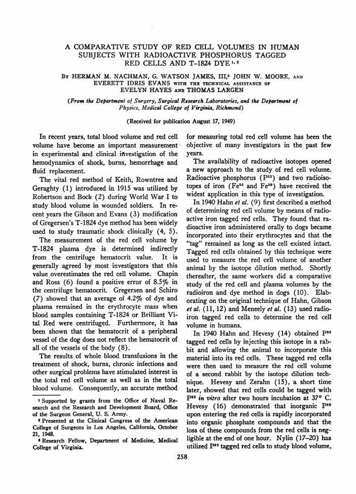
A COMPARATIVESTUDYOF RED CELL VOLUMESIN HUMANSUBJECTSWITH RADIOACTIVE PHOSPHORUSTAGGED
REDCELLS ANDT-1824 DYE1, 2
By HERMANM. NACHMAN,G. WATSONJAMES, III,8 JOHN W. MOORE,ANDEVERETT IDRIS EVANSWITH THE TECHNICAL ASSISTANCE OF
EVELYNHAYESANDTHOMASLARGEN
(From the Department of Surgery, Surgical Research Laboratories, and the Department ofPhysics, Medical College of Virginia, Richmond)
(Received for publication August 17, 1949)
In recent years, total blood volume and red cellvolume have become an important measurementin experimental and clinical investigation of thehemodynamics of shock, burns, hemorrhage andfluid replacement.
The vital red method of Keith, Rowntree andGeraghty (1) introduced in 1915 was utilized byRobertson and Bock (2) during World War I tostudy blood volume in wounded soldiers. In re-cent years the Gibson and Evans (3) modificationof Gregersen's T-1824 dye method has been widelyused to study traumatic shock clinically (4, 5).
The measurement of the red cell volume byT-1824 plasma dye is determined indirectlyfrom the centrifuge hematocrit value. It isgenerally agreed by most investigators that thisvalue overestimates the red cell volume. Chapinand Ross (6) found a positive error of 8.5 % inthe centrifuge hematocrit. Gregersen and Schiro(7) showed that an average of 4.2%o of dye andplasma remained in the erythrocyte mass whenblood samples containing T-1824 or Brilliant Vi-tal Red were centrifuged. Furthermore, it hasbeen shown that the hematocrit of a peripheralvessel of the dog does not reflect the hematocrit ofall of the vessels of the body (8).
The results of whole blood transfusions in thetreatment of shock, burns, chronic infections andother surgical problems have stimulated interest inthe total red cell volume as well as in the totalblood volume. Consequently, an accurate method
1 Supported by grants from the Office of Naval Re-search and the Research and Development Board, Officeof the Surgeon General, U. S. Army.
2 Presented at the Clinical Congress of the AmericanCollege of Surgeons in Los Angeles, California, October21, 1948.
8 Research Fellow, Department of Medicine, MedicalCollege of Virginia.
for measuring total red cell volume has been theobjective of many investigators in the past fewyears.
The availability of radioactive isotopes openeda new approach to the study of red cell volume.Radioactive phosphorus (P82) and two radioiso-topes of iron (Fe55 and Fe59) have received thewidest application in this type of investigation.
In 1940 Hahn et al. (9) first described a methodof determining red cell volume by means of radio-active iron tagged red cells. They found that ra-dioactive iron administered orally to dogs becameincorporated into their erythrocytes and that the"tag" remained as long as the cell existed intact.Tagged red cells obtained by this technique wereused to measure the red cell volume of anotheranimal by the isotope dilution method. Shortlythereafter, the same workers did a comparativestudy of the red cell and plasma volumes by theradioiron and dye method in dogs (10). Elab-orating on the original technique of Hahn, Gibsonet al. (11, 12) and Meneely et al. (13) used radio-iron tagged red cells to determine the red cellvolume in humans.
In 1940 Hahn and Hevesy (14) obtained p82tagged red cells by injecting this isotope in a rab-bit and allowing the animal to incorporate thismaterial into its red cells. These tagged red cellswere then used to measure the red cell volumeof a second rabbit by the isotope dilution tech-nique. Hevesy and Zerahn (15), a short timelater, showed that red cells could be tagged withp82 in vitro after two hours incubation at 370 C.Hevesy (16) demonstrated that inorganic P82upon entering the red cells is rapidly incorporatedinto organic phosphate compounds and that theloss of these compounds from the red cells is neg-ligible at the end of one hour. Nylin (17-20) hasutilized p82 tagged red cells to study blood volume,
258
259COMPARATIVESTUDY WITH pt2 TAGGEDCELLS AND T-1824
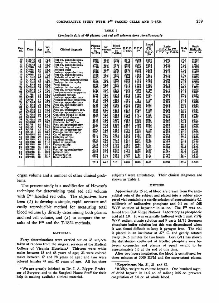
TABLE I
Composite data of 40 plasma and red cell volumes done simultaneously
_ IPlasma Blood l |Bloodvolume volume R.C.V. R.C.V. vol. Ratiood RatioE-xp. Date Age in Clinical diagnosis venous by T-1824 P .V. R.C.V. by P2 Bhotdy Bodyht._o. | _|_ kgm. T-by24 ht. T-1824 R.C.V. R.C.V. by T-1824 t Av. V. ht.
10 5/26148 38 72.6 Post-op. appendectomy 3085 48.2 5960 2875 2004 5089 0.697 39.3 0.81511 5/28/48 58 57.3 Post-op. herniorraphy 3330 40.9 5577 2247 1720 5050 0.765 34.0 0.83112 5/31/48 18 55.9 Post-op. appendectomy 2975 40.3 5440 2465 2089 5064 0.847 41.2 1.02;13 6/2/48 37 68.2 Bilateral ing. hernia 3290 47.9 6319 3029 2965 6255 0.978 47.4 0.98914 6/4/48 67 54.6 Infected wrist 2862 43.0 5002 2140 2008 4870 0.938 41.2 0.95815 6/7/48 30 81.5 Post-op. appendectomy 4940 40.1 8250 3310 2378 7318 0.718 32.4 0.80716 6/9/48 56 70.5 Post-op. appendectomy 2658 45.3 4859 2201 1563 4221 0.710 37.0 0.81617 6/10/48 67 69.1 Diabetic ulcer of toe 2615 40.2 4379 1764 1450 4065 0.821 35.6 0.88518 6/14/48 34 72.7 Stab wound-pneumothorax 2597 49.1 5100 2503 1755 4352 0.701 40.3 0.82019 6/15/48 36 54.6 Post-op. herniorraphy 3018 42.8 5270 2252 2115 5133 0.939 41.2 0.96321 6/18148 68 63.6 Fecal fistula 1675 61.5 4350 2675 1540 321S 0.576 47.9 0.77922 6/23/48 34 53.6 Post-op. herniorraphy 2800 40.1 4670 1910 1885 4685 0.987 40.2 1.00223 6/24/48 17 57.7 Post-op. herniorraphy 2700 49.4 5330 2630 2050 4750 0.779 43.2 0.87524 6/28/48 47 45.4 Postpneumonic empyema 2390 26.6 3300 910 664 3054 0.730 21.7 0.81725 7/1/48 18 63.0 Lacerated rectum 2795 45.7 5143 2348 1789 4584 0.762 39.0 0.85426 7/2/48 28 69.7 Post-op. appendectomy 2772 49.6 5502 2730 1679 4451 0.615 37.7 0.76027 7/6/48 47 42.0 Postpneumonic empyema 2418 28.6 3385 967 781 3199 0.807 24.4 0.85329 7/13/48 20 62.7 Post-op. appendectomy 2361 47.5 4486 2125 1690 4051 0.795 41.7 0.87830 7/14/48 20 63.2 Post-op. appendectomy 3245 45.6 5960 2715 1988 5233 0.732 38.0 0.83431 7/16/48 26 81.8 Osteo of hip 2923 49.2 5750 2827 2339 5262 0.827 44.5 0.90533 7/21/48 64 46.8 Post-op. exploratory lap. 2565 51.0 5238 2673 1925 4490 0.720 42.9 0.84135 7/26/48 16 61.1 Post-op. appendectomy 2401 50.1 4802 2401 2291 4692 0.954 48.8 0.97436 7/27/48 30 67.1 Gun shot wound of chest 2634 42.3 4562 1928 1771 4405 0.919 40.2 0.95137 7/28/48 37 67.3 Ischio-rectal abscess 2776 49.4 5480 2704 2117 4893 0.783 43.3 0.87738 7/29/48 60 65.0 Buerger's disease 2345 46.8 4409 2064 1698 4043 0.823 42.0 0.89739 7/30/48 37 58.0 Diabetes; inf. of foot 3228 37.1 5129 1901 1695 4923 0.891 34.4 0.92741 8/3/48 18 58.0 Post-op. appendectomy 2722 49.6 5405 2683 2330 5052 0.868 46.1 0.92942 8/4/48 64 55.8 Squamous ca. of mouth 3170 44.2 5681 2511 2113 5283 0.841 40.0 0.90543 8/5/48 42 91.5 Post-op. hysterectomy 2492 44.4 4483 1991 1698 4190 0.853 40.5 0.91144 8/6/48 25 68.8 Post-op. herniorraphy 2690 48.8 5256 2566 2392 5082 0.932 47.1 0.96545 8/10/48 78 49.3 Ca. of stomach 2619 40.9 4431 1812 1433 4052 0.791 35.4 0.86646 8/11/48 29 75.9 Cellulitis of foot 2717 47.2 5150 2433 1775 4492 0.730 39.5 0.83647 8/12/48 42 55.8 Inguinal hernia 2146 46.6 4022 1871 1695 3841 0.906 44.1 0.94648 8/12/48 48 51.0 Osteo of mandible 2685 34.9 4126 1441 1004 3689 0.697 27.2 0.77949 8/13 /48 40 72.7 Fractured hip 3228 43.5 5710 2482 1984 5212 0.799 38.1 0.87650 8/17/48 65 70.5 Ca. of colon 3569 41.6 6103 2534 2034 5603 0.803 36.3 0.87451 8/18/48 25 63.6 Post-op. appendectomy 2934 48.5 5698 2764 2286 5220 0.827 43.8 0.90352 8/18/48 24 70.0 Infected hip 3123 47.1 5906 2783 1829 4952 0.657 36.9 0.78353 8/19/48 38 63.5 Fractured ribs 2394 51.0 4884 2490 1783 4177 0.716 42.7 0.83754 8/19/48 20 55.7 Renal calculus 2571 45.6 4723 2152 1624 4195 0.755 38.7 0.849
2811 44.8 5131 2320 1848 4659 0.800 39.4 0.880
organ volume and a number of other clinical prob-lems.
The present study is a modification of Hevesy'stechnique for determining total red cell volumewith p.I labelled red cells. The objectives havebeen (1) to develop a simple, rapid, accurate andeasily reproducible method for measuring totalblood volume by directly determining both plasmaand red cell volume, and (2) to compare the re-sults of the pS2 and the T-1824 methods.
MATERIAL
Forty determinations were carried out on 38 subjectstaken at random from the surgical services of the MedicalCollege of Virginia Hospitals.4 Thirteen were whitemales between 18 and 68 years of age; 23 were coloredmales between 17 and 78 years of age; and two werecolored females 47 and 42 years of age. All but three
4 Weare greatly indebted to Dr. I. A. Bigger, Profes-sor of Surgery, and to the Surgical House Staff for theirhelp in making available clinical material.
subjects5 were ambulatory. Their clinical diagnoses areshown in Table I.
METHOD
Approximately 15 cc. of blood are drawn from the ante-cubital vein of the subject and placed into a rubber stop-pered vial containing a sterile solution of approximately 0.1millicurie of radioactive phosphate and 0.1 cc. of .068W/V solution of heparin6 in saline. The P3 was ob-tained from Oak Ridge National Laboratory as phosphoricacid pH 3.0. It was originally buffered with 1 part 2.5%W/V sodium citrate solution and 9 parts M/15 Sorensenphosphate buffer solution but this was discontinued whenit was found difficult to keep it pyrogen free. The vialis placed in an incubator at 370 C. and gently rotatedevery 10-15 minutes for two hours. Levi (21) has shownthe distribution coefficient of labelled phosphate ions be-tween corpuscles and plasma of equal weight to beapproximately 1.0 at the end of this time.
After two hours incubation, the blood is centrifuged forthree minutes at 3000 RPMand the supernatant plasma
6 Experiments No. 21, 31, and 52.6 0.068%o weight to volume heparin. One hundred mgm.
of dried heparin in 14.5 cc. of saline; 0.05 cc. preventscoagulation of 5.0 cc. of whole blood.
H. M. NACHMAN,G. W. JAMES, III, J. W. MOORE, AND E. I. EVANS
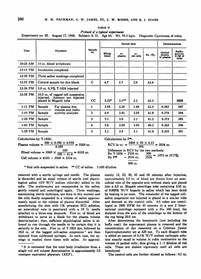
TABLE II
Protocol of a typical experimentExperiment no. 50. August 17, 1948. Subject: S. D. Age 65. Wt. 70.5 kgm. Diagnosis: Carcinoma of colon
Sample data Determinations
Time Procedure Sample Rino. Whole CC. Ht..optical Radio-blood plasma red cells (%) density (C/M Ofof plasma cells)
10:25 AM 15 cc. blood withdrawn
12:15 PM Incubation completed
12:50 PM Three saline washings completed
12:55 PM Control sample for dye blank C 4.7 2.7 2.0 42.6
12:56 PM 5.0 cc. 0.5% T-1824 injected
12:58 PM 10.0 cc. of tagged cell suspensioninjected. Amount not injectedplaced in Magath tube CC 5.25* 3.1** 2.2 42.3 3808
1:11 PM Sample For plasma dye 1 3.98 2.29 1.69 42.5 0.382 387volume and radio-
1:19 PM Sample activity analyses 2 4.9 2.81 2.05 41.8 0.378 394
1:28 PM Sample 3 5.1 3.0 2.1 41.2 0.373 385
1:43 PM Sample 4 5.0 2.98 2.02 40.2 0.363 396
1:58 PM Sample 5 5.1 3.0 2.1 41.0 0.355 391
Calculations by T-1824:500 X 0.590 X 4.958Plasma volume - -0.59 3569 cc.0.389
100Blood volume - 3569 X 100 - 41.6 6103 cc.
Cell volume = 6103 - 3569 = 2534 cc.
* Red cells suspended in saline. ** CCof saline. 1:1(
removed with a sterile syringe and needle. The plasmais discarded and an equal volume of sterile iced physio-logical saline (0.9 W/V sodium chloride) added to thecells. The erythrocytes are resuspended in the saline,gently rotated and centrifuged again. Three washings,maintaining sterile technique, are done in this manner andthe cells finally suspended in a volume of saline approxi-mately equal to the volume of plasma discarded. Afteranesthetizing the skin with 1%6 procaine HCO solution,an antecubftal vein is punctured with a 19 G. needleattached to a three-way stopcock. Five cc. of blood arewithdrawn to serve as a blank for the plasma volumedetermination; then, sufficient physiological saline is al-lowed to run through the needle to be certain that it issecurely in the vein. Five cc. of T-1824 dye, followed by10.0 cc. of the tagged cell-saline suspension s are theninjected from calibrated syringes. In each case, the sy-ringe is washed three times with saline. At approxi-
7 It is estimated that the total body irradiation from asingle red cell volume determination is approximately 3.0roentgen equivalent physicals (REP).
Calculations by P':
RCVin cc. 5 = 2034 cc.395
Difference in RCVby the two methods.By T-1824 - 2534 cc. 500 17 r1.%By Pa - 2034 cc. 2534 1973 or 19.7%.
500 cc.00 dilution.
mately 12, 23, 30, 45 and 60 minutes after injection,approximately 5.0 cc. of blood are drawn from an ante-cubital vein of the opposite arm without stasis and placedinto a 6.0 cc. Magath centrifuge tube containing 0.05 cc.of 0.068% W/V heparin in saline which has been driedby heating in an oven. The remainder of the tagged cell-saline suspension not injected is placed in a similar tubeand denoted as the control cells. All tubes are centri-fuged at 3000 RPM for 45 minutes in a size 2 Inter-national centrifuge equipped with a No. 240 head, thedistance from the axis of the centrifuge to the bottom ofthe cup being 20.0 cm.
After determining the hematocrit (not including thebuffy coat) the supernatant plasma is removed and theconcentration of dye measured on a Coleman JuniorSpectrophotometer set at 620 mis. To each Magath tubeis added an amount of 0.1% W/V sodium carbonate solu-tion exactly equal in volume in cubic centimeters to thevolume of packed cells; thus giving a 1: 1 dilution of redcells. These are shaken vigorously until all cells arehemolyzed.
The control cells are further diluted as follows: 0.1 cc.
260
COMPARATIVESTUDY WITH p32 TAGGEDCELLS AND T-1824
of the 1:1 dilution is made up to 5.0 cc. with distilledwater, giving a 1: 100 dilution. Then the radioactivity ofthe control cells and the other cells is determined in thefollowing manner: Exactly 0.1 cc. of hemolyzed cell solu-tion from each tube is carefully pipetted onto a copperplanchet 1" in diameter and evenly spread by adding onedrop of 10% aerosol OT8 solution. These are dried atroom temperature for approximately 30 minutes. Thesame pipette is used throughout but is carefully washedand dried between samples.
A Geiger tube and scaling circuit are used to measurethe radioactivity of each sample. The Geiger tubes areof the self-quenching variety, have overall dimension of3Vi" X 1%" diameter and have mica windows rangingfrom 2.5-3.5 mgm./cm!. These are shielded by 2" oflead and their background counts range from 25-35 countsper minute. They are standardized at regular intervalsto determine changes in plateau and counting efficiency.The sample to window distance is 2 mm. and the overallgeometric reproducibility is claimed by the manufacturerto be on the order of 0.1%. 4096 counts are totaled foreach sample and the result expressed in terms of countsper minute.
CALCULATION OF RED CELL VOLUME
From the dilutions of the red cells and the radioactivityanalyses the red cell volume is calculated as follows:
(1) RCVin cc.C/M/cc.' injected X cc. of red cells injected
C/M/cc. recovered from subject(2) C/M/cc. injected = C/M/0.1 cc. X 10 X 100(3) C/M/cc. recovered = C/M/0.1 cc. X 10 X 2(4) cc. of cell injected = cc. of saline suspension
X hematocritSubstituting in (1) therefore
RCVin cc.C/M/0.1 cc. X 10 X 100 X cc. of red cells injected
C/M/0.1 cc. X 10 X 2RCVin cc.
C/M of control sample X 50.0 X cc. of cells injectedC/M test cells from subject
Table II is a protocol of a typical experiment.To determine the technical error in pipetting and count-
ing, 10 samples from a tube of hemolyzed cells were pre-pared and each sample counted 10 times. The coefficientof variation's ranged from a minimum of 0.36% to amaximum of 3.2% with an average for the 100 determina-tions of 2.2%.
RESULTS
The tabulated results of the 40 determinationsare shown in Table I. The T-1824 dye method
8 Dioctyl sodium sulfosuccinate.9 C/M/cc. = counts per minute per cubic centimeter of
red cells.
0 Coefficient of variation = S X 100 Coefficient of
variation denotes the standard deviation as a percentageof the mean. N has already been taken into considerationin determining S (standard deviation).
gave a mean red cell volume of 2320 -+ 79 cc.; withthe p82 method the mean red cell volume was 1850--+ 66 cc. These two mean values are significantlydifferent (d = 470 + 102 cc., t = 4.6, PL.01).In every case, the red cell volume as determined bythe p82 method was less than the comparable valueobtained with T-1824 dye. The table shows theratio of the RCVby p82 to the RCVby T-1824for each case. The mean value obtained from these40 ratios was 0.800 -+- 0.153. None of the indi-vidual ratios fell outside the range of the meanratio plus or minus two and a half times itsstandard error.
Weassume the expression P + RCV by P32to be the relationship of red cells to plasma in theentire body (Body ht.) The average bodyhematocrit of the entire series was 39.4 while theaverage venous hematocrit was 44.8. The averagevenous hematocrit is found by averaging the he-matocrit of the six blood samples taken withouta tourniquet in the course of the experiment. No.correction factor is applied for the occluded plasma.
DISCUSSION
Determination of the red cell volume by meansof tagged erythrocytes is based upon the followingassumptions: (1) the tag remains essentially in-tact with the red cell for the time of the experi-ment, (2) the physical principle of dilution oftagged particulate matter is accurate for measuringchanges in volume of similar particulate matter,and (3) all of the tagged cells become completelymixed with the untagged cells of the subject.
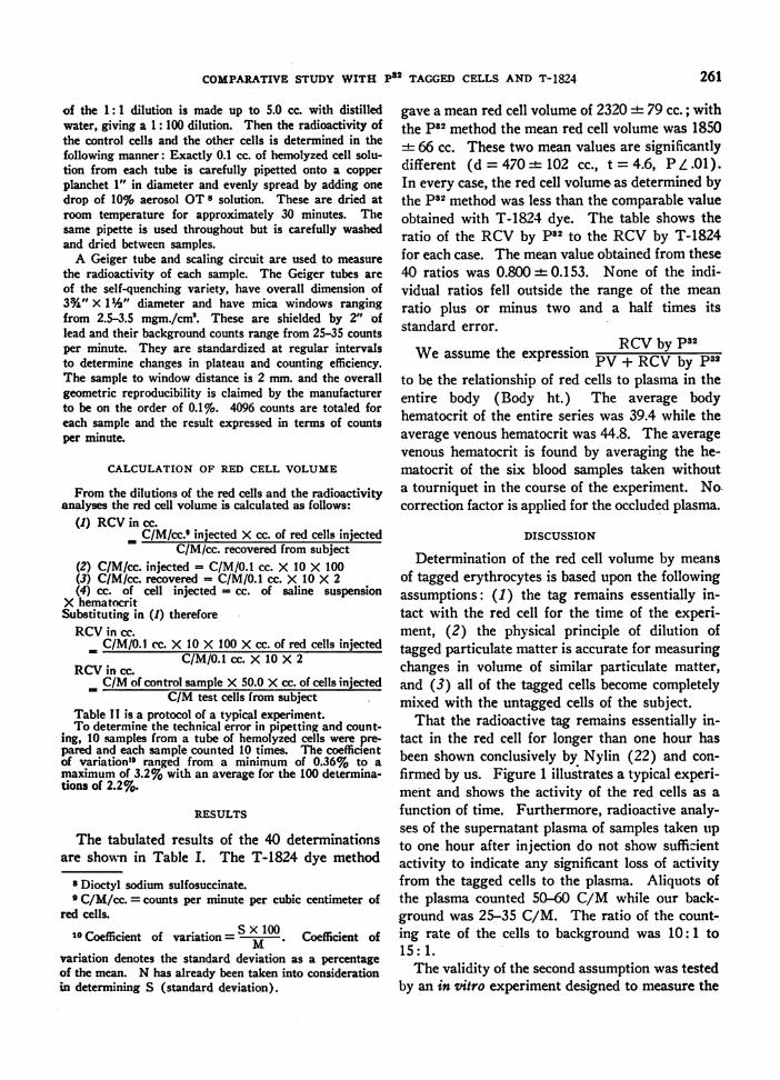
That the radioactive tag remains essentially in-tact in the red cell for longer than one hour hasbeen shown conclusively by Nylin (22) and con-firmed by us. Figure 1 illustrates a typical experi-ment and shows the activity of the red cells as afunction of time. Furthermore, radioactive analy-ses of the supernatant plasma of samples taken upto one hour after injection do not show sufficientactivity to indicate any significant loss of activityfrom the tagged cells to the plasma. Aliquots ofthe plasma counted 50-60 C/M while our back-ground was 25-35 C/M. The ratio of the count-ing rate of the cells to background was 10: 1 to15: 1.
The validity of the second assumption was testedby an in vitro experiment designed to measure the
261
H. M. NACHMAN,G. W. JAMES, III, J. W. MOORE, AND E. I. EVANS
1000
900
800
700
600
Soo5
6-
z
w
10
4001.
3001
6 12 18 24 30 36 42 48 54 60TIME IN MINUTES
FIG. 1. COUNTSPER MINUTE CONTAINEDIN RED CELLSAS A FUNCTION OF TIME
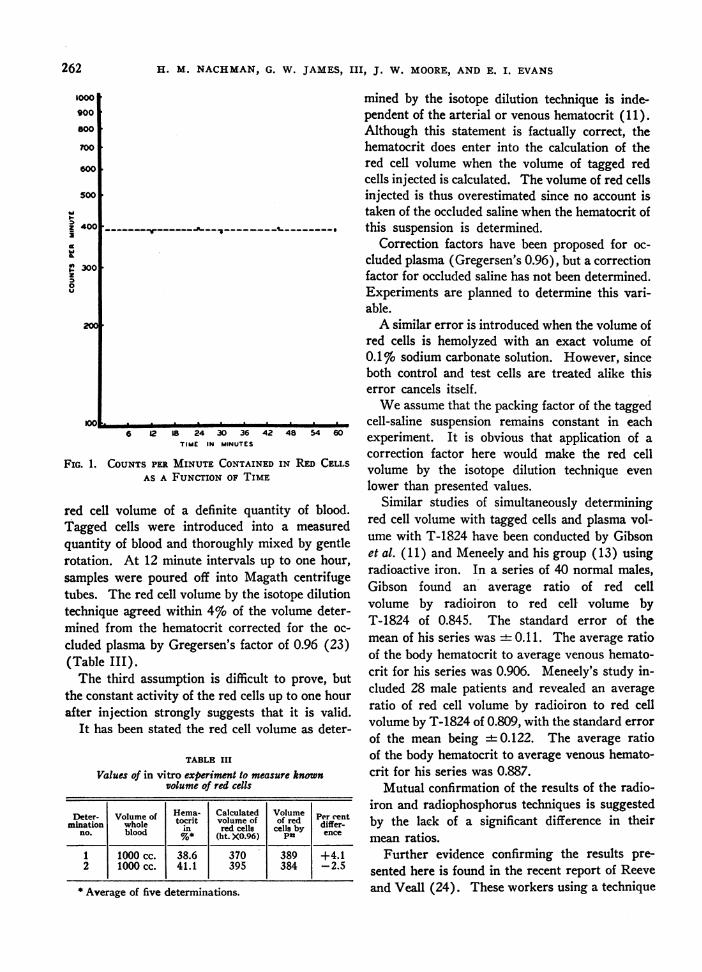
red cell volume of a definite quantity of blood.Tagged cells were introduced into a measuredquantity of blood and thoroughly mixed by gentlerotation. At 12 minute intervals up to one hour,samples were poured off into Magath centrifugetubes. The red cell volume by the isotope dilutiontechnique agreed within 4%o of the volume deter-mined from the hematocrit corrected for the oc-cluded plasma by Gregersen's factor of 0.96 (23)(Table III).
The third assumption is difficult to prove, butthe constant activity of the red cells up to one hourafter injection strongly suggests that it is valid.
It has been stated the red cell volume as deter-
TABLE IIIValues of in vitro experiment to measure known
volume of red cells
Deter- Volume of Hema. Calculated Volume Prcnminatio whole tocrit volume of of red Pifer-enmLina~tionl bw~hole~d | in red cells cells by differno. blood 4%* (ht. XO.96) Pu ence
1 1000 cc. 38.6 370 389 +4.12 1000 cc. 41.1 395 384 -2.5
* Average of five determinations.
mined by the isotope dilution technique is inde-pendent of the arterial or venous hematocrit (11).Although this statement is factually correct, thehematocrit does enter into the calculation of thered cell volume when the volume of tagged redcells injected is calculated. The volume of red cellsinjected is thus overestimated since no account istaken of the occluded saline when the hematocrit ofthis suspension is determined.
Correction factors have been proposed for oc-cluded plasma (Gregersen's 0.96), but a correctionfactor for occluded saline has not been determined.Experiments are planned to determine this vari-able.
A similar error is introduced when the volume ofred cells is hemolyzed with an exact volume of0.1%o sodium carbonate solution. However, sinceboth control and test cells are treated alike thiserror cancels itself.
Weassume that the packing factor of the taggedcell-saline suspension remains constant in eachexperiment. It is obvious that application of acorrection factor here would make the red cellvolume by the isotope dilution technique evenlower than presented values.
Similar studies of simultaneously determiningred cell volume with tagged cells and plasma vol-ume with T-1824 have been conducted by Gibsonet al. (11) and Meneely and his group (13) usingradioactive iron. In a series of 40 normal males,Gibson found an average ratio of red cellvolume by radioiron to red cell volume byT-1824 of 0.845. The standard error of themean of his series was + 0.11. The average ratioof the body hematocrit to average venous hemato-crit for his series was 0.906. Meneely's study in-cluded 28 male patients and revealed an averageratio of red cell volume by radioiron to red cellvolume by T-1824 of 0.809, with the standard errorof the mean being +- 0.122. The average ratioof the body hematocrit to average venous hemato-crit for his series was 0.887.
Mutual confirmation of the results of the radio-iron and radiophosphorus techniques is suggestedby the lack of a significant difference in theirmean ratios.
Further evidence confirming the results pre-sented here is found in the recent report of Reeveand Veall (24). These workers using a technique
262
COMPARATIVESTUDY WITH p'2 TAGGEDCELLS AND T-1824
similar to ours found the mean ratio ofred cell volume by p82
red cell volume by T-1824to be 0.87. Hematocrit values in their study werecorrected by a factor of 0.95.
Correction of the hematocrit for trapped plasmawas purposely omitted in the basic calculations(Table I), to facilitate a more fundamental com-parison of methods. If Gregersen's correction fac-tor (0.96) is applied to the hematocrits in TableI, the mean ratio of
red cell volume by p82red cell volume by T-1824
is 0.87.Mayerson, Lyons et al. (25) report a mean ra-
tio of red cell volume by p82 ored cell volume by T-1824
unusually high ratio is probably due to (1) dissimi-lar basic techniques and (2) the use of a hematocritcorrection factor of 0.915 (26).
The p82 method as described here is easily doneand has many advantages over radioiron. It isnecessary to produce a donor of radioactive cellswhen iron is used, but when p32 is used, the sub-ject's own red cells may be conveniently tagged.Furthermore, the concern of blood type and Rhcompatibility is eliminated. Separated isotopes ofradioiron are difficult to produce in either thecyclotron or the chain reacting pile. Some long-life iron is usually present in the most carefullyseparated isotopes of iron. Concern has been ex-pressed over the use of long-life isotopes such asFe 5 (half-life, four years) in human subjects.The use of p82 (half-life, 14.8 days) eliminatesthis concern. The preparation of blood samplescontaining iron for radioactive analysis is a com-plicated procedure. The red cells are wet ashedand the iron precipitated; the iron must then beredissolved and electrolytically deposited on cop-per discs in a special apparatus before the activitycan be measured. Radioactivity analysis of p82tagged red cells can be simply carried out on hemo-lyzed red cells by the method described.
CONCLUSIONS
1. A comparative study of the red cell andplasma volumes of 38 surgical patients as meas-ured by p82 tagged red cells and T-1824 plasmadye is presented.
2. On the average, the red cell volume as meas-ured with p32 tagged red cells is 20.09 less thanthe value obtained with T-1824 plasma dye.
3. This discrepancy is probably due to the dif-ference in the hematocrit of blood in peripheralveins and blood in other vessels of the body andto the intrinsic error of the centrifuge hematocrit.
4. The body hematocrit on the average, is 12.07o%less than the average venous hematocrit.
5. The hematocrit value enters into the calcula-tion of the red cell volume by the isotope dilutiontechniques and probably results in slight overesti-mation of the red cell volume.
6. Statistically, results of the present study donot differ significantly from similar studies usingradioiron tagged red cells.
7. The advantages of the p82 method over theradioiron method are outlined.
BIBLIOGRAPHY
1. Keith, N. M., Rowntree, L. G., and Geraghty, J. T.,A method for determination of plasma and bloodvolume. Arch. Int. Med., 1915, 16, 547.
2. Robertson, 0. H., and Bock, A. V., Blood volume inwounded soldiers. I. Blood volume and relatedblood changes after hemorrhage. J. Exper. Med.,1919, 29, 139.
3. Gibson, J. G., 2nd, and Evans, W. A., Jr., Clinicalstudies of the blood volume. I. Clinical applicationof a method employing the azo dye "Evans Blue"and the spectrophotometer. J. Clin. Invest., 1937,16, 301.
4. Evans, E. I., Hoover, M. J., James, G. W., III, andAlm, T., Studies on traumatic shock I. Bloodvolume changes in traumatic shock. Ann. Surg.,1944, 119, 64.
5. Noble, R. P., and Gregersen, M. I., Blood volume inclinical shock. II. The extent and cause of bloodvolume reduction in traumatic, hemorrhagic andburn shock. J. Clin. Invest., 1946, 25, 172.
6. Chapin, M. A., and Ross, J. F., The determination ofthe true cell volume by dye dilution, by proteindilution, and with radioactive iron. The error ofthe centrifuge hematocrit. Am. J. Physiol., 1942,137, 447.
7. Gregersen, M. I., and Schiro, H., The behavior of thedye T-1824 with respect to its absorption by redblood cells and its fate in blood undergoing coagu-lation. Am. J. Physiol., 1938, 121, 284.
8. Gibson, J. G., 2nd, Seligman, A. M., Peacock, W. C.,Aub, J. C., Fine, J., and Evans, R. D., The distri-bution of red cells and plasma in large and minutevessels of the normal dog, determined by radio-active isotopes of iron and iodine. J. Clin. Invest,1946, 25, 848.
263
H. M. NACHMAN,G. W. JAMES, III, J. W. MOORE, AND E. I. EVANS
9. Hahn, P. F., Balfour, W. M., Ross, J. F., Bale, W. F.,and Whipple, G. H., Red cell volume circulatingand total as determined by radioiron. Science,1941, 93, 87.
10. Hahn, P. F., Ross, J. F., Bale, W. F., Balfour, W. M.,and Whipple, G. H., Red cell and plasma volumes(circulating and total) as determined by radioironand by dye. J. Exper. Med., 1942, 75, 221.
11. Gibson, J. G., 2nd, Peacock, W. C., Seligman, A. M.,and Sack, T., Circulating red cell volume measuredsimultaneously by the radioactive iron and dyemethods. J. Clin. Invest., 1946, 25, 838.
12. Gibson, J. G., 2nd, Weiss, S., Evans, R. D., Peacock,W. C., Irvine, J. W., Jr., Good, W. M., and Kip,A. F., The measurement of the circulating red cellvolume by means of two radioactive isotopes ofiron. J. Clin. Invest., 1946, 25, 616.
13. Meneely, G. R., Wells, E. B., and Hahn, P. F., Appli-cation of the radioactive red cell method for deter-mination of blood volume in humans. Am. J.Physiol., 1947, 148, 531.
14. Hahn, L., and Hevesy, G., A method of blood volumedetermination. Acta physiol. Scandinav., 1940,1, 3.
15. Hevesy, G., and Zerahn, K., Determination of the redcorpuscle content. Ibid, 1942, 4, 376.
16. Hevesy, G., Radioactive Indicators; Their Applicationin Biochemistry, Animal Physiology, and Pathol-ogy. Interscience Publishers, Inc., New York,1948.
17. Nylin, G., Blood volume determination with radio-active phosphorus. Brit. Heart J., 1945, 7, 81.
18. Nylin, G., Circulatory blood volume of some organs.Am. Heart J., 1947, 34, 174.
19. Nylin, G., The effect of heavy muscular work on thevolume of circulating red corpuscles in man. Am.J. Physiol., 1947, 149, 180.
20. Nylin, G., and Blorck, G., Circulatory corpuscle andblood volume in a case of patent ductus arteriosusbefore and after ligation. Acta med. Scandinav.,1947, 127, 434.
21. Levi, H., The action of honey bee venom on redcorpuscles, especially on their ionic permeability.Arkiv Kemi, Mineral. och Geol., 1945, 21A, 1.
22. Nylin, G., Studies on the circulation with the aid ofblood corpuscles labelled with radioactive phos-phorus compounds. Arkiv Kemi, Mineral. ochGeol., 1945, 20A, 1.
23. Gregersen, M. I., A practical method for the deter-mination of blood volume with the dye T-1824. J.Lab. & Clin. Med., 1944, 29, 1266.
24. Reeve, E. B., and Veall, N., A simplified method forthe determination of circulating red cell volumewith radioactive phosphorus. J. Physiol., 1949,108, 12.
25. Mayerson, H. S., Lyons, C., Parson, W., Nieset, R.T., and Trautman, W. V., Jr., Comparison of re-sults of measurement of red blood cell volume bydirect and indirect technics. Am. J. Physiol., 1948,155, 232.
26. Nieset, R. T., Porter, B., Trautman, W. V., Jr., Bell,R. M., Parson, W., Lyons, C., and Mayerson, H. S.,Determination of circulating red blood cell volumewith radioactive phosphorus. Am. J. Physiol.,1948, 155, 226.
264