Embed Size (px)
Citation preview
01/2020
Accepted Article
Title: Comparing the antileishmanial activity of gold(I) and gold(III)compounds in L. amazonensis and L. braziliensisin vitro
Authors: Karen Minori, Leticia B. Rosa, Riccardo Bonsignore, AngelaCasini, and Danilo C. Miguel
This manuscript has been accepted after peer review and appears as anAccepted Article online prior to editing, proofing, and formal publicationof the final Version of Record (VoR). This work is currently citable byusing the Digital Object Identifier (DOI) given below. The VoR will bepublished online in Early View as soon as possible and may be differentto this Accepted Article as a result of editing. Readers should obtainthe VoR from the journal website shown below when it is publishedto ensure accuracy of information. The authors are responsible for thecontent of this Accepted Article.
To be cited as: ChemMedChem 10.1002/cmdc.202000536
Link to VoR: https://doi.org/10.1002/cmdc.202000536
COMMUNICATION
1
Comparing the antileishmanial activity of gold(I) and gold(III) compounds in L. amazonensis and L. braziliensis in vitro Karen Minori[a], Letícia B. Rosa[a], Riccardo Bonsignore[b], Angela Casini[b]* and Danilo C. Miguel[a]* [a] MSc K. Minori, MSc L. B. Rosa, Prof. D.C. Miguel
Department of Animal Biology, Biology Institute University of Campinas (UNICAMP) Rua Monteiro Lobato, 255, 13083-862. Campinas, SP, Brazil * E-mail: [email protected]
[b] Dr. R. Bonsignore, Prof. A. Casini Department of Chemistry Technical University of Munich (TUM), Lichtenbergstr. 4, 85748 Garching b. München, Germany * E-mail: [email protected]
Supporting information for this article is given via a link at the end of the document.
Abstract: A series of mononuclear coordination or organometallic Au(I)/ Au(III) complexes (1-9) have been comparatively studied for their antileishmanial activity against promastigotes and amastigotes, the clinically relevant parasite form, of L. amazonensis and L. braziliensis in vitro. One of the cationic Au(I) bis-N-heterocyclic carbenes (3) features low EC50 values (ca. 4 µM) in promastigotes cells and no toxicity in host macrophages. Together with other two Au(III) complexes (6-7), the compound is also extremely effective in intracellular amastigotes from L. amazonensis. Initial mechanistic studies included the evaluation of the gold complexes on L. amazonensis’ plasma membrane integrity.
Leishmaniasis is a vector-borne disease caused by Leishmania protozoan parasites that leads to cutaneous (localized, mucosal, diffuse, disseminated) or visceral manifestations, depending on the species, immune status of the host and geographic distribution.1 Leishmania amazonensis and L. braziliensis are important species related to cutaneous leishmaniasis in the New World, especially in Brazil, where about 73k to 120k people are infected every year.2 While vaccines for humans are not available, antimony-based compounds, such as sodium stibogluconate (Pentostamâ, Figure 1), amphotericin B (AMB), paromomycin, miltefosine, and pentamidine have been used for treating patients for several decades, despite their severe side effects and long-term parenteral administration in the majority of the cases.3 In this context, a key area in leishmaniasis research is focused on the identification of less toxic new therapeutic schemes. Moreover, a large increase in cases of resistance to treatment with antimonials has been reported.4,5,6
Due to the necessity to overcome the limitations of the actual chemotherapy, in the last 30 years other metal-based
compounds, targeted to different parasite relevant pathways, have been investigated for their anti-leishmanial activity.7,8 In this context, gold compounds have recently attracted attention since they are thiophilic agents recognized by their anti-inflammatory properties, which already hold promise as anticancer, antiparasitic and antibacterial agents.9-16 Thus, some coordination Au(I) and Au(III) compounds featuring phosphane ligands have been assessed on different strains of Leishmania, often showing interesting antiparasitic activity, but different selectivity ratios based on the macrophage toxicity in vitro.
For example, the antirheumatic Au(I) complex auranofin (Fig. 1) showed bioactivity against promastigotes cultures, and the X‐ray structure of the metallodrug bound to Leishmania infantum trypanothione reductase (TR) revealed a dual mode of inhibition.17 Moreover, organometallic Au(I) complexes with quinoline functionalized N-heterocyclic carbene (NHCs) ligands are active against L. infantum promastigote and amastigotes.18,19 Interestingly, a heteronuclear complex featuring both Au(I) NHC and ferrocene moieties showed the highest activities (nM level) against L. major amastigotes.20
Recently, Monte-Neto and coworkers reported on the effects on promastigote and amastigote proliferation from L. infantum and L. braziliensis of a series of Au(I) phosphane complexes with thiol-containing ligands.21,22 Moreover, some of the reported derivatives were effective for experimental cutaneous leishmaniasis in animal models.22 The mode of action of these compounds involves oxidative damage via TR inhibition and mitochondrial damage.22
10.1002/cmdc.202000536
Acc
epte
d M
anus
crip
t
ChemMedChem
This article is protected by copyright. All rights reserved.
COMMUNICATION
2
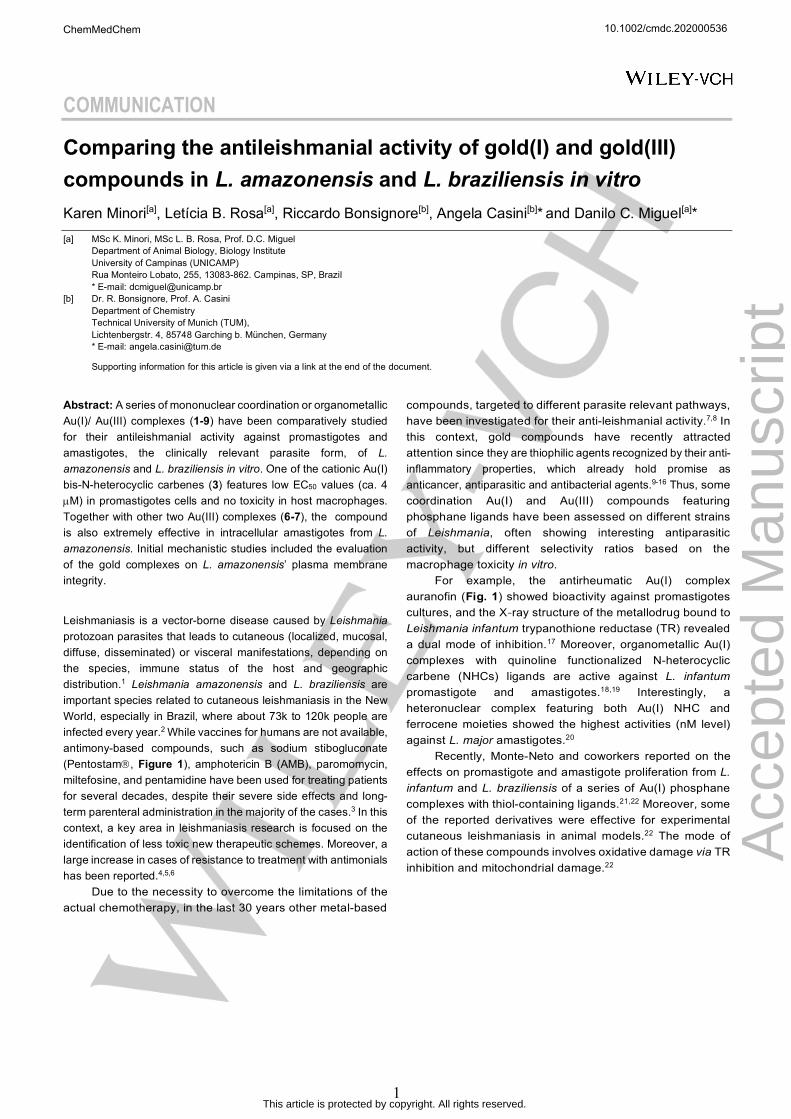
Figure 1. Structures of the first-line treatment for leishmaniasis, the Sb(V) complex with N-methyl-D-glucamine (sodium stibogluconate), of the oral anti-arthritis Au(I) complex auranofin, and of the Au(I) and Au(III) complexes (1-9) reported in this study.
The present study aims at extending the investigation to different families of Au(I) and Au(III) compounds to identify possible structure-activity relationships. Thus, nine gold compounds, previously characterized for their anticancer effects, were synthesized according to reported procedures, including Au(I) mono- and bis-NHC complexes (1-4),23,24,25 Au(III) complexes coordinated to a bidentate N^N scaffold (5, 6),26,27,28 and three cyclometallated Au(III) complexes with C^N ligands (7-9)29,30,31 (Fig. 1). Initially, the compounds were tested in vitro against promastigote cultures of L. amazonensis and L. braziliensis, following standard procedures (see Experimental for details, supplementary information available). The compounds were also tested against a model of host macrophages, namely BALB/c mouse primary macrophages (BMDM) to assess their selectivity. After 24 h incubation, the MTT colorimetric assay32 was performed and dose-response curves for parasites and host cells were obtained. The activities of gold-compounds in terms of 50% effective concentrations for promastigotes of L. amazonensis and L. braziliensis (EC50) and 50% cytotoxic concentrations (CC50) for BMDM are presented in Table 1.
Within the series, the cationic [AuI(1-benzyl-3,7,9-trimethylxanthin-8-ylidene)2]BF4 complex 3 shows the lowest EC50 values for both species and a remarkably favourable selectivity index (SI=CC50/EC50)>23.5, higher than those previously reported for other Au(I) NHCs,18 and with respect to the reference second-line anti-leishmanial drug AMB (Table 1). Compound 3 is also markedly more active than the two analogues [AuI(9-methylcaffeine-8-ylidene)2]BF4 2 and [AuI((9-methylcaffein-8-ylidene)(ethynylphenyl))] 4. The neutral [AuI(9-methylcaffein-8-ylidene)I] complex 1 has some activity against both parasite species as complex 3 (Table 1), but it is also more toxic against BMDM cells. The latter effect may be due to the different reactivity of 1 with respect to the bis-NHC complexes, particularly with
respect to the propensity to undergo ligand exchange reactions with different nucleophiles, which may lead to macrophage cell damage.
Table 1. Gold-compounds’ 50% effective (EC50) and cytotoxic (CC50) concentrations against Leishmania promastigotes and BALB/c mouse primary macrophages (BMDM) after 24h. SD: standard deviation; CI 95%: 95% confidence interval; ND: not determined. Two independent experiments were performed in triplicates.
EC50± SD (CI 95%) µM
EC50± SD (CI 95%) µM
CC50 ± SD (CI 95%) µM
Selectivity index
(CC50/EC50)
Comp L. amazonensis
L. braziliensis BMDM
L. amazonensis
/ L. braziliensis
1 13.96 ± 0.46 (13.6 - 14.3)
4.73 ± 0.20 (4.57 - 4.89)
23.53 ± 0.61 (25.0 - 26.0) 1.7 / 5.0
2
53.88 ± 0.21 (53.7 - 54.0)
50.36 ± 0.14 (50.2 - 50.5)
87.85 ±0.45 (87.4 - 88.3)
1.6 / 1.7
3
4.24 ± 0.23 (4.08 - 4.4)
4.25 ± 0.17 (4.11 - 4.39)
>100 (ND)
>23.6 / >23.5
4
117.30± 0.21
(117.1 - 117.5)
113.20± 0.10
(113.1 - 113.3)
156.60±0.10
(156.5 - 156.7) 1.3 / 1.4
5
37.02 ± 0.29 (36.8 - 37.3)
33.2± 0.31
(33.0 - 33.4)
117.30 ±0.20
(117.2 - 117.4) 3.2 / 3.5
6
9.44 ± 0.22 (9.26 - 9.62)
8.42 ± 0.20 (8.26 - 8.58)
59.34 ±0.20 (59.2 - 59.5)
6.3 / 7.1
7
15.89 ± 0.43 (15.5 - 16.2)
17.49 ± 0.21 (17.3 - 17.7)
51.90± 0.33 (51.6 - 52.2)
3.3 / 3.0
8
48.63 ± 0.29 (48.4 - 48.9)
19.96 ± 0.19 (19.8 - 20.1)
87.54 ±0.47 (87.2 - 87.9)
1.8 / 4.4
9
24.95 ± 0.38 (24.7 - 25.3)
5.12 ± 0.15 (5.00 - 5.24)
7.08 ± 0.19 (6.92 - 7.24)
0.3 / 1.4
AMB
0.53 ± 0.11 (0.4 - 0.6)
0.29 ± 0.09 (0.2 - 0.3)
1.97 ± 0.29 (1.7 - 2.2)
3.7 / 6.8
AuCl Cl
N
N
NAu
Cl Cl
N
N
N
PF6-
1
NAu
ClCl
NAu
ClCl
N
HN
AuClCl
O
AuN
N
N
N
O
O N
N
N
N
O
O
BF4-+
AuN
N
N
N
O
OI Au
N
N
N
N
O
O N
N
N
N
O
O
BF4-+
AuN
N
N
N
O
O2 3 4
5 6 7 8 9
OAcOAcO
OAcS
OAc
Au PEt3
auranofin
OSbO O
SbOHO
HO H
COO- COO-
OHOH
OOH
sodium stibogluconate
O-
OO
Na+
Na+
Na+
10.1002/cmdc.202000536
Acc
epte
d M
anus
crip
t
ChemMedChem
This article is protected by copyright. All rights reserved.
COMMUNICATION
3
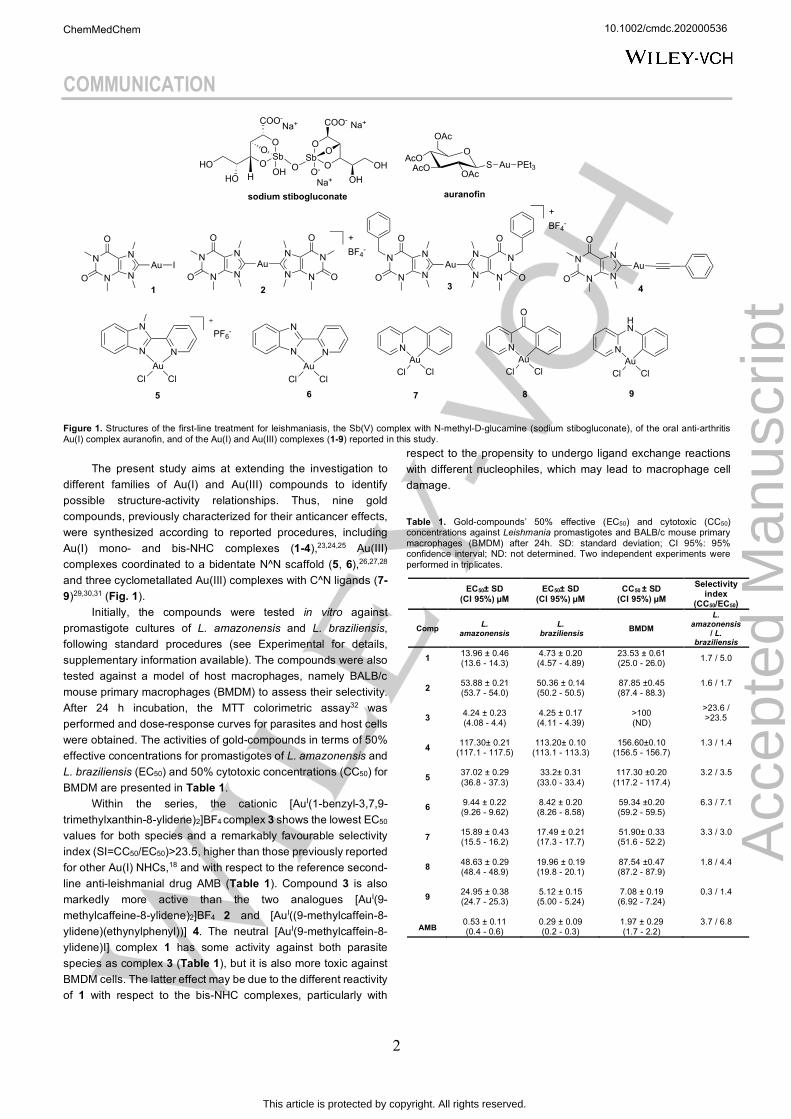
Figure 2. Gold complexes against Leishmania-infected BALB/c mouse primary macrophages (BMDM). (A) Cartoon representation of macrophagic infection by Leishmania promastigotes (left) and established intracellular infection by amastigotes residing in parasitophorous vacuoles (right). In vitro infections were established (Multiplicity of infection (MOI) = 2 for L. amazonensis and MOI = 5 for L. braziliensis) and kept in fresh culture medium with 3, 6 and 7 at different concentrations every 24 h up to 48 h. (B) Representative image of one optical field showing untreated (left) and treated with 6 (22.5 µM) infected cells (right) with L. braziliensis, as described in the experimental section. Methanol-fixed BMDM were stained with Instant Prov Kit (NewProv) and images were obtained using Invitrogen EVOS XL Core Cell Imaging System microscope. Arrows point to intracellular amastigotes. Scale bar = 6 µm. Effects on the infection rate (C) and amastigote burden (D) were calculated in relation to untreated control infections (100%) for two independent assays run in triplicates. p values: *<0.05; **<0.01; ***<0.001; ****<0.0001; treated infection vs. untreated infections.
Concerning Au(III) complexes, both coordination and organometallic compounds show good to moderate activity against promastigotes of both species; being compounds [AuIII(2-[(pyridin-2-yl)-benzimidazole)Cl2] 6 (SI = 6.3 and 7.0) and [AuIII(2-benzylpyridine)Cl2] 7 (SI = 3.3 and 3.0) the most active and selective derivatives. The Au(III) C^N complexes [AuIII (2-benzoylpyridine)Cl2] 8 and [AuIII(N-phenylpyridin-2-amine)Cl2] 9 show some selectivity towards the L. braziliensis, but overall their SI is less favourable than that of the related compound 7. Finally, [AuIII(1-methyl-2-(pyridin-2-yl)-benzimidazole)Cl2]PF6 5 was among the least effective, in line with its scarce cytotoxic activity against human cancer cells.27 The efficacy of the most promising compounds, 3, 6 and 7, was further assessed against intracellular amastigotes, parasitic forms involved in the pathogenesis of leishmaniasis, as they replicate within macrophages (Fig 2). To this aim, BMDM were cultivated in glass coverslips in 24-well plates followed by the infection with stationary-phase promastigotes of L. amazonensis and L. braziliensis (Fig. 2A; left). After 24 h, established infections (Fig. 2A; right and Fig. 2B) were incubated with each compound at increasing concentrations (see Experimental for details). Next, two in vitro infection parameters were assessed: the infection rate
(Fig. 2C) and the amastigote burden (Fig. 2D), respectively. AMB was used as a positive control drug for Leishmania killing in BMDM. At variance with the promastigote studies, in this assay L. amazonensis amastigotes were more sensitive to 3 and 6 than L. braziliensis even at the lowest tested concentrations. In the case of 7, the number of infected cells was drastically reduced only at the highest tested concentration (25 µM). Despite requiring slightly increased concentrations, L. braziliensis replication was also significantly inhibited by the compounds. In general, the gold complexes were able to reduce more than 90% of infections and intracellular parasites’ number (Fig. 2C,D), in some cases at markedly lower concentrations when compared with the EC50 values obtained for promastigotes. It has been shown that infected-BMDM treated with meglumine antimoniate for 6 days, inhibits 50% of the amastigotes load between 39 and 196 µM for L. braziliensis, and between 296 and 407 µM for L. amazonensis in different field isolates.33 Our data show that 3, 6 and 7 are markedly more active at lower concentrations and do not require prolonged incubation times, suggesting a more immediate effect on the parasites, without affecting the host cell (Fig. 2B).
10.1002/cmdc.202000536
Acc
epte
d M
anus
crip
t
ChemMedChem
This article is protected by copyright. All rights reserved.
COMMUNICATION
4
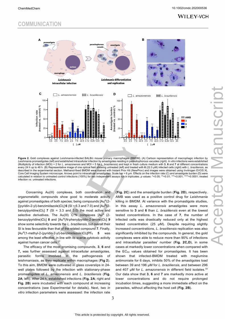
In an attempt to shed light into our compounds’ mechanism of action against Leishmania, their possible direct effect regarding L. amazonensis’ plasma membrane integrity was investigated in promastigotes and in the clinically relevant stage for the disease; i.e., the amastigote stage. Thus, both parasite forms incubated with 3, 6 and 7 were tested for their permeabilization to ethidium bromide.34 Control assays were carried out using AMB, as it is known that this polyene antibiotic disrupts Leishmania’s plasma membrane integrity.35 Fig. 3A shows that all gold compounds increased the permeabilization of promastigotes after short (3 h) and prolonged time points (24 h), with compound 6 being the most effective. In fact, incubation with 6 led to ~50% increased fluorescence signals of ethidium bromide upon nucleic acid binding compared to the AMB control in the same conditions. The membrane permeabilization possibly contributes to the triggering of the observed antiproliferative effects.
In parallel, assays were run using amastigotes incubated in acidic medium (pH 5.2) to mimic the intracellular milieu of the parasitophorous vacuole. Notably, ethidium bromide incorporation was higher for all the compounds, especially for 3 and 7, and equivalent levels to the fluorescence detected for AMB were observed after 24 h. Amastigotes treated with compound 6 showed similar levels of fluorescence signal at both 3 and 24 h time points. In general, the gold compounds were able to destabilise the parasite's membrane permeability control already after 3 h incubation (Fig. 3B), time at which we can exclude cytotoxic effects by the tested compounds’ concentrations.
Figure 3. L. amazonensis parasite’s plasma membrane permeabilization induced by treatment with gold complexes. (A) 4x10⁶ promastigotes and (B) 8x10⁶ axenic amastigotes were incubated with EC50 of AMB, 3, 6 and 7 at neutral and acidic pH, respectively, for 3 h (dashed lines) and 24 h (solid lines). (I): addition of 10 mM ethidium bromide; (II): addition of digitonin at 100 mM.
In conclusion, our findings further highlight the potential of gold complexes as antileishmanial agents with low toxicity to host cells. In both the promastigote and amastigote assays, the cationic bis-NHC complex 3 featuring a caffeine-like scaffold is certainly the best performer with the highest activity towards the two Leishmania species and selectivity with respect to the host macrophages. We hypothesize that an ideal combination of hydrophilic/lipophilic character and reduced metallodrug speciation due to the high stability of the Au-C bonds may be responsible for the overall observed effects of 3 in the parasite. Noteworthy, caffeine-based metal complexes have already shown promising pharmacological properties as anticancer and antimicrobial agents. 36,37,38 Complex 6 is one of the few Au(III) complexes with N-donor ligands reported so far for their anti-promastigote effects 8,39. Its reduced stability towards Au(III) reduction in biological environment with respect to the other reported organometallic Au(III) C^N compounds,26 may lead to parasite death via oxidative damage.
The cyclometallated compound 7, showing promising activity, is the first of this family of organometallics to be studied for the antileishmanial activity. The robustness of the C^N scaffold may be exploited to functionalize the compound with parasite targeting moieties. Moreover, 7 was recently reported to selectively arylate cysteine residues in protein domains via Au(III)-templated reductive elimination,40,41 leading to irreversible modification of the protein structure. We cannot exclude that a similar reactivity could account for the observed antiparasitic effects.
Our data on compounds 3, 6 and 7, showing the remarkable effect of these compounds in in vitro infections, combined with their activity against axenic amastigotes cultured in acidic medium, suggest that the leishmanicidal effect is preserved in the vacuolar environment; representing a pharmacological requirement of relevance for the development of novel leishmanicidal candidates.3
Further studies are certainly necessary to elucidate the mechanisms of action of the gold compounds in the Leishmania parasites, although knowledge of their targets against cancer cells is already available in the literature.42 For example, pharmacologically relevant DNA secondary structures, G-quadruplexes, have been targeted by cationic Au(I) bis-NHC compounds (2-3).23,43 Oxidative damage by gold complexes, also affecting membrane permeabilization, may also be relevant. Moreover, specific membrane protein channels and zinc-binding proteins are inhibited by the herewith investigated coordination and cyclometallated Au(III) complexes (5-7).26,44,45 While these studies focused mostly on the human targets, leishmanial homologues systems could also be considered.46
Acknowledgements
KM, LBR and DCM are grateful to Coordenação de Aperfeiçoamento de Pessoal de Nível Superior, Brasil (CAPES) for the PhD (KM) and Master’s (LBR) fellowship (Programa Demanda Social) and CAPES-PRINT Visiting Professor
10.1002/cmdc.202000536
Acc
epte
d M
anus
crip
t
ChemMedChem
This article is protected by copyright. All rights reserved.
COMMUNICATION
5
Fellowship at the Technische Universität München (#88887.468349/2019-00) (DCM).
Keywords: leishmaniasis • gold compounds • promastigotes • amastigotes• antiprotozoal agents References
[[1] World Health Organization: WHO. Leishmaniasis [Internet]; 2018; cited on 2020 June 06. Available from: https://www.who.int/leishmaniasis/burden/en/.
[2] J. Alvar, I. D. Vélez, C. Bern, M. Herrero, P. Desjeux, J. Cano, J. Jannin, M. den Boer, WHO Leishmaniasis Control Team, PloS One, 2012, 7(5):e35671, DOI: 10.1371/journal.pone.0035671.
[3] L. M. Alcântara, T. C. S. Ferreira, F. R. Gadelha, D. C. Miguel, Int. J. Parasitol. Drugs Drug Resist., 2018, 8(3), 430-439.
[4] A. Ponte-Sucre, F. Gamarro, J-C. Dujardin, M. P. Barrett, R. López-Vélez, R. García-Hernández, A. W. Pountain, R. Mwenechanya, R. Papadopoulou, PLoS Negl. Trop. Dis., 2017, 11(12), DOI: 10.1371/journal.pntd.0006052.
[5] S. M. Landfear in Drug Resistance in Leishmania Parasites, Vol. 1 (Eds.: A. P. Sucre, E. Diaz, M. P. Nieves), Springer-Verlag Wien, Caracas, Venezuela, 2013, pp.259-284.
[6] S. Sundar, Trop. Med. Int. Health: TM & IH, 2001, 6(11), 849-854.
[7] Y. C. Ong, S. Roy, P. C. Andrews, G. Gasser, Chem. Rev., 2019, 119(2), 730-796.
[8] M. Navarro, C. Hernández, I. Colmenares, P. Hernández, M. Fernández, A. Sierraalta, E. Marchán, J. Inorg. Biochem., 2007, 101, 111-116.
[9] S. Nobili, E. Mini, I. Landini, C. Gabbiani, A. Casini, L. Messori, Med. Res. Rev., 2010, 30(3), 550-580.
[10] M. Mora, M. C. Gimeno, R. Visbal, Chem. Soc. Rev., 2019, 48(2), 447-462.
[11] B. Bertrand, A. Casini, Dalton Trans., 2014, 43(11), 4209-4219.
[12] P. F. Salas, C. Herrmann, C. Orvig, Chem. Rev., 2013, 113(5), 3450-3492.
[13] A. R. Sannella, A. Casini, C. Gabbiani, L. Messori, A. R. Bilia, F. F. Vincieri, G. Majori, C. Severini, FEBS Lett., 2008, 582(6), 844-847.
[14] J. P. Owings, N. N. McNair, Y. F. Mui, T. N. Gustafsson, A. Holmgren, M. Contel, J. B. Goldberg, J. R. Mead, FEMS Microbiol. Lett., 2016, 363(14), DOI: 10.1093/femsle/fnw148.
[15] A. Frei, J. Zuegg, A. G. Elliott, M. Baker, S. Braese, C. Brown, F. Chen, C. G. Dowson, G. Dujardin, N. Jung, A. P. King, A. M. Mansour, M. Massi, J. Moat, H. A. Mohamed, A. K. Renfrew, P. J. Rutledge, P. J. Sadler, M. H. Todd, C. E. Willans, J. J. Wilson, M. A. Cooper, M. A. T. Blaskovich, Chem. Sci., 2020, 11(10), 2627-2639.
[16] C. Schmidt, B. Karge, R. Misgeld, A. Prokop, R. Franke, M. Brönstrun, I. Ott, Chemistry, 2017, 23(8), 1869-1880.
[17] A. Ilari, P. Baiocco, L. Messori, A. Fiorillo, A. Boffi, M. Gramiccia, T. Di Muccio, G. Colotti, Amino Acids, 2012, 42(2-3), 803-811.
[18] L. Paloque, C. Hemmert, A. Valentin, H. Gornitzka, Eur. J. Med. Chem., 2015, 94, 22-29.
[19] C. Zhang, S. B. Delmas, A. F. Álvarez, A. Valentin, C. Hemmert, H. Gornitzka, Eur. J. Med. Chem., 2018, 143, 1635-1643.
[20] W. S. Koko, J. Jentzsch, H. Kalie, R. Schobert, K. Ersfeld, I. S. Al Nasr, T. A. Khan, B. Biersack, Arch. Pharm., 2020, 353(5), DOI: 10.1002/ardp201900363.
[21] J. D. S. Chaves, L. G. Tunes, C. H. J. Franco, T. M. Francisco, C. C. Corrêa, S. M. F. Murta, R. L. Monte-Neto, H. Silva, A. P. S. Fontes, M. V. de Almeida, Eur. J. Med. Chem., 2017, 127, 727-739.
[22] L. G. Tunes, R. E. Morato, A. Garcia, V. Schmitz, M. Steindel, J. D. Corrêa-Junior, H. F. dos Santos, F. Frézard, M. V. de Almeida, H. Silva, N. S. Moretti, A. L. B. de Barros, R. L. do Monte-Neto, ACS Infect. Dis., 2020, 6(5), 1121-1139.
[23] B. Bertrand, L. Stefan, M. Pirrotta, D. Monchaud, E. Bodio, P. Richard, P. Le Gendre, E. Warmerdam, M. H. de Jager, G. M. M. Groothuis, M. Picquet, A. Casini, Inorg. Chem., 2014, 53(4), 2296-2303.
[24] S. M. Meier-Menches, B. Aikman, D. Döllerer, W. T. Klooster, S. J. Coles, N. Santi, L. Luk, A. Casini, R. Bonsignore, J. Inorg. Biochem., 2020, 202, DOI: 10.1016/j.jinorgbio.2019.110844.
[25] J. Oberkofler, B. Aikman, R. Bonsignore, A. Pöthig, J. Platts, A. Casini, F. E. Kühn, Eur. J. Inorg. Chem., 2020, 2020(11-12), 1040-1051.
[26] A. de Almeida, A. F. Mósca, D. Wragg, M. Wenzel, P. Kavanagh, G. Barone, S. Leoni, G. Soveral, A. Casini, Chem. Commun (Camb.)., 2017, 53(27), 3830-3833.
[27] B. Aikman, M. N. Wenzel, A. F. Mósca, A. de Almeida, W. T. Klooster, S. J. Coles, G. Soveral, A. Casini, Inorganics, 2018, 6(4), DOI: 10.3390/inorganics6040123.
[28] M. Serratrice, M. A. Cinellu, L. Maiore, M. Pilo, A. Zucca, C. Gabbiani, A. Guerri, I. Landini, S. Nobili, E. Mini, L. Messori, Inorg. Chem., 2012, 51(5), 3161-3171.
[29] M. A. Cinellu, A. Zucca, S. Stoccoro, G. Minghetti, M. Manassero, M. Sansoni, J. Chem. Soc., Dalton Trans., 1995, (17), 2865-2872.
[30] Y. Zhu, B. R. Cameron, R. Mossi, V. Anastassov, J. Cox, L. Qin, Z. Santucci, M. Metz, R. T. Skerlj, S. P. Fricker, J. Inorg. Biochem., 2011, 105(5), 754-762.
[31] B. Bertrand, S. Spreckelmeyer, E. Bodio, F. Cocco, M. Picquet, P. Richard, P. Le Gendre, C. Orvig, M. A. Cinellu, A. Casini, Dalton Trans., 2015, 44(26), 11911-11918.
[32] B. Mendes, J. R. Almeida, N. Vale, P. Gomes, F. R. Gadelha, S. L. da Silva, D. C. Miguel, Comp. Biochem. Physiol. C Toxicol. Pharmacol., 2019, 226, DOI: 10.1016/j.cbpc.2019.108612.
[33] R. C. Zauli-Nascimento, D. C. Miguel, J. K. U. Yokoyama-Yasunaka, L. I. A. Pereira, M. A. P. de Oliveira, F. Ribeiro-Dias, M. L. Dorta, S. R. B. Uliana, Trop. Med. Int. Health, 2010, 15(1), 68-76.
10.1002/cmdc.202000536
Acc
epte
d M
anus
crip
t
ChemMedChem
This article is protected by copyright. All rights reserved.
COMMUNICATION
6
[34] I. P. Beletsky, S. R. Umansky, J. Immunol. Methods, 1990, 134(2), 201-205.
[35] A. K. Saha, T. Mukherjee, A. Bhaduri, Mol. Biochem. Parasitol., 1986, 19(3), 195-200.
[36] J. J. Zhang, M. A. Abu El Maaty, H. Hoffmeister,C. Schmidt, J. K. Muenzner, R. Schobert, S. Wölf, I. Ott, Angew. Chem. Int. Ed. Engl., 2020, DOI: 10.1002/anie.202006212.
[37] J. J. Zhang, C. Che-Ming, I. Ott, J. Organomet. Chem., 2015, 782, 37-41.
[38] A. Kascatan-Nebioglu, A. Melaiye, K. Hindi, S. Durmus, M. J. Panzner, L. A. Hogue, R. J. Mallett, C. E. Hovis, M. Coughenour, S. D. Crosby, A. Milsted, D. L. Ely, C. A. Tessier, C. L. Cannon, W. J. Youngs, J. Med. Chem., 2006, (49)23, 6811-6818.
[39] L. Massai, L. Messori, N. Micale, T. Schirmeister, L. Maes, D. Fregona, M. A. Cinellu, C. Gabbiani, Biometals, 2017, 30(2), 313-320.
[40] M. N. Wenzel, R. Bonsignore, S. R. Thomas, D. Bourissou, G. Barone, A. Casini, Chemistry, 2019, 25(32), 7628-7634.
[41] S. R. Thomas, R. Bonsignore, J. E. Sanchez, S. M. Meier-Menches, C. Barone, L. Y. Luk, A. Casini, ChemBioChem., 2020, 21, DOI: 10.1002/cbic.202000262.
[42] A. de Almeida, B. L. Oliveira, J. D. G. Correia, G. Soveral, A. Casini, Coord. Chem. Rev., 2013, 257(19-20), 2689-2704.
[43] D. Wragg, A. de Almeida, R. Bonsignore, F. E. Kühn, S. Leoni, A. Casini, Angew Chem. Int. Ed. Engl., 2018, 57(44), 14524-14528.
[44] M. N. Wenzel, A. F. Mósca, V. Graziani, B. Aikman, S. R. Thomas, A. de Almeida, J. A. Platts, N. Re, C. Coletti, A. Marrone, G. Soveral, A. Casini, Inorg. Chem., 2019, 58(3), 2140-2148.
[45] M. N. Wenzel, S. M. Meier-Menches, T. L. Williams, E. Rämisch, G. Barone, A. Casini, Chem. Comm. (Camb.), 2018, 54(6), 611-614.
[46] N. Smargiasso, V. Gabelica, C. Damblon, F. Rosu, E. De Pauw, M-P. Teulade-Fichou, J. A. Rowe, A. Claessens, BMC Genomics, 2009, 10(362), DOI: 10.1186/1471-2164-10-362.
10.1002/cmdc.202000536
Acc
epte
d M
anus
crip
t
ChemMedChem
This article is protected by copyright. All rights reserved.
COMMUNICATION
7
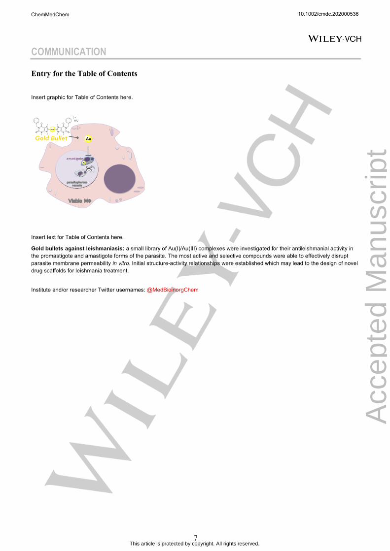
Entry for the Table of Contents
Insert graphic for Table of Contents here.
Insert text for Table of Contents here.
Gold bullets against leishmaniasis: a small library of Au(I)/Au(III) complexes were investigated for their antileishmanial activity in the promastigote and amastigote forms of the parasite. The most active and selective compounds were able to effectively disrupt parasite membrane permeability in vitro. Initial structure-activity relationships were established which may lead to the design of novel drug scaffolds for leishmania treatment.
Institute and/or researcher Twitter usernames: @MedBioinorgChem
Viable MΦ
parasitophorousvacuole
Gold Bullet
amastigote
Au
AuN
N
N
N
O
O N
N
N
N
O
O
BF4-+
Au
10.1002/cmdc.202000536
Acc
epte
d M
anus
crip
t
ChemMedChem
This article is protected by copyright. All rights reserved.


![Antileishmanial Properties of Moroccan Medicinal Plants and ......Centaurium erythraea Rafn. Gentianaceae Korsatlhaya Flowering top - Skin diseases [38] - Digestive system and kidney](https://img.pdfslide.net/doc/110x75/60ff7e00b5f77b412e69d940/antileishmanial-properties-of-moroccan-medicinal-plants-and-centaurium-erythraea.jpg)