Embed Size (px)
Citation preview
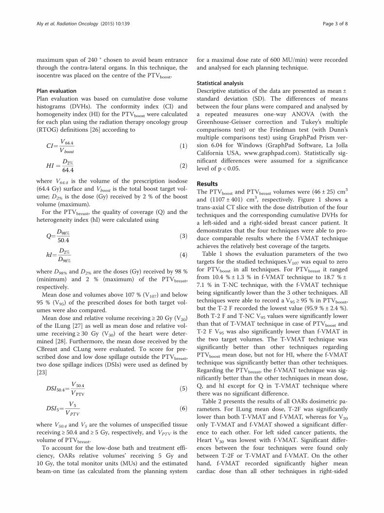
Aly et al. Radiation Oncology (2015) 10:139 DOI 10.1186/s13014-015-0452-2
RESEARCH Open Access
Comparison of breast simultaneous integratedboost (SIB) radiotherapy techniques
Moamen M.O.M. Aly1,2†, Gerhard Glatting1,3*†, Lennart Jahnke3, Frederik Wenz3 and Yasser Abo-Madyan3,4Abstract
Purpose: To dosimetrically evaluate different breast SIB techniques with respect to target coverage and organs atrisk (OARs) doses.
Methods: Four IMRT techniques were compared in 12 patients. Three techniques employ tangential whole breastirradiation with either two coplanar fields (T-2F), or four non-coplanar fields (T-NC), or one Volumetric ModulatedArc Therapy (T-VMAT) for the boost volume. The fourth technique is a fully-modulated VMAT technique (f-VMAT).Dosimetric parameters were compared for the boost and breast target volumes as well as OARs. Delivery efficiencywas analysed based on number of monitor units (MUs) and estimated delivery time.
Results: T-VMAT and f-VMAT ranked highest with respect to integral assessment of boost and breast treatmentquality measures. T-VMAT significantly outperformed f-VMAT with respect to ipsi-lateral lung and left-sidedpatients’ heart volumes ≥ 5 Gy (35 % ± 5 % vs. 52 % ± 6 % and 11 % ± 5 % vs. 22 % ± 6 %, respectively). f-VMATsignificantly outperformed T-VMAT with respect to ipsi-lateral lung volume ≥ 20 Gy (13 % ± 2 % vs. 15 % ± 3 %)and heart volume ≥ 30 Gy in left breast cancer (0 % ± 0 % vs. 1 % ± 1 %). T-VMAT and f-VMAT needed 442 ± 58and 1016 ± 152 MUs, respectively.
Conclusions: The hybrid T-VMAT is considered the technique of choice due to its balance of quality, efficiencyand dose to OARs.
Keywords: Breast Cancer, Simultaneous integrated Boost (SIB), Intensity Modulated Radiotherapy (IMRT),Volumetric Modulated Arc Therapy (VMAT)
BackgroundBreast cancer is the most common cancer in womenworldwide as it is also the main cause of cancer deathamong women globally [1]. The use of radiotherapy inthe adjuvant setting has shown to improve both localcontrol and overall survival in early stage breast cancerpatients [2]. The most common and traditional wholebreast radiotherapy technique uses two tangential fieldsdue to its efficiency in terms of sparing nearby organs atrisk (OARs) as well as technical simplicity in whichwedge filters are used to compensate patient’s surface ir-regularity and reach a homogenous dose distribution.
* Correspondence: [email protected]†Equal contributors1Medical Radiation Physics/Radiation Protection, UniversitätsmedizinMannheim, Medical Faculty Mannheim, Heidelberg University, Mannheim,Germany3Department of Radiation Oncology, Universitätsmedizin Mannheim, MedicalFaculty Mannheim, Heidelberg University, Mannheim, GermanyFull list of author information is available at the end of the article
© 2015 Aly et al. This is an Open Access articl(http://creativecommons.org/licenses/by/4.0),provided the original work is properly creditedcreativecommons.org/publicdomain/zero/1.0/
This technique has evolved over the last decade with theintroduction of multi-leaf collimators (MLC) to deliverfield-in-field (FIF) three-dimensional conformal (3D-CRT)[3–6] or intensity modulated radiation therapy (IMRT)variants [7–11].Dose escalation to the tumour-bed by a sequential
boost reduces local recurrence [12] but prolongs thetreatment duration and significantly increases the risk ofmoderate to severe breast fibrosis [13]. Alternatively,simultaneously integrated boost (SIB) using a higherdose per fraction to the tumour bed was shown to bedosimetrically advantageous especially regarding doseconformity of the boost volume [14, 15], more conveni-ent due to the shorter treatment time and was recentlyshown to be very well tolerated on the short andmedium terms [16–18].Different radiotherapy delivery techniques were pro-
posed for SIB, including 3D-CRT with wedges or FIF
e distributed under the terms of the Creative Commons Attribution Licensewhich permits unrestricted use, distribution, and reproduction in any medium,. The Creative Commons Public Domain Dedication waiver (http://) applies to the data made available in this article, unless otherwise stated.

Aly et al. Radiation Oncology (2015) 10:139 Page 2 of 8
technique [19, 20], IMRT [21, 22], helical tomotherapy[19], or volumetric modulated arc therapy (VMAT)[23, 24]. A thorough comparison of all these techniquesis yet to be performed. In this planning study we comparethe dosimetric outcomes of three inversely planned tech-niques for SIB delivery based on the standard two tan-gential whole breast fields plus two coplanar boost fields(T-2F), or four non-coplanar boost fields (T-NC), or oneboost VMAT arc (T-VMAT) as well as a fully modu-lated VMAT (f-VMAT) for both the whole breast andintegrated boost volumes.
Materials and methodsPatient selection and image dataTwelve female breast cancer patients (6 right-sided and6 left-sided), who were recently treated in the Depart-ment of Radiation Oncology, University Medical CentreMannheim, Heidelberg University, were retrospectivelyrandomly selected. The computed tomography (CT)data-sets were acquired on a CT-simulator (BrillianceCT Big Bore, Philips, Cleveland, OH, USA) according tothe institution’s standard protocol in 5 mm slice thick-ness, in supine position with the use of a wing board forarm positioning above the head.
Target volumes and organs at risk delineationBoth breast volumes (the affected side, and the contra-lateral breast (CBreast)) were delineated and cropped5 mm inside the skin contour. Also, the ipsi-lateral lung(ILung), contra-lateral lung (CLung), and heart were de-lineated. The boost clinical target volume was delineatedby an experienced physician according to the scar, preand post-operative radiological changes within the breasttissue, the surgical report and/or the presence of surgicalclips. A setup safety margin of 5 mm was automaticallyadded to this boost volume to create the boost planningtarget volume (PTVboost). This safety margin was con-strained to 5 mm behind the skin contour. The wholebreast volume subtracting the PTVboost was consideredthe breast planning target volume (PTVbreast).
Beam setup and plan prescriptionFor each patient, four different IMRT plans were gener-ated using a treatment planning system that employs aMonte Carlo calculation algorithm (Monaco v3.3, ElektaAB, Stockholm, Sweden). A prescribed dose of 64.4 Gyto the PTVboost and 50.4 Gy to the PTVbreast in 28 frac-tions was planned. The plans were created for a 6 MVphoton beam Elekta Synergy® linear accelerator with anMLCi2. Except for the VMAT techniques, all other tech-niques/beams were planned for step and shoot IMRTdelivery. Optimization was performed to get the bestplan for each technique for each individual patient. Theoptimization prescription aimed to deliver at least 92 %
of the prescribed dose to 95 % of the target volumes andto minimize the volume receiving ≥ 107 % of the boostdose. Having reached these criteria for the targets, add-itional effort was made to reduce dose to OARs indi-vidually for each patient and planning technique startingfrom the proper choice of gantry angles to the fine-tuning of the prescription cost functions and tighteningthe constraints to OARs. Our initial planning objectivesfor the OARs were a mean dose below 5 Gy to the heartfor left sided cases, below 3 Gy for contralateral breastand lung, a V20 below 22 % for ipsilateral lung. All planswere normalized to deliver a median of 64.4 Gy to thePTVboost volume.For the first technique, two tangential beams (medial
and lateral tangents) were assigned to the PTVbreast andanother two coplanar oblique beams assigned to thePTVboost with individually selected gantry angles to pre-vent any unnecessary dose to OARs especially the ipsi-lateral lung. These four fields were optimized together ina single plan (T-2F).The second technique consisted of the same tangential
beams assigned to the PTVbreast with four non-coplanarbeams assigned to the PTVboost (two gantry angles werechosen for each of two extra couch angles, 45° and 315°)aiming to further reduce OARs exposure, this, as anadaptation from the technique described by Baglan et al.2003 [25]. These six fields were optimized in a singleplan (T-NC).The third technique was generated by creating a
hybrid of tangential IMRT and VMAT deliveries in a sin-gle plan by assigning a single VMAT partial arc to thePTVboost. The arc typically starts at the same gantryangle assigned for the medial tangential beam and spans(depending on the shape of the thoracic wall, PTVbreast
and location of the PTVboost) to a maximum of 240 °. Inthis technique, the boost VMAT arc was firstly opti-mized separately to deliver 14 Gy to the PTVboost andthen the resulted plan was used as a biased dose to thetangential plan. The bias-dose option allows loading thedose brought by the VMAT boost arc into the tangentialplan to account for it in the optimization of the tangen-tial plan. This strategy leads to the reduction of breastintegral dose outside the boost volume. The combinedplan was named T-VMAT.In these three techniques, each beam was assigned to
a specific target (i.e. tangential beams to the PTVbreast
and all other beams or arcs only to the PTVboost). Thus,it was possible to prevent the inverse planning systemfrom using the non-tangential beams to target the wholebreast which avoids overexposure to the OARs. All theseplans were mono-isocentric with the isocentre placed inthe structure centre of the PTVbreast.The fourth technique was generated using a fully mod-
ulated VMAT (f-VMAT) partial double arc, over a
Aly et al. Radiation Oncology (2015) 10:139 Page 3 of 8
maximum span of 240 ° chosen to avoid beam entrancethrough the contra-lateral organs. In this technique, theisocentre was placed on the centre of the PTVboost.
Plan evaluationPlan evaluation was based on cumulative dose volumehistograms (DVHs). The conformity index (CI) andhomogeneity index (HI) for the PTVboost were calculatedfor each plan using the radiation therapy oncology group(RTOG) definitions [26] according to
CI¼ V 64:4
Vboostð1Þ
HI ¼ D2%
64:4ð2Þ
where V64.4 is the volume of the prescription isodose(64.4 Gy) surface and Vboost is the total boost target vol-ume; D2% is the dose (Gy) received by 2 % of the boostvolume (maximum).For the PTVbreast, the quality of coverage (Q) and the
heterogeneity index (hI) were calculated using
Q¼D98%
50:4ð3Þ
hI¼ D2%
D98%ð4Þ
where D98% and D2% are the doses (Gy) received by 98 %(minimum) and 2 % (maximum) of the PTVbreast,respectively.Mean dose and volumes above 107 % (V107) and below
95 % (V95) of the prescribed doses for both target vol-umes were also compared.Mean dose and relative volume receiving ≥ 20 Gy (V20)
of the ILung [27] as well as mean dose and relative vol-ume receiving ≥ 30 Gy (V30) of the heart were deter-mined [28]. Furthermore, the mean dose received by theCBreast and CLung were evaluated. To score for pre-scribed dose and low dose spillage outside the PTVbreast,two dose spillage indices (DSIs) were used as defined by[23]
DSI50:4¼ V 50:4
V PTVð5Þ
DSI5¼ V 5
VPTVð6Þ
where V50.4 and V5 are the volumes of unspecified tissuereceiving ≥ 50.4 and ≥ 5 Gy, respectively, and VPTV is thevolume of PTVbreast.To account for the low-dose bath and treatment effi-
ciency, OARs relative volumes’ receiving 5 Gy and10 Gy, the total monitor units (MUs) and the estimatedbeam-on time (as calculated from the planning system
for a maximal dose rate of 600 MU/min) were recordedand analysed for each planning technique.
Statistical analysisDescriptive statistics of the data are presented as mean ±standard deviation (SD). The differences of meansbetween the four plans were compared and analysed bya repeated measures one-way ANOVA (with theGreenhouse-Geisser correction and Tukey’s multiplecomparisons test) or the Friedman test (with Dunn’smultiple comparisons test) using GraphPad Prism ver-sion 6.04 for Windows (GraphPad Software, La JollaCalifornia USA, www.graphpad.com). Statistically sig-nificant differences were assumed for a significancelevel of p < 0.05.
ResultsThe PTVboost and PTVbreast volumes were (46 ± 25) cm3
and (1107 ± 401) cm3, respectively. Figure 1 shows atrans-axial CT slice with the dose distribution of the fourtechniques and the corresponding cumulative DVHs fora left-sided and a right-sided breast cancer patient. Itdemonstrates that the four techniques were able to pro-duce comparable results where the f-VMAT techniqueachieves the relatively best coverage of the targets.Table 1 shows the evaluation parameters of the two
targets for the studied techniques.V107 was equal to zerofor PTVboost in all techniques. For PTVbreast it rangedfrom 10.4 % ± 1.3 % in f-VMAT technique to 18.7 % ±7.1 % in T-NC technique, with the f-VMAT techniquebeing significantly lower than the 3 other techniques. Alltechniques were able to record a V95 ≥ 95 % in PTVboost,but the T-2 F recorded the lowest value (95.9 % ± 2.4 %).Both T-2 F and T-NC V95 values were significantly lowerthan that of T-VMAT technique in case of PTVboost andT-2 F V95 was also significantly lower than f-VMAT inthe two target volumes. The T-VMAT technique wassignificantly better than other techniques regardingPTVboost mean dose, but not for HI, where the f-VMATtechnique was significantly better than other techniques.Regarding the PTVbreast, the f-VMAT technique was sig-nificantly better than the other techniques in mean dose,Q, and hI except for Q in T-VMAT technique wherethere was no significant difference.Table 2 presents the results of all OARs dosimetric pa-
rameters. For ILung mean dose, T-2F was significantlylower than both T-VMAT and f-VMAT, whereas for V20
only T-VMAT and f-VMAT showed a significant differ-ence to each other. For left sided cancer patients, theHeart V30 was lowest with f-VMAT. Significant differ-ences between the four techniques were found onlybetween T-2F or T-VMAT and f-VMAT. On the otherhand, f-VMAT recorded significantly higher meancardiac dose than all other techniques in right-sided

Figure 1 A trans-axial CT slice and the corresponding DVHs of the breast two tangential fields with: two coplanar fields (T-2F), four non-coplanarfields (T-NC), and a VMAT arc (T-VMAT) for the boost volume and a fully modulated VMAT (f-VMAT) techniques for a right-sided (right) and left-sided (left) patient. The DVH line colours correspond to the structure colour
Table 1 The targets coverage multi-comparison analysis for all studied techniques (mean ± SD). Mean values with ǂ symbol demonstratethat the corresponding technique did not pass the normality test. Friedman test of significance (p < 0.05) was used in these cases;otherwise, repeated measures ANOVA significance test was used
N = 12 T-2F T-NC T-VMAT f-VMAT
PTVboost V107 (%) 0* a 0* a 0* a 0* a
V95 (%) 95.9 ± 2.4c 98.0 ± 0.9ǂ bc 99.3 ± 0.6a 98.6 ± 1.7ab
mean (Gy) 64.1 ± 0.1b 64.2 ± 0.0b 64.3 ± 0.0a 64.1 ± 0.1b
CI 0.50 ± 0.00a 0.50 ± 0.00a 0.50 ± 0.00a 0.50 ± 0.00a
HI 1.03 ± 0.01† a 1.03 ± 0.01† a 1.03 ± 0.01† a 1.02 ± 0.01† b
PTVbreast V107 (%) 18.1 ± 3.9a 18.7 ± 7.1ǂ a 18.7 ± 4.2a 10.4 ± 1.3b
V95 (%) 94.5 ± 1.8b 94.6 ± 1.9ab 95.5 ± 2.3ab 96.6 ± 1.6a
mean (Gy) 52.2 ± 0.3a 52.2 ± 0.5ǂ a 52.3 ± 0.5a 51.5 ± 0.2b
Q 0.91 ± 0.02b 0.91 ± 0.02b 0.92 ± 0.03ab 0.94 ± 0.02a
hI 1.36 ± 0.03† a 1.37 ± 0.04† a 1.34 ± 0.05† a 1.30 ± 0.03† b
a,b,c Values having the same superscript in the same horizontal line are not significantly different.*All the 12 patients have a 0 value.†Note that although these values look similar and have a comparatively small SD (between patients), the differences are significant due to individual patient’svariability (i.e. when looking at the paired data)V107 and V95 are volumes receiving 107 % and 95 % of prescribed dose respectively; CI, conformity index as defined by equation (1); HI, homogeneity index asdefined by equation (2); Q is the quality of coverage as defined by equation (3); hI, heterogeneity index as defined by equation (4)
Aly et al. Radiation Oncology (2015) 10:139 Page 4 of 8
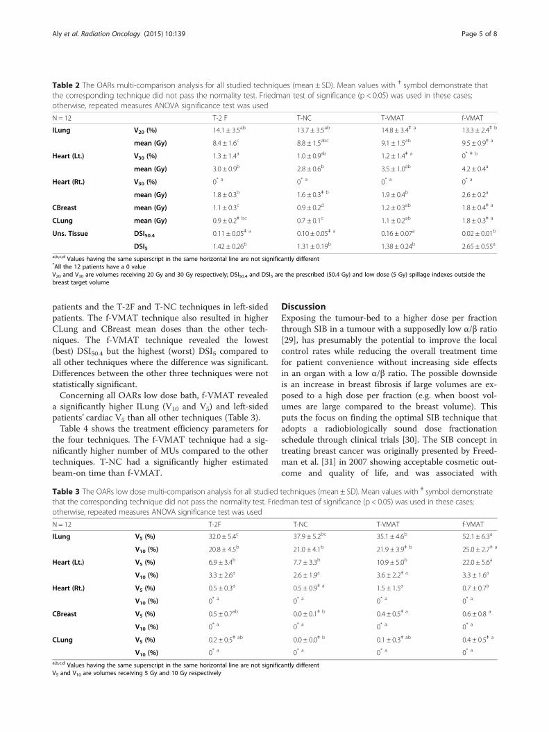
Table 2 The OARs multi-comparison analysis for all studied techniques (mean ± SD). Mean values with ǂ symbol demonstrate thatthe corresponding technique did not pass the normality test. Friedman test of significance (p < 0.05) was used in these cases;otherwise, repeated measures ANOVA significance test was used
N = 12 T-2 F T-NC T-VMAT f-VMAT
ILung V20 (%) 14.1 ± 3.5ab 13.7 ± 3.5ab 14.8 ± 3.4ǂ a 13.3 ± 2.4ǂ b
mean (Gy) 8.4 ± 1.6c 8.8 ± 1.5abc 9.1 ± 1.5ab 9.5 ± 0.9ǂ a
Heart (Lt.) V30 (%) 1.3 ± 1.4a 1.0 ± 0.9ab 1.2 ± 1.4ǂ a 0* ǂ b
mean (Gy) 3.0 ± 0.9b 2.8 ± 0.6b 3.5 ± 1.0ab 4.2 ± 0.4a
Heart (Rt.) V30 (%) 0* a 0* a 0* a 0* a
mean (Gy) 1.8 ± 0.3b 1.6 ± 0.3ǂ b 1.9 ± 0.4b 2.6 ± 0.2a
CBreast mean (Gy) 1.1 ± 0.3c 0.9 ± 0.2d 1.2 ± 0.3ab 1.8 ± 0.4ǂ a
CLung mean (Gy) 0.9 ± 0.2ǂ bc 0.7 ± 0.1c 1.1 ± 0.2ab 1.8 ± 0.3ǂ a
Uns. Tissue DSI50.4 0.11 ± 0.05ǂ a 0.10 ± 0.05ǂ a 0.16 ± 0.07a 0.02 ± 0.01b
DSI5 1.42 ± 0.26b 1.31 ± 0.19b 1.38 ± 0.24b 2.65 ± 0.55a
a,b,c,d Values having the same superscript in the same horizontal line are not significantly different*All the 12 patients have a 0 valueV20 and V30 are volumes receiving 20 Gy and 30 Gy respectively; DSI50.4 and DSI5 are the prescribed (50.4 Gy) and low dose (5 Gy) spillage indexes outside thebreast target volume
Aly et al. Radiation Oncology (2015) 10:139 Page 5 of 8
patients and the T-2F and T-NC techniques in left-sidedpatients. The f-VMAT technique also resulted in higherCLung and CBreast mean doses than the other tech-niques. The f-VMAT technique revealed the lowest(best) DSI50.4 but the highest (worst) DSI5 compared toall other techniques where the difference was significant.Differences between the other three techniques were notstatistically significant.Concerning all OARs low dose bath, f-VMAT revealed
a significantly higher ILung (V10 and V5) and left-sidedpatients’ cardiac V5 than all other techniques (Table 3).Table 4 shows the treatment efficiency parameters for
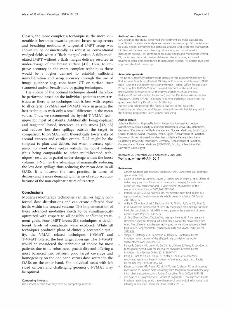
the four techniques. The f-VMAT technique had a sig-nificantly higher number of MUs compared to the othertechniques. T-NC had a significantly higher estimatedbeam-on time than f-VMAT.
Table 3 The OARs low dose multi-comparison analysis for all studiedthat the corresponding technique did not pass the normality test. Frieotherwise, repeated measures ANOVA significance test was used
N = 12 T-2F
ILung V5 (%) 32.0 ± 5.4c
V10 (%) 20.8 ± 4.5b
Heart (Lt.) V5 (%) 6.9 ± 3.4b
V10 (%) 3.3 ± 2.6a
Heart (Rt.) V5 (%) 0.5 ± 0.3a
V10 (%) 0* a
CBreast V5 (%) 0.5 ± 0.7ab
V10 (%) 0* a
CLung V5 (%) 0.2 ± 0.5ǂ ab
V10 (%) 0* a
a,b,c,d Values having the same superscript in the same horizontal line are not significV5 and V10 are volumes receiving 5 Gy and 10 Gy respectively
DiscussionExposing the tumour-bed to a higher dose per fractionthrough SIB in a tumour with a supposedly low α/β ratio[29], has presumably the potential to improve the localcontrol rates while reducing the overall treatment timefor patient convenience without increasing side effectsin an organ with a low α/β ratio. The possible downsideis an increase in breast fibrosis if large volumes are ex-posed to a high dose per fraction (e.g. when boost vol-umes are large compared to the breast volume). Thisputs the focus on finding the optimal SIB technique thatadopts a radiobiologically sound dose fractionationschedule through clinical trials [30]. The SIB concept intreating breast cancer was originally presented by Freed-man et al. [31] in 2007 showing acceptable cosmetic out-come and quality of life, and was associated with
techniques (mean ± SD). Mean values with ǂ symbol demonstratedman test of significance (p < 0.05) was used in these cases;
T-NC T-VMAT f-VMAT
37.9 ± 5.2bc 35.1 ± 4.6b 52.1 ± 6.3a
21.0 ± 4.1b 21.9 ± 3.9ǂ b 25.0 ± 2.7ǂ a
7.7 ± 3.3b 10.9 ± 5.0b 22.0 ± 5.6a
2.6 ± 1.9a 3.6 ± 2.2ǂ a 3.3 ± 1.6a
0.5 ± 0.9ǂ a 1.5 ± 1.5a 0.7 ± 0.7a
0* a 0* a 0* a
0.0 ± 0.1ǂ b 0.4 ± 0.5ǂ a 0.6 ± 0.8 a
0* a 0* a 0* a
0.0 ± 0.0ǂ b 0.1 ± 0.3ǂ ab 0.4 ± 0.5ǂ a
0* a 0* a 0* a
antly different
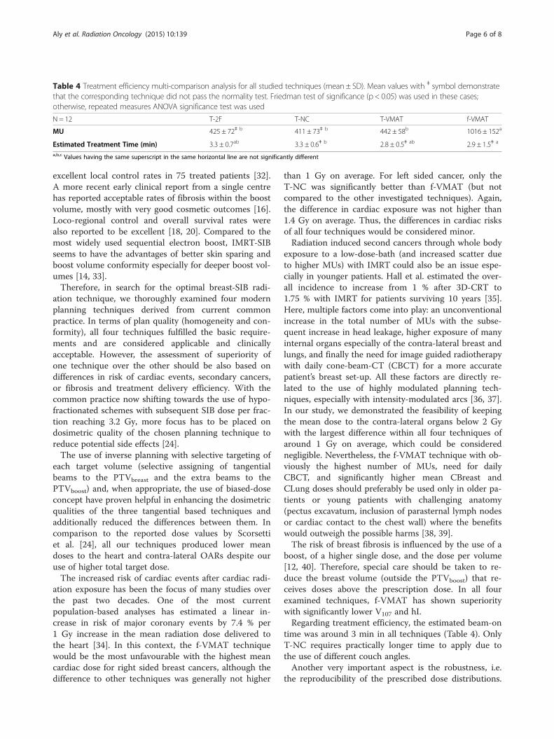
Table 4 Treatment efficiency multi-comparison analysis for all studied techniques (mean ± SD). Mean values with ǂ symbol demonstratethat the corresponding technique did not pass the normality test. Friedman test of significance (p < 0.05) was used in these cases;otherwise, repeated measures ANOVA significance test was used
N = 12 T-2F T-NC T-VMAT f-VMAT
MU 425 ± 72ǂ b 411 ± 73ǂ b 442 ± 58b 1016 ± 152a
Estimated Treatment Time (min) 3.3 ± 0.7ab 3.3 ± 0.6ǂ b 2.8 ± 0.5ǂ ab 2.9 ± 1.5ǂ a
a,b,c Values having the same superscript in the same horizontal line are not significantly different
Aly et al. Radiation Oncology (2015) 10:139 Page 6 of 8
excellent local control rates in 75 treated patients [32].A more recent early clinical report from a single centrehas reported acceptable rates of fibrosis within the boostvolume, mostly with very good cosmetic outcomes [16].Loco-regional control and overall survival rates werealso reported to be excellent [18, 20]. Compared to themost widely used sequential electron boost, IMRT-SIBseems to have the advantages of better skin sparing andboost volume conformity especially for deeper boost vol-umes [14, 33].Therefore, in search for the optimal breast-SIB radi-
ation technique, we thoroughly examined four modernplanning techniques derived from current commonpractice. In terms of plan quality (homogeneity and con-formity), all four techniques fulfilled the basic require-ments and are considered applicable and clinicallyacceptable. However, the assessment of superiority ofone technique over the other should be also based ondifferences in risk of cardiac events, secondary cancers,or fibrosis and treatment delivery efficiency. With thecommon practice now shifting towards the use of hypo-fractionated schemes with subsequent SIB dose per frac-tion reaching 3.2 Gy, more focus has to be placed ondosimetric quality of the chosen planning technique toreduce potential side effects [24].The use of inverse planning with selective targeting of
each target volume (selective assigning of tangentialbeams to the PTVbreast and the extra beams to thePTVboost) and, when appropriate, the use of biased-doseconcept have proven helpful in enhancing the dosimetricqualities of the three tangential based techniques andadditionally reduced the differences between them. Incomparison to the reported dose values by Scorsettiet al. [24], all our techniques produced lower meandoses to the heart and contra-lateral OARs despite ouruse of higher total target dose.The increased risk of cardiac events after cardiac radi-
ation exposure has been the focus of many studies overthe past two decades. One of the most currentpopulation-based analyses has estimated a linear in-crease in risk of major coronary events by 7.4 % per1 Gy increase in the mean radiation dose delivered tothe heart [34]. In this context, the f-VMAT techniquewould be the most unfavourable with the highest meancardiac dose for right sided breast cancers, although thedifference to other techniques was generally not higher
than 1 Gy on average. For left sided cancer, only theT-NC was significantly better than f-VMAT (but notcompared to the other investigated techniques). Again,the difference in cardiac exposure was not higher than1.4 Gy on average. Thus, the differences in cardiac risksof all four techniques would be considered minor.Radiation induced second cancers through whole body
exposure to a low-dose-bath (and increased scatter dueto higher MUs) with IMRT could also be an issue espe-cially in younger patients. Hall et al. estimated the over-all incidence to increase from 1 % after 3D-CRT to1.75 % with IMRT for patients surviving 10 years [35].Here, multiple factors come into play: an unconventionalincrease in the total number of MUs with the subse-quent increase in head leakage, higher exposure of manyinternal organs especially of the contra-lateral breast andlungs, and finally the need for image guided radiotherapywith daily cone-beam-CT (CBCT) for a more accuratepatient’s breast set-up. All these factors are directly re-lated to the use of highly modulated planning tech-niques, especially with intensity-modulated arcs [36, 37].In our study, we demonstrated the feasibility of keepingthe mean dose to the contra-lateral organs below 2 Gywith the largest difference within all four techniques ofaround 1 Gy on average, which could be considerednegligible. Nevertheless, the f-VMAT technique with ob-viously the highest number of MUs, need for dailyCBCT, and significantly higher mean CBreast andCLung doses should preferably be used only in older pa-tients or young patients with challenging anatomy(pectus excavatum, inclusion of parasternal lymph nodesor cardiac contact to the chest wall) where the benefitswould outweigh the possible harms [38, 39].The risk of breast fibrosis is influenced by the use of a
boost, of a higher single dose, and the dose per volume[12, 40]. Therefore, special care should be taken to re-duce the breast volume (outside the PTVboost) that re-ceives doses above the prescription dose. In all fourexamined techniques, f-VMAT has shown superioritywith significantly lower V107 and hI.Regarding treatment efficiency, the estimated beam-on
time was around 3 min in all techniques (Table 4). OnlyT-NC requires practically longer time to apply due tothe use of different couch angles.Another very important aspect is the robustness, i.e.
the reproducibility of the prescribed dose distributions.
Aly et al. Radiation Oncology (2015) 10:139 Page 7 of 8
Clearly, the more complex a technique is, the more vul-nerable it becomes towards patient, breast setup errorsand breathing motions. A tangential IMRT setup wasshown to be dosimetrically as robust as conventionalwedged fields when a “flash margin” exists. A fully mod-ulated IMRT without a flash margin delivery resulted inunder-dosage of the breast surface [41]. Thus, to im-prove accuracy in the more complex techniques therewould be a higher demand to establish sufficientimmobilization and setup accuracy through the use ofimage guidance (e.g. cone-beam CT or surface laserscanners) and/or breath-hold or gating techniques.The choice of the optimal technique should therefore
be performed based on the individual patient’s character-istics; as there is no technique that is best with respectto all criteria. T-VMAT and f-VMAT were in general thebest techniques with only a small difference in the meanvalues. Thus, we recommend the hybrid T-VMAT tech-nique for most of patients. Additionally, being coplanarand tangential based, increases its robustness [42, 43]and reduces low dose spillage outside the target incomparison to f-VMAT with theoretically lower risks ofsecond cancers and cardiac events. T-2F might be thesimplest to plan and deliver, but when inversely opti-mised to avoid dose spikes outside the boost volume(thus being comparable to other multi-beamed tech-niques) resulted in partial under-dosage within the boostvolume. T-NC has the advantage of marginally reducingthe low dose spillage thus reducing the mean dose in allOARs. It is however the least practical in terms ofdelivery and is more demanding in terms of setup accuracybecause of the non-coplanar nature of its setup.
ConclusionsModern radiotherapy techniques can deliver highly con-formal dose distributions and can create different doselevels within the treated volume. The implementation ofthese advanced modalities needs to be simultaneouslyoptimized with respect to all possibly conflicting treat-ment goals. Four IMRT breast-SIB techniques with dif-ferent levels of complexity were explored. While alltechniques produced plans of clinically acceptable qual-ity, the VMAT related techniques, f-VMAT andT-VMAT, offered the best target coverage. The T-VMATwould be considered the technique of choice for mostpatients due to its robustness, practicality and offering amost balanced mix between good target coverage andhomogeneity on the one hand versus dose scatter to theOARs on the other hand. For individual cases with leftsided cancers and challenging geometry, f-VMAT maybe optimal.
Competing interestsThe authors declare that they have no competing interests.
Authors’ contributionsMA, designed the study, performed the treatment planning calculations,contributed on statistical analysis and wrote the manuscript. GG, contributedto study design, performed the statistical analysis, and wrote the manuscript.LJ, checked the treatment planning calculations, and contributed tomanuscript writing. FW, contributed to study design and manuscript writing.YA, contributed to study design, delineated the structures, approvedtreatment plans, and contributed to manuscript writing. All authors read andapproved the final manuscript.
AcknowledgementsThe authors gratefully acknowledge grants by the Bundesministerium fürBildung und Forschung (Federal Ministry of Education and Research, BMBF01EZ1130) and Bundesamt für Strahlenschutz (Federal Office for RadiationProtection, BfS 3608S04001) for the establishment of the endowedprofessorship Medizinische Strahlenphysik/Strahlenschutz (MedicalRadiation Physics/Radiation Protection) and the Deutscher AkademischerAustausch-Dienst (DAAD – German Academic exchange service) for thegrant being held by Dr. Moamen M.O.M. Aly.Authors also acknowledge the financial support of the DeutscheForschungsgemeinschaft and Ruprecht-Karls-Universität Heidelberg withinthe funding programme Open Access Publishing.
Author details1Medical Radiation Physics/Radiation Protection, UniversitätsmedizinMannheim, Medical Faculty Mannheim, Heidelberg University, Mannheim,Germany. 2Department of Radiotherapy and Nuclear Medicine, South EgyptCancer Institute, Assiut University, Assiut, Egypt. 3Department of RadiationOncology, Universitätsmedizin Mannheim, Medical Faculty Mannheim,Heidelberg University, Mannheim, Germany. 4Department of RadiationOncology and Nuclear Medicine (NEMROCK), Faculty of Medicine, CairoUniversity, Cairo, Egypt.
Received: 25 December 2014 Accepted: 2 July 2015
References1. Cancer Incidence and Mortality Worldwide: IARC CancerBase No. 10 [http://
globocan.iarc.fr/]2. Clarke M, Collins R, Darby S, Davies C, Elphinstone P, Evans E, et al. Effects of
radiotherapy and of differences in the extent of surgery for early breastcancer on local recurrence and 15-year survival: an overview of therandomised trials. Lancet. 2005;366:2087–106.
3. Hamza HA, Aly MMOM, Soliman MA. Asymmetric open field-in-field canreplace wedged fields in tangential whole breast irradiation. GB Cancer.2011;10:250–5.
4. Al-Rahbi ZS, Al Mandhari Z, Ravichandran R, Al-Kindi F, Davis CA, Bhasi S,et al. Dosimetric comparison of intensity modulated radiotherapy isocentricfield plans and field in field (FIF) forward plans in the treatment of breastcancer. J Med Phys. 2013;38:22–9.
5. Jin GH, Chen LX, Deng XW, Liu XW, Huang Y, Huang XB. A comparativedosimetric study for treating left-sided breast cancer for small breast sizeusing five different radiotherapy techniques: conventional tangential field,filed-in-filed, tangential-IMRT, multi-beam IMRT and VMAT. Radiat Oncol.2013;8:89.
6. Vaegler S, Bratengeier K, Beckmann G, Flentje M. Conformal breastirradiation with the arm of the affected side parallel to the body.Strahlenther Onkol. 2014;190:100–5.
7. Farace P, Deidda MA, Lamundo De Cumis I, Deiana E, Farigu R, Lay G, et al.Bi-tangential hybrid IMRT for sparing the shoulder in whole breastirradiation. Strahlenther Onkol. 2013;189:967–71.
8. Hong L, Hunt M, Chui C, Spirou S, Forster K, Lee H, et al. Intensity-modulated tangential beam irradiation of the intact breast. Int J RadiatOncol, Biol, Phys. 1999;44:1155–64.
9. Kestin LL, Sharpe MB, Frazier RC, Vicini FA, Yan D, Matter RC, et al. Intensitymodulation to improve dose uniformity with tangential breast radiotherapy:initial clinical experience. Int J Radiat Oncol, Biol, Phys. 2000;48:1559–68.
10. van Asselen B, Raaijmakers CP, Hofman P, Lagendijk JJ. An improved breastirradiation technique using three-dimensional geometrical information andintensity modulation. Radiother Oncol. 2001;58:341–7.

Aly et al. Radiation Oncology (2015) 10:139 Page 8 of 8
11. Abo-Madyan Y, Polednik M, Rahn A, Schneider F, Dobler B, Wenz F, et al.Improving dose homogeneity in large breasts by IMRT: efficacy anddosimetric accuracy of different techniques. Strahlenther Onkol.2008;184:86–92.
12. Bartelink H, Horiot JC, Poortmans PM, Struikmans H, Van den Bogaert W,Fourquet A, et al. Impact of a higher radiation dose on local control andsurvival in breast-conserving therapy of early breast cancer: 10-year resultsof the randomized boost versus no boost EORTC 22881–10882 trial. J ClinOncol. 2007;25:3259–65.
13. Collette S, Collette L, Budiharto T, Horiot JC, Poortmans PM, Struikmans H, et al.Predictors of the risk of fibrosis at 10 years after breast conserving therapy forearly breast cancer: a study based on the EORTC Trial 22881–10882 ‘boostversus no boost’. Eur J Cancer. 2008;44:2587–99.
14. Alford SL, Prassas GN, Vogelesang CR, Leggett HJ, Hamilton CS. Adjuvantbreast radiotherapy using a simultaneous integrated boost: clinical anddosimetric perspectives. J Med Imaging Radiat Oncol. 2013;57:222–9.
15. Van Parijs H, Reynders T, Heuninckx K, Verellen D, Storme G, De Ridder M.Breast conserving treatment for breast cancer: dosimetric comparison ofdifferent non-invasive techniques for additional boost delivery. Radiat Oncol.2014;9:36.
16. Bantema-Joppe EJ, Schilstra C, de Bock GH, Dolsma WV, Busz DM, LangendijkJA, et al. Simultaneous integrated boost irradiation after breast-conservingsurgery: physician-rated toxicity and cosmetic outcome at 30 months’ follow-up. Int J Radiat Oncol, Biol, Phys. 2012;83:e471–7.
17. Van Parijs H, Miedema G, Vinh-Hung V, Verbanck S, Adriaenssens N,Kerkhove D, et al. Short course radiotherapy with simultaneous integratedboost for stage I-II breast cancer, early toxicities of a randomized clinicaltrial. Radiat Oncol. 2012;7:80.
18. Bantema-Joppe EJ, Vredeveld EJ, de Bock GH, Busz DM, Woltman-van IerselM, Dolsma WV, et al. Five year outcomes of hypofractionated simultaneousintegrated boost irradiation in breast conserving therapy; patterns ofrecurrence. Radiother Oncol. 2013;108:269–72.
19. Hijal T, Fournier-Bidoz N, Castro-Pena P, Kirova YM, Zefkili S, Bollet MA, et al.Simultaneous integrated boost in breast conserving treatment of breastcancer: a dosimetric comparison of helical tomotherapy and three-dimensional conformal radiotherapy. Radiother Oncol. 2010;94:300–6.
20. Bantema-Joppe EJ, van der Laan HP, de Bock GH, Wijsman R, Dolsma WV,Busz DM, et al. Three-dimensional conformal hypofractionated simultaneousintegrated boost in breast conserving therapy: results on local control andsurvival. Radiother Oncol. 2011;100:215–20.
21. Guerrero M, Li XA, Earl MA, Sarfaraz M, Kiggundu E. Simultaneous integratedboost for breast cancer using IMRT: a radiobiological and treatmentplanning study. Int J Radiat Oncol, Biol, Phys. 2004;59:1513–22.
22. Cendales R, Vasquez J, Arbelaez JC, Bobadilla I, Espanol R, Torres F, et al.Intensity modulated radiotherapy (IMRT) with simultaneous integratedboost (SIB) in a patient with left breast cancer and pectus excavatum. ClinTransl Oncol. 2012;14:747–54.
23. Nicolini G, Clivio A, Fogliata A, Vanetti E, Cozzi L. Simultaneous integratedboost radiotherapy for bilateral breast: a treatment planning and dosimetriccomparison for volumetric modulated arc and fixed field intensitymodulated therapy. Radiat Oncol. 2009;4:27.
24. Scorsetti M, Alongi F, Fogliata A, Pentimalli S, Navarria P, Lobefalo F, et al.Phase I-II study of hypofractionated simultaneous integrated boost usingvolumetric modulated arc therapy for adjuvant radiation therapy in breastcancer patients: a report of feasibility and early toxicity results in the first 50treatments. Radiat Oncol. 2012;7:145.
25. Baglan KL, Sharpe MB, Jaffray D, Frazier RC, Fayad J, Kestin LL, et al.Accelerated partial breast irradiation using 3D conformal radiation therapy(3D-CRT). Int J Radiat Oncol, Biol, Phys. 2003;55:302–11.
26. Shaw E, Kline R, Gillin M, Souhami L, Hirschfeld A, Dinapoli R, et al. Radiationtherapy oncology group: Radiosurgery quality assurance guidelines. Int JRadiat Oncol, Biol, Phys. 1993;27:1231–9.
27. Blom Goldman U, Wennberg B, Svane G, Bylund H, Lind P. Reduction ofradiation pneumonitis by V20-constraints in breast cancer. Radiat Oncol.2010;5:99.
28. Lohr F, Heggemann F, Papavassiliu T, El-Haddad M, Tome O, Dinter D, et al.Is cardiotoxicity still an issue after breast-conserving surgery and could it bereduced by multifield IMRT? Strahlenther Onkol. 2009;185:222–30.
29. Bentzen SM, Agrawal RK, Aird EG, Barrett JM, Barrett-Lee PJ, Bliss JM, et al.The UK Standardisation of Breast Radiotherapy (START) Trial A of
radiotherapy hypofractionation for treatment of early breast cancer: arandomised trial. Lancet Oncol. 2008;9:331–41.
30. Sedlmayer F, Sautter-Bihl ML, Budach W, Dunst J, Feyer P, Fietkau R, et al. Isthe simultaneously integrated boost (SIB) technique for early breast cancerready to be adopted for routine adjuvant radiotherapy? Statement of theGerman and the Austrian Societies of Radiooncology (DEGRO/OGRO).Strahlenther Onkol. 2013;189:193–6.
31. Freedman GM, Anderson PR, Goldstein LJ, Ma C-M, Li J, Swaby RF, et al.Four-Week Course of Radiation for Breast Cancer Using HypofractionatedIntensity Modulated Radiation Therapy With an Incorporated Boost. Int JRadiat Oncol, Biol, Phys. 2007;68:347–53.
32. Freedman GM, Anderson PR, Bleicher RJ, Litwin S, Li T, Swaby RF, et al. Five-year local control in a phase II study of hypofractionated intensity modulatedradiation therapy with an incorporated boost for early stage breast cancer. IntJ Radiat Oncol, Biol, Phys. 2012;84:888–93.
33. Peters S, Schiefer H, Plasswilm L. A treatment planning study comparingElekta VMAT and fixed field IMRT using the varian treatment planningsystem eclipse. Radiat Oncol. 2014;9:153.
34. Darby SC, Ewertz M, McGale P, Bennet AM, Blom-Goldman U, Brønnum D,et al. Risk of Ischemic Heart Disease in Women after Radiotherapy for BreastCancer. N Engl J Med. 2013;368:987–98.
35. Hall EJ, Wuu C-S. Radiation-induced second cancers: the impact of 3D-CRTand IMRT. Int J Radiat Oncol, Biol, Phys. 2003;56:83–8.
36. Donovan EM, James H, Bonora M, Yarnold JR, Evans PM. Second cancerincidence risk estimates using BEIR VII models for standard and complexexternal beam radiotherapy for early breast cancer. Med Phys.2012;39:5814–24.
37. Abo-Madyan Y, Aziz MH, Aly MM, Schneider F, Sperk E, Clausen S, et al.Second cancer risk after 3D-CRT, IMRT and VMAT for breast cancer.Radiother Oncol. 2014;110:471–6.
38. Thilmann C, Zabel A, Kuhn S, Bendl R, Rhein B, Wannenmacher M, et al.Inversely planned intensity modulated radiotherapy for irradiation of awoman with breast cancer and funnel chest. Strahlenther Onkol.2002;178:637–43.
39. Lohr F, El-Haddad M, Dobler B, Grau R, Wertz HJ, Kraus-Tiefenbacher U, et al.Potential effect of robust and simple IMRT approach for left-sided breastcancer on cardiac mortality. Int J Radiat Oncol, Biol, Phys. 2009;74:73–80.
40. Poortmans PM, Collette L, Horiot J-C, Van den Bogaert WF, Fourquet A, KutenA, et al. Impact of the boost dose of 10Gy versus 26Gy in patients with earlystage breast cancer after a microscopically incomplete lumpectomy: 10-yearresults of the randomised EORTC boost trial. Radiother Oncol. 2009;90:80–5.
41. van Mourik A, van Kranen S, den Hollander S, Sonke JJ, van Herk M, vanVliet-Vroegindeweij C. Effects of setup errors and shape changes on breastradiotherapy. Int J Radiat Oncol, Biol, Phys. 2011;79:1557–64.
42. Hurkmans CW, Dijckmans I, Reijnen M, van der Leer J, van Vliet-Vroegindeweij C, van der Sangen M. Adaptive radiation therapy for breastIMRT-simultaneously integrated boost: three-year clinical experience. Radio-ther Oncol. 2012;103:183–7.
43. Sijtsema NM, Van Dijk-Peters FB, Langendijk JA, Maduro JH, Veld AA V ’T.Electronic portal images (EPIs) based position verification for the breastsimultaneous integrated boost (SIB) technique. Radiother Oncol.2012;102:108–14.
Submit your next manuscript to BioMed Centraland take full advantage of:
• Convenient online submission
• Thorough peer review
• No space constraints or color figure charges
• Immediate publication on acceptance
• Inclusion in PubMed, CAS, Scopus and Google Scholar
• Research which is freely available for redistribution
Submit your manuscript at www.biomedcentral.com/submit