Embed Size (px)
Citation preview
Research ArticleComparison of Common Methods for Precision VolumeMeasurement of Hematoma
Minhong Chen,1 Zhong Li ,1 Jianping Ding,2 Xingqi Lu,2 Yinan Cheng,3 and Jiayun Lin 1
1College of Science, Zhejiang Sci-Tech University, Hangzhou, Zhejiang, China 3100182The Affiliated Hospital of Hangzhou Normal University, Hangzhou, Zhejiang, China 3100153College of Science, Southern University of Science and Technology, Shenzhen, Guangdong, China 518055
Correspondence should be addressed to Zhong Li; [email protected] and Jiayun Lin; [email protected]
Received 21 March 2020; Accepted 18 May 2020; Published 17 July 2020
Guest Editor: Lei Chen
Copyright © 2020 Minhong Chen et al. This is an open access article distributed under the Creative Commons Attribution License,which permits unrestricted use, distribution, and reproduction in any medium, provided the original work is properly cited.
Purpose. Our aim is to conduct analysis and comparison of somemethods commonly used to measure the volume of hematoma, forexample, slice method, voxelization method, and 3D-Slicer software method (projection method).Method. In order to validate theaccuracy of the slice method, voxelization method, and 3D-Slicer method, these three methods were first applied to measure twoknown volumetric models, respectively. Then, a total of 198 patients diagnosed with spontaneous intracerebral hemorrhage(ICH) were recruited. The patients were split into 3 different groups based on the hematoma size: group 1: volume < 10ml(n = 89), group 2: volume between 10 and 20ml (n = 59), and group 3: volume > 20ml (n = 50). And the shape of the hematomawas classed into regular (round to ellipsoid) with smooth margins (n = 76), irregular with frayed margins (n = 85), andmultilobular (n = 37). The slice method, voxelization method, and 3D-Slicer method were adopted to measure the volume ofhematoma, respectively, considering the nonclosed models and the models which may contain inaccurate normal informationduring CT scan. Moreover, the results were compared with the 3D-Slicer method for closed models. Results. There was asignificant estimation error (P < 0:05) using these three methods to calculate the volume of the closed hematoma model. Theestimated hematoma volume was calculated to be 14:2086743 ± 0:900559087ml, 14:2119130 ± 0:900851812ml, and 14:2123825± 0:900835916ml using slice method 1, slice method 2, and the voxelization method, respectively, compared to 14:212656 ±0:900992371ml using the 3D-Slicer method. The mean estimation error was -0.00398172ml, -0.00074303ml, and-0.00027354ml caused by slice method 1, slice method 2, and voxelization method, respectively. There was a significantestimation error (P < 0:05), applying these three methods to calculate the volume of the nonclosed hematoma model. Theestimated hematoma volume was calculated to be 14:1928246 ± 0:902210314ml using the 3D-Slicer method. The meanestimation error was calculated to be -0.00402121ml, -0.00078237ml, -0.00031288ml, and -0.01983136ml using slice method 1,slice method 2, voxelization method, and 3D-Slicer method, respectively. Conclusions. The 3D-Slicer software method isconsidered as a stable and capable method of high precision for the calculation of a closed hematoma model with correctnormal direction, while it would be inappropriate for the nonclosed model nor the model with incorrect normal direction. Theslice method and voxelization method can be the supplement and improvement of the 3D-Slicer software method, for thepurpose of achieving precision medicine.
1. Introduction
Intracerebral hemorrhage (ICH) has been identified as a sig-nificant cause of death and disability around the world [1].The increasing incidence of cerebral hemorrhage can causeprogression of the disease. In addition, the amount of cere-bral hemorrhage, or the cerebral hematoma volume, can betaken as a major indicator of early mortality at the time of
admission. It is also among the most effective indicators ofthe degree of neurological recovery within 90 days of theonset of the disease [2–6].
The diversity of hematoma shapes is one of the primarycauses of errors in applying volume assessment methods. Inpractice, there will be brain lesions with inconspicuouslesions, irregular borders, discontinuities, and high noise.The shape of hypertensive cerebral hemorrhage can be
HindawiComputational and Mathematical Methods in MedicineVolume 2020, Article ID 6930836, 11 pageshttps://doi.org/10.1155/2020/6930836

categorized into kidney shape, round shape, oval shape, fusi-form shape, and irregular shape, as shown in Figure 1. Thediversity of hematoma shapes (Figure 1) makes it necessaryto apply volumetric calculation methods that ensure bothaccuracy and robustness. Therefore, in order to facilitatethe accurate diagnosis and treatment of disease, choosingan accurate, simple, and noninvasive approach to the mea-surement of intracranial hematoma volume is definitely con-ducive to the selection of treatment options, evaluation ofclinical outcomes, and prediction of disease progression.
There are various methods to measure the volume ofhematoma, and they are mainly classed into four categories,including the mathematical formula method, tool measure-ment method, CT machine measurement method, and soft-ware method. Among them, the Tada formula method isone of most commonly used formula methods. The formulais V = 1/2 × A × B × C, where A indicates the long diameter,B represents the broad diameter, and C denotes the numberof hematoma layers. The Tada formula has been extensivelyapplied to assess the volume of intracerebral hematoma.Since the Tada formula in theory is derived from the ellipsoidvolume formula, when the shape of an intracranial hema-toma shows similarity to an ellipsoid, which has a regularshape, a hematoma such as a “ball” shape can be calculatedusing this method. However, when the shape of an intra-cranial hematoma is distant from the ellipsoid, that is,irregular hematoma or lobular hematoma, the Tada for-mula performs poorly [7]. In order to address this draw-back, some improved ball volume formulas [8] wereproposed based on the Tada formula. In spite of this,the accuracy of calculation for the volume formularemains associated with the shape of the hematoma. Themore irregular the hematoma morphology, the more sig-nificant the error in the calculation results.
As computer technology progresses at a fast pace, thehematoma model can be measured and analyzed using differ-ent software methods. The 3D-Slicer method is one of thesoftware methods purposed to measure the volume of ahematoma. It provides a free open source software platformfor biomedical research to be conducted (http://www.slicer.org). With regard to the measurement principle, it is similarto the computer-aided volume analysis. The software is capa-ble of identifying hematoma pixels based on CT data in cere-bral hemorrhage images and reconstructing blood clots in athree-dimensional manner. Besides, it is free from restrictionby hematoma morphology and bleeding sites. The 3D-Slicermethod could ensure both accuracy and simplicity for hema-toma assessment [9], which makes it gradually accepted as aneffective measurement method [10–12]. In addition, the 3D-Slicer software method has been demonstrated to be fasterand less user-intensive compared to manual delineation,which makes it suitable as a standard method. Xu et al. [7]analyzed the accuracy of the Tada formula by comparingwith the 3D-Slicer software method, which led to theconclusion that hematoma assessment with software 3D-Slicer is a low-cost, accurate, and effective technique for themeasurement of ICH volume. However, the stability of the3D-Slicer software method has not yet been included in dis-cussion. As for measurement of ICH volume, some other
methods can be analyzed and applied as well, such as the slicemethod and voxelization method.
In this paper, our aim is to improve the accuracy ofhematoma assessment. The stability of the 3D-Slicer methodwas analyzed, and a comparison was performed between the3D-Slicer method and two other methods. It was found outthat, when the three-dimensional hematoma model is non-closed or the surface normal of the hematomamodel is incor-rect, the 3D-Slicer method will give rise to some errors, whichcan be rectified by two other methods, the slice method andthe voxelization method.
2. Commonly Used Methods
2.1. 3D-Slicer Method (Projection Method). Jointly developedby Harvard University Brigham and Women’s Hospital andthe Massachusetts Institute of Technology, 3D-Slicer soft-ware represents a free open source software platform for bio-medical research. Hematoma is reconstructed using theoriginal DICOM format data in 3D-Slicer software accordingto CT scanning, which ensures an accurate measurement forhematoma. Besides, the triangular mesh model is used for thevolume measurement of hematoma by the 3D-Slicer method,slice method, and voxelization method.
2.1.1. Operation. Run 3D-Slicer software (3D-Slicer 4.6.2,Harvard University, USA), import the CT data of the patientin DICOM format, adjust the size of image, and proceed asfollows: run Editor→ Threshold→Apply. The CT thresholdrange is manually set, while the software automatically iden-tifies and marks the pixels that constitute the hematoma. Ifnecessary, editing is continued to completely separate thehematoma from the surrounding normal brain tissue. RunMakeModel→Models. Then, the three-dimensional shapeof the hematoma and the volume of the hematoma can bedetermined, as shown in Figure 2.
2.1.2. Principle. 3D-Slicer software, as developed for the pro-cessing of image visualization and image analysis, is premisedon VTK, ITK, Teem, QT, and other open source software [9,13]. The principle of volume measurement is similar to thecomputer-aided volume analysis. The hematoma is seg-mented using the GrowCut method [9]. The hematomavolume is calculated following the three-dimensional recon-struction of hematoma. This method is simple, accurate,and resistant to the impact made by the shape and locationof hematoma [14].
Its volume calculation is performed by referencing thevolume calculation formula in the open source softwareVTK, where the major class for the calculation of volumeand area in VTK is vtkMassProperties [15]. The principleof this method is premised on the triangulation projection,which means that the model volume refers to the algebraicsum of the convex polyhedral volume enclosed by all triangu-lar patches and the projection plane.
It is assumed that the coordinates of each triangle vertexare P0ðx0, y0, z0Þ, P1ðx1, y1, z1Þ, P2ðx2, y2, z2Þ, the length ofthe triangle edge are a, b, and c, the normal of the triangularpatch is u ðux , uy , uzÞ, and the center of gravity of the
2 Computational and Mathematical Methods in Medicine

triangular patch is avgðx, y, zÞ. Then, the projection volumeis expressed as
Vx = area · ux · avgx ,
Vy = area · uy · avgz ,
Vz = area · uz · avgz ,
ð1Þ
where area =ffiffiffiffiffiffiffiffiffiffiffiffiffiffiffiffiffiffiffiffiffiffiffiffiffiffiffiffiffiffiffiffiffiffiffiffiffiffiffiffiffiffiffiffiffiffiffiffiffiffijs · ðs − aÞ · ðs − bÞ · ðs − cÞjp
means the trian-gular area and s = ða + b + cÞ/2. Therefore, the calculationformula for model volume is written as
Figure 2: The hematoma model was reconstructed using 3D-Slicersoftware.
Regular(a)
Nonregular(b)
Lobular(c)
Regular(d)
Nonregular(e)
Lobular(f)
Figure 1: Hematoma shape classification.
3Computational and Mathematical Methods in Medicine
V = kx · Vx + ky · Vy + kz · Vz
�� ��, ð2Þ
where Vx, Vy, and Vz denote the sums of the projection vol-umes for the triangular patches, while kx, ky, and kz representthe weights of each projection direction.
The measurement by 3D-Slicer software provides anaccurate and simple method for the hematoma volume basedon CT data. As shown in experiment, intracranial hematomaclearance (only about 2.71ml left in average) is performed incombination with 3D-Slicer software, which achieves a 93.8%clearance rate [16]. However, it is discovered that the hema-tomamodel required for the 3D-Slicer software method mustbe the closed triangular mesh model, and accurate normalinformation of the model surface needs to be known inadvance. In some cases, the hematoma model may be non-closed or with incorrect normal information before the vol-ume measurement. For example, when the boundary of atumor surrounds that of the hematoma data, there is a possi-bility that the hematoma model is not closed. When theMarching Cube algorithm is applied to reconstruct thethree-dimensional hematoma model, it will also give rise tothe situation where the surface normal is inaccurate. There-fore, measuring the volume with the 3D-Slicer method inthese cases will result in a significant error.
2.2. Slice Method. This method firstly performs layering onthe three-dimensional hematoma model, then calculates thearea of the corresponding section, and estimates the modelvolume based on the distance between adjacent planes. The
idea of slicing is to measure the volume of hematoma bythe sum of quantitative measurement between consecutivesections; that is, the hematoma volume calculation formulais obtained as V =∑Si × h, where Si indicates the area of eachCT slice and h denotes the thickness of the CT slice. The vol-ume is determined based on the accumulation, which meansthat the three-dimensionally reconstructed hematoma issliced, the adjacent section is supposed to form a round table,and the volume of all the sliced round tables is added as thetotal volume of the model. Different formulas can beobtained to calculate the volume of hematoma by applyingdifferent methods to calculate the volume of the round tableVi. For example, see the following.
Slice method 1: formula for each sliced round tablevolume is expressed as
Vi = Si1 + Si2 +ffiffiffiffiffiffiffiffiffiffiSi1Si2
p� � step3
, ð3Þ
where Si1 represents the upper floor area, Si2 indicates thelower floor area, and step refers to the interval between twoslices.
Slice method 2: formula for each sliced round tablevolume is shown as follows:
Vi = Si1 + Si2ð Þ step2 , ð4Þ
where Si1 indicates the upper floor area, Si2 refers to the lowerfloor area, and step denotes the interval between two slices.
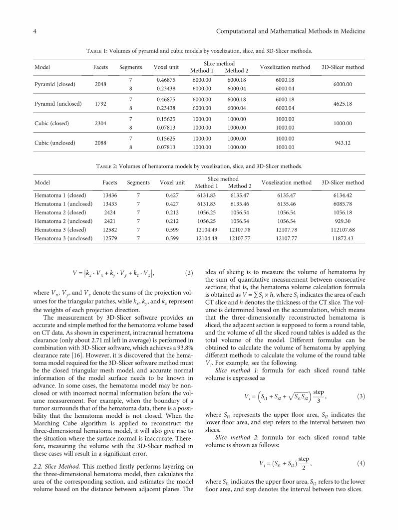
Table 1: Volumes of pyramid and cubic models by voxelization, slice, and 3D-Slicer methods.
Model Facets Segments Voxel unitSlice method
Voxelization method 3D-Slicer methodMethod 1 Method 2
Pyramid (closed) 20487 0.46875 6000.00 6000.18 6000.18
6000.008 0.23438 6000.00 6000.04 6000.04
Pyramid (unclosed) 17927 0.46875 6000.00 6000.18 6000.18
4625.188 0.23438 6000.00 6000.04 6000.04
Cubic (closed) 23047 0.15625 1000.00 1000.00 1000.00
1000.008 0.07813 1000.00 1000.00 1000.00
Cubic (unclosed) 20887 0.15625 1000.00 1000.00 1000.00
943.128 0.07813 1000.00 1000.00 1000.00
Table 2: Volumes of hematoma models by voxelization, slice, and 3D-Slicer methods.
Model Facets Segments Voxel unitSlice method
Voxelization method 3D-Slicer methodMethod 1 Method 2
Hematoma 1 (closed) 13436 7 0.427 6131.83 6135.47 6135.47 6134.42
Hematoma 1 (unclosed) 13433 7 0.427 6131.83 6135.46 6135.46 6085.78
Hematoma 2 (closed) 2424 7 0.212 1056.25 1056.54 1056.54 1056.18
Hematoma 2 (unclosed) 2421 7 0.212 1056.25 1056.54 1056.54 929.30
Hematoma 3 (closed) 12582 7 0.599 12104.49 12107.78 12107.78 112107.68
Hematoma 3 (unclosed) 12579 7 0.599 12104.48 12107.77 12107.77 11872.43
4 Computational and Mathematical Methods in Medicine

From the aforementioned volume calculation formulas, itcan be known that the calculation of the hematoma volume isrelated to the slice thickness (slice interval). A small thicknesscan improve accuracy, but this incurs more computationcosts. Conversely, a large thickness reduces computationcosts, but this causes accuracy to be compromised. Therefore,how to identify the appropriate slice thickness (interval) is amajor problem facing the use of the slice method.
2.3. Voxelization Method. Voxelization provides a modelingmethod that approximates the geometric shape of a three-dimensional model by using spatial voxel units. These spatialvoxels show similarity to pixels in a two-dimensional imageand can be regarded as the expansion from a two-dimensional square area to a three-dimensional cube unit.
The realization of the voxelization method for the volumemeasurement involves two aspects. The octree operation isfirstly implemented, and then, the calculation of boundaryvoxel volume is optimized. The major details are as follows:
(1) Implementation of the Octree Operation. (a) Thebounding box of the models is computed. (b) Theoctree is subdivided, the voxel with no intersectionwith the model mesh as a leaf voxel is marked, andthe nonleaf voxel is subdivided again. (c) All leaf vox-els are determined as either inside or outside themodel. (d) The volume is defined as the sum of thevolume of all inside leaf voxels and boundary voxels(i.e., the lowest nonleaf voxels).
(2) Optimization of the Boundary Voxel Volume. Thevolume of the boundary voxel (the lowest nonleafvoxel) can be calculated using the slice method.
According to the voxelization method, spatial voxel unitsare required to approximate the three-dimensional model,and the computational complexity is higher compared tothe 3D-Slicer software method (projection method) and theslice method. The computational accuracy of the voxelizationmethod is determined by the size of the voxel unit and the
45
40
35
30
Hem
atom
a vol
umes
by
3D-S
licer
met
hod
25
5
20
15
10
45403530
y = x
25Hematoma volumes by slicing method 2
5 201510
(a)
40
35
30
Hem
atom
a vol
umes
of c
lose
d m
odel
sby
3D
-Slic
er m
etho
d
25
5
20
15
10
40353025Hematoma volumes of nonclosed models
by slicing method 2
5 201510
y = x
(b)
Figure 4: Comparisons of slice method 2 for measuring closed and nonclosed hematoma with the 3D-Slicer method.
45
40
35
30
Hem
atom
a vol
umes
by
3D-S
licer
met
hod
25
5
20
15
10
45403530
y = x
25Hematoma volumes by slicing method 1
5 201510
(a)
45
40
35
30
Hem
atom
a vol
umes
of c
lose
d m
odel
by 3
D-S
licer
met
hod
25
5
20
15
10
4540353025Hematoma volumes of nonclosed model
by slicing method 1
5 201510
y = x
(b)
Figure 3: Comparisons of slice method 1 for measuring closed and nonclosed hematoma with the 3D-Slicer method.
5Computational and Mathematical Methods in Medicine

volume calculation of the boundary voxel. When the modelvolume is unknown, the results obtained by the voxelizationmethod can be taken as the reference to compare the accu-racy between the slice method and the 3D-Slicer softwaremethod.
2.4. Comparison of Three Measurement Methods
2.4.1. Standard Models with Known Volume. Firstly, a com-parison is performed between the 3D-Slicer method, the slicemethod, and the voxelization method for the volume mea-surement of standard models, the volumes of which areknown. For the slice method and voxelization method, wefirstly calculate the volume with a large interval and a largevoxel unit, respectively, and then reduce the interval andthe voxel unit by a certain value until the calculated volumeis relatively stable.
(1) Volume Measurement for Quadrangular PyramidModel. It is assumed that the length of a pyramid is l = 30,the width is w = 30, and the height is h = 20. Then, the vol-ume of a pyramid is calculated to be 6000 using the quadran-
gular pyramid volume formula. The model is triangulated toconstruct a nonclosed 3D model and a closed 3D model withdifferent triangular facets, respectively. Then, the 3D-Slicermethod, the slice method, and the voxelization method areapplied to measure the quadrilateral pyramid models, respec-tively. The results are indicated in Table 1.
(2) Volume Measurement for Cube. Suppose the length ofthe cube is a = 10, it can be known intuitively that the volumeis 1000. Similarly, the model is triangulated to obtain a non-closed 3D model and a closed 3D model with different trian-gular facets. Then, the 3D-Slicer method, the slice method,and the voxelization method are employed to measure thevolumes, respectively. The results are presented in Table 1as well.
2.4.2. Nonstandard Models with Unknown Volume. Two 3Dmodels of hematomas stemming from the patients were firstreconstructed using 3D-Slicer software. Besides, the 3D-Slicer method, the slice method, and the voxelization methodare applied to measure the volumes of the nonclosed 3Dmodel and the closed 3D model, respectively. The resultsare shown in Table 2.
2.4.3. Discussion
(1) As for the closed cube model, the 3D-Slicer method iscapable of ensuring accuracy. In comparison with the3D-Slicer method, the results obtained by the slice
Table 3: Mean errors of closed hematoma (grouping by size) byslice and voxelization methods compared with the 3D-Slicermethod.
nSlice method
1Slice method
2Voxelizationmethod
First group 89 0.00125517 0.00220450 0.00082404
Secondgroup
59 0.00405220 0.00067305 0.00067305
Third group 50 0.08751800 0.00175580 0.00175580
35
30
Hem
atom
a vol
umes
of c
lose
d m
odel
sby
3D
-Slic
er m
etho
d
25
20
15
10
353025Hematoma volumes of nonclosed models
by 3D-Slicer method
201510
y = x
Figure 6: Comparisons of the 3D-Slicer method for measuringclosed and nonclosed hematoma.
45
40
35
30
Hem
atom
a vol
umes
by
3D-S
licer
met
hod
25
5
20
15
10
45403530
y = x
25Hematoma volumes by voxelization method
5 201510
(a)
45
40
35
30
Hem
atom
a vol
umes
of c
lose
d m
odel
sby
3D
-Slic
er m
etho
d
25
5
20
15
10
4540353025Hematoma volumes of nonclosed models
by voxelization method
5 201510
y = x
(b)
Figure 5: Comparisons of the voxelization method for measuring closed and nonclosed hematoma with the 3D-Slicer method.
6 Computational and Mathematical Methods in Medicine

method and the voxelization method show theiraccuracy and minor errors
(2) The slice method and the voxelization method areconsistent for either closed or nonclosed models.In addition, the results as obtained by the slicemethod and the voxelization method show similar-ity to the 3D-Slicer method applied for the closedmodel. However, the 3D-Slicer software methodcould result in a significant estimation error forthe nonclosed model
(3) The voxelization method and the slice methodexhibit a low level of sensitivity to the number oftriangular facets, and the volumes are identicalfor the model with different facets. The 3D-Slicermethod shows sensitivity to the closeness of themodel, and a small reduction of the facets will leadto a large error
3. Hematoma Volume Measurement andComparison Analysis
For all patients, the 3D hematoma models were recon-structed using the 3D-Slicer software. Then, volumemeasurements were performed using the 3D-Slicermethod, the slice method, and the voxelization method,respectively.
3.1. Materials and Methods
3.1.1. Patients. In this study, the patients admitted to theAffiliated Hospital of Hangzhou Normal University betweenDecember 2017 and January 2018 with diagnosis of sponta-neous ICH were recruited. A total of 198 consecutive patientswere recruited, including 132 male patients and 66 femalepatients, with the average age of 56:2 ± 28:8. The patientswith multiple sites of ICH were excluded from this study.
Table 4: Mean errors of nonclosed hematoma (grouping by size) by slice, voxelization methods, and 3D-Slicer method, compared with the3D-Slicer method for closed models.
n Slice method 1 Slice method 2 Voxelization method 3D-Slicer method for nonclosed models
First group 89 0.00128517 0.00025000 0.00079449 0.01426753
Second group 59 0.00405797 0.00067915 0.00036763 0.04990034
Third group 50 0.00888480 0.00185180 0.00131520 0.00574640
0.10
0.08
0.06
0.04
0.02
0.00
–0.02
Slicingmethod 1
Mea
sure
men
t err
ors o
ffir
st gr
oup
Voxelizationmethod
⁎
⁎
⁎⁎
0.01
0.0000
–0.01
–0.02
–0.03
–0.04
Slicingmethod 1
Mea
sure
men
t err
ors o
fse
cond
gro
up
Voxelizationmethod
⁎
⁎⁎
Slicingmethod 2
⁎
⁎
0.02
0.0000
–0.06
–0.08
–0.10
–0.12
Slicingmethod 1
Mea
sure
men
t err
ors o
fth
ird g
roup
Voxelizationmethod
⁎
⁎
⁎⁎
⁎
⁎⁎
⁎
Slicingmethod 2
–0.04
–0.02 ⁎
⁎
⁎
Figure 7: Distribution of measurement errors of closed hematoma models by slice method 1, slice method 2, voxelization method, and 3D-Slicer method (grouping by size).
7Computational and Mathematical Methods in Medicine

All cases were included to the standard that the onset to headCT examination time is less than 24 hours.
3.1.2. Imaging. A total of 198 brain computed tomographicimage data sets were acquired according to the hospital PACSsystem with the digital imaging standard in medicine format.
3.1.3. Patient Groups. The patients were split into 3 differentgroups depending on the hematoma size. Group 1 was com-prised of 89 patients with volume < 10ml, group 2 consisted
of 59 patients with volume ranging from 10 to 20ml, andgroup 3 was made up of 50 patients with volume > 20ml.Based on the maximal slice, the shape of the hematoma wasclassed into regular (round to ellipsoid) with smooth margins(76 cases), irregular with frayed margins (85 cases), and mul-tilobular (37 cases).
3.1.4. Statistical Analysis. All of the statistical analyses wereconducted with SPSS Statistics 21 (IBM Corporation, Amer-ica). Moreover, GraphPad Prism was applied to draw charts.The relationship between the hematoma volume and themeasurement method was analyzed by applying the simplelinear correlation. Subsequent to the confirmation of distri-bution, the data were indicated as the mean ± SD, andunpaired t-test or 1-way ANOVA was conducted for com-parison between different methods and groups, while theLSD method was applied to compare the two groups. A valueof P < 0:05 was treated as statistically significant.
3.2. Results. We set the volumes of the closed models mea-sured by the 3D-Slicer method as the standard values. Theslice method (slice methods 1 and 2), voxelization method,and 3D-Slicer software method were compared using theclosed hematoma and nonclosed hematoma models. For
0.2
0.3
0.0000
–0.1
0.1
–0.2
Slicingmethod 1
Slicingmethod 2
Mea
sure
men
t err
ors o
f non
close
dm
odel
s of t
hird
gro
up
Voxelizationmethod
3D-Slicermethod
⁎⁎⁎
⁎
⁎⁎
⁎
⁎
⁎⁎
⁎⁎⁎
0.1
0.2
0.0000
–0.1
–0.2
–0.3
Slicingmethod 1
Slicingmethod 2
Mea
sure
men
t err
ors o
f non
close
dm
odel
s of fi
rst g
roup
Voxelizationmethod
3D-Slicermethod
⁎
⁎
⁎
⁎⁎
0.2
0.0000
–0.6
–0.4
–0.2
–0.8
–1.0
Slicingmethod 1
Slicingmethod 2
Mea
sure
men
t err
ors o
f non
close
dm
odel
s of s
econ
d gr
oup
Voxelizationmethod
3D-Slicermethod
⁎ ⁎
⁎
⁎⁎
Figure 8: Distribution of measurement errors of nonclosed hematoma models by slice method 1, slice method 2, voxelization method, and3D-Slicer method (grouping by size).
Table 5: Mean errors of closed hematoma (grouping by shape) byslice and voxelization methods compared with the 3D-Slicermethod.
nSlice method
1Slice method
2Voxelizationmethod
Regulargroup
76 0.00188145 0.00019487 0.00019487
Irregulargroup
85 0.00601259 0.00155753 0.00046388
Lobulargroup
37 0.00363027 0.00000216 0.00000216
8 Computational and Mathematical Methods in Medicine

different methods, a simple correlation analysis was con-ducted under different models.
Figures 3–6 show the comparison results obtained byslice method 1, slice method 2, voxelization method, and3D-Slicer method for the closed and nonclosed hematomamodels. The results displayed in (a) are those for closedhematoma models. The scatter plots shown in Figures 3–6have demonstrated that the results obtained from slicemethod 1, slice method 2, and voxelization methods are lin-early related to those from the 3D-Slicer method. Moreover,their correlation is close to one. As revealed by the linearcorrelation analysis carried out by SPSS, the correlation coef-ficients between the slice methods 1 and 2, the voxelizationmethod, and the 3D-Slicer method for the closed hematomamodel were r = 1. There were statistically significant differ-ences (t = −5:627, P < 0:01) observed for the results betweenthe slice method, the voxelization method, and the 3D-Slicermethod. From the results in Figures 3–5, we can see that thefigures in (b) are similar to the results in the figures in (a).
That means the slice methods 1 and 2 and voxelizationmethod are stable when the hematomamodel was nonclosed,and the measurement results conform to those of the 3D-Slicer method when the hematomamodel is closed. However,large errors will be caused by applying the 3D-Sclicer methodto the nonclosed hematoma model.
3.3. Analysis. When the patients are split into groups basedon hematoma size, the statistical analyses are shown inTables 3 and 4 and Figures 7 and 8. We can see that the meanerrors of the results obtained by using the slice methods 1 and2 and the voxelization method for closed and nonclosedhematoma measurements are broadly the same. The meanerror of the voxelization method is less significant comparedto the mean error of the slice method. The 3D-Slicer softwaremethod measures the nonclosed hematoma model with a sig-nificantly higher error than the slice method and the voxeli-zation method. Specifically, for the first group, the errorcaused by the 3D-Slicer measurement for the nonclosed
Table 6: Mean errors of nonclosed hematoma (grouping by shape) by slice, voxelization methods, and 3D-Slicer method, compared with the3D-Slicer method for closed models.
n Slice method 1 Slice method 2 Voxelization method 3D-Slicer method for nonclosed models
Regular group 76 0.00193513 0.00024842 0.00013171 0.01995789
Irregular group 85 0.00605059 0.00159624 0.00109388 0.01590706
Lobular group 37 0.00364405 0.00000946 0.00002892 0.02858676
⁎
0.012
0.010
0.008
0.006
0.004
0.000
0.002
–0.02
Slicingmethod 1
Slicingmethod 2
Mea
sure
men
t err
ors o
fre
gula
r gro
up
Voxelizationmethod
⁎
⁎
⁎
⁎
⁎
⁎
5.0E–4
0.0E0
–5.0E–4
–1.0E–3
–1.5E–3
–2.5E–3
–2.0E–3
Slicingmethod 1
Mea
sure
men
t err
ors o
fno
nreg
ular
gro
upVoxelization
method
⁎
⁎
⁎
⁎
Slicingmethod 2
⁎
5.0E–4
0.0E0
–5.0E–4
–1.0E–3
–1.5E–3
Slicingmethod 1
Mea
sure
men
t err
ors o
flo
bula
r gro
up
Voxelizationmethod
⁎⁎
⁎
⁎
⁎⁎
⁎
⁎
Slicingmethod 2
⁎
⁎⁎
Figure 9: Distribution of measurement errors of closed hematoma models by slice method 1, slice method 2, voxelization method, and 3D-Slicer method (grouping by shape).
9Computational and Mathematical Methods in Medicine

model is 18 times that of the voxelization method. For thesecond group, the error by the 3D-Slicer measurement forthe nonclosed model exceeds 100 times that of the voxeliza-tion method.
When the hematoma is classed by shape, the statisticalanalyses are shown in Tables 5 and 6 and Figures 9 and 10.It can be found out that slice methods 1 and 2 and voxeliza-tion methods are unaffected by the shape of the hematoma,and the measurement results obtained by these methods donot cause significant errors due to the irregular shape ofhematoma. The mean errors as measured by slice method 1and voxelization method show the same order of magnitudefor the regular group and the irregular group. Besides, thevoxelization method measures the hematoma of the lobu-lated group with less error, with its order of magnitudereaching 10-6. Compared with the voxelization method, the3D-Slicer method measures the nonclosed hematoma withsignificant errors. The error of the regular group, irregulargroup, and lobular group measured by the 3D-Slicer methodis shown to be 151 times, 15 times, and nearly 1000 times thatof the voxelization method, respectively.
4. Discussion
The accurate measurement of hematoma volume is of clinicalsignificance as hematoma volume has been commonly usedto correlate with treatment strategy, functional outcome,and mortality. It is inevitable for an inaccurately assessedhematoma volume to exert influence on the initial treatmentdecisions, thus leading to an undesirable outcome. Mean-while, hematoma volume plays a crucial role in the prognosisof patients. The measurement of hematoma volume aftercerebral hemorrhage can be taken as a potential indicatorfor prediction, which is of great significance to the clinicaldevelopment of a sensible treatment. There are various formsof cerebral hemorrhage, especially for the presence of irregu-lar hematoma, which makes it necessary to find an accuratemethod to determine the size of the volume based on differ-ent hematoma morphologies.
At present, the widely used methods to measure the vol-ume of hematoma include the 3D-Slicer method and theTada formula method. The Tada formula method is consid-ered to be a rough calculation of hematoma due to its
0.05
0.0000
–0.05
–0.10
–0.15
Slicingmethod 1
Slicingmethod 2
Mea
sure
men
t err
ors o
f non
close
dm
odel
s of l
obul
ar g
roup
Voxelizationmethod
3D-Slicermethod
⁎ ⁎⁎⁎
0.2
0.0000
–0.6
–0.4
–0.2
–0.8
–1.0
Slicingmethod 1
Slicingmethod 2
Mea
sure
men
t err
ors o
f non
close
dm
odel
s of r
egul
ar g
roup
Voxelizationmethod
3D-Slicermethod
⁎ ⁎
⁎
⁎⁎
⁎
0.3
0.0000
–0.1
0.1
0.2
–0.2
–0.3
Slicingmethod 1
Slicingmethod 2
Mea
sure
men
t err
ors o
f non
close
dm
odel
s of n
onre
gula
r gro
up
Voxelizationmethod
3D-Slicermethod
⁎⁎⁎⁎
⁎⁎⁎⁎
⁎
⁎⁎
⁎
⁎
⁎
⁎
⁎⁎⁎
Figure 10: Distribution of measurement errors of nonclosed hematoma models by slice method 1, slice method 2, voxelization method, and3D-Slicer method (grouping by shape).
10 Computational and Mathematical Methods in Medicine

inaccuracy in measuring irregular hematoma [7]. Moreover,the 3D-Slicer method is unaffected by the shape and locationof the hematoma. Due to its real-time efficiency and lowrequirements on the operator, the 3D-Slicer method has beenwidely applied to measure the volume of hematoma.
For precision medicine, the 3D-Slicer method and otherpopular approaches to the volume measurement of hema-toma were studied in this paper. The 3D-Slicer methodcaused a significant error in the measurement of the non-closed hematoma model or the model with wrong surfacenormal information. Nevertheless, the slice method andvoxelization method were unaffected by closeness of thehematoma model nor the model with wrong normal infor-mation. Therefore, they can be treated as effective supple-ment methods of the 3D-Slicer method to measure thevolume of hematoma. The drawbacks shown by slice andvoxelization methods are the slice interval and the divisionof voxel units which affect both efficiency and accuracy. Ifthere are significant errors between the 3D-Slicer methodand the slice method (or the voxelization method), the voxe-lization method (or slice method) can be applied to validatethe accuracy of these methods of measurement.
Data Availability
The data used to support the findings of this study areincluded within the article.
Conflicts of Interest
The authors declare that they have no conflicts of interest.
Acknowledgments
This work was supported by the National Natural ScienceFoundation of China under Grant Nos. 11671009 and11801513 and Zhejiang Provincial Natural Science Founda-tion of China under Grant Nos. LZ19A010002 andLQ18A010008.
References
[1] S. Sacco, C. Marini, D. Toni, L. Olivieri, and A. Carolei, “Inci-dence and 10-year survival of intracerebral hemorrhage in apopulation-based registry,” Stroke, vol. 40, no. 2, pp. 394–399, 2009.
[2] J. P. Broderick, T. G. Brott, J. E. Duldner, T. Tomsick, andG. Huster, “Volume of intracerebral hemorrhage. A powerfuland easy-to-use predictor of 30-day mortality,” Stroke,vol. 24, no. 7, pp. 987–993, 1993.
[3] M. T. C. Poon, A. F. Fonville, and R. al-Shahi Salman, “Long-term prognosis after intracerebral haemorrhage: systematicreview and meta-analysis,” Journal of Neurology, Neurosur-gery, and Psychiatry, vol. 85, no. 6, pp. 660–667, 2014.
[4] S. Morris, R. M. Hunter, A. I. G. Ramsay et al., “Impact of cen-tralising acute stroke services in English metropolitan areas onmortality and length of hospital stay: difference-in-differencesanalysis,” BMJ, vol. 349, no. aug04 4, article g4757, 2014.
[5] J. C. Hemphill III, S. M. Greenberg, C. S. Anderson et al.,“Guidelines for the management of spontaneous intracerebral
hemorrhage: a guideline for healthcare professionals from theAmerican Heart Association/American Stroke Association,”Stroke, vol. 46, no. 7, pp. 2032–2060, 2015.
[6] F. Milletari, “TOMAAT: volumetric medical image analysis asa cloud service,” 2018, http://arxiv.org/abs/1803.06784.
[7] X. Xu, X. Chen, J. Zhang et al., “Comparison of the Tada for-mula with software slicer precise and low-cost method for vol-ume assessment of intracerebral hematoma,” Stroke, vol. 45,no. 11, pp. 3433–3435, 2014.
[8] X. Lu and W. Lu, “Measurement of intracranial hematomausing the improved cubature formula,” Chinese Journal ofForensic Medicine, vol. 26, no. 3, pp. 177–180, 2010.
[9] A. Fedorov, R. Beichel, J. Kalpathy-Cramer et al., “3D Slicer asan image computing platform for the quantitative imagingnetwork,” Magnetic Resonance Imaging, vol. 30, no. 9,pp. 1323–1341, 2012.
[10] Z. Ma, X. Chen, Y. Huang et al., “MR diffusion-weightedimaging-based subcutaneous tumour volumetry in a xeno-grafted nude mouse model using 3D Slicer: an accurate andrepeatable method,” Scientific Reports, vol. 5, no. 1, p. 15653,2015.
[11] G. Z. Cheng, R. San Jose Estepar, E. Folch, J. Onieva,S. Gangadharan, and A. Majid, “Three-dimensional printingand 3D slicer: powerful tools in understanding and treatingstructural lung disease,” Chest, vol. 149, no. 5, pp. 1136–1142, 2016.
[12] D. M. T. Devakumar, D. Devakumar, B. Sasidharan, S. R.Bowen, D. K. Heck, and E. J. J. Samuel, “Hybrid positron emis-sion tomography segmentation of heterogeneous lung tumorsusing 3D Slicer: improved GrowCut algorithm with thresholdinitialization,” Journal of Medical Imaging, vol. 4, no. 1, article011009, 2017.
[13] J. Egger, T. Kapur, A. Fedorov et al., “GBM volumetry usingthe 3D Slicer medical image computing platform,” ScientificReports, vol. 3, no. 1, pp. 1–7, 2013.
[14] D. Petrovic, D. Mihailovic, S. Petrovic et al., “Asymptomaticflow of Rosai-Dorfman disease,” Vojnosanitetski Pregled,vol. 71, no. 8, pp. 780–783, 2014.
[15] https://www.vtk.org/gitweb?p=VTK.git.
[16] G. Q. Xie, W. Shi, S. J. Chen et al., “Application of 3D-slicer inneuroendoscopic surgery for hypertensive intracerebral hem-orrhage (in Chinese),” Chinese Journal of Minimally InvasiveNeurosurgery, vol. 22, no. 3, pp. 109–111, 2017.
11Computational and Mathematical Methods in Medicine