Embed Size (px)
Citation preview
Comparison of Morphological Variation and
Shear Bond Strength Between Conventional Acid
Etchant at Different Etch Times and Self Etching
Primer - An in vitro Study
M. J. Ravindranath R. M. D. C & H, Annamalai University, Annamalai Nagar, Chidambaram – 608002, Tamilnadu, India
Email: [email protected]
Little Mahendra Dept. of Periodontology, R. M. D. C & H, Annamalai University
K. Rajasigamani and Kurinchi Kumaran Dept. of Orthodontics & Dentofacial Orthopedics, R. M. D. C & H, Annamalai University
Abstract—The bonding of orthodontic attachments to acid-
etched enamel is an accepted clinical procedure. Phosphoric
acid is the commonly used acid for etching before bonding.
This in vitro study compared the enamel etch patterns
achieved on the orthodontic bonding area of extracted
premolars treated with 37% wt/wt phosphoric acid applied
for 15, 30seconds and self etching primer. The etch patterns
were viewed with a scanning electron microscope and
assessed. The statistical analysis indicates that the self
etching primer and 37% phosphoric acid at 15 & 30
seconds etched tooth produced variable etching pattern.
Application of 37% phosphoric acid was more effective at
producing a good etch pattern at 30 seconds than 15
seconds. Shear bond strength study shows that the 30
seconds etched tooth surface with 37% wt/wt phosphoric
acid shows relatively higher bond strength when compared
with 15 seconds etched tooth. While comparing with the 30
seconds of 37% wt/wt phosphoric acid and self etching
primer group comparison shows there was no significant
difference in bond strength, and comparison of 15 seconds
of etching with phosphoric acid and self etching shows that
self etching primer etching was effective than the 15 seconds
etching with phosphoric acid.
Index Terms—orthodontic bonding, acid etching, etching
pattern, self-etching primer, shear bond strength.
I. INTRODUCTION
The introduction of the acid etching technique by
Buonocore in 1955, have greatly influenced and
revolutionized orthodontic practice. A key factor in
bonding is the enamel to composite interface.
Manuscript received October 10, 2014; revised December 12, 2014.
For application of orthodontic appliances to a dental
structure surface, the etchants, primers, and adhesives are
typically applied in a step-wise fashion. Often between
such steps, one or more rinsing and drying steps are used.
As a result the application of orthodontic appliances
typically involves multi-step procedures2, 5, 14
.
To simplify orthodontic procedures, it would be
desirable to provide a single composition that
accomplishes both etching and priming self-etching
primer was introduced, for improved bonding of
adhesives to a substrate surface which eliminated the
conventional post-etching rinsing and drying steps and
also helps in prevention of contamination of the etched
surface2, 5, 7
.
II. MATERIALS & METHODS
This study was performed in the Department of
Orthodontics and Dento-Facial Orthopedics,
R.M.D.C&H and in collaboration with Sophisticated
Test and Instrumentation Centre (STIC) Cochin
University, Kochi.
Seventy eight freshly extracted teeth for orthodontic
purpose (maxillary and mandibular premolars) were
collected for the study. The teeth were clinically sound.
Following extraction, residue on the teeth was removed
and washed away with tap water. They were then stored
in normal saline at room temperature to prevent
dehydration and bacterial growth.
A. Materials Used
Maxillary and mandibular premolar stainless steel
brackets (American Orthodontics®).
37% phosphoric acid gel.
Transbond XT light cure adhesive & primer (3M
Unitek).
International Journal of Pharma Medicine and Biological Sciences Vol. 4, No. 1, January 2015
©2015 Int. J. Pharm. Med. Biol. Sci. 11
Transbond TM plus Self Etching Primer (3M
Unitek).
Universal testing machine (Lloyd Universal
testing machine –Model No. L.R. 100K)
Scanning electron microscope (JOEL Model No-
JSM 6390LA)
B. Methodology
The buccal surface of the teeth were cleaned with a
pumice and water with the use of rotary brush in a dental
hand piece, they were rinsed with water for 30 seconds
and dried with oil and water free compressed air for 30
seconds following which they were mounted on acrylic
blocks such that the roots were completely embedded
into the acrylic up to the cemento-enamel junction
leaving the crown exposed.
The teeth were randomly divided into three groups.
Group 1, 2 & 3 respectively. Each group contains 20
teeth.
Group 1 etched with 37% phosphoric acid for 15
seconds.
Group 2 etched with 37% phosphoric acid for 30
seconds.
Group 3 etched with self etching primer.
The blocks were color coded for easy identification.
Acid etching was done on the buccal surface of the teeth
with 37% phosphoric acid gel for 15 & 30 seconds. The
teeth was again washed and dried with oil and water free
compressed air. A thin coat of primer was applied to the
acid-etched enamel.
The self-etch primer which contains both the acid and
the primer, For activation, the 2 components are
squeezed together, and the resulting mix can be applied
directly on the tooth surface etchant placed on the enamel
of 20 teeth for 5 seconds and gently evaporated with oil
and moisture free air for 1-2 seconds, Following the
enamel conditioning the teeth were bonded with
premolar brackets (American Orthodontics®) using
Transbond XT light cure adhesive (3M Unitek) the
excess material was removed using sickle scaler and
cured for 40 seconds using a visible light cure unit.
C. Bond Strength Testing
Bond strength testing was performed with universal
testing machine (Lloyd Universal testing machine –
Model No. L.R. 100K) at a crosshead speed of 1mm/min.
The shear force was applied with chisel shaped rod from
the occlusal side parallel to the bracket surface. The
embedded teeth and brackets were aligned in the testing
apparatus to ensure consistency for the point of force
application and direction of the debonding force for all
samples. The load at failure was recorded in a computer
in terms of Newtons. This was converted into Mega
Pascal as the ratio of debond force to the surface area of
the bracket.
Bond strength MPa = Force (Newton) / Surface area
of the bracket (mm)2
III. SEM ANALYSIS
A. Sample Preparation
Eighteen teeth were used for the analysis and
randomly divided into three groups contains six teeth
each.
Group 1 etched with 37% phosphoric acid for 15
seconds.
Group 2 etched with 37% phosphoric acid for 30
seconds.
Group 3 etched with self etching primer.
The crown part of the premolars were sectioned at the
cemento-enamel junction and they were mounted on the
acrylic blocks the crowns of the premolars were oriented
with their buccal surfaces facing uppermost and the
samples were etched and washed they were prepared for
scanning electron microscopy (SEM) the teeth was gold
sputtered and examined in the SEM microscope at 10kv
and 2000x magnification. Photograph was recorded from
the central region of each etched area with a standardized
orientation technique to ensure uniformity between
specimens the etch pattern was compared using the
following 3-grade scale12
.
Poor etch pattern;
Moderate etch pattern;
Good etch pattern.
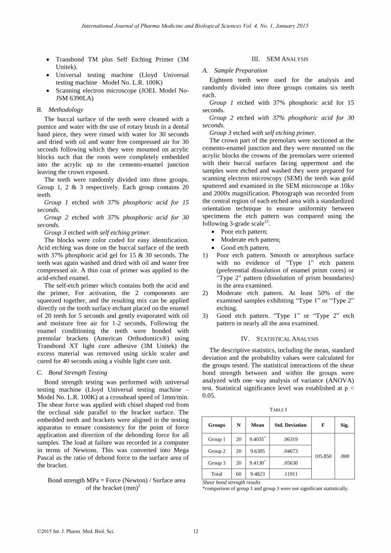
1) Poor etch pattern. Smooth or amorphous surface
with no evidence of "Type 1" etch pattern
(preferential dissolution of enamel prism cores) or
"Type 2" pattern (dissolution of prism boundaries)
in the area examined.
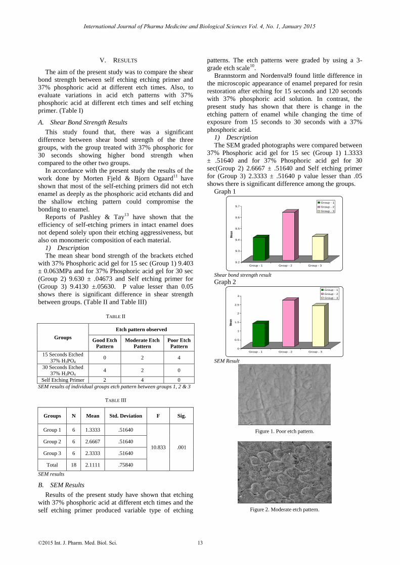
2) Moderate etch pattern. At least 50% of the
examined samples exhibiting “Type 1” or “Type 2”
etching.
3) Good etch pattern. “Type 1” or “Type 2” etch
pattern in nearly all the area examined.
IV. STATISTICAL ANALYSIS
The descriptive statistics, including the mean, standard
deviation and the probability values were calculated for
the groups tested. The statistical interactions of the shear
bond strength between and within the groups were
analyzed with one–way analysis of variance (ANOVA)
test. Statistical significance level was established at p <
0.05.
TABLE I
Groups N Mean Std. Deviation F Sig.
Group 1 20 9.4035* .06319
105.850 .000 Group 2 20 9.6305 .04673
Group 3 20 9.4130* .05630
Total 60 9.4823 .11911
Shear bond strength results
*comparison of group 1 and group 3 were not significant statistically.
International Journal of Pharma Medicine and Biological Sciences Vol. 4, No. 1, January 2015
©2015 Int. J. Pharm. Med. Biol. Sci. 12
V. RESULTS
The aim of the present study was to compare the shear
bond strength between self etching etching primer and
37% phosphoric acid at different etch times. Also, to
evaluate variations in acid etch patterns with 37%
phosphoric acid at different etch times and self etching
primer. (Table I)
A. Shear Bond Strength Results
This study found that, there was a significant
difference between shear bond strength of the three
groups, with the group treated with 37% phosphoric for
30 seconds showing higher bond strength when
compared to the other two groups.
In accordance with the present study the results of the
work done by Morten Fjeld & Bjorn Ogaard11
have
shown that most of the self-etching primers did not etch
enamel as deeply as the phosphoric acid etchants did and
the shallow etching pattern could compromise the
bonding to enamel.
Reports of Pashley & Tay13
have shown that the
efficiency of self-etching primers in intact enamel does
not depend solely upon their etching aggressiveness, but
also on monomeric composition of each material.
1) Description
The mean shear bond strength of the brackets etched
with 37% Phosphoric acid gel for 15 sec (Group 1) 9.403
± 0.063MPa and for 37% Phosphoric acid gel for 30 sec
(Group 2) 9.630 ± .04673 and Self etching primer for
(Group 3) 9.4130 ±.05630. P value lesser than 0.05
shows there is significant difference in shear strength
between groups. (Table II and Table III)
TABLE II
Groups
Etch pattern observed
Good Etch
Pattern
Moderate Etch
Pattern
Poor Etch
Pattern
15 Seconds Etched
37% H3PO4 0 2 4
30 Seconds Etched 37% H3PO4
4 2 0
Self Etching Primer 2 4 0
SEM results of individual groups etch pattern between groups 1, 2 & 3
TABLE III
Groups N Mean Std. Deviation F Sig.
Group 1 6 1.3333 .51640
10.833 .001 Group 2 6 2.6667 .51640
Group 3 6 2.3333 .51640
Total 18 2.1111 .75840
SEM results
B. SEM Results
Results of the present study have shown that etching
with 37% phosphoric acid at different etch times and the
self etching primer produced variable type of etching
patterns. The etch patterns were graded by using a 3-
grade etch scale10
.
Brannstorm and Nordenval9 found little difference in
the microscopic appearance of enamel prepared for resin
restoration after etching for 15 seconds and 120 seconds
with 37% phosphoric acid solution. In contrast, the
present study has shown that there is change in the
etching pattern of enamel while changing the time of
exposure from 15 seconds to 30 seconds with a 37%
phosphoric acid.
1) Description
The SEM graded photographs were compared between
37% Phosphoric acid gel for 15 sec (Group 1) 1.3333
± .51640 and for 37% Phosphoric acid gel for 30
sec(Group 2) 2.6667 ± .51640 and Self etching primer
for (Group 3) 2.3333 ± .51640 p value lesser than .05
shows there is significant difference among the groups.
Graph 1
9.2
9.3
9.4
9.5
9.6
9.7
Me
an
Group - 1 Group - 2 Group - 3
Group - 1
Group - 2
Group - 3
Shear bond strength result
Graph 2
0
0.5
1
1.5
2
2.5
3
Mea
n
Group - 1 Group - 2 Group - 3
Group - 1
Group - 2
Group - 3
SEM Result
Figure 1. Poor etch pattern.
Figure 2. Moderate etch pattern.
International Journal of Pharma Medicine and Biological Sciences Vol. 4, No. 1, January 2015
©2015 Int. J. Pharm. Med. Biol. Sci. 13
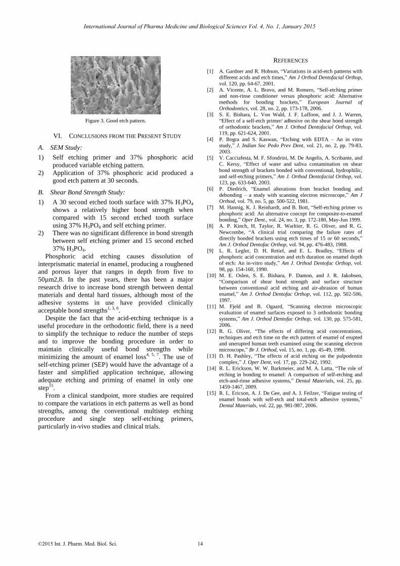
Figure 3. Good etch pattern.
VI. CONCLUSIONS FROM THE PRESENT STUDY
A. SEM Study:
1) Self etching primer and 37% phosphoric acid
produced variable etching pattern.
2) Application of 37% phosphoric acid produced a
good etch pattern at 30 seconds.
B. Shear Bond Strength Study:
1) A 30 second etched tooth surface with 37% H3PO4
shows a relatively higher bond strength when
compared with 15 second etched tooth surface
using 37% H3PO4 and self etching primer.
2) There was no significant difference in bond strength
between self etching primer and 15 second etched
37% H3PO4.
Phosphoric acid etching causes dissolution of
interprismatic material in enamel, producing a roughened
and porous layer that ranges in depth from five to
50µm2,8. In the past years, there has been a major
research drive to increase bond strength between dental
materials and dental hard tissues, although most of the
adhesive systems in use have provided clinically
acceptable bond strengths1, 3, 6
.
Despite the fact that the acid-etching technique is a
useful procedure in the orthodontic field, there is a need
to simplify the technique to reduce the number of steps
and to improve the bonding procedure in order to
maintain clinically useful bond strengths while
minimizing the amount of enamel loss4, 5, 7
. The use of
self-etching primer (SEP) would have the advantage of a
faster and simplified application technique, allowing
adequate etching and priming of enamel in only one
step15
.
From a clinical standpoint, more studies are required
to compare the variations in etch patterns as well as bond
strengths, among the conventional multistep etching
procedure and single step self-etching primers,
particularly in-vivo studies and clinical trials.
REFERENCES
[1] A. Gardner and R. Hobson, “Variations in acid-etch patterns with different acids and etch times,” Am J Orthod Dentofacial Orthop,
vol. 120, pp. 64-67, 2001.
[2] A. Vicente, A. L. Bravo, and M. Romero, “Self-etching primer and non-rinse conditioner versus phosphoric acid: Alternative
methods for bonding brackets,” European Journal of
Orthodontics, vol. 28, no. 2, pp. 173-178, 2006. [3] S. E. Bishara, L. Von Wald, J. F. Laffoon, and J. J. Warren,
“Effect of a self-etch primer/ adhesive on the shear bond strength
of orthodontic brackets,” Am J. Orthod Dentofacial Orthop, vol. 119, pp. 621-624, 2001.
[4] P. Bogra and S. Kaswan, “Etching with EDTA – An in vitro study,” J. Indian Soc Pedo Prev Dent, vol. 21, no. 2, pp. 79-83,
2003.
[5] V. Cacciafesta, M. F. Sfondrini, M. De Angelis, A. Scribante, and C. Kersy, “Effect of water and saliva contamination on shear
bond strength of brackets bonded with conventional, hydrophilic,
and self-etching primers,” Am J. Orthod Dentofacial Orthop, vol. 123, pp. 633-640, 2003.
[6] P. Diedrich, “Enamel alterations from bracket bonding and
debonding – a study with scanning electron microscope,” Am J Orthod, vol. 79, no. 5, pp. 500-522, 1981.
[7] M. Hannig, K. J. Reinhardt, and B. Bott, “Self-etching primer vs
phosphoric acid: An alternative concept for composite-to-enamel bonding,” Oper Dent., vol. 24, no. 3, pp. 172-180, May-Jun 1999.
[8] A. P. Kinch, H. Taylor, R. Warltier, R. G. Oliver, and R. G.
Newcombe, “A clinical trial comparing the failure rates of directly bonded brackets using etch times of 15 or 60 seconds,”
Am J. Orthod Dentofac Orthop, vol. 94, pp. 476-483, 1988.
[9] L. R. Legler, D. H. Retief, and E. L. Bradley, “Effects of phosphoric acid concentration and etch duration on enamel depth
of etch: An in-vitro study,” Am J. Orthod Dentofac Orthop, vol.
98, pp. 154-160, 1990. [10] M. E. Oslen, S. E. Bishara, P. Damon, and J. R. Jakobsen,
“Comparison of shear bond strength and surface structure
between conventional acid etching and air-abrasion of human enamel,” Am J. Orthod Dentofac Orthop, vol. 112, pp. 502-506,
1997.
[11] M. Fjeld and B. Ogaard, “Scanning electron microscopic evaluation of enamel surfaces exposed to 3 orthodontic bonding
systems,” Am J. Orthod Dentofac Orthop, vol. 130, pp. 575-581,
2006. [12] R. G. Oliver, “The effects of differing acid concentrations,
techniques and etch time on the etch pattern of enamel of erupted
and unerupted human teeth examined using the scanning electron microscope,” Br J. Orthod, vol. 15, no. 1, pp. 45-49, 1998.
[13] D. H. Pashley, “The effects of acid etching on the pulpodentin
complex,” J. Oper Dent, vol. 17, pp. 229-242, 1992. [14] R. L. Erickson, W. W. Barkmeier, and M. A. Latta, “The role of
etching in bonding to enamel: A comparison of self-etching and
etch-and-rinse adhesive systems,” Dental Materials, vol. 25, pp. 1459-1467, 2009.
[15] R. L. Ericson, A. J. De Gee, and A. J. Feilzer, “Fatigue testing of
enamel bonds with self-etch and total-etch adhesive systems,” Dental Materials, vol. 22, pp. 981-987, 2006.
International Journal of Pharma Medicine and Biological Sciences Vol. 4, No. 1, January 2015
©2015 Int. J. Pharm. Med. Biol. Sci. 14