Embed Size (px)
Citation preview
Available online at www.sciencedirect.com
Acta Biomaterialia 5 (2009) 693–706
www.elsevier.com/locate/actabiomat
Comparison of the structure and mechanical properties of bovinefemur bone and antler of the North American elk
(Cervus elaphus canadensis)
P.-Y. Chen a, A.G. Stokes b, J. McKittrick a,b,*
a Materials Science and Engineering Program, UC San Diego, La Jolla, CA 92093-0411, USAb Department of Mechanical and Aerospace Engineering, UC San Diego, La Jolla, CA 92093-0411, USA
Received 20 February 2008; received in revised form 3 September 2008; accepted 15 September 2008Available online 2 October 2008
Abstract
Antler and limb bone have a similar microstructure and chemical composition. Both are primarily composed of type I collagen and amineral phase (carbonated apatite), arranged in osteons in compact (cortical bone) sections and a lamellar structure in the cancellous(spongy or trabecular bone) sections. The mineral content is lower in antler bone and it has a core of cancellous bone surrounded bycompact bone running through the main beam and tines. The mineral content is higher in the compact compared with the cancellousbone, although there is no difference in ratios of the mineral elements with calcium. Mechanical tests (bend and compression) on lon-gitudinal and transverse orientations of dry and rehydrated compact bone of North American elk (Cervus elaphus canadensis) antlersare compared with known data on other antlers as well as bovine femora. Both dry and rehydrated bones are highly anisotropic, withthe bending and compressive strength and elastic modulus higher in the longitudinal than in the transverse direction. There is no signif-icant difference between the bend strength and elastic modulus between dry and rehydrated samples tested in the transverse direction.The elastic modulus measured from the bending tests is compared with composite models. The elastic modulus and bend strengthsare lower in the rehydrated condition, but the strain to failure and fracture toughness is much higher compared with dry samples.All antler bone mechanical properties are lower than that of bovine femora. The antler has a much higher fracture toughness comparedwith bovine femora, which correlates with their main function in intraspecific combat as a high impact resistant, energy absorbent mate-rial. A model of compression deformation is proposed, which is based on osteon sliding during shear.� 2008 Acta Materialia Inc. Published by Elsevier Ltd. All rights reserved.
Keywords: Antler; Bone; Mechanical properties; Deformation
1. Introduction
Antlers are the bony protuberances that form on theheads on animals from the family Cervidae (deer) and havebeen in recorded existence for over 25 million years [1]. Elk(wapiti), reindeer (caribou) and moose are included in the40 species of deer that have antlers, and there are an addi-tional four species that are antlerless. Normally, antlers
1742-7061/$ - see front matter � 2008 Acta Materialia Inc. Published by Else
doi:10.1016/j.actbio.2008.09.011
* Corresponding author. Address: Materials Science and EngineeringProgram, UC San Diego, La Jolla, CA 92093-0411, USA. Tel.: +1 858 5345425; fax: +1 858 534 5698.
E-mail address: [email protected] (J. McKittrick).
grow on male deer with the exception of reindeer, whereantlers appear on both males and females. Blood supplyto the growing antler arises from two sources: from thehighly vascularized tissue (the velvet) on the surface ofthe antler and internally through the base of the antler(pedicle) [1], which results in an extraordinary growth rate.The interior supply is important as ligation of the velvetdoes not affect antler growth [1]. Antlers are one of the fast-est growing tissues in the animal kingdom, growing asmuch as 14 kg in 6 months, with a peak growth rate of2–4 cm day�1 [2,3]. Once antlers are fully grown, the velvetis shed leaving the antler bare. Most antlers start growingin the spring (March–April) and reach full maturity in
vier Ltd. All rights reserved.
694 P.-Y. Chen et al. / Acta Biomaterialia 5 (2009) 693–706
the fall at the start of the rut (September–November) [1].Antlers are deciduous and are cast off (dropped) at theend of the rut. The antler is the only mammalian bone thatis capable of regeneration; thus it offers unique insights intobone mineralization and growth.
The function of antlers is in some dispute, and it has beensuggested that antlers are superfluous and will eventuallydisappear [3]. Stonehouse [4] proposed that the primaryfunction of antlers is as a cooling mechanism, due to thepresence of the velvet during the summer months. However,the main consensus is that antlers have two primary func-tions: they serve as visual signs of social rank within bach-elor groups [5–9] and they are used in combat, as both ashield and a weapon [5,8–10]. On the other hand, Lincoln[7] observed that male red deer (Cervus elaphus) withoutantlers (from amputation) seem to suffer no catastrophicconsequences in terms of competition for and defense of aharem. They were found to be quite capable at both tasks.
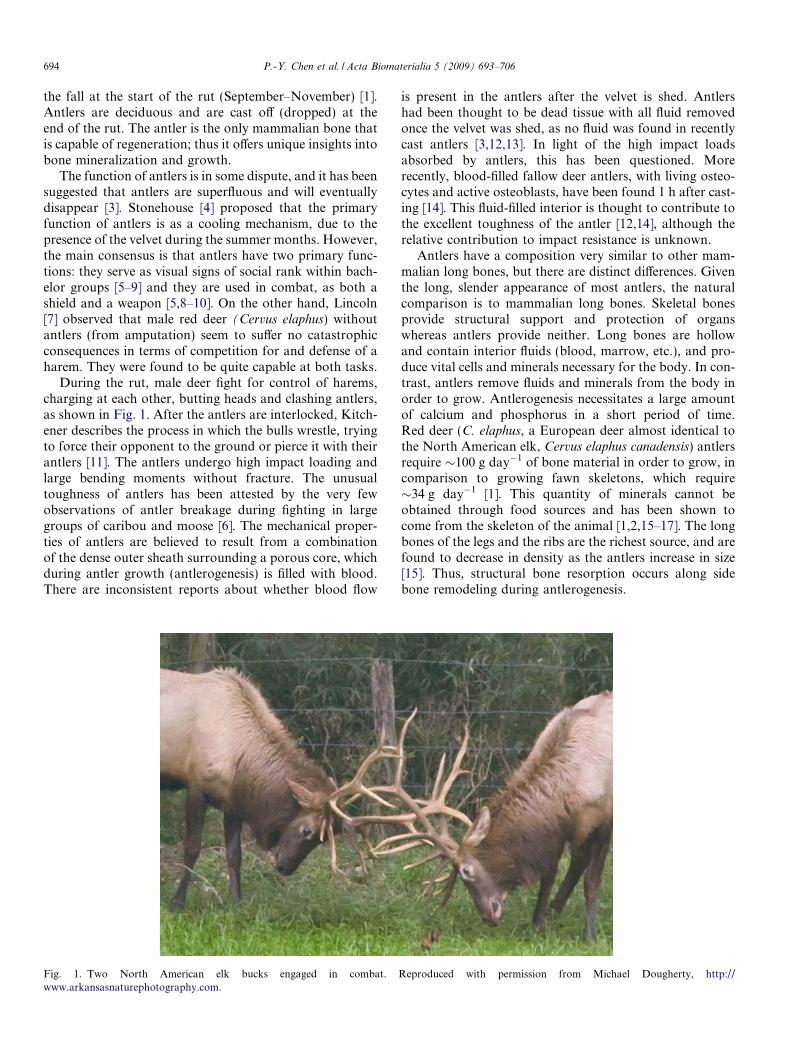
During the rut, male deer fight for control of harems,charging at each other, butting heads and clashing antlers,as shown in Fig. 1. After the antlers are interlocked, Kitch-ener describes the process in which the bulls wrestle, tryingto force their opponent to the ground or pierce it with theirantlers [11]. The antlers undergo high impact loading andlarge bending moments without fracture. The unusualtoughness of antlers has been attested by the very fewobservations of antler breakage during fighting in largegroups of caribou and moose [6]. The mechanical proper-ties of antlers are believed to result from a combinationof the dense outer sheath surrounding a porous core, whichduring antler growth (antlerogenesis) is filled with blood.There are inconsistent reports about whether blood flow
Fig. 1. Two North American elk bucks engaged in combat.www.arkansasnaturephotography.com.
is present in the antlers after the velvet is shed. Antlershad been thought to be dead tissue with all fluid removedonce the velvet was shed, as no fluid was found in recentlycast antlers [3,12,13]. In light of the high impact loadsabsorbed by antlers, this has been questioned. Morerecently, blood-filled fallow deer antlers, with living osteo-cytes and active osteoblasts, have been found 1 h after cast-ing [14]. This fluid-filled interior is thought to contribute tothe excellent toughness of the antler [12,14], although therelative contribution to impact resistance is unknown.
Antlers have a composition very similar to other mam-malian long bones, but there are distinct differences. Giventhe long, slender appearance of most antlers, the naturalcomparison is to mammalian long bones. Skeletal bonesprovide structural support and protection of organswhereas antlers provide neither. Long bones are hollowand contain interior fluids (blood, marrow, etc.), and pro-duce vital cells and minerals necessary for the body. In con-trast, antlers remove fluids and minerals from the body inorder to grow. Antlerogenesis necessitates a large amountof calcium and phosphorus in a short period of time.Red deer (C. elaphus, a European deer almost identical tothe North American elk, Cervus elaphus canadensis) antlersrequire �100 g day�1 of bone material in order to grow, incomparison to growing fawn skeletons, which require�34 g day�1 [1]. This quantity of minerals cannot beobtained through food sources and has been shown tocome from the skeleton of the animal [1,2,15–17]. The longbones of the legs and the ribs are the richest source, and arefound to decrease in density as the antlers increase in size[15]. Thus, structural bone resorption occurs along sidebone remodeling during antlerogenesis.
Reproduced with permission from Michael Dougherty, http://
P.-Y. Chen et al. / Acta Biomaterialia 5 (2009) 693–706 695
2. Background
2.1. Structure
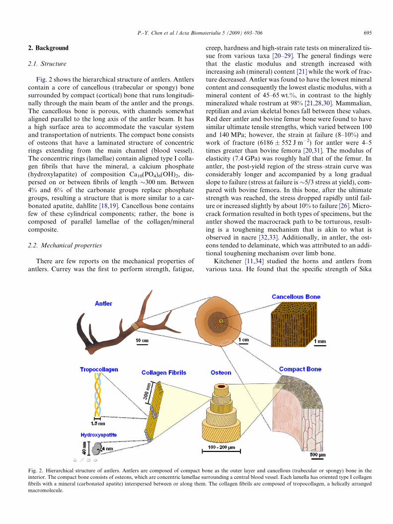
Fig. 2 shows the hierarchical structure of antlers. Antlerscontain a core of cancellous (trabecular or spongy) bonesurrounded by compact (cortical) bone that runs longitudi-nally through the main beam of the antler and the prongs.The cancellous bone is porous, with channels somewhataligned parallel to the long axis of the antler beam. It hasa high surface area to accommodate the vascular systemand transportation of nutrients. The compact bone consistsof osteons that have a laminated structure of concentricrings extending from the main channel (blood vessel).The concentric rings (lamellae) contain aligned type I colla-gen fibrils that have the mineral, a calcium phosphate(hydroxylapatite) of composition Ca10(PO4)6(OH)2, dis-persed on or between fibrils of length �300 nm. Between4% and 6% of the carbonate groups replace phosphategroups, resulting a structure that is more similar to a car-bonated apatite, dahllite [18,19]. Cancellous bone containsfew of these cylindrical components; rather, the bone iscomposed of parallel lamellae of the collagen/mineralcomposite.
2.2. Mechanical properties
There are few reports on the mechanical properties ofantlers. Currey was the first to perform strength, fatigue,
Fig. 2. Hierarchical structure of antlers. Antlers are composed of compact binterior. The compact bone consists of osteons, which are concentric lamellae sufibrils with a mineral (carbonated apatite) interspersed between or along themmacromolecule.
creep, hardness and high-strain rate tests on mineralized tis-sue from various taxa [20–29]. The general findings werethat the elastic modulus and strength increased withincreasing ash (mineral) content [21] while the work of frac-ture decreased. Antler was found to have the lowest mineralcontent and consequently the lowest elastic modulus, with amineral content of 45–65 wt.%, in contrast to the highlymineralized whale rostrum at 98% [21,28,30]. Mammalian,reptilian and avian skeletal bones fall between these values.Red deer antler and bovine femur bone were found to havesimilar ultimate tensile strengths, which varied between 100and 140 MPa; however, the strain at failure (8–10%) andwork of fracture (6186 ± 552 J m�2) for antler were 4–5times greater than bovine femora [20,31]. The modulus ofelasticity (7.4 GPa) was roughly half that of the femur. Inantler, the post-yield region of the stress–strain curve wasconsiderably longer and accompanied by a long gradualslope to failure (stress at failure is�5/3 stress at yield), com-pared with bovine femora. In this bone, after the ultimatestrength was reached, the stress dropped rapidly until fail-ure or increased slightly by about 10% to failure [26]. Micro-crack formation resulted in both types of specimens, but theantler showed the macrocrack path to be torturous, result-ing is a toughening mechanism that is akin to what isobserved in nacre [32,33]. Additionally, in antler, the ost-eons tended to delaminate, which was attributed to an addi-tional toughening mechanism over limb bone.
Kitchener [11,34] studied the horns and antlers fromvarious taxa. He found that the specific strength of Sika
one as the outer layer and cancellous (trabecular or spongy) bone in therrounding a central blood vessel. Each lamella has oriented type I collagen. The collagen fibrils are composed of tropocollagen, a helically arranged
696 P.-Y. Chen et al. / Acta Biomaterialia 5 (2009) 693–706
and hog deer antlers were higher than that of mild steel anddetermined that antlers appear to be structured to resistdeflection [34]. Tropical deer, such as the muntjak, hogand spotted deer, have a lower fraction of cancellous bonethan other deer, resulting in a higher elastic modulus (11–14 GPa) compared with other deer species (5–7 GPa) (seeTable 1) [11].
Rajaram and Ramanathan [13] examined the antlersfrom the spotted deer (Axis axis) that had an ash contentof 55 wt.%. The tensile strength was 188 MPa, the elasticmodulus was 17.1 GPa, the strain to failure was 1.5%and the work of fracture was 13.5 kJ m�3. The high workof fracture and modulus of elasticity is surprising, giventhat the ash content is similar to that of the Currey’s reddeer, which had roughly half the value of the fracturetoughness and elastic modulus. They also observed a dis-tinct plastic region. Fracture surfaces showed that the ost-eons delaminated, corroborating Piekarski’s observation,compared with bovine femora in which whole osteons arepulled out intact [35].
Blob et al. [36–38] studied the elastic moduli of bothmoose (Alces alces) and white-tailed deer (Odocoileus vir-ginianus). They found no correlation of the elastic modulusas a function of the position along the antler, suggestingthat other mechanical properties may not be influencedby the location of the test specimen. Moose antlers had ahigher elastic modulus (11.6 GPa) compared with thewhite-tailed deer (6.8 GPa). The difference was attributedto the different fighting behavior between moose and thewhite-tailed deer as a consequence (or a predicator) ofthe different antler structure. Moose have large palmateantlers, with small prongs. Deer have a long antler beamwith prongs extending from this central beam. As a result,
Table 1Summary of the properties of the compact bone from antlers of various spstrength, ef = strain to failure, WOF = work of fracture, ash = percentage of
Species E (GPa) rb (MPa) ru
Cervus elaphus (red deer) 7.4 1797.2 15
Axis axis (spotted deer) 17.1 1811.6 233
Rangifer tarandus (reindeer) 8.1 956.4
Cervus nippon (Sika deer) 13.7 239Cervus porcinus (hog deer) 12.7 246Alces alces (moose) 11.8Odocoileus virginanus (white-tailed deer) 6.8Cervus elaphus hispanicus (Iberian red deer) 5.3 81.9Muntiacus muntjak (muntjac deer) 11.4Capreolus capreolus (roe deer) 2.2Elaphrus davidianus (Pere David deer) 12Cervus elaphus canadensis (North American elk) 7.6 145Bovine femur 13.5
Work of fracture calculated from a notched 3-point bend sample. The area und[20].
a Corresponds to published volume fractions of 0.3.b 0.287 using 1.35 g cm�3 for collagen and 3.15 g cm�3 for hydroxyapatite.c Values taken from integrating the area under the stress–strain curve, in kJ
fighting moose cannot interlock their antlers and are thussubjected to higher bending moments.
Landete-Castillejos et al. [39] studied antlers from free-range and captive Iberian red deer (Cervus elaphus hispani-
cus). The captive deer antlers had a higher elastic modulus;bend strength and work of fracture; yet the ash and Cacontents were not different. There were small but measur-able differences in Mg, Na, K, Zn, Fe and Si, and the dif-ference in mechanical properties was attributed to this, notthe Ca or ash content. For both antlers, a higher elasticmodulus, bending strength and work of fracture was foundfor specimens taken closer to the pedical compared withones taken from further along the beam, in contrast toBlob et al. [38]. This group had previously suggested thatthere is a chemical composition difference from the pedicalto the end of the beam, resulting from increasingly betternutrition of the animal during antlerogenesis. Table 1 sum-marizes mechanical property data found measured on var-ious antlers, along with a comparison to bovine femur.
In this study, we report on the structure and mechanicalproperties of an antler from the North American elk, C. ela-
phus canadensis. This is the first comprehensive report onthese antlers, providing chemical and microstructural analy-sis as well as mechanical behavior studies, and the first toreport on the compressive properties, transverse bendingproperties and fracture toughness values of antler. Addition-ally, we report dry and rehydrated properties, side by side.Our ultimate aim is to produce bioinspired antler-like,energy absorbent, tough materials. To this end, a thoroughunderstanding of all of the mechanical properties is neces-sary. The compressive properties are important to under-stand deformation of the antler under bending loads,which is how they are loaded during combat. Although the
ecies. E = elastic modulus, rb = bending strength, ruts = ultimate tensileash, q = density
ts (MPa) ef (%) WOF (kJ m�2) Ash (%) q (g cm�3) Ref.
6.2 59.3 1.86 [20]8 11.4 93,000c 48b 0.91 [29]8 1.46 13.5 55 1.86 [13]
9 1.9 [11]5.1 32,000c 50a 0.83 [29]
[23]9 1.9 [34]10 2 [11,34]
[36]61–64 [38]
18.2 61.6 [39]2.99 [22]2.99 [22]
63.3 1.87 [55]13.3 13.9 56.9 1.72 This work
1.7 66.7 2.06 [20]
er the load–deformation curve was divided by twice the cross-sectional area
m�3.
P.-Y. Chen et al. / Acta Biomaterialia 5 (2009) 693–706 697
first loading condition is impact, when the antlers are inter-locked static loads are present. Bending produces both ten-sile and compressive stresses in the antler bone, anddamage can accumulate (microbuckling) under repeatedcompressive loads, inducing stress concentrators. Becausethe mechanical properties of antler are relatively unknown,we have compared our values to bovine femora whereverpossible, as well as to reported properties of antlers fromother species. The elastic modulus of elk antler is discussedand compared with the predicted values from various com-posite models.
3. Materials and methods
The North American elk (Cervus canadensis) antler waspurchased from Into the Wilderness Trading Company(Pinedale, WY). The antler, from a large, mature bull,was shed approximately 1 year before we obtained it fortesting. The length of the antler was 1.05 m and it had sixtines. The thickest cross-sectional diameter at the pedicalwas 7.2 cm. The tines ranged in diameter from 3.5 to6.0 cm. The main beam was cut into sections (�10 cm)using a band saw, as was a bovine femur, which was pur-chased from a local butcher. The slaughter age of the cattlewas approximately 18 months. Samples used for bendingand compressive tests were obtained from the centralregion of the beam portion of the antler. For bending tests,samples from compact region of the antler were dissectedinto rectangles (30 mm � 3 mm � 2 mm) with a diamondsaw and lightly sanded. Twenty-four pieces were cut to pre-pare two equal sets of samples, one in the dry conditionand one in the rehydrated condition. Rehydration wasaccomplished by immersion of the samples in Hank’s bal-anced saline solution (Mediatech Inc., VA) at room tem-perature for 24 h, which were then weighed beforetesting. Each set of samples was further divided into twogroups, six to be tested in the longitudinal direction andsix in the transverse direction. The transverse direction istaken as perpendicular to both the radial and longitudinaldirection. For compressive tests, samples were cut intocubes with an edge of 0.5 cm. Twenty-four cubes were pre-pared for the two testing groups: longitudinal and trans-verse compression. The density was calculated fromweighing and measuring the dimensions of the compressiontest samples. For fracture toughness measurements, 12samples (six in dry and six in rehydrated conditions) weredissected into rectangles (25 mm � 3 mm � 4 mm) with adiamond saw and notches �1 mm in length were madeusing a wire saw. The ash content was obtained from0.6 g powder samples from the antler. Samples were firstdried in a muffle furnace at 105 �C for 4 h and weighed.They were then ashed at 550 �C for 24 h and the ashedweight was measured. The ash content was calculated bydividing the weight of the ashed antler by the weight ofthe dried antler. X-ray diffraction (XRD) was performedon the antler powder by a Rigaku MiniFlexTM II benchtopXRD system (Rigaku Company, Texas, USA).
The bending tests were performed on a laboratory-designed fixture consisting of three knife edges, such thatthe specimen is place on top of two knife edges with a spanof 20 mm while the third knife edge applies stress from thetop. A universal testing machine (Instron 3346 Single Col-umn Testing Systems, Instron, MA, USA) equipped with a500 N load cell was used. The crosshead speed was0.3 mm min�1, which corresponded to a strain rate of1.5 � 10�4 s�1. Compressive tests were conducted on thecubic samples in the longitudinal and transverse directions.A universal testing machine (Instron 3367 Dual ColumnTesting Systems, Instron, MA, USA) equipped with a30 kN load cell was used. Specimens were tested at a0.03 mm min�1 crosshead speed, which translated to astrain rate of 1 � 10�4 s�1, close to that of the bendingtests. All samples were tested to failure. Fracture toughnesswas measured by the ASTM C1421 pre-cracked beammethod using a four-point bend fixture [40]. Bending andfracture toughness tests were performed on dry and rehy-drated antler. Compression testing was performed on dryantler only.
The fracture surface was characterized by using a fieldemission scanning electron microscope (SEM) equippedwith electron dispersive spectroscopy (EDS) (FEI-XL30,FEI Company, OR). The fractured samples were mountedon aluminum sample holders, air dried for 5 min andcoated with 10 nm of gold in a sputter coater. They werethen observed in the secondary electron mode at 20 kVaccelerating voltage. Samples prepared for optical micros-copy were observed under a Zeiss Axio imager equippedwith a CCD camera (Zeiss MicoImaging Inc., Thornwood,New York, USA). Transmission electron microscopy(TEM) was performed on a 200 kV microscope equippedwith a LaB6 electron gun (Technai Sphera, FEI Company,Oregon, USA). The TEM samples were prepared followingthe procedures developed by Weiner and Price [41].
4. Results and discussion
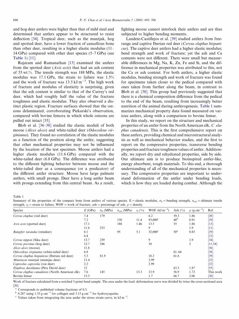
A cross-section, perpendicular to the growth of an ant-ler, is shown in Fig. 3, identifying the four main regionsradiating outward from the center: cancellous bone, a tran-sition zone between cancellous and compact bone, compactbone and subvelvet [14]. Optical micrographs show thesubvelvet to be 100–150 lm thick, which has layered struc-ture. Beneath the subvelvet is the compact bone. Movingfrom the compact bone to the cancellous bone shows anincrease in the size of the porosity, with the pore size rang-ing from 300 lm at the compact/cancellous interface toseveral millimeters at the interior of the cancellous region.
The cross-sectional and longitudinal microstructures incompact antler bone are shown in Fig. 4(a) and (b), respec-tively. In Fig. 4(a), osteons (100–225 lm diameter), Volk-mann canals, vascular channels (15–25 lm diameter) andlacunae spaces (�10 lm diameter) are observed. Themajority of the osteons appear to be aligned along thegrowth direction. Depending on the age of the bone,
Fig. 3. Antler cross-section showing optical micrographs of the (a) subvelvet/compact interface, (b) compact and (c) transition (compact to cancellous)zone, (d) a SEM micrograph of the cancellous bone.
698 P.-Y. Chen et al. / Acta Biomaterialia 5 (2009) 693–706
human osteons range from 200 to 300 lm [42], substan-tially larger than what is found in the antler. This is likelydue to the age difference between the reported values forhuman bone, typically taken from adults, as opposed towhat is found in the relatively young antler. In Fig. 4(b),the Volkmann canals are roughly perpendicular to andhave smaller diameters than the vascular channels. TheVolkmann canals also appear to have a somewhat circularlaminated structure, similar to the longitudinal osteons.
Two types of osteons can be present in bone: primaryand secondary. Primary osteons contain vascular channelssurrounded by concentric bone lamellae. Primary osteonsare generally smaller and do not have a cement line sur-rounding them. Secondary osteons result from boneremodeling, often intersect each other and have a morerounded, uniform shape than primary osteons. Currey
and others have shown that bone with primary osteons isstronger than bone with secondary osteons [18,42,43].Using quantitative backscattered electron imaging andEDS analysis, Skedros et al. [44] recently showed thatcement lines are highly mineralized, in contrast to earlierconclusions that they were poorly mineralized [45–48].The hypermineralized cement lines are thought to play animportant role in enhancing mechanical properties byattenuating the propagation of microcracks [43,49]. Wecould not completely distinguish between the two types inthe micrograph; however, the majority appears to be pri-mary osteons, since they show a somewhat distortedcross-section. Additionally, based on observations in back-scattered electron images, Skedros et al. [49] have pointedout that antlers undergo limited secondary osteon remodel-ing. Although primary osteons do not develop cement lines
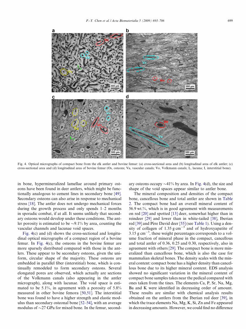
Fig. 4. Optical micrographs of compact bone from the elk antler and bovine femur: (a) cross-sectional area and (b) longitudinal area of elk antler; (c)cross-sectional area and (d) longitudinal area of bovine femur (Os, osteons; Va, vascular canals; Vo, Volkmann canals; L, lacuna; I, interstitial bone).
P.-Y. Chen et al. / Acta Biomaterialia 5 (2009) 693–706 699
in bone, hypermineralized lamellae around primary ost-eons have been found in deer antlers, which might be func-tionally analogous to cement lines in secondary bone [49].Secondary osteons can also arise in response to mechanicalstress [18]. The antler does not undergo mechanical forcesduring the growth process and only spends 1–2 monthsin sporadic combat, if at all. It seems unlikely that second-ary osteons would develop under these conditions. The ant-ler porosity is estimated to be �9.1% by area, counting thevascular channels and lacunae void spaces.
Fig. 4(c) and (d) shows the cross-sectional and longitu-dinal optical micrographs of a compact region of a bovinefemur. In Fig. 4(c), the osteons in the bovine femur aremore sparsely distributed compared with those in the ant-lers. These appear to be secondary osteons, given the uni-form, circular shape of the majority. These osteons areembedded in parallel fiber (interstitial) bone, which is con-tinually remodeled to form secondary osteons. Severalelongated pores are observed, which actually are sectionsof the Volkmann canals (also appearing in the antlermicrograph), along with lacunae. The void space is esti-mated to be 5.1%, in agreement with a porosity of 5.8%measured in other bovine femora [50,51]. The interstitialbone was found to have a higher strength and elastic mod-ulus than secondary osteonal bone [52–54], with an averagemodulus of �27 GPa for mixed bone. In the femur, second-
ary osteons occupy �41% by area. In Fig. 4(d), the size andshape of the void spaces appear similar to antler bone.
The mineral composition and densities of the compactbone, cancellous bone and total antler are shown in Table2. The compact bone had an overall mineral content of56.9 wt.%, which is in good agreement with measurementson red [20] and spotted [13] deer, somewhat higher than inreindeer [29] and lower than in white-tailed [38], Iberianred [39] and Pere David deer [55] (see Table 1). Using a den-sity of collagen of 1.35 g cm�3 and of hydroxyapatite of3.15 g cm�3, these weight percentages corresponds to a vol-ume fraction of mineral phase in the compact, cancellousand total antler of 0.36, 0.25 and 0.30, respectively, also inagreement with others [29]. The compact bone is more min-eralized than cancellous bone, which is also the case formammalian skeletal bones. The density scales with the min-eral content: compact bone has a higher density than cancel-lous bone due to its higher mineral content. EDS analysisshowed no significant variation in the mineral content ofcompact bone samples takes near the pedical compared withones taken from the tines. The elements Ca, P, Sr, Na, Mg,Ba and K were identified in decreasing order of amount.These results are similar with chemical analysis resultsobtained on the antlers from the Iberian red deer [39], inwhich the trace elements Na, Mg, K, Si, Zn and Fe appearedin decreasing amounts. However, we could find no difference
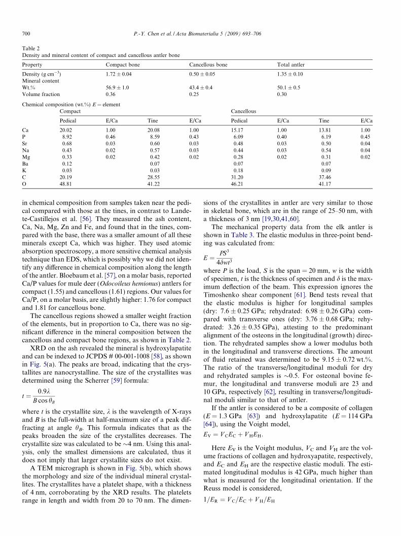
Table 2Density and mineral content of compact and cancellous antler bone
Property Compact bone Cancellous bone Total antler
Density (g cm�3) 1.72 ± 0.04 0.50 ± 0.05 1.35 ± 0.10Mineral contentWt.% 56.9 ± 1.0 43.4 ± 0.4 50.1 ± 0.5Volume fraction 0.36 0.25 0.30
Chemical composition (wt.%) E = elementCompact Cancellous
Pedical E/Ca Tine E/Ca Pedical E/Ca Tine E/Ca
Ca 20.02 1.00 20.08 1.00 15.17 1.00 13.81 1.00P 8.92 0.46 8.59 0.43 6.09 0.40 6.19 0.45Sr 0.68 0.03 0.60 0.03 0.48 0.03 0.50 0.04Na 0.43 0.02 0.57 0.03 0.44 0.03 0.54 0.04Mg 0.33 0.02 0.42 0.02 0.28 0.02 0.31 0.02Ba 0.12 0.07 0.07 0.07K 0.03 0.03 0.18 0.09C 20.19 28.55 31.20 37.46O 48.81 41.22 46.21 41.17
700 P.-Y. Chen et al. / Acta Biomaterialia 5 (2009) 693–706
in chemical composition from samples taken near the pedi-cal compared with those at the tines, in contrast to Lande-te-Castillejos et al. [56]. They measured the ash content,Ca, Na, Mg, Zn and Fe, and found that in the tines, com-pared with the base, there was a smaller amount of all theseminerals except Ca, which was higher. They used atomicabsorption spectroscopy, a more sensitive chemical analysistechnique than EDS, which is possibly why we did not iden-tify any difference in chemical composition along the lengthof the antler. Bloebaum et al. [57], on a molar basis, reportedCa/P values for mule deer (Odocoileus hemionus) antlers forcompact (1.55) and cancellous (1.61) regions. Our values forCa/P, on a molar basis, are slightly higher: 1.76 for compactand 1.81 for cancellous bone.
The cancellous regions showed a smaller weight fractionof the elements, but in proportion to Ca, there was no sig-nificant difference in the mineral composition between thecancellous and compact bone regions, as shown in Table 2.
XRD on the ash revealed the mineral is hydroxylapatiteand can be indexed to JCPDS # 00-001-1008 [58], as shownin Fig. 5(a). The peaks are broad, indicating that the crys-tallites are nanocrystalline. The size of the crystallites wasdetermined using the Scherrer [59] formula:
t ¼ 0:9kB cos hB
where t is the crystallite size, k is the wavelength of X-raysand B is the full-width at half-maximum size of a peak dif-fracting at angle hB. This formula indicates that as thepeaks broaden the size of the crystallites decreases. Thecrystallite size was calculated to be �4 nm. Using this anal-ysis, only the smallest dimensions are calculated, thus itdoes not imply that larger crystallite sizes do not exist.
A TEM micrograph is shown in Fig. 5(b), which showsthe morphology and size of the individual mineral crystal-lites. The crystallites have a platelet shape, with a thicknessof 4 nm, corroborating by the XRD results. The plateletsrange in length and width from 20 to 70 nm. The dimen-
sions of the crystallites in antler are very similar to thosein skeletal bone, which are in the range of 25–50 nm, witha thickness of 3 nm [19,30,41,60].
The mechanical property data from the elk antler isshown in Table 3. The elastic modulus in three-point bend-ing was calculated from:
E ¼ PS3
4dwt3
where P is the load, S is the span = 20 mm, w is the widthof specimen, t is the thickness of specimen and d is the max-imum deflection of the beam. This expression ignores theTimoshenko shear component [61]. Bend tests reveal thatthe elastic modulus is higher for longitudinal samples(dry: 7.6 ± 0.25 GPa; rehydrated: 6.98 ± 0.26 GPa) com-pared with transverse ones (dry: 3.76 ± 0.68 GPa; rehy-drated: 3.26 ± 0.35 GPa), attesting to the predominantalignment of the osteons in the longitudinal (growth) direc-tion. The rehydrated samples show a lower modulus bothin the longitudinal and transverse directions. The amountof fluid retained was determined to be 9.15 ± 0.72 wt.%.The ratio of the transverse/longitudinal moduli for dryand rehydrated samples is �0.5. For osteonal bovine fe-mur, the longitudinal and transverse moduli are 23 and10 GPa, respectively [62], resulting in transverse/longitudi-nal moduli similar to that of antler.
If the antler is considered to be a composite of collagen(E = 1.3 GPa [63]) and hydroxylapatite (E = 114 GPa[64]), using the Voight model,
EV ¼ V CEC þ V HEH:
Here EV is the Voight modulus, VC and VH are the vol-ume fractions of collagen and hydroxyapatite, respectively,and EC and EH are the respective elastic moduli. The esti-mated longitudinal modulus is 42 GPa, much higher thanwhat is measured for the longitudinal orientation. If theReuss model is considered,
1=ER ¼ V C=EC þ V H=EH

Fig. 5. (a) X-ray diffraction patterns from the compact antler bone. Allpeaks correspond to JCPDS file 00-001-1008 for hydroxylapatite [58] and(b) TEM micrograph of the hydroxylapatite crystals.
P.-Y. Chen et al. / Acta Biomaterialia 5 (2009) 693–706 701
the modulus is estimated to be 2 GPa, lower than what ismeasured for the transverse orientations. However, otherlimb bones also fall between these two values, indicatinga mechanical similarity. Others researchers have investi-gated modified composite models to predict the elasticmodulus of bone and have found that the mineral orienta-tion, shape of the mineral [63], presence of lamellae [63],collagen fiber orientation [50,65,66], porosity [50,65,66],mineralization [50,65,66], osteon orientation [67] and frac-tion of secondary osteons [66,68] affect the mechanicalproperties. All of these cause deviations from the Voightand Reuss models.
Considering porosity alone, Bonfield and Clark [63]have expressed a modified Mackenzie [69] equation toaccount for porosity as:
E ¼ Eoð1� 1:9p þ 0:9p2Þwhere E = measured elastic modulus, Eo = elastic modulusof sample containing no porosity and p = porosity volumefraction. Using this expression, the calculated value for Eo
is 8.6 GPa, which is higher, but still not close to the com-posite value of 40 GPa estimated from the Voigt model,indicating that porosity is not the only factor that has influ-ence on the elastic modulus. Carter and Hayes [54] haveproposed an expression for the elastic modulus that is pro-portional to (1 – p)3, and this expression also does notapproximate the measured elastic modulus. Thus, the elas-tic modulus of elk antlers is a complex function of the vari-ables listed above. Another important point is that thetransverse modulus is relatively high when compared withthe longitudinal modulus. Taking the ratio of the Reuss/Voight moduli, a value of 0.05 is calculated, which is an or-der of magnitude lower that what is found for antler andlimb bone. This further demonstrates the inadequacy ofusing these two models to describe the elastic modulus oflongitudinal compact antler bone.
As shown in Table 3, the longitudinal modulus value isin the middle of the range of longitudinal moduli for otherdeer (2.2–17.1 GPa). What is surprising is that the spotteddeer modulus (17.1 GPa [13]) is over twice that of the elk,even though the mineral content varies by <1%. The roedeer (Capreolus capreolus) has a dry low elastic modulusof 2.2 GPa [23] but, since no mineral data were provided,it is difficult to make any statements about this unusuallylow value. The rehydrated longitudinal elastic modulus issimilar to that of red [29] and white-tailed [38] deer andreindeer [22,23].
Fig. 6 shows a representative stress–strain curve for athree-point bend test in the longitudinal and transversedirections for both dry and rehydrated antler. The failureswere graceful and many specimens never completely brokein half. The bending strength was calculated from:
r ¼ 3PS2wt2
The longitudinal strength (dry: 197.3 ± 24.0 MPa; rehy-drated: 145.1 ± 9.0 MPa) was higher than the transversestrength (dry: 66.7 ± 10.7 MPa; rehydrated: 64.9 ± 6.8MPa), also indicative of osteon alignment in the longitudi-nal direction. The hydrated longitudinal bend strength isless than that of other deer species and bovine femora(238 MPa [70]). For the dry longitudinal specimen, thecurve shows a linear elastic region and a plastic region witha gradual increase in stress until fracture, which occurs at astrain of 6.5%. The rehydrated antler shows a much largerstrain to failure (12.3%), an increase of 83% over that ofdry antler. This indicates that a rehydrated antler can with-stand much more deflection during fighting than dry antler,suggesting that antlers are not dead tissue, but are livingorgans during combat. The rehydrated bend strength islower than what is reported for most other deer. The differ-ence between the transverse strengths in the dry and rehy-
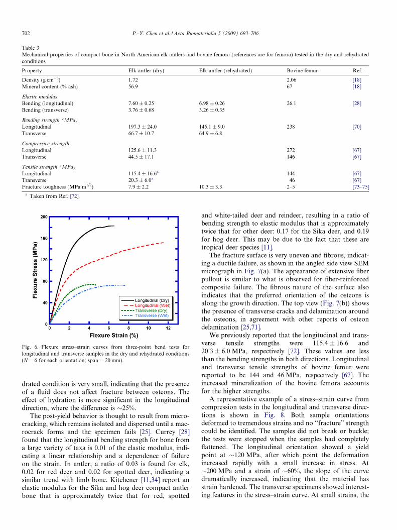
Table 3Mechanical properties of compact bone in North American elk antlers and bovine femora (references are for femora) tested in the dry and rehydratedconditions
Property Elk antler (dry) Elk antler (rehydrated) Bovine femur Ref.
Density (g cm�3) 1.72 2.06 [18]Mineral content (% ash) 56.9 67 [18]
Elastic modulus
Bending (longitudinal) 7.60 ± 0.25 6.98 ± 0.26 26.1 [28]Bending (transverse) 3.76 ± 0.68 3.26 ± 0.35
Bending strength (MPa)
Longitudinal 197.3 ± 24.0 145.1 ± 9.0 238 [70]Transverse 66.7 ± 10.7 64.9 ± 6.8
Compressive strength
Longitudinal 125.6 ± 11.3 272 [67]Transverse 44.5 ± 17.1 146 [67]
Tensile strength (MPa)
Longitudinal 115.4 ± 16.6a 144 [67]Transverse 20.3 ± 6.0a 46 [67]Fracture toughness (MPa�m1/2) 7.9 ± 2.2 10.3 ± 3.3 2–5 [73–75]
a Taken from Ref. [72].
Fig. 6. Flexure stress–strain curves from three-point bend tests forlongitudinal and transverse samples in the dry and rehydrated conditions(N = 6 for each orientation; span = 20 mm).
702 P.-Y. Chen et al. / Acta Biomaterialia 5 (2009) 693–706
drated condition is very small, indicating that the presenceof a fluid does not affect fracture between osteons. Theeffect of hydration is more significant in the longitudinaldirection, where the difference is �25%.
The post-yield behavior is thought to result from micro-cracking, which remains isolated and dispersed until a mac-rocrack forms and the specimen fails [25]. Currey [28]found that the longitudinal bending strength for bone froma large variety of taxa is 0.01 of the elastic modulus, indi-cating a linear relationship and a dependence of failureon the strain. In antler, a ratio of 0.03 is found for elk,0.02 for red deer and 0.02 for spotted deer, indicating asimilar trend with limb bone. Kitchener [11,34] report anelastic modulus for the Sika and hog deer compact antlerbone that is approximately twice that for red, spotted
and white-tailed deer and reindeer, resulting in a ratio ofbending strength to elastic modulus that is approximatelytwice that for other deer: 0.17 for the Sika deer, and 0.19for hog deer. This may be due to the fact that these aretropical deer species [11].
The fracture surface is very uneven and fibrous, indicat-ing a ductile failure, as shown in the angled side view SEMmicrograph in Fig. 7(a). The appearance of extensive fiberpullout is similar to what is observed for fiber-reinforcedcomposite failure. The fibrous nature of the surface alsoindicates that the preferred orientation of the osteons isalong the growth direction. The top view (Fig. 7(b)) showsthe presence of transverse cracks and delamination aroundthe osteons, in agreement with other reports of osteondelamination [25,71].
We previously reported that the longitudinal and trans-verse tensile strengths were 115.4 ± 16.6 and20.3 ± 6.0 MPa, respectively [72]. These values are lessthan the bending strengths in both directions. Longitudinaland transverse tensile strengths of bovine femur werereported to be 144 and 46 MPa, respectively [67]. Theincreased mineralization of the bovine femora accountsfor the higher strengths.
A representative example of a stress–strain curve fromcompression tests in the longitudinal and transverse direc-tions is shown in Fig. 8. Both sample orientationsdeformed to tremendous strains and no ‘‘fracture” strengthcould be identified. The samples did not break or buckle;the tests were stopped when the samples had completelyflattened. The longitudinal orientation showed a yieldpoint at �120 MPa, after which point the deformationincreased rapidly with a small increase in stress. At�200 MPa and a strain of �60%, the slope of the curvedramatically increased, indicating that the material hasstrain hardened. The transverse specimens showed interest-ing features in the stress–strain curve. At small strains, the
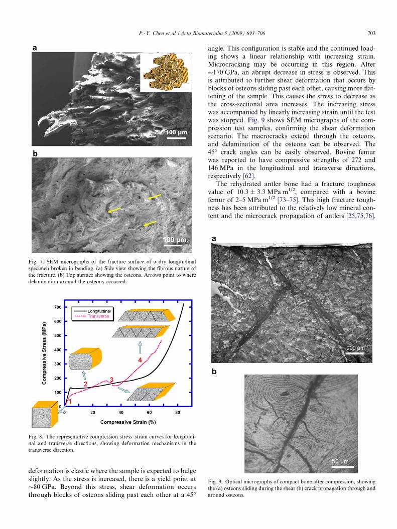
Fig. 7. SEM micrographs of the fracture surface of a dry longitudinalspecimen broken in bending. (a) Side view showing the fibrous nature ofthe fracture. (b) Top surface showing the osteons. Arrows point to wheredelamination around the osteons occurred.
Fig. 8. The representative compression stress–strain curves for longitudi-nal and transverse directions, showing deformation mechanisms in thetransverse direction.
Fig. 9. Optical micrographs of compact bone after compression, showingthe (a) osteons sliding during the shear (b) crack propagation through andaround osteons.
P.-Y. Chen et al. / Acta Biomaterialia 5 (2009) 693–706 703
deformation is elastic where the sample is expected to bulgeslightly. As the stress is increased, there is a yield point at�80 GPa. Beyond this stress, shear deformation occursthrough blocks of osteons sliding past each other at a 45�
angle. This configuration is stable and the continued load-ing shows a linear relationship with increasing strain.Microcracking may be occurring in this region. After�170 GPa, an abrupt decrease in stress is observed. Thisis attributed to further shear deformation that occurs byblocks of osteons sliding past each other, causing more flat-tening of the sample. This causes the stress to decrease asthe cross-sectional area increases. The increasing stresswas accompanied by linearly increasing strain until the testwas stopped. Fig. 9 shows SEM micrographs of the com-pression test samples, confirming the shear deformationscenario. The macrocracks extend through the osteons,and delamination of the osteons can be observed. The45� crack angles can be easily observed. Bovine femurwas reported to have compressive strengths of 272 and146 MPa in the longitudinal and transverse directions,respectively [62].
The rehydrated antler bone had a fracture toughnessvalue of 10.3 ± 3.3 MPa m1/2, compared with a bovinefemur of 2–5 MPa m1/2 [73–75]. This high fracture tough-ness has been attributed to the relatively low mineral con-tent and the microcrack propagation of antlers [25,75,76].
704 P.-Y. Chen et al. / Acta Biomaterialia 5 (2009) 693–706
The dry fracture toughness was 7.9 ± 2.2 MPa m1/2, lowerthan the rehydrated samples but higher than wet bovinefemora. Both limb and antler bone derive their toughnessby forming microcracks during the process of crack prop-agation, yet in the case of antler bone, microcracks aremore isolated and dispersed from each other than thosein limb bone before the formation of the fatal macrocrack.The macrocracks, once formed, follow a much more tortu-ous route, resulting in rougher fracture surfaces comparedwith limb bone [25]. Another possible toughening mecha-nism is the hypermineralized peripheral lamellae in primaryosteons, which may enhance the fracture toughness byattenuating the propagation of microcracks [49].
5. Conclusions
Physical and mechanical properties were measured onan antler from the North American elk (C. elaphus canad-
ensis) and compared with published values for bovine fem-ora. The major findings are:
� Elk antler consists of an interior core of cancellous boneand exterior sheath of compact bone. Osteons, vascularchannels, Volkmann canals, lacunae voids and interlam-inar bone are observed in the compact bone region, whichhas �9.1% porosity accounting for all the void space.� The mineral content in the compact bone regions (56.9%
ash) is higher than the cancellous bone regions (43.4%ash) but lower than bovine femora (67% ash). TheEDS results show no significant difference in the chemi-cal composition between the compact and cancellousbone or along the length of the antler, when normalizedto calcium. The fraction of the mineral phase is similarto that of red and spotted deer, somewhat higher thanthat of reindeer, but lower than that of white-tailed, Ibe-rian red and Pere David deer.� For the first time, the mineral phase in antler was
imaged by TEM. The mineral phase can be indexed tohydroxylapatite and has a plate-like morphology, witha thickness of 4 nm and length and width between 20and 70 nm, similar to the mineral size found in skeletalbone.� The mechanical properties are highly anisotropic: the
longitudinal elastic modulus and bending, tensile andcompressive strengths are higher than in the transversedirection in both dry and rehydrated conditions. Thisis the first report on the transverse properties.� For the first time, dry and rehydrated antler bone data
are presented side by side. The rehydrated longitudinalelastic modulus is comparable to that of red andwhite-tailed deer and reindeer but lower than that ofother deer species. The rehydrated bend strength islower than in most other deer species.� The elastic modulus cannot be modeled by the Voigt or
Reuss models and appears to be a complex function ofmineral content, mineral orientation, osteon orientationand collagen fiber orientation.
� Longitudinal bending failure is characterized by a largestrain to failure and is ductile with a fibrous fracture sur-face. The rehydrated samples have lower bendingstrengths in both directions compared with dry samples;however, the strain to failure is increased by 83%.� The difference between the transverse strengths in the
dry and rehydrated condition is very small, indicatingthat the presence of a fluid does not affect fracturebetween osteons.� Compressive properties are reported for the first time. A
compression failure mechanism is proposed: compres-sive failure in the transverse direction occurred by shearand involved the successive movement of block-like seg-ments along 45� angles.� The fracture toughness of rehydrated antler bone is over
twice that of bovine femora. The dry fracture toughnessis also higher than that of femora. This is the first reporton fracture toughness values on antler bone.
Acknowledgements
We thank Dr. Norm Olson for TEM assistance. Weacknowledge the use of the UCSD Cryo-Electron Micros-copy Facility, which is supported by NIH Grants1S10RR20016 and GM033050 to Dr. Timothy S. Bakerand a gift from the Agouron Institute to UCSD. Dr. Da-mon Toroin and Prof. Paul Price helped with TEM samplepreparation and advice, and Jonathan Tao collected theXRD spectrum. Students Buay Deng, Matthew Chase,York Chang, Steve Rendenez, Kadija Amba and GloriaChukwueke designed and built the three-point bend fixtureand made preliminary measurements on the antlers.Thanks go to Prof. John Skedros (University of Utah)for his helpful comments on the structure of skeletal boneand antler. Prof. Marc Meyers is especially thanked for hisvaluable insights, enthusiasm and support of this project.This research was funded by the National Science Founda-tion, Division of Materials Research, Biomaterials Pro-gram (Grant DMR 0510138).
References
[1] Chapman DI. Antlers–bones of contention. Mamm Rev1975;5:121–72.
[2] Goss RJ. Deer antlers: regeneration, function and evolution. NewYork: Academic Press; 1983.
[3] Modell W. Horns and antlers. Sci Am 1969;220:114–22.[4] Stonehouse B. Thermoregulatory function of growing antlers. Nature
1968;218:870–2.[5] Geist V. The evolution of horn-like organs. Behavior
1966;27:175–214.[6] Henshaw J. Antlers – the unbrittle bones of contention. Nature
1971;231:469.[7] Lincoln GA. The role of antlers in the behavior of red deer. J Exp
Zool 1972;182:233–49.[8] Lincoln GA. The biology of antlers. J Zool Lond 1992;226:517–28.[9] Clutton-Brock TH. The function of antlers. Behavior 1982;79:108–
124.
P.-Y. Chen et al. / Acta Biomaterialia 5 (2009) 693–706 705
[10] Leslie Jr DM, Jenkins KJ. Rutting mortality among male Rooseveltelk. J Mammol 1985;66:163–4.
[11] Kitchener AC. The evolution and mechanical design of horns andantlers. In: Rayner JMV, Wootton RJ, editors. Biomechanics andevolution. Cambridge: Cambridge University Press; 1991.
[12] Chapman DI. Antler structure and function – a hypothesis. JBiomech 1981;14:195–7.
[13] Rajaram A, Ramanathan N. Tensile properties of antler bone. CalcifTissue Int 1982;34:301–5.
[14] Rolf HJ, Enderle A. Hard fallow deer antler: a living bone till antlercasting. Anat Rec 1999;255:69–77.
[15] Meister W. Changes in biological structure of the long bones ofwhite-tailed deer during the growth of antlers. Anat Rec1956;124:709–21.
[16] Harvey PH, Bradbury JW. Sexual selection. In: Krebs JR, DaviesNB, editors. Behavioural ecology: an evolutionaryapproach. Oxford: Blackwell Scientific; 1991. p. 203–33.
[17] Muir PD, Sykes AR, Barrell GK. Calcium metabolism in red deer(Cervus elaphus) offered herbages during antlerogenesis: kinetic andstable balance studies. J Agric Sci Camb 1987;109:357–64.
[18] Currey JD. Bones: structure and mechanics. Princeton, NJ: PrincetonUniversity Press; 2002.
[19] Weiner S, Wagner HD. The material bone: structure–mechanicalfunction relations. Annu Rev Mater Sci 1998;28:271–98.
[20] Currey JD. Mechanical properties of bone tissues with greatlydiffering functions. J Biomech 1979;12:313–9.
[21] Currey JD. Effects of differences in mineralization on the mechanicalproperties of bone. Philos Trans R Soc Lond B 1984;304:509–18.
[22] Currey JD. The evolution of the mechanical properties of amniotebone. J Biomech 1987;20:1035–44.
[23] Currey JD. The effect of porosity and mineral content on the Young’smodulus of elasticity of compact bone. J Biomech 1988;21:131–9.
[24] Currey JD. The mechanical consequences of variation in the mineralcontent of bone. J Biomech 1969;2:1–11.
[25] Zioupos P, Currey JD, Sedman AJ. An examination of themicromechanics of failure of bone and antler by acoustic emissiontests and laser scanning confocal microscopy. Med Eng Phys1994;16:203–12.
[26] Currey JD. Strain rate dependence of the mechanical properties ofreindeer antler and the cumulative damage model of bone fracture. JBiomech 1989;22:469–75.
[27] Zioupos P, Wang XT, Currey JD. Experimental and theoreticalquantification of the development of damage in fatigue tests of boneand antler. J Biomech 1996;29:989–1002.
[28] Currey JD. The design of mineralized hard tissues for theirmechanical functions. J Exp Biol 1999;202:3285–94.
[29] Currey JD. Physical characteristics affecting the tensile failureproperties of compact bone. J Biomech 1990;23:837–44.
[30] Rho JY, Kuhn-Spearing L, Zioupos P. Mechanical properties and thehierarchical structure of bone. Med Eng Phys 1998;20:92–103.
[31] Currey JD. The mechanical adaptations of bones. Princeton,NJ: Princeton University Press; 1984.
[32] Lin AYM, Meyers MA, Vecchio KS. Mechanical properties andstructure of Strombus gigas, Tridacna gigas and Haliotis rufescens seashells: a comparative study. Mater Sci Eng C 2006;26:1380–9.
[33] Jackson AP, Vincent JFV, Turner RM. The mechanical design ofnacre. Proc R Soc Lond B 1988;234:415–40.
[34] Kitchener AC. Fighting and the mechanical design of horns andantlers. In: Domenici P, Blake RW, editors. Biomechanics in AnimalBehaviour. Oxford: BIOS Scientific Publishers; 2000.
[35] Piekarski KR. Fracture in bone. In: Fracture, vol. I. Waterloo,Canada: International Conference on Fracture; 1977. p. 607–42.
[36] Blob RW, Snelgrove JM. Antler stiffness in moose (Alces alces):correlated evolution of bone function and material properties? JMorphol 2006;267:1075–86.
[37] Snelgrove JM, Blob RW. Antler stiffness in moose: patterns ofvariation in the material properties of antler bone. Integr Comp Biol2003;43:1026.
[38] Blob RW, LaBarbera M. Correlates of variation in deer antlerstiffness: age, mineral content, intra-antler location, habitat andphylogeny. Biol J Linn Soc 2001;74:113–20.
[39] Landete-Castillejos T, Currey JD, Estevez JA, Gaspar-Lopez E,Garcia A, Gallego L. Influence of physiological effort of growth andchemical composition on antler bone mechanical properties. Bone2007;41:794–803.
[40] American Society for Testing and Materials. ASTM C1421-01b:Standard test methods for determination of fracture toughness ofadvanced ceramics at ambient temperature. In: Annual Book ofASTM, vol. 15.01. West Conshohocken, PA: ASTM; 2006. p. 626–59.
[41] Weiner S, Price PA. Disaggregation of bone into crystals. CalcifTissue Int 1986;39:365–75.
[42] Martin RB, Burr DB. Skeletal tissue mechanics. New York: Springer-Verlag; 1998.
[43] Saha S, Hayes WC. Relations between tensile impact properties andmicrostructure of compact bone. Calcif Tissue Res 1977;24:65–72.
[44] Skedros JG, Holmes JI, Vajda EG, Bloebaum RD. Cement lines ofsecondary osteons in human bone are not mineral-deficient: new datain a historical perspective. Anat Rec A 2006;286:781–803.
[45] Lakes RS, Saha S. Cement line motion in bone. Science1979;204:501–3.
[46] Frasca P, Harper R, Katz JL. Strain and frequency dependence ofshear storage modulus for human single osteons and cortical bonemicrosamples: size and hydration effects. J Biomech 1981;14:679–90.
[47] Burr DB, Schaffler MB, Frederickson RG. Composition of thecement line and its possible mechanical role as a local interface inhuman compact bone. J Biomech 1988;21:939–45.
[48] Schaffler MB, Burr DB, Frederickson RG. Morphology of theosteonal cement line in human bone. Anat Rec 1987;217:223–8.
[49] Skedros JG, Durand P, Bloebaum RD. Hypermineralized peripherallamellae in primary osteons of deer antler: potential analogues ofcement lines in mammalian secondary bone. J Bone Miner Res1995;10(Suppl 1):441.
[50] Martin RB, Boardman DL. The effects of collagen fiber orientation,porosity, density and mineralization on bovine cortical bone bendingproperties. J Biomech 1993;26:1047–54.
[51] Kim JH, Niinomi M, Akahori T. Influence of bone structure onmechanical properties of bovine and swine compact bones. Mater SciForum 2005;475–479:2407–10.
[52] Reilly DT, Burstein AH, Frankel VH. The elastic modulus for bone. JBiomech 1974;7:271–5.
[53] Bonfield W, Datta PK. Young’s modulus of compact bone. JBiomech 1974;7:147–9.
[54] Carter DR, Hayes WC. The compressive behavior of bone as a two-phase porous structure. J Bone Joint Surg Am 1977;59:954–62.
[55] H-Ch Spatz, O’Leary EJ, Vincent JFV. Young’s moduli and shearmoduli in cortical bone. Proc R Soc Lond 1996;263:287–94.
[56] Landete-Castillejos T, Estevez JA, Martinez A, Ceacero F, Garcia A,Gallego L. Does chemical composition of antler bone reflect thephysiological effort made to grow it? Bone 2007;40:1095–102.
[57] Bloebaum RD, Skedros JG, Vajda EG, Bachus KN, Constantz BR.Determining mineral content variations in bone using backscatteredelectron imaging. Bone 1997;20:485–90.
[58] Joint Committee on Powder Diffraction Standards, Associateship atthe National Bureau of Standards. Powder diffraction data: no. 00-001-1008. Swarthmore, PA: JCPDS; 1976.
[59] Cullity BD. Elements of X-ray diffraction. 2nd ed. Reading,MA: Addison-Wesley; 1978.
[60] Ziv V, Weiner S. Bone crystal sizes: a comparison of transmissionelectron microscopic and X-ray diffraction line width broadeningtechniques. Connect Tissue Res 1994;30:165–75.
[61] Gere JM, Timoshenko SP. Mechanics of materials. CA: Brooks andCole Publishing; 1984.
[62] Martin RB, Burr DR. Structure, function, and adaptation of compactbone. New York: Raven Press; 1989, p. 72.
706 P.-Y. Chen et al. / Acta Biomaterialia 5 (2009) 693–706
[63] Bonfield W, Clark EA. Elastic deformation of compact bone. J MaterSci 1973;8:1590–4.
[64] Hard KatzJL. Hard tissue as a composite material I. Bounds on theelastic behavior. J Biomech 1971;4:244–473.
[65] Currey JD. Comparative mechanical properties and histology ofbone. Am Zool 1984;24:5–12.
[66] Martin RB. Determinants of the mechanical properties of bone. JBiomech 1991;24:79–88.
[67] Reilly DT, Burstein AH. The elastic and ultimate properties ofcompact bone and tissue. J Biomech 1975;8:393–405.
[68] Wagner HD, Weiner S. On the relationship between the micro-structure of bone and its mechanical stiffness. J Biomech 1992;11:1311–20.
[69] Mackenzie JK. The elastic constants of a solid containing sphericalholes. Proc Phys Soc Lond B 1950;63:2–11.
[70] Reilly DT, Burnstein AH. The mechanical properties of cortical bone.J Bone Joint Surg A 1974;56:1001–22.
[71] Boyce TM, Fyhrie DP, Glotkowski MC, Radin EL, Schaffler MB.Damage type and strain mode associations in human compact bonebending fatigue. J Ortho Res 1998;16:322–9.
[72] Chen P-Y, Lin AYM, Lin Y-S, Seki Y, Stokes AG, Peyras J, et al.Structure and mechanical properties of selected biomaterials. J MechBehav Biomed Mater 2008;1:208–26.
[73] Wright TM, Hayes WC. Fracture mechanics parameters for compactbone – effect of density and specimen thickness. J Biomech1977;10:419–30.
[74] Bonfield W, Grynpas MD, Young RJ. Crack velocity and the fractureof bone. J Biomech 1978;11:473–9.
[75] Lucksanambool P, Higgs WAJ, Higgs RJED, Swain MW. Fracturetoughness of bovine bone: influence of orientation and storage media.Biomater 2001;22:3127–32.
[76] Vashishth D. Rising crack-growth-resistance behavior in corticalbone: implications for toughness measurements. J Biomech2004;37:943–6.