Embed Size (px)
Citation preview

1
Comparison of the Success and Survival Rates of Implant Supported Crowns and
Endodontically Treated Teeth – An Updated Systematic Review
Matthew G. Healy, DDS
2
1 PREAMBLE
In recent years, technical advances in endodontics have allowed the seriously
compromised tooth—which would in the past have been extracted—to be treated
and restored to full function. In addition, single tooth implants have emerged as a
treatment for the replacement of these seriously compromised teeth. In the
individual case of a compromised tooth, which of these treatments should be
used? The decision should, as in all evidence-based practice, be based on the
best available evidence. The paramount concern is prognosis. A comparison of
the long-term outcomes of both procedures was made five years ago by
Torabinejad, Anderson and colleagues, published as a systematic review[1].
Unfortunately, the evidence available at that time yielded inconclusive results and
the need for more clinical trials that were prospective in nature, long-term, and of
large sample size. Since publication of the original review in 2007, more data
has been added to the knowledge base such that it might be possible to conduct
a more definitive comparison of implant and endodontic outcomes.

3
2 AIM
To extend a systematic review initially conducted by Torabinejad, Anderson, and
colleagues in 2007 and determine whether sufficient evidence has been added to
the literature base since 2006 to more completely answer:
1) In patients with periodontally sound teeth that have pulpal and/or
periradicular pathosis, does initial nonsurgical endodontic therapy (RCT)
result in a more beneficial or more harmful clinical, biological,
psychosocial, and/or economic outcome as compared to extraction without
replacement?
2) Does root canal therapy result in a more beneficial or more harmful
outcome compared to extraction and replacement of the missing tooth with
a fixed partial denture (FPD)?
3) Does root canal therapy result in a more beneficial or more harmful
outcome compared to extraction and replacement of the missing tooth with
an implant-supported single crown (ISC)?
4
3 INTRODUCTION
The seriously compromised tooth
In the United States oral disease is pandemic. It is estimated that 53 million
children and adults have untreated decay in their permanent teeth[2]. More than
84% of adults aged 18 or older have active or treated dental caries. A quarter of
all adults report difficulty in chewing, 20% report difficulty in sleeping, and 15%
limit their work and leisure habits—all because of dental pain[3]. Therefore, either
preserving or replacing a compromised tooth will have both functional and
cosmetic benefits that not only encompasses the teeth and gingiva, but also
encapsulates good nutrition, social well-being, and complete systemic health[3].
Teeth in danger of extraction have either necrotic pulps with associated
periapical disease or severely inflamed pulps in which the inflammation cannot
be controlled. For relatively intact teeth with necrotic pulps, irreversible pulpitis,
or apical periodontitis the treatment of choice is non-surgical endodontic therapy
followed by full-coverage restoration[4, 5]. If a tooth is severely broken down or
disease has recurred after endodontic treatment, the treatment plan is more
complex and removal of the tooth becomes one of the treatment choices. If the
tooth is removed it should be replaced with some type of prosthesis[1]. The
discovery of the biological compatibility of titanium alloys has led to the
successful development of implants to replace teeth. When a tooth is severely
compromised a choice has to be made whether to treat (or re-treat) it with
endodontics or to extract it and place an implant. There are limitations to both
5
approaches. A practitioner should be aware of these and balance them in
developing a treatment plan.
Limitations of endodontic therapy
The goal of clinical endodontics—and the mark of its success—is the prevention
and elimination of apical periodontitis[6-8]. While the quality of care provided by
specialist endodontists is very high, there remain some challenges that can limit
success.
One such limitation is the visualization of the field. In order to do well, the
clinician must be able to see well; this includes conspicuous and inconspicuous
canal anatomy alike. Many failures in the past—prior to the introduction of the
dental operating microscope—may have been due to missed canals and
unobserved fracture lines[9]. Indeed, the degree of success of endodontic
therapy has improved significantly since the use of the microscope has become
commonplace[10, 11], although to date there is still no definitive evidence to
vindicate the clinical advantages of the dental operating microscope in non-
surgical root canal therapy[12]. Nonetheless, the use of magnification has been
recommended as the standard of care[13].
Another limitation lies in the ability to adequately disinfect the canal system. The
complete removal of pathogenic bacteria is hampered by incomplete knowledge
of the bacteria present and the agents that would kill them. Teeth that still harbor
bacteria at the time of obturation have a much lower prognosis than teeth that
have been adequately cleaned and prepared[14-17].

6
Many other confounding factors influence the endodontic outcome. Certain
systemic diseases such as uncontrolled diabetes and hypertension may
negatively modulate periapical healing[18]. The treatment of teeth with apical
periodontitis shows a lower success rate than those where disease is limited to
the pulp[19]. Retreatment of failed endodontic therapy also shows lower than
ideal success rates. Restoration of the endodontically treated tooth can be
complex and limit the overall success. Iqbal and colleagues estimated higher
failure rates for inadequately restored teeth (85%) and teeth without full coverage
restorations (a failure rate six times as high as for full coverage restorations)[20].
Limitations of single tooth implant therapy
The limits of implant therapy can broadly be summarized into two categories:
biologic limitations, and technical limitations[21]. Biologic limitations include those
that have to do with systemic conditions, environmental factors, and the
supporting tissues; when they occur, complications of this type tend to be more
serious. Early biologic complications are those that relate to tissue-implant
integration and usually result in implant loss before loading. Late biologic
complications include problems such as peri-implantitis, vertical bone loss, or soft
tissue complications such as pain, swelling, or purulence. Surgical interventions
are required to treat these conditions, and ultimately may or may not result in loss
of the implant.
Many systemic and environmental conditions have been implicated in increased
implant failures; some of these are clear while others are more controversial.
Smoking detrimentally affects the outcome of implant therapy. A systematic
review of prospective and retrospective studies demonstrated lower survival and
success rates for smokers (89.7% and 77.0%) compared to non-smokers (93.3%

7
and 91.0%)[22]. A history of periodontitis will also negatively affect the outcome of
implant therapy. Like smoking, the impact is more significant for implant survival
than implant success[23].
Radiation therapy will influence the outcome of implants in a dose-dependent
manner as well as increase the risk for osteoradionecrosis. This increase in
failure appears to be significant only for radiation does in excess of 55Gy[24].
The risk association with diabetes is less clear. Diabetes mellitus potentially
influences wound healing and increases the susceptibility of the implant site to
infection. However there are few well-controlled human studies that are large
enough to draw conclusions as to whether this significantly plays a role in implant
outcome. No large studies to date have examined the effect of glycemic control
on implant outcome[22, 24].
Recent systematic reviews on the subject of bisphosphonates have shown no
significant influence on implant survival, and no significant increase in risk for
bisphosphonate-related osteonecrosis[25, 26]. However many medical and dental
organizations recommend avoiding elective surgical procedures to oral osseous
structures in patients with a history of intravenous bisphosphonates, citing in
particular the drugs’ extremely long half-lives[27]. Therefore the use of IV-
bisphosphonates remains a contraindication to treatment.
Other biologic limitations are related to the implant site. For instance,
significantly worse outcomes have been shown regarding the failure rates of
implants placed in severely resorbed bone, highly porous bone, or both[28].
The literature generally finds systemic and environmental risk factors more
influential on implant success than on implant survival. Therefore they remain
8
only as relative contraindications that must be considered as a whole on a
patient-by-patient basis.
Mechanical complications include those that relate to the function of the
prosthesis. Examples include implant fracture, abutment or crown fracture, and
loose screws. These tend to be quite frequent occurrences[21] and can range in
severity from nuisances to loss of implants.
Success versus Survival
Superimposed over these limitations on both endodontic and implant therapies
are the difficulties in the assessment of the outcomes. As previously stated, the
aim of endodontic therapy is prevention or elimination of periapical disease. The
most common way of assessing this is by periapical radiography[19], and
researchers have introduced radiographic criteria for doing so[29, 30]. However,
the radiograph is a questionable means of evaluating success or failure of
endodontic treatment. First, it is very difficult to get inter-examiner agreement
from looking at radiographs (less than 50%)[31]. Second, there must be
significant physical bone loss for a lesion to be apparent on a radiograph, and
periapical lesions confined to cancellous bone are usually not detected by
conventional radiograph unless very large[32, 33]. Subtle alterations in angulation
of the film can cause drastic changes in the image captured[19]. For these
reasons, radiographs are not good at depicting the healing—or resolution—of
periapical lesions. A cadaver study demonstrated that a normal, healthy
appearing periapical radiograph would reflect a healthy peri-apex only 74% of the
time[34].
9
Since all periapical lesions, whether large, small, or radiographically undetectable
contain inflammatory cells, clinical exam has been advocated for measurement of
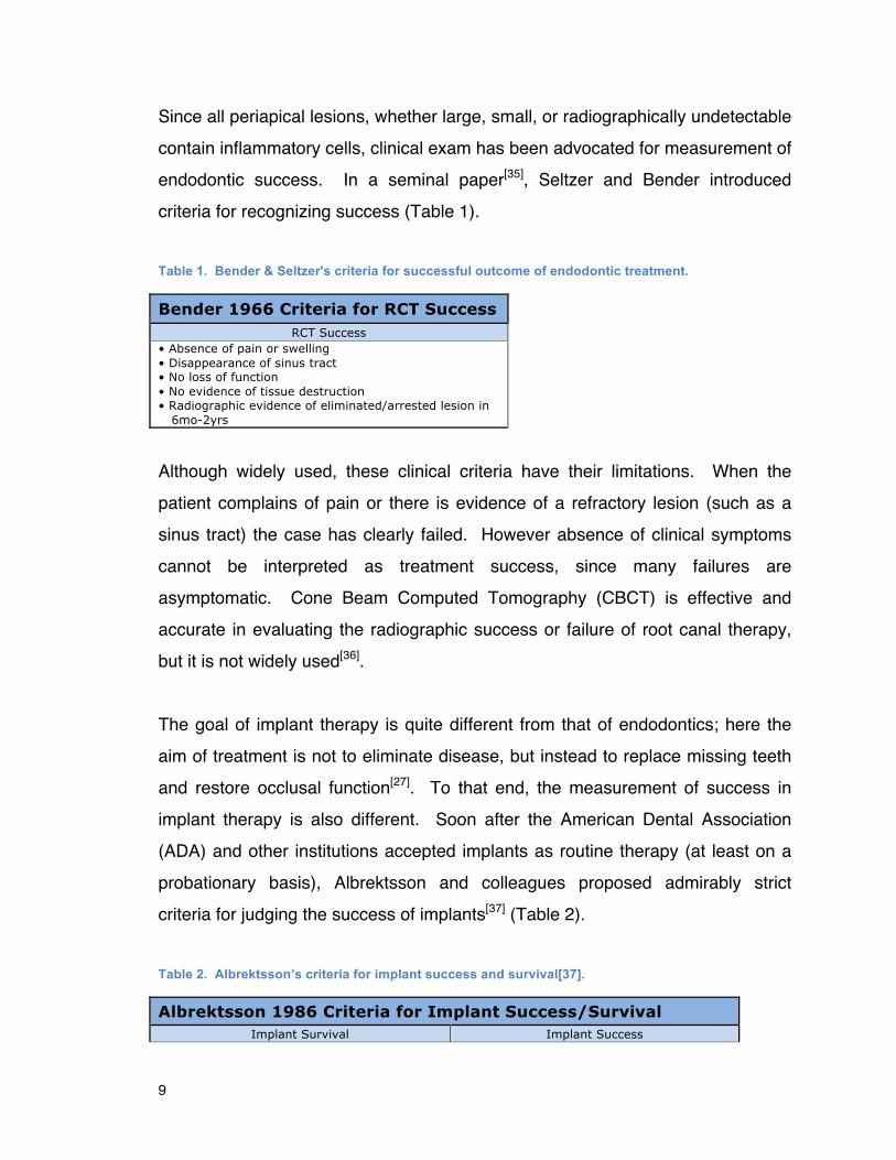
endodontic success. In a seminal paper[35], Seltzer and Bender introduced
criteria for recognizing success (Table 1).
Table 1. Bender & Seltzer's criteria for successful outcome of endodontic treatment.
Bender 1966 Criteria for RCT Success RCT Success
• Absence of pain or swelling • Disappearance of sinus tract • No loss of function • No evidence of tissue destruction • Radiographic evidence of eliminated/arrested lesion in
6mo-2yrs
Although widely used, these clinical criteria have their limitations. When the
patient complains of pain or there is evidence of a refractory lesion (such as a
sinus tract) the case has clearly failed. However absence of clinical symptoms
cannot be interpreted as treatment success, since many failures are
asymptomatic. Cone Beam Computed Tomography (CBCT) is effective and
accurate in evaluating the radiographic success or failure of root canal therapy,
but it is not widely used[36].
The goal of implant therapy is quite different from that of endodontics; here the
aim of treatment is not to eliminate disease, but instead to replace missing teeth
and restore occlusal function[27]. To that end, the measurement of success in
implant therapy is also different. Soon after the American Dental Association
(ADA) and other institutions accepted implants as routine therapy (at least on a
probationary basis), Albrektsson and colleagues proposed admirably strict
criteria for judging the success of implants[37] (Table 2).
Table 2. Albrektsson’s criteria for implant success and survival[37].
Albrektsson 1986 Criteria for Implant Success/Survival Implant Survival Implant Success

10
• Absence of mobility • Absence of peri-implant radiolucency • <0.2mm MBL per year, after first year • Absence of clinical or radiographic signs/symptoms
• All implant survival criteria met for 85% of implants over 5 years, or • for 80% of implants over 10 years
In addition, they placed an emphasis on the need to observe these parameters
over the long term: upwards of five and ten years. The Albrektsson criteria were
developed early in the evolution of dental implants, during a time when
successful osseointegration was the primary concern and not necessarily the
esthetic or functional outcomes. As such, Smith and Zarb revised the success
criteria three years later to include requirements for implant restorability and
patient esthetics[38] (Table 3). Other researchers have proposed alternative
criteria[39-41] (Table 4 through Table 6). Despite these standards being
reasonably stringent, the “success” of single-tooth implants has typically been
judged by their functionality and survival in the mouth[1, 20, 42].
Table 3. Smith and Zarb’s criteria for implant success and survival[38].
Smith & Zarb 1989 Criteria for Implant Success/Survival Implant Survival Implant Success
• Absence of mobility • Absence of peri-implant radiolucency • <0.2mm MBL per year, after first year • Absence of clinical or radiographic signs/symptoms • Implant is restorable • Restoration is esthetic
• All implant survival criteria met for 85% of implants over 5 years, or • for 80% of implants over 10 years
Table 4. Buser’s criteria for a successful outcome of implant therapy[39].
Buser 1990 Criteria for Implant Success Implant Success
• Absence of complaints, pain, or foreign body sensation • No recurrent peri-implant infection or suppuration • Absence of mobility • No continuous peri-implant radiolucency • Implant is restorable

11
Table 5. Glauser’s criteria for a successful outcome of implant therapy[40].
Glauser 2003 Criteria for Implant Success Implant Success
• Absence of a radiolucent zone around the implant • Implant acts as an anchor for the functional prosthesis • Confirmed individual implant stability • Absence of suppuration, pain, or ongoing pathologic processes
Table 6. Misch’s criteria for implant success and survival[41].
Misch 2008 Criteria for Implant Success/Survival Implant Success Satisfactory Survival Compromised Survival Failure
• No pain on function • No mobility • <2mm MBL from initial
surgery • No history of exudate
• No pain on function • No mobility • 2-4mm of MBL •No history of exudate
• Pain on function • No mobility • >4mm MBL • History of exudate
• Pain on function • Mobility • MBL > ½ the length of
the implant • Uncontrolled exudate • Implant not present
Current status of decision making
When treatment planning a patient case, the general dentist has many different
strategies to choose from in his or her arsenal. Viable treatment options for
severely compromised teeth include, but are not limited to, root canal therapy
and restoration (RCT), extraction and replacement with an implant-supported
single crown (ISC), extraction with replacement by a fixed partial denture (FPD),
or extraction with no replacement (Ext)[1]. If left only up to clinician preference,
the prescribed treatment may not be the best treatment. It stands to reason that
some teeth that are extracted could have been successfully treated with
endodontic therapy, and some teeth that receive endodontic therapy probably
should have been extracted[1].
According to the ADA, the clinician must rely not only on personal preference and
past clinical experience, but also on the best available scientific evidence[43]. As
there are many scientific articles published annually[44], decision-making can be
facilitated if the information can be ranked according to quality; this is achieved

12
through evidence-based medicine. The Centre for Evidence-Based Medicine
(based in the United Kingdom) defines evidence-based medicine as “the
conscientious, explicit, and judicious use of current best evidence in making
decisions about the care of individual patients”[45]. Decision-making can be
further enhanced if all of the best evidence can be summarized for the clinician.
Systematic reviews as a tool for prognosis
Clinical trials are useful for asking simple yet clinically relevant questions. They
assemble a group of individuals, assign them to alternative treatments, and then
follow them over time to assess their outcomes. There is a hierarchy to these
trials. For interventions, randomized, controlled clinical trials are the gold
standard and provide the best evidence for judging outcomes. Less rigorous
cohort studies or observational studies may still provide a good level of evidence,
but also may exaggerate the effects of treatment and introduce selection bias[46,
47].
Literature reviews have the potential to sit at the peak of the hierarchy and offer
the best evidence because they assemble multiple trials for analysis. This allows
the researcher or clinician can glean information from a broader pool of
knowledge[48]. Two different methods are widely used to summarize the scientific
literature: the narrative literature review, and the systematic review. Traditional
narrative reviews (also known as ‘topical’ reviews) are typically performed by a
single examiner and tend to explore a broad range of issues on a particular topic.
Because the author decides which studies to include and how to interpret them,
they are likely to be more subjective and more susceptible to bias. Worse,
narrative reviews lag behind and even contradict the best available evidence[49].

13
A systematic review on the other hand attempts to address a very narrow
question in great detail by collecting data from many individual published studies.
Which studies are to be included in the review is carefully and explicitly thought
out before hand, with criteria that are methodological in nature and therefore are
reproducible. As a best practice, systematic reviews are designed and
conducted by a multidisciplinary team of experts[50, 51] (Table 7) to ensure that the
maximum number of potentially valid studies is included. The same team then
assesses the results of the search, analyzes the data, and interprets the findings.
By design, this strategy helps eliminate bias and potential error [52].
Table 7. Multidisciplinary team of experts for a systematic review[50, 51].
Systematic Review Team Principal Examiner • Initiates, selects, and defines the topic Clinical Expert(s) • Partners and collaborators representing each of the relevant disciplines in effort to reduce bias Librarian • Ensures process quality and methodological oversight for the literature searching process Statistician • Ensures process quality and methodological oversight for the analysis and synthesis of data Healthcare Consumer • Provides insight into the priorities for research and acts as an information liaison between consumers and clinicians
If the studies included in the systematic review are sufficiently similar, and if the
resulting data from those studies are sufficiently homogenous, then a meta-
analysis can be performed. Meta-analyses strengthen the level of evidence by
pooling the data from all of the studies, which increases the sample size and
narrows the confidence interval. However, this must be done with great care
under the supervision of a statistician. While it is rarely inappropriate to
undertake a systematic review, it can be inappropriate to apply a meta-analysis.
If the study data is too heterogeneous, then erroneous and invalid conclusions
can be drawn. On the other hand, if too many studies are excluded in the name
of achieving a homogenous study sample, then the results may be too narrow to
offer any useful generalization for the clinician’s decision making[49].

14
Meta-analysis of direct, head-to-head comparative trials remains the gold
standard for summarizing and assessing the outcomes of health care
interventions. However, when the number and types of interventions grow, direct
comparisons of every possible treatment combination may not be possible and
indirect comparisons must instead be made[53]. Since these comparisons have
not been directly tested in controlled, randomized trials, their validity is based on
the assumption that the various interventions are similar[54]. Inferences from such
comparisons must therefore be made with caution.
When carefully designed and executed, systematic reviews—even without a
meta-analysis—are of the highest level of evidence[46]. However there is a
fundamental flaw in the methodology of a systematic review. There is an
assumption that the evidence base being searched is complete; it is in fact not
complete. First, it is well known that studies that do make it into publication often
suffer from publication bias[49]. This is the tendency to publish studies with
positive results over those with less flattering outcomes. Second, studies that get
declined for publication—along with those studies that are never even submitted
for publication—get left out of the evidence base. Finally, systematic reviews that
only consider articles published in English leave behind other articles that are
published in foreign languages. Whenever a medical discipline is attempting to
use a systematic review to more completely and clearly define itself, broadening
the pool of potential articles is of upmost importance. The systematic review
process may be the best the scientific community has, but it is not the best
possible. “Best available evidence” should never be interpreted as “Absolutely
correct”. Systematic reviews are thus only as good as the individual studies
included[20], and therefore the quality and types of trials that are included in them
must be considered. Findings and conclusions from the reviews can—and
should be—questioned. And, they should always be applied with experienced,
clinical expertise and judgment.

15
The Cochrane Collaboration is a not-for-profit international organization whose
mission is ‘Improving healthcare decision-making globally.’ They aim to improve
the evidence base for healthcare interventions by generating and disseminating
high-quality systematic reviews on the effects of healthcare[48]. According to the
Cochrane Handbook, the key characteristics of a systematic review are as
follows[55]:
• A clearly stated set of objectives with pre-defined eligibility criteria for studies;
• An explicit, reproducible methodology;
• A systematic search that attempts to identify all studies that would meet the
eligibility criteria;
• An assessment of the validity of the findings of the included studies, for
example through the assessment of risk of bias; and
• A systematic presentation, and synthesis, of the characteristics and findings
of the included studies.
The quality of reporting in the review is of upmost importance. In the hope of
protecting the integrity of the systematic review, the PRISMA (Preferred
Reporting Items for Systematic reviews and Meta-Analyses) guidelines were
recently released by an international group of researchers and clinicians[56].
These guidelines consist of a 27-item checklist for authors to consider when
formulating and reporting their review (Appendix A).
Summary of the present reviews
As mentioned previously, neither the evidence base nor the systematic reviews
upon which the evidence base is based are perfect. A survey of systematic
reviews published in the last decade suggests that many of the outcomes studies
published on endodontic therapy may be flawed[8]. First, they are based largely
16
on radiographic assessment, which has been shown to be much less consistent
at judging apical status than CBCT[33]. Secondly, they have increasingly been
based on Ørstavik’s PAI, which as a one-size-fits-all approach, and has
questionable validity[8]. And finally, many of these studies have short follow-ups
and poor recall rates (averaging less than 53%)[8].
Recently, two teams of examiners conducted systematic reviews that compared
the outcomes of endodontic therapy to that of implants. Iqbal and colleagues
attempted to examine the outcomes of single tooth implants and endodontically
treated teeth by comparing the survival of endodontically treated teeth to the
survival of implants[20]. To do this, they considered only those endodontic studies
where teeth were restored with full coverage restorations. For all included
studies, they recalculated the survival rate of the endodontically treated teeth
using their own criteria (essentially, teeth were said to be surviving if they were
present in the mouth at the time of the study). They also used their own
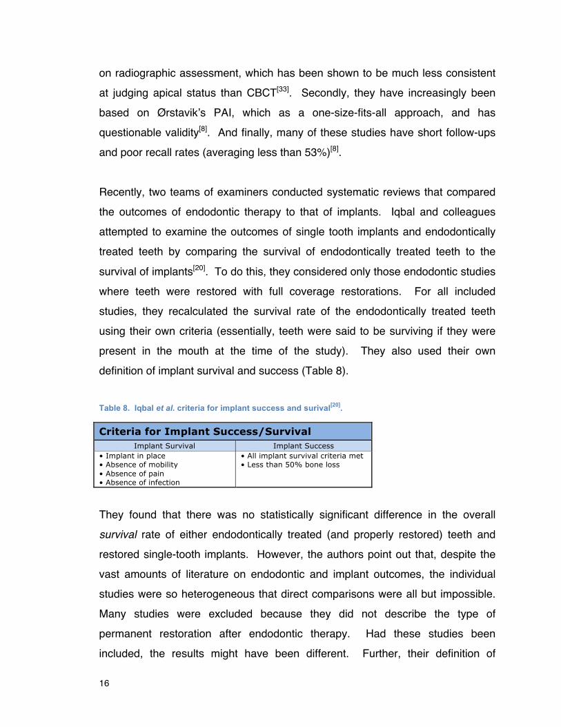
definition of implant survival and success (Table 8).
Table 8. Iqbal et al. criteria for implant success and surival[20].
Criteria for Implant Success/Survival Implant Survival Implant Success
• Implant in place • Absence of mobility • Absence of pain • Absence of infection
• All implant survival criteria met • Less than 50% bone loss
They found that there was no statistically significant difference in the overall
survival rate of either endodontically treated (and properly restored) teeth and
restored single-tooth implants. However, the authors point out that, despite the
vast amounts of literature on endodontic and implant outcomes, the individual
studies were so heterogeneous that direct comparisons were all but impossible.
Many studies were excluded because they did not describe the type of
permanent restoration after endodontic therapy. Had these studies been
included, the results might have been different. Further, their definition of

17
endodontic survival seems extremely lenient, with little similarity to the implant
criteria. Finally, despite having a criteria set for implant success, they did not
examine success as an outcome.
Torabinejad and colleagues also conducted a systematic review on the subject[1].
Their analysis included more endodontic outcome studies because they did not
exclude studies that failed to describe coronal restoration. In addition, they
broadened the comparison of alternative treatment modalities to include
extracted teeth replaced with a fixed partial denture and extracted teeth with no
replacement. They also considered other data that enters the clinician’s decision
tree such as economics and psychosocial effects (i.e. patient satisfaction of
treatment outcome) of each of the treatments. Unlike the Iqbal study,
Torabinejad’s team did not attempt to create their own success/survival criteria or
pool the results, since they found the various criteria in the included studies far
too heterogeneous for meaningful comparisons. The findings of the review
showed no statistical difference in survival between single-tooth implants and
root canal therapy (both 97%) but higher success rates for implants (95%) versus
root canal treated teeth (84%). However the main conclusion from the review
was that the existing literature base was problematic. The outcomes studies
varied widely in study design, sample size, evaluation criteria, and follow-up
period. Complications were incompletely described, and direct comparisons of
the treatments were absent among the included studies. Therefore, only indirect
comparisons were possible. The clinical extrapolations were hardly definitive,
and less than helpful.
18
The need for further review
While these two reviews have been valuable in validating both endodontic and
implant therapy as sound dental treatments, they have not been able to
demonstrate that either treatment carries a superior clinical outcome for patients.
Most importantly, they have shed light on the problems with the existing literature
base. The outcome of endodontic therapy has been shown to vary depending on
the technique used (e.g.: single-visit or multi-visit, type of material used), pre-
operative circumstances (e.g.: presence or absence of apical periodontitis), or
type of post-operative restoration (e.g.: full coverage crown or MOD filling).
While it would make sense for the outcome of each of these scenarios to be
separately evaluated and directly compared to the outcomes of dental implants,
the reality is that the literature base has simply not been robust enough to make
such an analysis possible[20]. Even more heterogeneity exists in the form of
operator experience, sample size, recall rate, and follow-up interval[19].
Furthermore, the shelf life of any given systematic review is limited. New
evidence will emerge, technology will improve, and caveats in established studies
will continue to be found. This can substantially change the conclusions drawn
from the existing evidence base. Shojania and colleagues monitored a cohort of
100 systematic reviews among rapidly changing fields of medicine and found
them to remain clinically relevant an average of 5.5 years. Twenty-three percent
of them required updating just two years after publishing and 15% after one year.
Seven percent of the reviews were obsolete before they were even published[57].
Indeed, since the aforementioned implant/root canal studies have been
published, more literature on the subject has become available. It is possible
that with an updated review of the literature, we may be closer to the answer to
the question: “Should a tooth be retained through root canal treatment and
restoration, or should it be extracted and replaced with a dental implant?”
19
4 HYPOTHESIS
There has been a sufficient addition to the literature base of single-tooth implant
and endodontic therapy outcomes that:
1.) An update of the Torabinejad systematic review is needed.
2.) A more definitive answer will be obtained.
20
5 METHODS
The 2007 Torabinejad systematic review was replicated, encapsulating the
literature that has been published since its release. It was the authors’ intent to
remain true to the methodology of the original review as much as possible.
However, due to constraints in time and manpower, an exact recapitulation was
simply not possible. Any deviations from the original review are clearly indicated.
The same PICO (Patient Population, Intervention, Comparison, and Outcome)
framework was used to formulate the basis of the systematic review. The three
questions to be addressed were:
1) In patients with periodontally sound teeth that have pulpal and/or
periradicular pathosis, does initial nonsurgical endodontic therapy result in
a more beneficial or more harmful clinical, biological, psychosocial, and/or
economic outcome as compared to extraction without replacement?
2) Does root canal therapy result in a more beneficial or more harmful
outcome compared to extraction and replacement of the missing tooth with
a fixed partial denture (FPD)?
3) Does root canal therapy result in a more beneficial or more harmful
outcome compared to extraction and replacement of the missing tooth with
an implant-supported single crown (ISC)?
Inclusion and exclusion criteria were the same as the original systematic review,
except for the dates of publication. Where Torabinejad et al. considered articles
published between 1966 and 2006, this review encompassed the years 2006
through 2011. The inclusion and exclusion criteria are summarized in Table 9.

21
The types of studies considered were comparative or non-comparative,
prospective or retrospective, longitudinal data related to clinical, biological,
psychosocial, or economic outcomes of initial RCT, extraction without
replacement (EXT), extraction and replacement of missing tooth with an FPD, or
extraction and replacement of missing tooth with an ISC. In an effort to limit
publication bias, issuance in a peer-reviewed journal was not considered a
criterion for inclusion. However, like the Torabinejad review, so-called ‘gray’
literature such as proceedings from conferences, meetings and lectures not listed
in MEDLINE, EMBASE, and Cochrane databases were excluded.
Table 9. Inclusion and exclusion criteria for both the original Torabinejad and the current systematic reviews.
Inclusion Criteria Exclusion Criteria • Articles published in English between January
2006 and December 2011
• Adult subjects
• Secondary teeth
• Initial treatments
• Implant-supported single crowns
• Threaded or cylindrical implants regardless of
surface type, placement & loading protocols, or
platform switching
• Minimum of 2-year follow-up
- RCT—from obturation;
- ISC—from implant placement;
- FPD—from cementation
• Treatment as being described as:
- RCT teeth (not roots or canals);
- an individual, non-splinted ISC;
- a short-span FPD (3- or 4-units);
• Minimum of 25 treatments (not patients)
• Studies that failed to meet the inclusion criteria
• 2006 studies previously reported by Torabinejad
et al
• RCTs due to trauma
• Treatment modalities not currently being used
• Moderate or severe periodontal disease
• Multiple-unit implant restorations
• Cantilevered FPDs
• Implant studies on completely edentulous
patients
The same electronic search strategies were employed, as well as the same
methods for hand searching articles. Accordingly, the search strategies
designed by the library expert (PFA) for the three disciplines of ISCs, FPDs, and

22
RCTs were executed in MEDLINE, EMBASE, and COCHRANE and are
presented in Appendix B.
The hand search consisted of reviewing the same relevant endodontic and
prosthodontics journals as from the Torabinejad review. The tables of contents
from every issue published in the last two years of the study (2010 and 2011)
were hand searched; these journals are presented in Table 10. Citation mining
was also performed in any systematic or narrative review uncovered by the
search. Time and labor constraints prevented the hand searching of textbooks.
Table 10. Relevant endodontic and prosthodontics journals hand-searched in both the Torabinejad and current systematic reviews.
Journals Included In The Two-Year Hand Search American Journal of Dentistry Clinical Implant Dentistry & Related Research Clinical Oral Implants Research Dental Materials Dental Traumatology Implant Dentistry International Endodontic Journal International Journal of Oral and Maxillofacial Implants International Journal of Periodontics and Restorative Dentistry International Journal of Prosthodontics Journal of Dentistry Journal of Endodontics Journal of Periodontology The Journal of Prosthetic Dentistry Journal of Oral and Maxillofacial Surgery Journal of Oral Rehabilitation Operative Dentistry Oral Surgery Oral Medicine Oral Pathology Oral Radiology and Endodontics Quintessence International
Studies were qualified for inclusion by two independent reviewers (MGH, GRH)
as follows: First, irrelevant articles were discarded via a title-only review that was
blinded of authors, dates, and publication journal. Next, surviving citations had
their abstracts reviewed for inclusion or disqualification, again with blinding of
authors, dates, and publication journals. For surviving articles that appeared to
meet the inclusion criteria, or for those where there was insufficient data in the
title and abstract to make a clear decision, full text copies were obtained and
reviewed in detail for inclusion or exclusion. All disputes were settled by
23
consensus agreement. A log of excluded articles along with reasons for their
exclusion was kept. If more information was required in order to make a decision
on whether to include or disqualify an article, authors were contacted and
requested to provide it.
Relevant details of methodology and resulting data for each study were recorded
in a data abstraction form (see Appendix C) by the principal investigator (MGH):
• Clinical Setting (private practice, general hospital, teaching hospital, dental school)
• Sample Size (and method, i.e. patients, teeth, roots, units) • Gender • Whether socioeconomic status was stated • Single Center or Multiple Center • Type of Operator (general practitioner, specialist, resident, or dental
student) • Type of Tooth (anterior, premolar, molar) • Assessment Method (radiographic, clinical, questionnaire) • Follow-up Interval • Primary Study Outcome (number and percentage for success, survival,
and failure) • Measure of Effect (confidence intervals, P-values, survival curves,
odds/risk, etc.) • Whether pain was stated • Psychosocial Outcomes (pre- and post-Tx anxiety, post-Tx satisfaction,
pain relief, complications) • Whether economics were addressed • Statistical Analysis Used
The quality of each included article was assessed concerning the type of study,
stated sample size, stated operator experience, stated patient demographics,
complete description of treatment modality, blinded evaluators, stated recall loss,
description of treatment complications, description of outcome evaluation
methods, and appropriateness of statistics. The assessment was performed
using the 17-point system proposed by the Torabinejad team (Table 11).

24
Table 11. 17-point quality rating system for assessing included articles[1].
Quality Rating
Criterion Points
Study Type
Randomized Controlled Clinical Trial
Non-randomized clinical trial
Clinical trial with no controls
Observational cohort study
Case-control study
Case series
Unable to classify
4
5
2
2
1
1
0
Sample Size
Total number of enrolled subjects stated
Predetermined with a power analysis
1
1
Operator experience stated 1
Demographic description included 1
Treatment procedures completely described 1
Evaluator different then operator 1
Complete description of subject loss 1
Treatment Complications
Complications reported as a percent of outcomes
Complication included as failures
Categorized with frequencies
1
1
1
Quality of Clinical Evaluation
Measurements standardized
Statistics described and appropriate
Stratification appropriate
1
1
1
• Total Possible 17
In addition, strict reporting criteria[58] were applied to the included articles:
• Recalls should be scheduled, and it should be clearly stated how many patients appeared for recalls. All dropouts must be accounted for, and if there are no dropouts, this should be stated.
• For studies reporting survival, then criteria for survival should be defined, as well as a frank criterion for failure.
• For studies reporting success, a reference should be provided for the success criteria used.
• For implant studies reporting success, marginal bone levels must be reported, specifying precisely how many implants encountered more bone

25
loss than the referenced criteria allowed. It is not permissible to simply report mean marginal bone loss levels.
• When different materials are used in different patients within the same study, these differences should be clearly described, with numbers of each type specified.
Data Analysis
The data from the included articles were analyzed, and each study was classified
as to success, survival, or both. Life tables obtained from the articles were used
to construct estimates of the survivor function (i.e., the proportion of ISCs, RCTs,
or FPDs that did not fail before a given time) and standard errors for the survivor
function. The survivor function and its standard error were calculated using the
Kaplan-Meier estimator. This approach attempts to estimate survival rates for a
given sample, and its main advantage is that it can take into account censored
patients (patients who withdraw from a study by failing to show for recall) before
the final event (success/failure/presence/absence) is observed[59].
Ninety-five per cent confidence intervals were also calculated using a margin of
error of 1.96 standard errors. In certain cases the success and survival rates had
to be reinterpreted, such as those where stated outcomes criteria were
inappropriately applied, or when only a particular subset of data met the inclusion
criteria. In other cases the rates were not provided at all and were calculated.
Where more information was required to interpret the data and include the study,
authors were contacted for an opportunity to provide it.
In an attempt to summarize what authors of different length studies were
reporting, the results were grouped into short-term (two- to four-year recall),
medium-term (six-year recall or less), and long-term (more than six-year recall)
stratifications for each of the three treatment modalities, and for both of the
outcomes of success and survival. These were the same stratifications used by

26
Torabinejad et al. The rates were pooled within these groups using a simple
inverse-proportion weighting system. For those studies reporting success or
survival rates of 100%, the mean standard error of all included studies was used
in place of a standard error of zero. This was done so as not to give these
studies too small of a standard error.
Finally, in an attempt to compare the success and survival rates among the
different treatment modalities, the results were pooled at yearly time points where
possible.
Results are presented in accordance with the PRISMA statement[56], and are
summarized both in table form and with forest plots. Forest plots were generated
using Forest Plot Viewer version 1.00[60].

27
6 RESULTS
Description of the Existing Literature
The electronic searches yielded an initial 10,412 citations for review. MEDLINE
produced the majority of these yielding 7,945 hits. EMBASE resulted in an
additional 1,727 hits, COCHRANE an additional 648, and the hand searches an
additional 76 citations (Figure 1). By comparison, the Torabinejad review, which
encompassed the years 1966-2006, resulted in an initial 13,099 hits. In other
words, of all the literature available at the close of 2011, 44% of it has been
published in its last five years (Figure 2).

28
Figure 1. Flow chart showing the number of citations screened, disqualified, and included for final
analysis. NB: 33 ISC articles were identified as eligible, but two of these were combined for
reporting purposes.
Figure 2. Comparison of the search results
yielded from this review, as compared to the original review by Torabinejad et al.
Figure 3. Comparison of the number of
articles included from this review, as compared to the original review by
Torabinejad et al.
A total of 568 articles survived the title-only exclusion process, and following
abstract review, 305 studies were considered for full text review. Of these, 61
fulfilled the inclusion/exclusion criteria: 32 ISC, 7 RCT, 5 FPD, and 17

29
psychosocial studies (Figure 1, above). By comparison, 144 articles made
inclusion in the Torabinejad review (Figure 3). No studies examining the effects
of tooth extraction without replacement were identified. Details are provided in
Figure 4 and Figure 5. It is notable that despite there being a 34% overlap[49] in
journal coverage neither EMBASE nor COCHRANE resulted in the admission of
any further studies.
Figure 4. Breakdown of articles included for
analysis in this review (2006-2011).
Figure 5. Breakdown of articles included for
analysis in Torabinejad et al. (1966-2006).
Implant studies clearly out-numbered the RCT and FPD studies. No outcome
studies involving the direct comparison of treatment modalities were identified.
Interventional studies tended to make comparisons among different treatment
protocols or materials used. As with the Torabinejad review, the included studies
were found to be extremely heterogeneous in nature. For all disciplines, there
was variance in terms of sample size, follow-up time, tooth/arch location,
operator experience, surgical/treatment protocols and most importantly the
definition of success and survival criteria used. Many authors who reported
success did not provide a reference for their success criteria, and of those that
did, some did not adhere to said criteria. Of authors that reported survival,
almost none provided a frank criterion for failure. When success and survival
rates were calculated, various methods were used, and most authors did not
account for subjects lost to recall.

30
For implant studies, there was wide variance in terms of the types, surface
coatings and sizes of implants placed, their time of placement post-extraction
and the time allotted for healing prior to loading. Only one-quarter of the studies
described the treatment protocols thoroughly, and only one-quarter of those
specified that an independent, blinded examiner evaluated the outcomes. The
majority (62.5%) of studies were prospective in nature, and of those 40% were
interventional. The other 38.5% of the studies were retrospective. In 40.6% of
the studies oral surgeons rendered treatment, while general dentists (9.4%),
periodontists (6.3%), and specialty residents (3.1%) provided treatment in a
minority of the studies. No studies included dental students as operators placing
implants, although some studies did permit dental students to restore the
implants. Alarmingly, 43.8% of studies did not specify the experience of the
operators. Most studies took place either in private practice (43.8%) or in a
dental school (37.5%).
The majority (71.9%) of implant studies looked solely at implant survival as an
outcome while 9.4% looked at success and 18.8% looked at both. When a
reference was given for outcomes criteria it was usually Albrektsson et al.[37]
(25.0%) or Buser et al.[39] (12.5%). Other referenced criteria were those of
Misch[41], Glauser[40], or Smith & Zarb[38] (Table 2 through Table 6). However,
these criteria seemed to be applied indiscriminately of whether the authors were
reporting success or survival.
Of the seven included RCT studies, there was wide variation in the treatment
protocols, and thorough descriptions of such treatment protocols were lacking in
71.4% of them. Forty-three percent of the studies occurred in private practice
and 57% in dental schools. Providers encompassed endodontists, general
dentists, residents, and dental students. Forty-three percent of the studies were
retrospective in nature, 57% prospective. There was one interventional trial.
31
Only two of the seven studies recorded RCT success as an outcome, and only
two of them had a blinded, independent evaluator.
The five FPD studies all took place in a dental school, and were all prospective
observational studies. Providers again ranged in level of specialty and
experience. While all of the studies reported on both success and survival, only
one of them had an independent evaluator, and only three described the
treatment protocols completely.
Overall, the available literature lacked many of the desirable traits required of an
outcomes study. Out of a possible quality score of 17, the mean score (and
standard deviation) was 8.6(2.7). FPD studies appeared to have the highest
rigor in study design, averaging 12.0±1.7 on a 17-point scale. RCT and ISC
studies were of lower quality, with RCT studies averaging slightly higher than ISC
(RCT average score 8.7±2.9; ISC average score 8.0±2.4; both groups median
score of 8.0). Out of all the included studies, eight appeared to have a conflict of
interest that was as minor as a vendor supplying the materials being investigated
or as critical as the principle investigator(s) receiving compensation from the
vendors.
The duration of the studies varied, but most (80.0%) tended to have five years of
follow-up or less. Only five studies had a follow-up of ten years or longer (three
ISC and two RCT studies). Sample sized also differed considerably, ranging
from 27 treatments to 30,843. Patient demographics were poorly described.
Studies reported on participant ages and genders, but not a single study provided
any further socioeconomic or demographic information.

32
Clinical Outcomes
Success and survival rates from the included studies are summarized in Table 12
through Table 17. Corresponding forest plots are provided in Figure 6 through
Figure 22. The studies are grouped into short-term (four-year or less), mid-term
(six-year or less), and long-term (more than six-years) stratifications with pooled
results and confidence intervals for each. Two studies by the same author
(Turkyilmaz 2006[61] and Turkyilmaz 2007[62]) were combined as these studies
reported the three- and four-year outcomes of the same sample. Four studies
reported outcomes in terms of a range of follow-up as opposed to having specific
recall intervals (Kan et al. 2011[63], Canullo 2007[64], de Chevigny et al. 2008[65],
and Avvanzo et al. 2009[66]). Because details were not provided for dropouts or
yearly outcomes, these four studies were not included in any further analysis.
For the remaining studies, the outcomes were combined by yearly intervals and
are summarized in Table 18. Corresponding graphs are provided in Figure 23
through Figure 27. Paradoxically, long-term (three-years and later) success rates
for implants were higher than that for survival rates. This reflects the limitations
of systematic reviews of heterogeneous literature.
It has been pointed out that many of the included studies—some of which contain
important data—are of poor quality simply because the authors have failed to
include pertinent observations and adhere to clinical reporting criteria. When
strict reporting criteria[58] were applied to the studies, more than three-quarters of
the studies dropped out. Only two success studies met the strict criteria: the
RCT study by Özer[67] and the FPD study by Schmitt et al[68]. Nine other survival
studies met the criteria: seven ISC (Canizzaro et al. 2008[69], Mangano et al.
2010[70], Bilhan et al. 2011[71], Lee et al. 2011[72], Bischof et al. 2006[73], Vigolo &
Givani 2009[74], Koo et al. 2010[75]), and two RCT (Özer 2009[67], Lumley et
al.2008[76]). In most cases, recalculating the weighted success and survival rates
resulted in a slightly lower rate. As the Özer study was the only RCT success

33
study in the 2-4 year bracket, it was not included in the recalculation. The results
are summarized in Table 19.

34
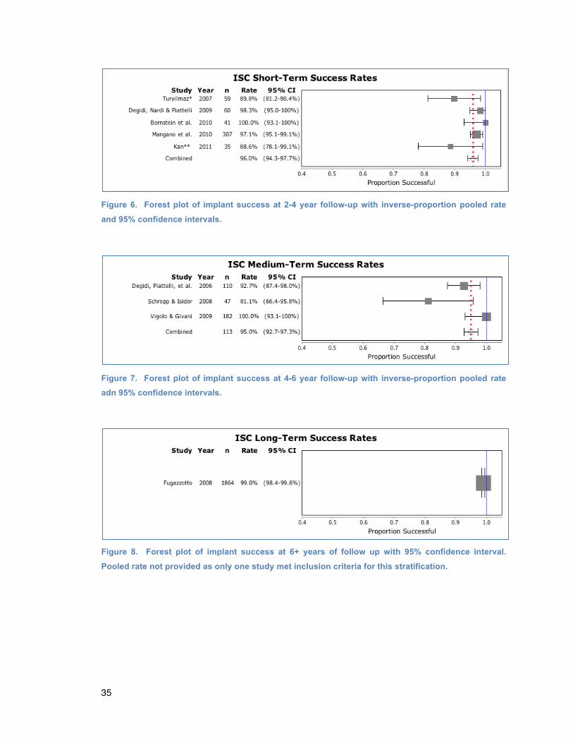
Table 12. Evidence table summary for single tooth implant success rates. Pooled and weighted
success rates were calculated using simple inverse-proportions.
ISC Success
Authors Year Published
Study Duration
Sample Size Failures Success
Rate Standard
Error 95% CI Quality Score
Turkyilmaz*[61, 62] 2007 2-4 59 6 89.8% 4.4% 81.2-98.4% 6
Degidi, Nardi & Piattelli[77] 2009 2-4 60 1 98.3% 1.7% 95.0-100% 13
Bornstein et al.[78] 2010 2-4 41 0 100.0% 3.5%§ 93.1-100%§ 10
Mangano et al.[70] 2010 2-4 307 9 97.1% 1.0% 95.1-99.1% 12
Kan**[63] 2011 2-4 35 4 88.6% 5.4% 78.1-99.1% 7
Weighted Success Rate: 96.0% 0.9% 94.3-97.7%
Degidi, Piattelli, et al.[79] 2006 4-6 110 8 92.7% 2.7% 87.4-98.0% 8
Schropp & Isidor[80] 2008 4-6 47 8 81.1% 7.5% 66.4-95.8% 10
Vigolo & Givani[74] 2009 4-6 182 0 100.0% 3.5%§ 93.1-100%§ 8
Weighted Success Rate: 95.0% 1.2% 92.7-97.3%
Fugazzotto[81] 2008 6+ 1864 15 99.0% 0.3% 98.4-99.6% 5
*Combines data from two published studies; 2006 & 2007 **Specific recall intervals not provided; data is reported in a range of follow-ups. §For studies reporting zero failures, the standard error and 95% confidence intervals are taken as an average of all included studies.
35
Figure 6. Forest plot of implant success at 2-4 year follow-up with inverse-proportion pooled rate
and 95% confidence intervals.
Figure 7. Forest plot of implant success at 4-6 year follow-up with inverse-proportion pooled rate adn 95% confidence intervals.
Figure 8. Forest plot of implant success at 6+ years of follow up with 95% confidence interval.
Pooled rate not provided as only one study met inclusion criteria for this stratification.

36
Table 13. Evidence table summary for single tooth implant survival rates. Pooled and weighted
success rates were calculated using simple inverse-proportions.
ISC Survival
Authors Year Published
Study Duration
Sample Size Failures Survival
Rate Standard
Error 95% CI Quality Score
Canullo**[64] 2007 2-4 30 0 100.0% 3.5%§ 93.1-100%§ 7
Cooper et al.[82] 2007 2-4 54 3 94.3% 3.4% 87.6-100% 9
Turkyilmaz*[61, 62] 2007 2-4 59 3 94.9% 3.0% 89.0-100% 6
Cannizzaro et al.[69] 2008 2-4 108 0 100.0% 3.5%§ 93.1-100%§ 9
Avvanzo**[66] 2009 2-4 282 18 93.6% 1.5% 90.7-96.5% 3
Degidi, Nardi & Piattelli[77] 2009 2-4 60 0 100.0% 3.5%§ 93.1-100%§ 13
Acocella et al.[83] 2010 2-4 68 3 95.6% 2.6% 90.5-100% 6
Crespi et al.[84] 2010 2-4 30 0 100.0% 3.5%§ 93.1-100%§ 10
Mangano et al.[70] 2010 2-4 307 5 98.4% 0.7% 97.0-99.8% 12
Rossi et al.[85] 2010 2-4 40 2 95.0% 3.6% 87.9-100% 6
Bilhan et al.[71] 2011 2-4 165 10 93.9% 2.0% 90.0-97.8% 8
Enkling et al.[86] 2011 2-4 42 0 100.0% 3.5%§ 93.1-100%§ 13
Kan**[63] 2011 2-4 35 0 100.0% 3.5%§ 93.1-100%§ 7
Lee et al.[72] 2011 2-4 207 2 98.4% 1.1% 96.2-100% 9
Weighted Success Rate: 96.8% 0.5% 95.9-97.7%
Bischof et al.[73] 2006 4-6 157 0 100.0% 3.5%§ 93.1-100%§ 7
Degidi, Piattelli, et al.[79] 2006 4-6 110 5 95.5% 2.1% 91.4-99.6% 8
Malo et al.[87] 2007 4-6 58 0 100.0% 3.5%§ 93.1-100%§ 5
Fugazzotto[81] 2008 4-6 341 2 98.9% 0.9% 97.1-100% 5
Jung et al.[88] 2008 4-6 305 6 98.0% 0.8% 96.4-99.6% 8
Schropp & Isidor[80] 2008 4-6 47 4 90.9% 4.8% 81.5-100% 10
Degidi, Iezzi, et al.[89] 2009 4-6 45 0 100.0% 3.5%§ 93.1-100%§ 11
Vigolo & Givani[74] 2009 4-6 182 0 100.0% 3.5%§ 93.1-100%§ 8
Koo et al.[75] 2010 4-6 521 15 95.6% 1.2% 93.2-98.0% 5
Zafiropoulos et al.[90] 2010 4-6 252 11 95.6% 1.3% 93.1-98.1% 9
Özkan et al.[91] 2011 4-6 93 0 100.0% 3.5%§ 93.1-100%§ 9
Prosper et al.[92] 2010 4-6 120 4 96.7% 1.7% 93.4-100% 9
Visser et al.[93] 2011 4-6 93 3 96.8% 1.9% 93.1-100% 10
Weighted Success Rate: 97.4% 0.3% 96.8-98.1%
Levin et al.[94] 2008 6+ 64 4 65.5% 30.4% 5.9-100% 7
Jemt[95] 2009 6+ 41 4 86.7% 7.2% 72.6-100% 4
Matarasso et al.[96] 2010 6+ 40 2 95.0% 3.6% 87.9-100% 6
Weighted Success Rate: 79.6% 3.3% 73.1-86.2%
*Combines data from two published studies; 2006 & 2007 **Specific recall intervals not provided; data is reported in a range of follow-ups. §For studies reporting zero failures, the standard error and 95% confidence intervals are taken as an average of all included studies.

37
Figure 9. Forest plot of implant survival at 2-4 year follow-up with inverse-proportion pooled rate
and 95% confidence intervals.
Figure 10. Forest plot of implant survival at 4-6 year follow-up with inverse-proportion pooled rate
and 95% confidence intervals.

38
Figure 11. Forest plot of implant survival at 6+ year follow-up with inverse-proportion pooled rate
and 95% confidence intervals.

39
Table 14. Evidence table summary for endodontic success rates.
RCT Success
Authors Year Published
Study Duration
Sample Size Failures Success
Rate Standard
Error 95% CI Quality Score
Özer[67] 2009 2-4 98 14 82.5% 5.1% 72.5-92.5% 8
de Chevigny**[65] 2008 4-6 1952 166 71.7% 1.0% 69.7-73.7% 14
**Specific recall intervals not provided; data is reported in a range of follow-ups.

40
Figure 12. Plot of endodontic success at 2-4 year follow-up with 95% confidence interval.
Figure 13. Plot of endodontic success at 4-6 year follow-up with 95% confidence interval.

41
Table 15. Evidence table summary for endodontic survival rates. Pooled and weighted success
rates were calculated using simple inverse-proportions.
RCT Survival
Authors Year Published
Study Duration
Sample Size Failures Survival
Rate Standard
Error 95% CI Quality Score
Ferrari et al.[97] 2007 2-4 240 17 92.9% 1.8% 89.4-96.4% 12
Alley et al.[98] 2008 2-4 100 11 89.0% 3.5% 82.1-95.9% 6
Özer[67] 2009 2-4 98 7 91.0% 3.5% 84.1-97.9% 8
Shafiei et al.[99] 2010 2-4 33 0 100.0% 3.5%§ 93.1-100%§ 7
Weighted Success Rate: 92.2% 1.2% 89.7-94.6%
de Chevigny**[65] 2008 4-6 1952 76 87.0% 0.8% 85.5-88.5% 14
Lumley et al.[76] 2008 6+ 30843 - 73.7% 0.3% 73.2-74.2% 6
Fokkinga et al.[100] 2008 6+ 98 14 79.4% 6.5% 66.7-92.1% 8
Weighted Success Rate: 73.7% 0.3% 73.2-74.2%
**Specific recall intervals not provided; data is reported in a range of follow-ups. §For studies reporting zero failures, the standard error and 95% confidence intervals are taken as an average of all included studies.

42
Figure 14. Forest plot of endodontic survival at 2-4 year follow-up with inverse-proportion pooled
rate and 95% confidence intervals.
Figure 15. Plot of endodontic survival at 4-6 year follow-up with 95% confidence interval.
Figure 16. Forest plot of endodontic survival at 6+ year follow-up with inverse-proportion pooled rate and 95% confidence intervals.

43
Table 16. Evidence table summary for fixed partial denture success rates. Pooled and weighted
success rates were calculated using simple inverse-proportions.
FPD Success
Authors Year Published
Study Duration
Sample Size Failures Success
Rate Standard
Error 95% CI Quality Score
Schmitt et al.[68] 2009 2-4 27 1 96.3% 3.8% 88.9-100% 14
Roediger et al.[101] 2010 2-4 99 38 62.0% 4.9% 52.4-71.6% 13
Weighted Success Rate: 69.4% 4.1% 61.3-77.4%
Sailer et al.[102] 2007 4-6 54 9 77.3% 8.7% 60.2-94.4% 10
Eschbach et al.[103] 2009 4-6 65 12 61.9% 19.3% 24.1-99.7% 10
Weighted Success Rate: 68.9% 4.2% 24.1-99.7%
Wolfart et al.[104] 2009 6+ 36 8 69.7% 13.5% 43.2-96.2 13

44
Figure 17. Forest plot of FPD success at 2-4 year follow-up with inverse-proportion pooled rate and
95% confidence intervals.
Figure 18. Forest plot of FPD success at 4-6 year follow-up with inverse-proportion pooled rate and
95% confidence intervals.
Figure 19. Plot of FPD success at 6+ year follow-up with 95% confidence interval.
45
Table 17. Evidence table summary for fixed partial denture survival rates. Pooled and weighted
success rates were calculated using simple inverse-proportions.
FPD Survival
Authors Year Published
Study Duration
Sample Size Failures Survival
Rate Standard
Error 95% CI Quality Score
Schmitt et al.[68] 2009 2-4 27 0 100.0% 3.5%§ 93.1-100%§ 14
Roediger et al.[101] 2010 2-4 99 7 94.0% 2.4% 89.3-98.7% 13
Weighted Success Rate: 95.3% 1.9% 91.6-99.0%
Eschback et al.[103] 2009 4-6 65 2 96.8% 2.3% 92.3-100% 10
Wolfart et al.[104] 2009 6+ 36 2 90.9% 6.7% 77.8-100% 13
§For studies reporting zero failures, the standard error and 95% confidence intervals are taken as an average of all included studies.

46
Figure 20. Forest plot of FPD survival at 2-4 year follow-up with inverse-proportion pooled rate and
95% confidence intervals.
Figure 21. Plot of FPD survival at 4-6 year follow-up with 95% confidence interval.
Figure 22. Plot of FPD survival at 6+ year follow up with 95% confidence interval.

47
Table 18. Pooled yearly success and survival rates (and standard errors) broken down by treatment
disciplines.
Pooled Success and Survival Rates
Success (SE) Survival (SE)
12mo
ISC 99.6% (0.1%) 99.6% (0.2%)
RCT § §
FPD 98.5% (1.6%) § 24mo
ISC 99.5% (0.2%) 99.5% (0.2%)
RCT § 96.5% (1.3%)
FPD 93.8% (3.2%) 98.4% (1.6%)
36mo
ISC 99.5% (0.2%) 98.9% (0.3%)
RCT 82.5% (5.1%) 93.4% (2.0%)
FPD 87.8% (3.6%) 96.8% (2.3%)
48mo
ISC 99.3% (0.2%) 98.7% (0.3%)
RCT § §
FPD 85.1% (5.5%) 96.8% (2.3%)
60mo
ISC 99.4% (0.2%) 98.6% (0.3%)
RCT 93.9% (2.6%) 97.7% (1.6%)
FPD 85.5% (4.4%) 98.4% (1.6%)
72mo
ISC 99.0% (0.3%) 98.3% (0.6%)
RCT § §
FPD 84.6% (7.5%) § 84mo
ISC § §
RCT § §
FPD § § 96mo
ISC § §
RCT § §
FPD § § §Pooled rates were not calculated, as no studies for this interval met inclusion criteria. Data was insufficient for pooling for the entire range of studies with follow up longer than 84 months.

48
Figure 23. Pooled yearly ISC success and survival rates with 95% confidence intervals.
Figure 24. Pooled yearly RCT success and survival rates with 95% confidence intervals.

49
Figure 25. Pooled yearly FPD success and survival rates with 95% confidence intervals.
Figure 26. Pooled yearly success rates for ISC, RCT, and FPDs with 95% confidence intervals.

50
Figure 27. Pooled yearly survival rates for ISC, RCT, and FPDs with 95% confidence intervals.

51
Table 19. Weighted success and survival rates with standard errors and 95% confidence intervals
for all included studies compared to only those with strict outcomes criteria applied.
Strict Success Criteria All included articles Strict Criteria Applied
Stratification Success Rate SE 95% CI Success
Rate SE 95% CI
FPD Success 2-4yr 69.4 4.1 61.3-77.4 96.3% 2.8% 88.9-100%
ISC Survival 2-4yr 96.8 0.5 95.9-97.7 97.7% 0.5% 96.6-98.7%
ISC Surival 4-6yr 97.4 0.3 96.8-98.1 97.3% 0.5% 96.2-98.4%
RCT Survival 2-4yr 92.2 1.2 89.7-94.6 91.0% 3.5% 84.1-97.9%
RCT Survival >6yr 73.7 0.3 73.2-74.2 73.7% 0.3% 73.2-74.2%
Psychosocial Outcomes
Seventeen studies were included that examined the psychosocial effects of
treatment; they are summarized in Table 20. Almost all of them are short-term in
duration, less than two years. As with the clinical outcomes studies, the majority
(68.4%) focused on ISCs. There were two RCT studies and one FPD study.
Two further studies made direct comparisons among treatment modalities
(Gatten et al. 2011[105] and Al-Quran et al. 2011[106]). The majority of the implant
studies examined patients’ satisfaction with their treatment received and
esthetics; overall patients were highly satisfied. The remaining implant studies
analyzed anxiety and post-operative pain, which typically was only mild to
moderate. The two RCT studies examined anxiety and post-operative pain,
which was minimal. The FPD study covered the motivating factors that drove
patients to accept or decline fixed bridge treatment.
Of the two direct comparison studies, one compared ISCs to RCTs, and the other
ISCs to FPDs or EXTs. Patients were generally satisfied with whatever treatment

52
they had received; yet they also generally felt that their respective treatments
were expensive. One study found that regardless of whether patients had
received ISC or RCT, they had a strong desire to preserve their natural dentition
when possible. The other study found that overall patients with ISCs were
happier with their treatment than patients with FPDs, who were in turn happier
than patients who had extractions without replacement.

53
Table 20. Evidence table for psychosocial effects of implant-supported single crowns, fixed partial
denture, and root canal therapies.
Psychosocial Outcomes
Study Field of Study Study Type Sample Relevant Findings
Hashem et al. 2006[107]
ISC Anxiety Post-op Pain
Prospective 1-week
30 implants 18 patients
Most patients reported pain and/or anxiety that only mildly to moderately interfered with their daily activities post-operatively. No patients reported high levels of any symptoms.
Urban & Wenzel 2010[108]
ISC Post-op Pain
Prospective 3-days
92 implants 92 patients
After placement of immediate molar implants in concert with local site augmentation, patients experienced little to moderate pain, which peaked 5-6 hours post-operatively.
Hsieh et al. 2010[109]
ISC Function
Cross-sectional
10 implants 10 patients
Patients have some proprioceptive awareness of implant loading, but it is less intense than for natural teeth.
Palmer et al. 2007[110]
ISC Outcomes
Retrospective 1-year
66 implants 66 patients
Patients were highly satisfied with the esthetics and function of single tooth implants, including the appearance of soft tissues. Clinicians were more critical of the restorations than were the patients.
Schropp & Isidor 2008[80]
ISC Outcomes
Prospective 5-year
45 implants 45 patients
Patients were highly satisfied with their implants and course of treatment. At 2-year follow-up, patients who had implants placed 10 days after extractions were more satisfied than patients who had implants placed 3 months after extractions. At 5-year follow-up, there was no significant difference. Older patients were more satisfied with esthetics, function and ease of care than younger patients.
Thierer et al. 2008[111]
ISC Outcomes
Prospective 5-year
245 implants 120 patients
Overwhelming majority of patients rated implant therapy as good to excellent with regards to implant function, implant esthetics, and ease of cleaning the prosthesis. They did not significantly change these assessments between year 1 and year 5. The small number of patients who rated implant treatment as fair to poor also did not significantly change their minds, nor differ between years 1 and 5.
Gallucci et al. 2011[112]
ISC Outcomes
RCCT 2-year
20 implants 20 patients
Patients had high satisfaction with the esthetics of anterior PFM or all-ceramic implant supported crowns with no significant difference between the 2 restorations.
Luo et al. 2011[113]
ISC Outcomes
Prospective 3-months
33 implants 31 patients
Median satisfaction of implants per a visual analog scale (VAS) was 88.5%. Pink Esthetic Scores significantly correlated to VAS scores.
Özkan et al. 2011[91]
ISC Outcomes
Retrospective 5-year
93 implants 83 patients
All patients rated esthetics, masticatory ability, phonetics, and cleansability as either good or excellent.
Ponsi et al. 2011[114]
ISC Outcomes
Prospective 3-month
131 implants 80 patients
Mean OHIP-14* severity scores decreased significantly, from 10.4 prior to uncovering the implants to 3.1 after restoration. Replacement of missing teeth with single dental implants in the anterior and premolar (but not molar) areas may significantly improve patients' subjective oral health.

54
Psychosocial Outcomes
Study Field of Study Study Type Sample Relevant Findings
van Lierde et al. 2011[115]
ISC Outcomes
Retrospective 1.5-year
14 implants 25 patients
Mean satisfaction of implants per VAS was 95%. 43% of patients perceived problems with function or physical or psychological comfort. All patients were capable of producing Dutch vowels and consonants; however 57% had phonetic disorders with the consonant [s]. Blowing, sucking, and swallowing patterns were all normal.
Vilhjálmsson et al. 2011[116]
ISC Outcomes
Retrospective 1-year
56 implants 50 patients
88% of patients were satisfied with the form of the crown, 84% were satisfied with the shade, and 72% were satisfied with the adjacent mucosa.
Lai et al. 2008[117]
RCT Anxiety
RCCT Non-Longitudinal
44 RCTs 44 patients
Patients who listened to soothing music during RCT had significantly less anxiety during treatment vs. patients who did not.
Wang et al. 2010[118]
RCT Post-op Pain
RCCT 1-week
89 RCTs 89 patients
The vast majority of patients reported no pain or slight pain after RCT. There was no significant difference in post-obturation pain between one-visit and two-visit RCT on teeth with vital pulps.
Leles et al. 2009[119]
FPD Motivation
Cross-sectional
63 FPDs 87 Exts 150 patients
The main motivating factor for patients electing FPDs was the desire for a fixed prosthesis. Main motivating factors for refusal of FPDs were cost, fear of the need for removal of remaining tooth structure, fear of the negative effect on remaining teeth, and difficulties with oral hygiene.
Gatten et al. 2011[105]
ISC & RCT Outcomes
Questionnaire 1-year
20 implants 17 RCTs 37 patients
Both RCT & ISC patients were satisfied with their respective treatments, however both groups expressed a desire to preserve their natural dentition whenever possible. Both RCT & ISC patients felt that their respective treatment was expensive. RCT patients complained about how long they had to keep their mouth open; ISC patients complained about how long total treatments took from extraction to crown delivery.
Al-Quran et al. 2011[106]
ISC, FPD, Ext Patient Factors
Questionnaire 1-year
50 ISCs 100 FPDs 50 Exts 150 patients
Monthly income was significantly higher in Patients who had ISCs vs. Exts. Patients with ISCs and FPDs felt they had more favorable relations with other people vs. patients who had extractions. Patients with ISCs were more satisfied with esthetics, function, and speech efficiency compared to patients with FPDs, who were more satisfied compared to patients with extractions without replacement.
*Oral Health Impact Profile; 14-item questionnaire that attempts to measure subjective oral health including comfort, function, speech, esthetics, self-image, physical pain, psychological discomfort, social disability, and handicap.

55
7 DISCUSSION
Key Findings and Their Limitations
The original Torabinejad review set out to answer the clinical question: Is initial
root canal therapy superior to extraction and replacement with an implant,
replacement with an FPD, or no replacement at all? The results of that review
revealed that both root canal therapy and implant therapy were superior to
extraction without replacement or extraction with FPD treatment. However the
literature base from which those conclusions were drawn was found to be
extremely problematic. The individual studies comprising the review varied
considerably in every aspect from study design to operator experience to follow-
up duration and even to the very definitions of success and survival.
The goal of this systematic review was to see if the recent additions to the
literature pool would allow a more definitive conclusion to be drawn. The authors
anticipated that many of the same trends would be observed. That was certainly
true with regards to the degree of inter-study heterogeneity. What was surprising
was how lacking the studies were of rigorous design and thorough clinical
reporting. Most of the ISC studies identified by the search strategies were
excluded because they involved multi-unit restorations. A high number of studies
for both RCT and ISC were disqualified because their follow-up was extremely
short-term, they did not specify patient ages, or there was insufficient data to
calculate success and survival rate.

56
The methodology behind this review was an effort to obtain the broadest capture
of the relevant literature—something that the original review team of 13
investigators excelled at. However, time and labor were significant constraints in
this review, and despite the authors’ best efforts some relevant literature may still
have been missed. Hand searching was limited to citation mining of relevant
reviews identified in the search and journal table of contents. Searching of
textbooks and other non-indexed literature could have been more exhaustive had
time permitted. Also, like the Torabinejad review, articles not published in
English were not considered for inclusion. This could be leaving a portion of the
evidence base behind.
The possible bias of study selection is always of concern when conducting a
literature review. In this review the authors have attempted to eliminate bias by
keeping the article inclusion process as objective as possible. The initial search
results began with titles only, blinded of authors and publication journal. This
progressed to full abstract review and finally a full-text review. Each stage of
article qualification was conducted in tandem by two different examiners (MGH
and GRH), and any disputes were resolved by examiner discussion. While
tedious, these efforts helped to greatly reduce bias that would have been
introduced by single-reviewer decision-making.
In the broadest sense, the clinician is (or should be) seeking a realistic success
rate for endodontic treatment, and a realistic survival rate for single-tooth implant
therapy. That is to say, he or she is seeking a rate that applies to the entire
population of every root canal performed and every implant placed. This is
impossible, and so the goal becomes to devise a realistic method for estimating
these rates based on much smaller samples, i.e., the individual studies that met
inclusion for this review. Each of these studies attempts to estimate the success
and survival rates for the population at large based upon a small group of
patients or treatments that they have sampled. In turn, the authors have

57
attempted to use those individual estimates to better estimate the success and
survival rates of the population at large. The standard errors and 95%
confidence intervals are measurements of how accurate our estimates are. An
experienced statistician supervised the data analysis to ensure proper use of
statistics. However, robust statistical analysis was not possible because of the
heterogeneity of the data.
In the course of this review, only three studies involving direct-comparison of
different treatment modalities were identified; none of them met inclusion criteria.
All three compared ISCs and RCTs. The first study was a cross-sectional
comparison of initial RCT and ISCs by Doyle and colleagues[120]. However, it
was already included in the initial Torabinejad review. Two other retrospective
studies were identified; both of these contained treatments with less than two-
year follow-up, and both contained patients under the age of 18. Hannahan et
al.[121] reported high survival rates for both treatments with no significant
difference (98.4% for ISC and 99.3% for RCT). Success rates were lower but
again not significantly different (87.6% for ISC and 90.2% for RCT). Laird[122]
found that single tooth implants placed adjacent to sound vital teeth had
significantly higher success and survival rates compared to implants placed
adjacent to endodontically treated teeth or edentulous spaces.
The findings in this review, therefore, are based upon indirect comparisons, i.e.
comparing the success and survival rates of one sort of study for one type of
treatment to another sort of study with another type of treatment. This severely
limits the degree to which the resulting evidence can be interpreted with any
significant clinical meaning[53, 54]. The conclusions drawn from any systematic
review must be interpreted with caution, and this review is no exception.

58
Literature Quality and Bias
Overall, the available literature lacked many of the desirable traits of an
outcomes study, coincident with the unexceptional quality scores observed (the
average score was 8.6±2.7 out of a possible 17 points). FPD studies appeared
to have the highest caliber in study design, with complete treatment protocol
descriptions, blinding of examiners, detailed accounts of subject loss, and
identification and description of treatment complications and how they affected
the outcomes. RCT and ISC studies tended to be less meticulous. They were
less likely to account for subject loss and more likely to omit details of treatment
complications. The lowest quality studies included in the review all happened to
be ISC studies.
This trend of low quality reporting of outcomes studies and trials is not a new
revelation. Indeed, it was a major conclusion of the original Torabinejad review
and something that has plagued the dental and medical literature at large. In
2001 the CONSORT Group (Consolidated Standards of Reporting Trials) was
formed with the aim of improving clinical reporting of randomized clinical trials. It
released guidelines to aid researchers in complete and transparent reporting of
trial findings, with the ultimate goal of improving the appraisal and interpretation
of the evidence base. Unfortunately, it appears that the dental community has
not improved its quality of reporting trials since the release and promotion of
these guidelines[47].
Whether or not clinical outcomes can be correlated with best practices in study
design is beyond the scope of this review. However with so few studies of high
quality methodological design (and most of them associated with the FPD
studies), the results of this review should be interpreted with caution.

59
The majority of included studies were ISC studies, which is reflective of the
rapidly evolving implant technology. In contrast, RCT and FPD represented a
minority of the studies. This may have skewed the results in favor of the implant
literature, although an effort was made to minimize this effect by comparing
weighted, pooled rates wherever possible.
Another significant source of bias is the experience of the treatment provider. In
the included studies, implants tended to be placed by specialists or “experienced”
general dentists. In a few cases they were placed by residents and in no case
were they placed by dental students (although, implants were restored by
supervised dental students in some instances). The providers performing RCT or
FPD therapies were less often specialists, and most commonly general dentists,
residents, and in a few cases, supervised dental students.
Study duration is also a major source of bias. Studies with the longest follow-up
also predictably suffer from the highest number of dropouts. Censoring patients
who do not attend recall examinations reduces the sample size, which inflates
the effect of failures. This effect was most notably seen in the ISC survival study
by Levin et al. 2008[94] (Table 21). The study followed 65 implants over 14 years,
and during that time there were only four failures. The first failure was not
recorded until five years into the study for a cumulative success rate of 98.4%.
One more failure was recorded in the sixth year. Yet 30 patients failed to show
for recall, nearly cutting the available sample size in half, and the cumulative
survival rate dropped to 95.3%. The next failure was seen two years later, but
with the loss of 19 more patients the study population had dwindled to 11
subjects, and the cumulative survival rate fell to 87.4%. The study continued for
six more years; and the fourth and final failure was not seen until year 14. By this
time, over 90% of the sample population had been censored with just four
patients presenting for recall. The survival rate during year 14 was just three in
four (75%), which drastically pulled the 14-year cumulative survival rate down to

60
65.5%. A simple absolute survival rate calculated over the entire 14 years tells
quite a different story: four failures out of 65 or a 95% survival rate. This
problem is endemic in long-term prospective studies, and almost certainly causes
the failure rate to be overstated. One alternative would be to assume that no
failures occurred in the patients failing to show for recall, but doing this would
simply overstate the survival rate. Numerous other methods have been
proposed to better deal with censored study participants, which arguably hold
merit. Yet because the included studies in this review varied in the extent and
details of the raw data provided or extractable, applying them in a consistent
manner would have been all but impossible.
Table 21. Life table analysis for Levin et al. demonstrating the exaggerated effect of failures as
subjects are lost to follow-up.
Levin et al. Life Table Analysis
Interval (yr)
Number Starting Interval
Number of Failures
Number of Dropouts
Number Ending Interval
Absolute Rate
Cumulative Rate
1 64 0 0 64 100.0% 100.0%
2 64 0 0 64 100.0% 100.0%
3 64 0 0 64 100.0% 100.0%
4 64 0 0 64 100.0% 100.0%
5 64 1 1 62 98.4% 98.4%
6 62 1 30 31 96.9% 95.3%
7 31 0 10 21 100.0% 95.3%
8 21 1 9 11 91.7% 87.4%
9 11 0 7 4 100.0% 87.4%
10 4 0 0 4 100.0% 87.4%
11 4 0 0 4 100.0% 87.4%
12 4 0 0 4 100.0% 87.4%
13 4 0 0 4 100.0% 87.4%
14 4 1 0 3 75.0% 65.5%

61
Clinical Outcomes: Success or Survival?
In terms of long-term survival, the original Torabinejad review[1] found that:
“…initial endodontic treatment has high long-term survival rate for periodontally sound teeth that have pulpal and/or periapical pathosis. Equivalent long-term survival rates have been also reported for extraction and replacement of the missing tooth with an implant-supported restoration. Substantially lower long-term survival rates have been reported for extraction and replacement of the missing tooth with fixed partial dentures.”
The findings of this review are not in close agreement with the Torabinejad
review. In terms of long-term survival, RCT teeth and ISCs were reported to
have worse rates than FPDs, while short-term survival rates for the three
treatments appeared to be similarly high. In terms of long-term success, ISCs
tended to have higher rates than did RCTs or FPDs, which were similar. This is
likely due to the relatively few included studies of long-term duration and the
immense variability in study design.
The treatment modalities examined in this review have completely different goals
of therapy. The aim of initial non-surgical endodontic treatment is to eliminate or
prevent disease, whether of pulpal origin, periapical origin, or both. On the other
hand, implant and partial denture therapies do not aim to cure disease. They aim
to restore esthetics and function from an already missing tooth, or one with a
hopeless prognosis.
As the goals of treatment are different, so too should be the criteria and
measures for evaluating that treatment. Outcomes of endodontic therapy ought
to measure the degree to which disease has been cured, that is to say, the
degree to which the body has healed, or if already healthy, not degenerated into
disease. Different criteria have been suggested for this. Outcomes of implant

62
therapy ought to measure the degree to which function and esthetics have been
restored or improved, and to some degree, to which the existing surrounding
tissues have remained healthy. Traditionally, endodontic literature reports its
outcomes as success, while the implant literature has normed to a term of
survival.
If the outcomes are to be reported in terms of survival, then a minimum, basic set
of criteria should be expected[58]. The tooth or prosthesis should be present, it
should be in function, and it should be asymptomatic. Rigorous studies will
define not only the criteria for survival, but also the criteria for failure (and nearly
two-thirds of studies reporting survival did not). If the outcomes are to be
reported in terms of success, then a reference for success should be provided,
one that has been validated by the (endodontic) or (implant) community. Most
studies in this review did not.
Only two endodontic studies provided references for their success criteria. As
part of the Toronto Study, de Chevigny et al.[65] used the PAI[30] as a radiographic
measure of success combined with the presence or absence of clinical signs and
symptoms; these parameters were used to classify the teeth as either healed or
diseased[6]. Özer[67] referenced Petersson’s criteria[123] for radiographic
outcomes, and combined those with clinical findings to classify the endodontic
treatment as either successful or failing. Only one other study reported success
(Alley et al. 2006[98]), but the authors did not provide a reference. The remaining
four RCT studies reported survival, but only two of them included a frank criterion
for failure.
For implants, nine of the 32 included studies reported success. Four of them
referenced Albrektsson[37], one Smith and Zarb[38], and one Buser[39]. The
remaining three studies did not provide references for success. None of the
studies adhered to the success criteria they provided, mostly, by failing to provide

63
data on marginal bone loss. This can probably be attributed to the fact that the
Albrektsson criteria (and other early implant criteria) emerged during the early
stages of implant technology when osseointegration was of key concern.
Whether or not implants have vindicated themselves in terms of preservation and
maintenance of the supporting alveolar bone and gingival tissues, the literature
appears to have moved on; detailed reporting of marginal bone loss is largely
absent. Of the remaining 23 studies reporting survival, 74% did not provide a
frank criterion for failure.
FPD studies did not fare much better. Although data in this review was re-
calculated to provide success rates for all five included studies, only three of the
authors reported success as an outcome. Schmitt et al. 2009[68] was the only
article to provide a reference for success, the California Dental Association’s
evaluation criteria[124]. The other two studies did not provide references or
criteria. However, for survival calculations, all authors provided a frank criterion
for failure.
The importance of using a clearly defined and widely accepted definition and
criteria for measuring outcomes cannot be under-emphasized. After all, the
whole point of conducting an outcomes study is to measure the outcome in a
meaningful way so that the patient and the clinician can make a well-informed
decision based on sound, clinical evidence. At best, the substance of the
reported results is open to interpretation, and at worst, the results are rendered
completely useless. Conducting a study and publishing a success or survival
rate that has no backbone is meaningless, and only serves to discredit the dental
profession.
When strict reporting criteria were applied (that is, for success: detailing out the
criteria and providing a reference for it; for survival: providing both a criteria for

64
survival and a criteria for failure) nearly all of the included studies in this review
were disqualified.
In summary, the methodology by which the dental profession appears to be
assessing the outcomes of its treatment remains problematic. Within the
endodontic, implant, and fixed partial denture treatment modalities, conventions
or standards are lacking. The endodontic literature does not tend to report
success, it has turned to the much more lax outcome of survival. The implant
literature tends to report both survival and success, often inter-changing the two.
Where success is reported, it is very often not in accordance with the very criteria
set out by leaders in the field.
Psychosocial Outcomes
Torabinejad’s original systematic review also examined the various psychosocial
aspects of treatment[1]; it found:
“…tooth retention through root canal therapy and restoration or tooth replacement with an implant or a fixed partial denture results in superior clinical outcomes, compared to extraction without replacement. The reasons for this were due to diminution of esthetics and psychological trauma associated with tooth loss…”
The findings from this review tend to be in agreement with the original review and
have been summarized in the results section and in Table 20. Overwhelmingly,
these studies tended to be of a short-term duration, with only four of the 17
studies examining effects for a two-year or longer duration. This reflects the
types of factors these studies were examining, namely post-operative pain,
patient anxiety, and satisfaction with the esthetics, function, and the overall

65
treatment experience. All but three of the studies analyzed implants, which can
be a source of potential bias. However, Gatten et al.[105] did find that both RCT
and ISC patients had a strong desire to preserve the natural dentition whenever
possible.

66
8 Conclusions
The existing literature describing the long-term outcomes of root canal therapy,
extraction with replacement of a single-tooth implant, extraction with replacement
of a fixed partial denture, or extraction without replacement remains problematic.
Studies drawing direct comparisons are lacking, success and survival are defined
in many different ways, subjects lost to follow-up are not uniformly accounted for,
and treatment complications are largely unaddressed. The extent of
heterogeneity in the data set makes applying statistical analysis all but
impossible. As such, the weighted and pooled success and survival rates
regrettably do not contribute much more clinical value than the individual studies
on their own. There is a dire need for the endodontic and implant communities to
identify and conform to a set of robust and thoughtful criteria for success and
survival. There is a further need for good quality, long-term outcomes studies for
both implants and endodontics that adhere to the CONSORT guidelines.
In terms of long-term outcome, there does not appear to be any significant
differences in survival between single-tooth implants and initial nonsurgical root
canal treatment in periodontally sound teeth (98.6 vs 97.7% at 5-years). The
long-term success of implants would appear to be slightly more optimistic in
implants (99.4%) as compared to root canal treatment (93.9%). However, as the
success of both RCT and ISC have been loosely and inconsistently defined,
these results may not be clinically relevant. Both treatment modalities appreciate
improved success and survival rates over fixed partial dentures. These findings
are in accordance with the existing literature to date.
67
What treatment to recommend to which patient remains a decision that must be
made on a case-by-case, tooth-by-tooth, and patient-by-patient basis. The
desire of some clinicians to attempt to cure disease by extracting teeth and
replacing them with implants would seem to be inappropriate and premature in
many cases. Likewise, heroic efforts to attempt to save severely compromised
teeth through endodontics may not be in the best interest of the patient, from
both a prognostic and cost-efficacy point-of-view.

68
9 Acknowledgements
PATRICIA ANDERSON, for her considerable efforts in designing the original
MEDLINE, EMBASE, and COCHRANE search strategies as well as updating
them for the purposes of this review, and also for her valuable and wise expertise
in designing, executing, and writing a systematic review.
TATIANA BOTERO, for her guidance and oversight relative to the endodontic
literature.
G. REX HOLLAND, for his exhaustive labors during the literature screening
process, as well as for his mentoring and feedback during the entire thesis
project.
MICHAEL RAZZOOG, for his guidance and oversight relative to the implantology
literature
KERBY SHEDDEN, for his statistical counsel and oversight of the data analysis.
BARBARA SHIPMAN, for her assistance in updating and executing the
EMBASE database search.
MAHMOUD TORABINEJAD and his team, for their great efforts undertaken in
the original systematic review, and the inspiration to continue it with this project.
69
REFERENCES
1. Torabinejad, M., et al., Outcomes of root canal treatment and restoration, implant-supported single crowns, fixed partial dentures, and extraction without replacement: A systematic review. The Journal of Prosthetic Dentistry, 2007. 98(4): p. 285-311.
2. CDC, N.C.C.f.C.D.P.a.H.P., Oral Health: Preventing cavities, gum disease, tooth loss, and oral cancers, C.f.D. Control, Editor 2010.
3. Satcher, D., Oral Health in America: A Report of the Surgeon General, U.D.o.H.a.H. Services, Editor 2000: Rockville, MD. p. 61--73.
4. Aquilino, S.A. and D.J. Caplan, Relationship between crown placement and the survival of endodontically treated teeth. J Prosthet Dent, 2002. 87(3): p. 256-63.
5. Siskin, M., Economics, indications and contraindications for endodontic therapy. J Am Dent Assoc, 1963. 66: p. 648-62.
6. Friedman, S., Considerations and concepts of case selection in the management of post-treatment endodontic disease (treatment failure). Endodontic Topics, 2002. 1: p. 54-78.
7. Trope, M., The vital tooth – its importance in the study and practice of endodontics. Endodontic Topics, 2003. 5(1): p. 1-1.
8. Wu, M.K., H. Shemesh, and P.R. Wesselink, Limitations of previously published systematic reviews evaluating the outcome of endodontic treatment. International Endodontic Journal, 2009. 42(8): p. 656-666.
9. Chalfin, H., M. Kellert, and C. Solomon, Treatment planning considerations for the endodontically compromised tooth. N Y State Dent J, 2004. 70(6): p. 30-4.
10. Saunders, W.P. and E.M. Saunders, Conventional endodontics and the operating microscope. Dent Clin North Am, 1997. 41(3): p. 415-28.
11. Song, M., S.-J. Shin, and E. Kim, Outcomes of Endodontic Micro-resurgery: A Prospective Clinical Study. Journal of Endodontics, 2011. 37(3): p. 316-320.
12. Del Fabbro, M., et al., Magnification devices for endodontic therapy. Cochrane Database Syst Rev, 2009(3): p. CD005969.
13. Kim, S., Modern endodontic practice: instruments and techniques. Dental clinics of North America, 2004. 48(1): p. 1-9.
14. Fabricius, L., et al., Influence of residual bacteria on periapical tissue healing after chemomechanical treatment and root filling of experimentally infected monkey teeth. Eur J Oral Sci, 2006. 114(4): p. 278-85.
15. Nair, P.N.R., et al., Microbial status of apical root canal system of human mandibular first molars with primary apical periodontitis after “one-visit” endodontic treatment. Oral Surgery, Oral Medicine, Oral Pathology, Oral Radiology, and Endodontology, 2005. 99(2): p. 231-252.

70
16. Sjogren, U., et al., Influence of infection at the time of root filling on the outcome of endodontic treatment of teeth with apical periodontitis. Int Endod J, 1997. 30(5): p. 297-306.
17. Sundqvist, G., et al., Microbiologic analysis of teeth with failed endodontic treatment and the outcome of conservative re-treatment. Oral Surgery, Oral Medicine, Oral Pathology, Oral Radiology, and Endodontology, 1998. 85(1): p. 86-93.
18. Mindiola, M.J., et al., Endodontic treatment in an American Indian population: a 10-year retrospective study. Journal of Endodontics, 2006. 32(9): p. 828-32.
19. Friedman, S., Treatment outcome and prognosis of endodontic therapy, in Essential Endodontology, T.P.-F. D Orstavik, Editor 1998, Blackwell Science Ltd: Cambridge. p. 367-401.
20. Iqbal, M.K. and S. Kim, For teeth requiring endodontic treatment, what are the differences in outcomes of restored endodontically treated teeth compared to implant-supported restorations? Int J Oral Maxillofac Implants, 2007. 22 Suppl: p. 96-116.
21. Berglundh, T., L. Persson, and B. Klinge, A systematic review of the incidence of biological and technical complications in implant dentistry reported in prospective longitudinal studies of at least 5 years. J Clin Periodontol, 2002. 29 Suppl 3: p. 197-212; discussion 232-3.
22. Klokkevold, P.R. and T.J. Han, How do smoking, diabetes, and periodontitis affect outcomes of implant treatment? Int J Oral Maxillofac Implants, 2007. 22 Suppl: p. 173-202.
23. Heitz-Mayfield, L.J. and G. Huynh-Ba, History of treated periodontitis and smoking as risks for implant therapy. Int J Oral Maxillofac Implants, 2009. 24 Suppl: p. 39-68.
24. Esposito, M., et al., Biological factors contributing to failures of osseointegrated oral implants. (II). Etiopathogenesis. Eur J Oral Sci, 1998. 106(3): p. 721-64.
25. Javed, F. and K. Almas, Osseointegration of Dental Implants in Patients Undergoing Bisphosphonate Treatment: A Literature Review. Journal of Periodontology, 2010. 81(4): p. 479-484.
26. Madrid, C. and M. Sanz, What impact do systemically administrated bisphosphonates have on oral implant therapy? A systematic review. Clinical Oral Implants Research, 2009. 20: p. 87-95.
27. IJOMI, 2010 Guidelines of the Academy of Osseointegration for the provision of dental implants and associated patient care. Int J Oral Maxillofac Implants, 2010. 25(3): p. 620-7.
28. Herrmann, I., et al., Evaluation of patient and implant characteristics as potential prognostic factors for oral implant failures. Int J Oral Maxillofac Implants, 2005. 20(2): p. 220-30.

71
29. Brynolf, I., A histological and roentgenological study of the periapical region of human upper incisors. Odontologisk revy,v. 18, suppl. 11, 19671967, Uppsala: Almqvist & Wiksell. 176 p.
30. Orstavik, D., K. Kerekes, and H.M. Eriksen, The periapical index: a scoring system for radiographic assessment of apical periodontitis. Endod Dent Traumatol, 1986. 2(1): p. 20-34.
31. Goldman, M., A.H. Pearson, and N. Darzenta, Endodontic success--who's reading the radiograph? Oral Surg Oral Med Oral Pathol, 1972. 33(3): p. 432-7.
32. Bender, I.B., Factors influencing the radiographic appearance of bony lesions. J Endod, 1982. 8(4): p. 161-70.
33. Paula-Silva, F.W.G.d., et al., Accuracy of Periapical Radiography and Cone-Beam Computed Tomography Scans in Diagnosing Apical Periodontitis Using Histopathological Findings as a Gold Standard. Journal of Endodontics, 2009. 35(7): p. 1009-1012.
34. Green, T.L., et al., Radiographic and histologic periapical findings of root canal treated teeth in cadaver. Oral Surg Oral Med Oral Pathol Oral Radiol Endod, 1997. 83(6): p. 707-11.
35. Bender, I.B., S. Seltzer, and W. Soltanoff, Endodontic success--a reappraisal of criteria. 1. Oral Surg Oral Med Oral Pathol, 1966. 22(6): p. 780-9.
36. Cotti, E. and G. Campisi, Advanced radiographic techniques for the detection of lesions in bone. Endodontic Topics, 2004. 7(1): p. 52-72.
37. Albrektsson, T., et al., The long-term efficacy of currently used dental implants: a review and proposed criteria of success. Int J Oral Maxillofac Implants, 1986. 1(1): p. 11-25.
38. Smith, D.E. and G.A. Zarb, Criteria for success of osseointegrated endosseous implants. J Prosthet Dent, 1989. 62(5): p. 567-72.
39. Buser, D., H.P. Weber, and N.P. Lang, Tissue integration of non-submerged implants. 1-year results of a prospective study with 100 ITI hollow-cylinder and hollow-screw implants. Clinical Oral Implants Research, 1990. 1(1): p. 33-40.
40. Glauser, R., et al., Immediate occlusal loading of Branemark TiUnite implants placed predominantly in soft bone: 1-year results of a prospective clinical study. Clinical implant dentistry and related research, 2003. 5 Suppl 1: p. 47-56.
41. Misch, C.E., et al., Implant success, survival, and failure: the International Congress of Oral Implantologists (ICOI) Pisa Consensus Conference. Implant dentistry, 2008. 17(1): p. 5-15.
42. Salinas, T.J. and S.E. Eckert, In patients requiring single-tooth replacement, what are the outcomes of implant- as compared to tooth-supported restorations? Int J Oral Maxillofac Implants, 2007. 22 Suppl: p. 71-95.

72
43. ADA. About EBD. 2011 [cited 2011 4/19/11]; Available from: http://ebd.ada.org/about.aspx.
44. Forrest, J.L. and S.A. Miller, The anatomy of evidence-based publications: article summaries and systematic reviews. Part I. J Dent Hyg, 2004. 78(2): p. 343-8.
45. Sackett, D.L., et al., Evidence based medicine: what it is and what it isn't. BMJ, 1996. 312(7023): p. 71-2.
46. Howick, J., et al. The Oxford 2011 Levels of Evidence. 2001; Available from: http://www.cebm.net/index.aspx?o=5653.
47. Savithra, P. and L.S. Nagesh, Have CONSORT guidelines improved the quality of reporting of randomised controlled trials published in public health dentistry journals? Oral health & preventive dentistry, 2013. 11(2): p. 95-103.
48. Worthington, H.V., et al., Using Cochrane reviews for oral diseases. Oral diseases, 2010. 16(7): p. 592-6.
49. Wright, R.W., et al., How to Write a Systematic Review. Clinical Orthopaedics and Related Research, 2007. 455: p. 23-29.
50. Anderson, P., Systematic Review Processes, Teams, Experiences, 2012. 51. The Cochrane Collaboration. The Cochrane Policy Manual. April 22, 2013
[cited 2013 July 30, 2013]. 52. Cook, D.J., C.D. Mulrow, and R.B. Haynes, Systematic reviews: synthesis
of best evidence for clinical decisions. Ann Intern Med, 1997. 126(5): p. 376-80.
53. Buti, J., et al., Network meta-analysis of randomised controlled trials: direct and indirect treatment comparisons. European journal of oral implantology, 2011. 4(1): p. 55-62.
54. Xiong, T., et al., Overall similarity and consistency assessment scores are not sufficiently accurate for predicting discrepancy between direct and indirect comparison estimates. Journal of clinical epidemiology, 2013. 66(2): p. 184-91.
55. Green, S., et al., Chapter 1: Introduction, in Cochrane Handbook for Systematic Reviews of Interventions, J. Higgins and G. S, Editors. 2011, The Cochrane Collaberation.
56. Moher, D., et al., Preferred reporting items for systematic reviews and meta-analyses: the PRISMA statement. BMJ, 2009. 339: p. b2535.
57. Shojania, K.G., et al., How quickly do systematic reviews go out of date? A survival analysis. Ann Intern Med, 2007. 147(4): p. 224-33.
58. Albrektsson, T. and G.A. Zarb, Determinants of correct clinical reporting. The International journal of prosthodontics, 1998. 11(5): p. 517-21.
59. Kaplan, E.L. and P. Meier, Nonparametric estimation from incomplete observations. J Amer Statist Assn, 1958. 53(282): p. 457-481.
60. Boyles, A. Forest Plot Viewer v1.00. 2013; Available from: http://ntp.niehs.nih.gov/go/tools_forestplotviewer.

73
61. Turkyilmaz, I., A 3-year prospective clinical and radiologic analysis of early loaded maxillary dental implants supporting single-tooth crowns. International Journal of Prosthodontics, 2006. 19(4): p. 389-90.
62. Turkyilmaz, I., et al., A 4-year prospective clinical and radiological study of maxillary dental implants supporting single-tooth crowns using early and delayed loading protocols. Clinical Implant Dentistry & Related Research, 2007. 9(4): p. 222-7.
63. Kan, J.Y.K., et al., Facial gingival tissue stability following immediate placement and provisionalization of maxillary anterior single implants: a 2- to 8-year follow-up. International Journal of Oral & Maxillofacial Implants, 2011. 26(1): p. 179-87.
64. Canullo, L., Clinical outcome study of customized zirconia abutments for single-implant restorations. International Journal of Prosthodontics, 2007. 20(5): p. 489-93.
65. de Chevigny, C., et al., Treatment outcome in endodontics: the Toronto study--phase 4: initial treatment. Journal of Endodontics, 2008. 34(3): p. 258-63.
66. Avvanzo, P., et al., Immediate placement and temporization of implants: three- to five-year retrospective results. Journal of Oral Implantology, 2009. 35(3): p. 136-42.
67. Ozer, S.Y. and B.O. Aktener, Outcome of root canal treatment using Soft-Core and cold lateral compaction filling techniques: a randomized clinical trial. Journal of Contemporary Dental Practice [Electronic Resource], 2009. 10(1): p. 74-81.
68. Schmitt, J., et al., Zirconia posterior fixed partial dentures: a prospective clinical 3-year follow-up. The International journal of prosthodontics, 2009. 22(6): p. 597-603.
69. Cannizzaro, G., et al., Immediate functional loading of implants placed with flapless surgery versus conventional implants in partially edentulous patients: a 3-year randomized controlled clinical trial. International Journal of Oral & Maxillofacial Implants, 2008. 23(5): p. 867-75.
70. Mangano, C., et al., Prospective clinical evaluation of 307 single-tooth morse taper-connection implants: a multicenter study. International Journal of Oral & Maxillofacial Implants, 2010. 25(2): p. 394-400.
71. Bilhan, H., et al., The evaluation of the success of immediately placed single implants: a retrospective clinical study. Implant dentistry, 2011. 20(3): p. 215-25.
72. Lee, D.J., et al., Three-year clinical outcomes of implant treatments provided at a predoctoral implant program. International Journal of Prosthodontics, 2011. 24(1): p. 71-6.
73. Bischof, M., et al., A five-year life-table analysis on wide neck ITI implants with prosthetic evaluation and radiographic analysis: results from a private practice. Clinical Oral Implants Research, 2006. 17(5): p. 512-20.

74
74. Vigolo, P. and A. Givani, Platform-switched restorations on wide-diameter implants: a 5-year clinical prospective study. International Journal of Oral & Maxillofacial Implants, 2009. 24(1): p. 103-9.
75. Koo, K.-T., et al., Evaluation of single-tooth implants in the second molar region: a 5-year life-table analysis of a retrospective study. Journal of Periodontology, 2010. 81(9): p. 1242-9.
76. Lumley, P.J., P.S. Lucarotti, and F.J. Burke, Ten-year outcome of root fillings in the General Dental Services in England and Wales. International Endodontic Journal, 2008. 41(7): p. 577-85.
77. Degidi, M., D. Nardi, and A. Piattelli, Immediate versus one-stage restoration of small-diameter implants for a single missing maxillary lateral incisor: a 3-year randomized clinical trial. Journal of Periodontology, 2009. 80(9): p. 1393-8.
78. Bornstein, M.M., et al., Early loading at 21 days of non-submerged titanium implants with a chemically modified sandblasted and acid-etched surface: 3-year results of a prospective study in the posterior mandible. Journal of Periodontology, 2010. 81(6): p. 809-18.
79. Degidi, M., et al., Five-year outcome of 111 immediate nonfunctional single restorations. Journal of Oral Implantology, 2006. 32(6): p. 277-85.
80. Schropp, L. and F. Isidor, Clinical outcome and patient satisfaction following full-flap elevation for early and delayed placement of single-tooth implants: a 5-year randomized study. International Journal of Oral & Maxillofacial Implants, 2008. 23(4): p. 733-43.
81. Fugazzotto, P.A., Shorter implants in clinical practice: rationale and treatment results. International Journal of Oral & Maxillofacial Implants, 2008. 23(3): p. 487-96.
82. Cooper, L.F., et al., Three-year evaluation of single-tooth implants restored 3 weeks after 1-stage surgery. International Journal of Oral & Maxillofacial Implants, 2007. 22(5): p. 791-800.
83. Acocella, A., R. Bertolai, and R. Sacco, Modified insertion technique for immediate implant placement into fresh extraction socket in the first maxillary molar sites: a 3-year prospective study. Implant Dentistry, 2010. 19(3): p. 220-8.
84. Crespi, R., P. Cappare, and E. Gherlone, Fresh-socket implants in periapical infected sites in humans. Journal of Periodontology, 2010. 81(3): p. 378-83.
85. Rossi, F., et al., Early loading of single crowns supported by 6-mm-long implants with a moderately rough surface: a prospective 2-year follow-up cohort study. Clinical Oral Implants Research, 2010. 21(9): p. 937-43.
86. Enkling, N., et al., Open or submerged healing of implants with platform switching: a randomized, controlled clinical trial. Journal of Clinical Periodontology, 2011. 38(4): p. 374-84.
87. Malo, P., M. de Araujo Nobre, and B. Rangert, Short implants placed one-stage in maxillae and mandibles: a retrospective clinical study with 1 to 9

75
years of follow-up. Clinical Implant Dentistry & Related Research, 2007. 9(1): p. 15-21.
88. Jung, U.-W., et al., Evaluation of mandibular posterior single implants with two different surfaces: a 5-year comparative study. Journal of Periodontology, 2008. 79(10): p. 1857-63.
89. Degidi, M., et al., Comparative analysis of immediate functional loading and immediate nonfunctional loading to traditional healing periods: a 5-year follow-up of 550 dental implants. Clinical Implant Dentistry & Related Research, 2009. 11(4): p. 257-66.
90. Zafiropoulos, G.-G.K., et al., Single-tooth implant placement and loading in fresh and regenerated extraction sockets. Five-year results: a case series using two different implant designs. Journal of Periodontology, 2010. 81(4): p. 604-15.
91. Ozkan, Y., B. Akoglu, and Y. Kulak-Ozkan, Five-year treatment outcomes with four types of implants in the posterior maxilla and mandible in partially edentulous patients: a retrospective study. The International journal of oral & maxillofacial implants, 2011. 26(3): p. 639-47.
92. Prosper, L., et al., Five-year follow-up of wide-diameter implants placed in fresh molar extraction sockets in the mandible: immediate versus delayed loading. International Journal of Oral & Maxillofacial Implants, 2010. 25(3): p. 607-12.
93. Visser, A., et al., Oral implants in dependent elderly persons: blessing or burden? Gerodontology, 2011. 28(1): p. 76-80.
94. Levin, L., et al., Long-term marginal bone loss around single dental implants affected by current and past smoking habits. Implant Dentistry, 2008. 17(4): p. 422-9.
95. Jemt, T., Cemented CeraOne and porcelain fused to TiAdapt abutment single-implant crown restorations: a 10-year comparative follow-up study. Clinical Implant Dentistry & Related Research, 2009. 11(4): p. 303-10.
96. Matarasso, S., et al., A 10-year retrospective analysis of radiographic bone-level changes of implants supporting single-unit crowns in periodontally compromised vs. periodontally healthy patients. Clinical Oral Implants Research, 2010. 21(9): p. 898-903.
97. Ferrari, M., et al., Post placement affects survival of endodontically treated premolars. Journal of Dental Research, 2007. 86(8): p. 729-34.
98. Alley, B.S., T.H. Buchanan, and P.D. Eleazer, Comparison of the success of root canal therapy in HIV/AIDS patients and non-infected controls. General Dentistry, 2008. 56(2): p. 155-7.
99. Shafiei, F., M. Memarpour, and M. Doozandeh, Three-year clinical evaluation of cuspal coverage with combined composite-amalgam in endodontically-treated maxillary premolars. Operative Dentistry, 2010. 35(6): p. 599-604.

76
100. Fokkinga, W.A., et al., Composite resin core-crown reconstructions: an up to 17-year follow-up of a controlled clinical trial. International Journal of Prosthodontics, 2008. 21(2): p. 109-15.
101. Roediger, M., et al., Prospective evaluation of zirconia posterior fixed partial dentures: four-year clinical results. The International journal of prosthodontics, 2010. 23(2): p. 141-8.
102. Sailer, I., et al., Five-year clinical results of zirconia frameworks for posterior fixed partial dentures. The International journal of prosthodontics, 2007. 20(4): p. 383-8.
103. Eschbach, S., et al., Clinical evaluation of all-ceramic posterior three-unit FDPs made of In-Ceram Zirconia. The International journal of prosthodontics, 2009. 22(5): p. 490-2.
104. Wolfart, S., et al., Clinical outcome of three-unit lithium-disilicate glass-ceramic fixed dental prostheses: up to 8 years results. Dental Materials, 2009. 25(9): p. e63-71.
105. Gatten, D.L., et al., Quality of life of endodontically treated versus implant treated patients: a University-based qualitative research study. Journal of Endodontics, 2011. 37(7): p. 903-9.
106. Al-Quran, F.A., R.F. Al-Ghalayini, and B.N. Al-Zu'bi, Single-tooth replacement: factors affecting different prosthetic treatment modalities. BMC oral health, 2011. 11: p. 34.
107. Hashem, A.A., N.M. Claffey, and B. O'Connell, Pain and anxiety following the placement of dental implants. International Journal of Oral & Maxillofacial Implants, 2006. 21(6): p. 943-50.
108. Urban, T. and A. Wenzel, Discomfort experienced after immediate implant placement associated with three different regenerative techniques. Clinical Oral Implants Research, 2010. 21(11): p. 1271-7.
109. Hsieh, W.W., et al., Sensory discrimination of teeth and implant-supported restorations. The International journal of oral & maxillofacial implants, 2010. 25(1): p. 146-52.
110. Palmer, R.M., et al., Astra Tech single-tooth implants: an audit of patient satisfaction and soft tissue form. Journal of Clinical Periodontology, 2007. 34(7): p. 633-8.
111. Thierer, T., et al., Five-year prospective clinical evaluation of highly crystalline HA MP-1-coated dental implants. Journal of Oral Implantology, 2008. 34(1): p. 39-46.
112. Gallucci, G.O., et al., Dimensional changes of peri-implant soft tissue over 2 years with single-implant crowns in the anterior maxilla. Journal of Clinical Periodontology, 2011. 38(3): p. 293-9.
113. Luo, Z., R. Zeng, and Z. Chen, Single implants in the esthetic zone: analysis of recent peri-implant soft tissue alterations and patient satisfaction. A photographic study. The International journal of oral & maxillofacial implants, 2011. 26(3): p. 578-86.
77
114. Ponsi, J., et al., Change in subjective oral health after single dental implant treatment. The International journal of oral & maxillofacial implants, 2011. 26(3): p. 571-7.
115. Van Lierde, K.M., et al., Impact of anterior single-tooth implants on quality of life, articulation and oromyofunctional behaviour: a pilot study. Journal of Oral Rehabilitation, 2011. 38(3): p. 170-5.
116. Vilhjalmsson, V.H., et al., Aesthetics of implant-supported single anterior maxillary crowns evaluated by objective indices and participants' perceptions. Clinical Oral Implants Research, 2011. 22(12): p. 1399-403.
117. Lai, H.L., et al., Randomised controlled trial of music on state anxiety and physiological indices in patients undergoing root canal treatment. Journal of clinical nursing, 2008. 17(19): p. 2654-60.
118. Wang, C., et al., Comparison of post-obturation pain experience following one-visit and two-visit root canal treatment on teeth with vital pulps: a randomized controlled trial. International Endodontic Journal, 2010. 43(8): p. 692-7.
119. Leles, C.R., et al., Discriminant analysis of patients' reasons for choosing or refusing treatments for partial edentulism. Journal of Oral Rehabilitation, 2009. 36(12): p. 909-15.
120. Doyle, S.L., et al., Retrospective cross sectional comparison of initial nonsurgical endodontic treatment and single-tooth implants. Journal of Endodontics, 2006. 32(9): p. 822-7.
121. Hannahan, J.P. and P.D. Eleazer, Comparison of success of implants versus endodontically treated teeth. Journal of Endodontics, 2008. 34(11): p. 1302-5.
122. Laird, B.S., et al., Incidence of endodontic implantitis and implant endodontitis occurring with single-tooth implants: a retrospective study. Journal of Endodontics, 2008. 34(11): p. 1316-24.
123. Petersson, K., et al., Technical quality of root fillings in an adult Swedish population. Endodontics & dental traumatology, 1986. 2(3): p. 99-102.
124. California Dental Association, Quality Evaluation for Dental Care: Guidelines for the Assessment of Clincial Quality and Professional Performance. 3 ed1995, Sacramento, California: California Dental Association.

78
Appendix A
PRISMA Checklist of Items To Include When Reporting Systematic Reviews Title Title 1 Identify the report as a systematic review, meta-analysis, or both
Abstract
Structured Summary 2
Provide a structured summary including, as applicable, background, objectives, data sources, study eligibility criteria, participants, interventions, study appraisal and synthesis methods, results, limitations, conclusions and implications of key findings, systematic review registration number
Introduction
Rationale 3 Describe the rationale for the review in the context of what is already known
Objectives 4 Provide an explicit statement of questions being addressed with reference to
participants, interventions, comparisons, outcomes, and study design (PICOS)
Methods
Protocol & Registration 5
Indicate if a review protocol exists, if and where it can be accessed (such as web address), and, if available, provide registration information including registration number
Eligibility Criteria 6
Specify study characteristics (such as PICOS, length of follow-up) and report characteristics (such as years considered, language, publication status) used as criteria for eligibility, giving rationale
Information Sources 7
Describe all information sources (such as databases with dates of coverage, contact with study authors to identify additional studies) in the search and date last searched
Search 8 Present full electronic search strategy for at least one database, including any
limits used, such that it could be repeated
Study Selection 9 State the process for selecting studies (that is, screening, eligibility, included
in systematic review, and, if applicable, included in the meta-analysis)
Data Collection Process 10
Describe method of data extraction from reports (such as piloted forms, independently, in duplicate) and any processes for obtaining and confirming data from investigators
Data items 11 List and define all variables for which data were sought (such as PICOS,
funding sources) and any assumptions and simplifications made
Risk of Bias Individual Studies
12 Describe methods used for assessing risk of bias of individual studies (including specification of whether this was done at the study or outcome level), and how this information is to be used in any data synthesis
Summary Measures 13 State the principal summary measures (such as risk ratio, difference in
means).
Synthesis of Results 14
Describe the methods of handling data and combining results of studies, if done, including measures of consistency (such as I2 statistic) for each meta-analysis
Risk of Bias Across Studies 15 Specify any assessment of risk of bias that may affect the cumulative
evidence (such as publication bias, selective reporting within studies)
Additional Analyses 16 Describe methods of additional analyses (such as sensitivity or subgroup
analyses, meta-regression), if done, indicating which were pre-specified
Results
Study Selection 17 Give numbers of studies screened, assessed for eligibility, and included in the
review, with reasons for exclusions at each stage, ideally with a flow diagram
Study Characteristics 18 For each study, present characteristics for which data were extracted (such as
study size, PICOS, follow-up period) and provide the citations
Risk of Bias Within Studies 19 Present data on risk of bias of each study and, if available, any outcome-level
assessment (see item 12).

79
Results of Iindividual Studies
20 For all outcomes considered (benefits or harms), present for each study (a) simple summary data for each intervention group and (b) effect estimates and confidence intervals, ideally with a forest plot
Synthesis of Results 21 Present results of each meta-analysis done, including confidence intervals and
measures of consistency
Risk of Bias Across Studies 22 Present results of any assessment of risk of bias across studies (see item 15)
Additional Analyses 23 Give results of additional analyses, if done (such as sensitivity or subgroup
analyses, meta-regression) (see item 16)
Discussion
Summary of Evidence 24
Summarize the main findings including the strength of evidence for each main outcome; consider their relevance to key groups (such as health care providers, users, and policy makers)
Limitations 25
Discuss limitations at study and outcome level (such as risk of bias), and at review level (such as incomplete retrieval of identified research, reporting bias)
Conclusions 26 Provide a general interpretation of the results in the context of other
evidence, and implications for future research
Funding
Funding 27 Describe sources of funding for the systematic review and other support (such
as supply of data) and role of funders for the systematic review

80
Appendix B
1. MEDLINE search strategy for single tooth implant studies, without limits.
(((((exp Dental Implants/ OR exp Dental Implantation/ OR Dental Prosthesis, Implant-Supported/) OR ((Osseointegration/ OR Implants, Experimental/ OR "Prostheses and Implants"/) AND (exp Jaw/ OR exp Jaw Diseases/ OR exp Jaw Abnormalities/ OR exp Mouth, Edentulous/))) OR ((dental or oral or maxillofacial or jaw) adj3 implant$1).mp. OR osseointegrat:.mp. OR (peri-implant: or periimplant:).mp. OR "implant-supported".mp. OR (implant adj (tooth or tissue) adj support:).mp. OR (implantology or implantologia or implantologie).mp.) OR (((hollow adj (screw$2 or cylinder$1)) or (HS or HC)).mp. AND (exp Jaw/ OR exp Jaw Diseases/ OR exp Jaw Abnormalities/ OR exp Mouth, Edentulous/)) OR ((surgi: adj3 dental adj3 prosthe:).mp. OR ((single-tooth or subperiosteal or endosseous or occlusal or periapical) adj3 implant:).mp.) OR ((branemark.ti,ab. OR 3i.mp. OR Anthogyr.mp. OR "Astra Tech".mp. OR Bicon.mp. OR BioHorizons.mp. OR BLB.mp. OR Calcitek.mp. OR conical.mp. OR transmucosal.mp. OR "conventional cast".mp. OR (friatec: or friadent or frialit:).mp. OR Impla-Med.mp. OR IMTEC.mp. OR IMZ.mp. OR ITI.mp. OR "laser-welded".mp. OR Lifecore.mp. OR ((Mk or Mark) adj (II or III or IV)).mp. OR (MKII or MKIII or MKIV).mp. OR Micro-Lok.mp. OR "morse taper".mp. OR novum.mp. OR Omnilock.mp. OR Paragon.mp. OR Restore.mp. OR screw-shaped.mp. OR "Screw Vent".mp. OR self-tapping.mp. OR splinted.mp. OR Stargrip.mp. OR Steri-Oss.mp. OR Sulzer.ti,ab. OR TBR.mp. OR Tenax.mp. OR TiUnite.mp. OR titanium.mp. OR unsplinted.mp. OR zygomaticus.mp. OR ((dental or implant) adj (protocol or system or framework)).mp.) AND (exp Jaw/ OR exp Jaw Diseases/ OR exp Jaw Abnormalities/ OR exp Mouth, Edentulous/))) AND (Clinical Protocols/ OR exp Clinical trial/ OR exp Patient Care Management/ OR Patient Selection/ OR Practice Guidelines/ OR clinic:.mp. OR (recall adj3 appointment$1).mp. OR ((patient or research) adj3 (recruitment or selection)).mp. OR (selection adj3 (criteria or treatment or subject$1)).mp. OR (treatment adj protocol$1).mp. OR ra.fs. OR radiograph:.mp. OR ah.fs. OR histolog:.mp. OR (nonsurg: or non-surg:).mp. OR exp "Quality of Life"/ OR ((surviv$3 or fail$3 or success$3) adj rate).mp. OR "Denture Retention"/ OR Dental prosthesis retention/ OR exp Wound Healing/) AND (exp Disease progression/ OR exp Morbidity/ OR exp Mortality/ OR exp "Outcome assessment (health care)"/ OR exp Patient satisfaction/ OR exp Prognosis/ OR exp Survival analysis/ OR exp Time factors/ OR exp Treatment outcome/ OR ((beneficial or harmful) adj3 effect$).mp. OR co.fs. OR course.mp. OR (inception adj cohort$1).mp. OR (natural adj history).mp. OR outcome$1.mp. OR predict$.mp. OR prognos$.mp. OR surviv$3.mp. OR fail$5.mp. OR longevity.mp. OR durability.mp. OR succes:.mp. OR random$.ti,ab. OR predispos$.ti,ab. OR causa$.ti,ab. OR exp Case-control studies/ OR (case$1 adj control$).ti,ab. OR exp Cohort studies/ OR exp "Comparative study"/ OR exp Epidemiological Studies/ OR odds ratio/ OR (odds adj ratio$1).ti,ab. OR exp Risk/ OR risk$.ti,ab. OR Meta-analysis/ OR Meta-analysis.pt. OR practice guideline.pt. OR exp Clinical Trial/ OR (randomized controlled trial or controlled clinical trial).pt. OR random$.ti,ab. OR (systematic adj review$1).mp. OR Retreatment/ OR Recurrence/ OR (retreat: or revis:).mp.))

81
2. MEDLINE search strategy for endodontic studies, without limits.
((((exp Endodontics/ OR exp Dental Pulp Diseases/ OR exp Periapical Diseases/ OR exp "Root Canal Filling Materials"/ OR Dental Pulp Test/ OR Dental Pulp/ OR Dental Pulp Cavity/) OR ("root canal".mp. OR apicectom:.mp. OR apicoectom:.mp. OR (dead adj3 (teeth or tooth)).mp. OR (dental adj3 pulp:).mp. OR endodont:.mp. OR endont:.mp. OR endosonic.mp. OR ((lateral or vertical) adj condensation).mp. OR ((non-vital or nonvital) adj3 (teeth or tooth)).mp. OR obtura.mp. OR obturation.mp. OR obturate.mp. OR (pulp adj3 (capping or therap: or extirpation:)).mp. OR (pulp adj (canal$1 or chamber$1)).mp. OR pulpectomy.mp. OR pulpotomy.mp. OR replantation.mp. OR ("root" adj end adj5 fill:).mp. OR ((silver or gutta) adj3 (percha or balata)).mp. OR (silver adj (cone$1 or point$1)).mp. OR thermafil.mp. OR trans-polyisoprene.mp. OR transpolyisoprene.mp. OR ultrafil.mp. OR ((periradicular or radicular or periapical or apical).mp. AND (exp tooth/ OR exp tooth components/))) NOT (*Apicoectomy/ OR *Dental Implantation, Endosseous, Endodontic/ OR *Retrograde Obturation/ OR *Tooth Replantation/)) AND (Clinical Protocols/ OR exp Clinical trial/ OR exp Patient Care Management/ OR Patient Selection/ OR Practice Guidelines/ OR clinic:.mp. OR (recall adj3 appointment$1).mp. OR ((patient or research) adj3 (recruitment or selection)).mp. OR (selection adj3 (criteria or treatment or subject$1)).mp. OR (treatment adj protocol$1).mp. OR ra.fs. OR radiograph:.mp. OR ah.fs. OR histolog:.mp. OR (nonsurg: or non-surg:).mp. OR exp "Quality of Life"/ OR ((surviv$3 or fail$3 or success$3) adj rate).mp. OR "Denture Retention"/ OR Dental prosthesis retention/ OR exp Wound Healing/)) AND (exp Disease progression/ OR exp Morbidity/ OR exp Mortality/ OR exp "Outcome assessment (health care)"/ OR exp Patient satisfaction/ OR exp Prognosis/ OR exp Survival analysis/ OR exp Time factors/ OR exp Treatment outcome/ OR ((beneficial or harmful) adj3 effect$).mp. OR co.fs. OR course.mp. OR (inception adj cohort$1).mp. OR (natural adj history).mp. OR outcome$1.mp. OR predict$.mp. OR prognos$.mp. OR surviv$3.mp. OR fail$5.mp. OR longevity.mp. OR durability.mp. OR succes:.mp. OR random$.ti,ab. OR predispos$.ti,ab. OR causa$.ti,ab. OR exp Case-control studies/ OR (case$1 adj control$).ti,ab. OR exp Cohort studies/ OR exp "Comparative study"/ OR exp Epidemiological Studies/ OR odds ratio/ OR (odds adj ratio$1).ti,ab. OR exp Risk/ OR risk$.ti,ab. OR Meta-analysis/ OR Meta-analysis.pt. OR practice guideline.pt. OR exp Clinical Trial/ OR (randomized controlled trial or controlled clinical trial).pt. OR random$.ti,ab. OR (systematic adj review$1).mp. OR Retreatment/ OR Recurrence/ OR (retreat: or revis:).mp.))
3. MEDLINE search strategy for fixed partial denture studies, without limits.
Reserved
4. Limits applied to all MEDLINE searches.
Reserved
5. MEDLINE search strategy for psychosocial outcomes, applied to all topic searches.
Reserved
6. EMBASE search strategy for single tooth implant studies, without limits.
'dental implant'/exp OR 'dental implants'/exp OR 'dental implantation'/exp OR 'tooth implantation'/exp OR 'tooth implant'/exp OR 'tooth prosthesis'/exp OR 'dental prosthesis'/exp OR 'dental prostheses'/exp OR 'dental prosthesis design'/exp OR 'dental prosthesis repair'/exp OR 'dental prosthesis retention'/exp OR 'dental prosthesis, implant-supported'/exp OR 'denture prosthesis'/exp OR 'denture prostheses' OR 'palatal obturator'/exp OR 'palatal obturators'/exp OR 'prosthodontics'/exp OR 'dental porcelain'/exp OR 'porcelain tooth'/exp OR 'porcelain teeth' OR 'teeth, porcelain' OR 'tooth, porcelain'/exp OR 'artificial tooth'/exp OR 'artificial teeth' OR 'tooth, artificial'/exp OR 'teeth, artificial' OR 'plastic tooth'/exp OR 'plastic teeth' OR 'tooth, plastic'/exp OR 'teeth, plastic' OR 'prostheses and orthoses'/exp OR 'reparative dentistry'/exp OR 'denture'/exp OR (osseointegra* OR 'tissue regeneration'/exp OR 'bone regeneration'/exp OR 'osseointegration'/exp OR 'implant'/exp OR 'bone implant'/exp OR 'bone implants' OR 'implant, bone'/exp
82
OR 'implant material'/exp OR 'implantation material'/exp OR 'implantable material'/exp OR 'implants, artificial'/exp OR 'artificial implants'/exp OR 'artificial implant'/exp OR 'experimental implants' OR 'experimental implant'/exp OR 'implants, experimental'/exp OR 'implant, experimental'/exp OR prosthodon* OR 'prostheses'/exp OR 'prosthesis'/exp OR 'prosthesis failure'/exp OR 'prosthesis defect'/exp OR 'prosthesis defects' OR 'prosthesis design'/exp OR 'prosthesis material'/exp OR protheti* OR 'bioprosthesis'/exp OR 'collagen implant'/exp OR 'knee endoprosthesis'/exp OR 'prostheses and orthoses'/exp OR 'bone prosthesis'/exp OR 'calcium phosphate ceramic'/exp OR 'ceramic prosthesis'/exp OR 'denture'/exp OR 'mandible prosthesis'/exp AND ('maxilla'/exp OR 'mandible'/exp OR 'jaw, edentulous, partially'/exp OR 'jaw'/exp OR 'skull'/exp OR 'alveolar process'/exp OR gnatholog* OR 'jaw bone'/exp OR 'dentition'/exp OR 'adenoid'/exp OR 'cheek mucosa'/exp OR 'gingiva'/exp OR 'cheek pouch'/exp OR 'cricopharyngeus muscle'/exp OR 'hard palate'/exp OR 'hypopharynx'/exp OR 'lower lip'/exp OR 'masticatory muscle'/exp OR 'minor saliva gland'/exp OR 'mouth cavity'/exp OR 'orbicularis oris muscle'/exp OR 'palatine tonsil'/exp OR 'parotid gland'/exp OR 'philtrum'/exp OR 'salivary gland duct'/exp OR 'soft palate'/exp OR 'salivary gland'/exp OR 'taste bud'/exp OR 'throat'/exp OR edentat* OR edento* OR edentul* OR 'dental loss'/exp OR 'dental migration'/exp OR 'dental mobility'/exp OR 'edentulism'/exp OR 'furcation defect' OR 'furcation defects'/exp OR 'mesial movement of teeth'/exp OR 'paradontal disease'/exp OR 'paradontopathy'/exp OR 'paraodontopathy'/exp OR 'paradontopathies' OR 'paraodontopathies' OR 'parodontal disease'/exp OR 'parodontium disease'/exp OR 'parodontive tissue disease'/exp OR 'peridontal disease'/exp OR 'peridontal tissue disease'/exp OR 'peridontium disease'/exp OR 'peridontal attachment loss' OR 'periodontal cyst'/exp OR 'periodontal cysts' OR 'periodontal diseases'/exp OR 'periodontal disease'/exp OR 'periodontal infection'/exp OR 'periodontal infections' OR 'periodontium disease'/exp OR 'periodontium diseases' OR 'periodontopathy'/exp OR 'periodontopathies' OR 'tooth loss'/exp OR 'tooth migration'/exp OR 'tooth mobility'/exp OR 'tooth movement'/exp OR 'tooth disease'/exp OR 'gingiva bleeding'/exp OR 'gingiva fibromatosis'/exp OR 'gingiva hyperplasia'/exp OR 'gingiva hypertrophy'/exp OR 'gingiva pain'/exp OR 'gingiva tumor'/exp OR 'gingiva ulcer'/exp OR 'periodontosis'/exp OR 'tooth periapical disease'/exp OR 'gingiva disease'/exp OR 'periodontitis'/exp OR 'edentulousness'/exp OR 'tooth malformation'/exp OR 'edentulous mandible'/exp OR 'edentulous mandibles' OR 'edentulous patient'/exp OR 'edentulous patients' OR 'edentulous state'/exp OR 'edentulous states' OR 'edentulous jaw'/exp OR 'edentulous jaws' OR 'jaw, edentulous'/exp)) AND [humans]/lim AND [english]/lim AND ([embase]/lim OR [embase classic]/lim) AND [2006-2011]/py NOT [medline]/lim 7. EMBASE search strategy for endodontic studies, without limits
‘endodontics’/exp OR 'apicoectomy'/exp OR 'dental pulp capping'/exp OR 'dental pulp exposure'/exp OR 'dental reimplantation'/exp OR 'endodontic surgery'/exp OR 'marginal adaptation' OR pulpectom* OR pulpotom* OR 'retrograde obturation'/exp OR 'tooth reimplantation'/exp OR 'tooth reinclusion'/exp OR 'tooth replantation'/exp OR 'tooth periapical abscess' OR 'tooth periapical disease'/exp OR 'periapical granuloma'/exp OR 'periapical infection'/exp OR 'canal, dental root'/exp OR 'canal, tooth root'/exp OR 'canalis radicis dentis'/exp OR 'dental canal'/exp OR 'dental pulp'/exp OR 'dental pulp cavity'/exp OR 'dental pulpa'/exp OR 'pulp vitality'/exp OR 'pulpa'/exp OR 'pulpa dens vitality'/exp OR 'pulpa dentis'/exp OR 'pulpal tissue'/exp OR 'pulp, tooth'/exp OR 'pulp crownwork'/exp OR 'pulp devitalization' OR 'tooth pulp extirpation'/exp OR 'dental surgery'/exp OR 'tooth pulp infection'/exp OR 'pulpitis'/exp OR 'tooth pulp inflammation'/exp OR 'pulp necrosis' OR 'tooth pulp pressure' OR 'tooth pulp stimulation' OR 'tooth pulp vitality'/exp OR 'tooth pulp'/exp OR 'tooth pulpa'/exp OR 'tooth pulpitis'/exp OR 'tooth pulpotomy'/exp OR 'dental pulp autolysis'/exp OR 'tooth pulp disease'/exp OR 'dental pulp calcification'/exp OR 'dental pulp disease'/exp OR 'dental pulp diseases'/exp OR 'dental pulp gangrene'/exp OR 'dental pulp necrosis'/exp OR 'dental pulp test'/exp OR 'dentin, secondary'/exp OR 'tooth pulp gangrene'/exp OR 'tooth, nonvital'/exp OR 'nonvital tooth' OR 'root canal'/exp OR 'tooth root canal'/exp OR 'root canal depth'/exp OR 'root canal filling material'/exp OR 'root canal irrigants'/exp OR 'biomedical and dental materials'/exp OR 'root canal obturation'/exp OR 'root canal preparation'/exp OR 'root canal sealant'/exp OR 'root canal therapy'/exp OR 'root canal, tooth'/exp OR 'dental root canal'/exp OR 'root dental canal'/exp 8. EMBASE search strategy for fixed partial denture studies, without limits.
'tooth prosthesis'/exp OR 'tooth prosthesis' OR 'dental prosthesis'/exp OR 'dental prosthesis' OR 'dental prostheses'/exp OR 'dental prostheses' OR 'dental prosthesis design'/exp OR 'dental prosthesis design' OR 'dental prosthesis repair'/exp OR 'dental prosthesis repair' OR 'dental prosthesis retention'/exp OR 'dental prosthesis retention' OR 'dental prosthesis, implant-supported'/exp OR 'dental prosthesis, implant-supported' OR 'denture prosthesis'/exp OR 'denture prosthesis' OR 'denture prostheses' OR 'palatal obturator'/exp OR 'palatal obturator' OR 'palatal obturators'/exp OR 'palatal obturators' OR 'prosthodontics'/exp OR 'prosthodontics' OR 'dental porcelain'/exp OR 'dental porcelain' OR 'porcelain tooth'/exp OR 'porcelain tooth' OR 'porcelain teeth' OR 'teeth, porcelain' OR 'tooth, porcelain'/exp OR 'tooth, porcelain' OR 'artificial tooth'/exp OR 'artificial tooth' OR 'artificial teeth' OR 'tooth, artificial'/exp OR 'tooth, artificial' OR 'teeth, artificial' OR 'plastic tooth'/exp OR 'plastic tooth' OR 'plastic teeth' OR 'tooth, plastic'/exp OR 'tooth, plastic' OR 'teeth, plastic' OR 'prostheses and orthoses'/exp OR 'prostheses and

83
orthoses' OR 'reparative dentistry'/exp OR 'reparative dentistry' OR 'denture'/exp OR 'denture') OR ('maxillofacial prosthesis'/de OR 'maxillofacial obturator'/de OR 'maxillofacial prosthesis' OR 'maxillofacial prostheses') OR ('denture'/exp OR 'tooth prosthesis'/exp OR 'dental abutment'/exp OR 'dental abutment' OR 'dental casting' OR 'dental clasp'/exp OR 'dental clasp' OR 'dental clasps' OR 'dental retainer' OR 'dental retainers' OR 'dental veneer' OR 'dental veneers' OR 'denture base' OR 'denture bases' OR 'denture design' OR 'denture liner' OR 'denture liners' OR 'denture precision attachment'/exp OR 'denture precision attachment' OR 'denture precision attachments' OR 'denture rebasing' OR 'denture repair' OR 'denture repairs' OR 'denture retention' OR 'complete denture' OR 'complete dentures' OR 'denture, complete' OR 'dentures, complete' OR 'complete immediate denture' OR 'complete immediate dentures' OR 'denture, complete, immediate' OR 'dentures, complete, immediate' OR 'complete lower denture' OR 'complete lower dentures' OR 'denture, complete, lower' OR 'dentures, complete, lower' OR 'complete upper denture' OR 'complete upper dentures' OR 'denture, complete, upper' OR 'dentures, complete, upper' OR 'overlay denture' OR 'overlay dentures' OR 'denture, overlay' OR 'dentures, overlay' OR 'fixed partial denture' OR 'fixed partial dentures' OR 'denture, partial, fixed' OR 'dentures, partial, fixed' OR 'denture, partial, fixed, resin-bonded' OR 'dentures, partial, fixed, resin-bonded' OR 'immediate partial denture' OR 'immediate partial dentures' OR 'denture, partial, immediate' OR 'dentures, partial, immediate' OR 'removable partial denture' OR 'denture, partial, removable' OR 'partial denture, removable' OR 'partial dentures, removable' OR 'removable partial dentures' OR 'denture, partial, temporary' OR 'dentures, partial, temporary' OR 'dentures' OR 'tooth casting') OR ('tooth implantation'/exp OR 'reparative dentistry'/exp OR apertognathi* OR 'blade implantation' OR 'dental implant' OR 'dental implants' OR 'dental implantation')) AND (('mouth'/exp OR 'buccal floor' OR 'mouth floor' OR 'mouth tissue' OR 'oral floor' OR 'oral tissue' OR 'stomatognathic system'/de OR 'mouth and teeth') OR ('tooth'/exp OR 'dentition'/de OR 'dental evolution' OR 'dental tissue' OR 'dental tissues' OR 'dentes' OR 'permanent tooth' OR teeth OR 'tooth auxiliary' OR 'tooth components' OR 'tooth component' OR 'tooth condition' OR 'tooth emergency' OR 'tooth emergencies' OR 'tooth tissue' OR 'tooth, unerupted' OR 'unerupted tooth' OR 'permanent teeth' OR 'tooth, permanent' OR 'teeth, permanent')) AND [humans]/lim AND [english]/lim AND ([embase]/lim OR [embase classic]/lim) AND [2006-2011]/py 9. Limits applied to all EMBASE searches.
(topic search) AND [humans]/lim AND [english]/lim AND ([embase]/lim OR [embase classic]/lim) AND [2006-2011]/py NOT [medline]/lim 10. COCHRANE search strategy for single tooth implant studies (no limits applied).
#1 MeSH descriptor Dental Implants explode all trees in MeSH products #2 MeSH descriptor Dental Implantation explode all trees in MeSH products #3 MeSH descriptor Dental Prosthesis, Implant-Supported explode all trees in MeSH products #4 MeSH descriptor Osseointegration explode all trees in MeSH products #5 MeSH descriptor Implants, Experimental explode all trees in MeSH products #6 MeSH descriptor Prostheses and Implants explode all trees in MeSH products #7 MeSH descriptor Jaw explode all trees in MeSH products #8 MeSH descriptor Jaw Diseases explode all trees in MeSH products #9 MeSH descriptor Jaw Abnormalities explode all trees in MeSH products #10 MeSH descriptor Mouth, Edentulous explode all trees in MeSH products #11 (#4 OR #5 OR #6) #12 (#7 OR #8 OR #9 OR #10) #13 (#11 AND #12) #14 (#1 OR #2 OR #3 OR #13)
11. COCHRANE search strategy for endodontic studies (no limits applied).
#1 MeSH descriptor Endodontics explode all trees in MeSH products #2 MeSH descriptor Dental Pulp Diseases explode all trees in MeSH products #3 MeSH descriptor Periapical Diseases explode all trees in MeSH products #4 MeSH descriptor Root Canal Filling Materials explode all trees in MeSH products #5 MeSH descriptor Dental Pulp Test explode all trees in MeSH products #6 MeSH descriptor Dental Pulp explode all trees in MeSH products #7 MeSH descriptor Dental Pulp Cavity explode all trees in MeSH products #8 (#1 OR #2 OR #3 OR #4 OR #5 OR #6 OR #7) 12. COCHRANE search strategy for fixed partial denture studies (no limits applied).
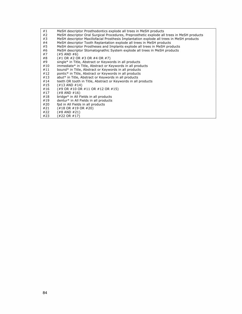
84
#1 MeSH descriptor Prosthodontics explode all trees in MeSH products #2 MeSH descriptor Oral Surgical Procedures, Preprosthetic explode all trees in MeSH products #3 MeSH descriptor Maxillofacial Prosthesis Implantation explode all trees in MeSH products #4 MeSH descriptor Tooth Replantation explode all trees in MeSH products #5 MeSH descriptor Prostheses and Implants explode all trees in MeSH products #6 MeSH descriptor Stomatognathic System explode all trees in MeSH products #7 (#5 AND #6) #8 (#1 OR #2 OR #3 OR #4 OR #7) #9 single* in Title, Abstract or Keywords in all products #10 immediate* in Title, Abstract or Keywords in all products #11 bound* in Title, Abstract or Keywords in all products #12 pontic* in Title, Abstract or Keywords in all products #13 abut* in Title, Abstract or Keywords in all products #14 teeth OR tooth in Title, Abstract or Keywords in all products #15 (#13 AND #14) #16 (#9 OR #10 OR #11 OR #12 OR #15) #17 (#8 AND #16) #18 bridge* in All Fields in all products #19 dentur* in All Fields in all products #20 fpd in All Fields in all products #21 (#18 OR #19 OR #20) #22 (#8 AND #21) #23 (#22 OR #17)

85
Appendix C
Data abstraction form, page 1.

86
Data Abstraction form, page 2.


![Disorders of intestinal rotation and fixation (‘‘malrotation’’)deepblue.lib.umich.edu/bitstream/handle/2027.42/46708/... · 2020. 2. 13. · consequences [4]. ‘‘Malrotation’’](https://img.pdfslide.net/doc/110x75/60afb5330f88520c4e13c968/disorders-of-intestinal-rotation-and-ixation-aamalrotationaa-2020-2.jpg)