Embed Size (px)
Citation preview
258 Korean J Radiol 5(4), December 2004
Comparison of Wet Radiofrequency Ablationwith Dry Radiofrequency Ablation andRadiofrequency Ablation Using HypertonicSaline Preinjection: Ex Vivo Bovine Liver
Objective: We wished to compare the in-vitro efficiency of wet radiofrequency(RF) ablation with the efficiency of dry RF ablation and RF ablation with preinjec-tion of NaCl solutions using excised bovine liver.
Materials and Methods: Radiofrequency was applied to excised bovine liversin a monopolar mode for 10 minutes using a 200 W generator and a perfused-cooled electrode with or without injection or slow infusion of NaCl solutions. Afterplacing the perfused-cooled electrode in the explanted liver, 50 ablation zoneswere created with five different regimens: group A; standard dry RF ablation,group B; RF ablation with 11 mL of 5% NaCl solution preinjection, group C; RFablation with infusion of 11 mL of 5% NaCl solution at a rate of 1 mL/min, groupD; RFA with 6 mL of 36% NaCl solution preinjection, group E; RF ablation withinfusion of 6 mL of 36% NaCl solution at a rate of 0.5 mL/min. In groups C and E,infusion of the NaCl solutions was started 1 min before RF ablation and thenmaintained during RF ablation (wet RF ablation). During RF ablation, we mea-sured the tissue temperature at 15 mm from the electrode. The dimensions of theablation zones and changes in impedance, current and liver temperature duringRF ablation were then compared between the groups.
Results: With injection or infusion of NaCl solutions, the mean initial tissueimpedance prior to RF ablation was significantly less in groups B, C, D, and E(43 75 ) than for group A (80 ) (p < 0.05). During RF ablation, the tissueimpedance was well controlled in groups C and E, but it was often rapidlyincreased to more than 200 in groups A and B. In group D, the impedancewas well controlled in six of ten trials but it was increased in four trials (40%) 7min after starting RF ablation. As consequences, the mean current was higher forgroups C, D, and E than for the other groups: 401 145 mA in group A, 287 32mA in group B, 1907 96 mA in group C, 1649 514 mA in group D, and 1968108 mA in group E (p < 0.05). In addition, the volumes of RF-induced coagulationnecrosis were greater in groups C and E than in group D, which was greater thanin groups A and B than in group E (p < 0.05); 14.3 3.0 cm3 in group A; 12.4 3.8cm3 in group B; 80.9 9.9 cm3 in group C; 45.3 11.3 cm3 in group D and 81.68.6 cm3 in group E. The tissue temperature measured at 15 mm from the elec-trode was higher in groups C, D and E than other groups (p < 0.05): 53 12 ingroup A, 42 2 in group B, 93 8 in group C; 79 12 in group D and 83
8 in group E.
Conclusion: Wet RF ablation with 5% or 36% NaCl solutions shows betterefficiency in creating a large ablation zone than does dry RF ablation or RF abla-tion with preinjection of NaCl solutions.
Jeong Min Lee, MD1, 2
Joon Koo Han, MD1, 2
Se Hyung Kim, MD1, 2
Kyung Sook Shin, MD3
Jae Young Lee, MD1, 2
Hee Sun Park, MD1, 2
Hurn Hur, MD1, 2
Byung Ihn Choi, MD1, 2
Index terms:Experimental studyInterventional proceduresLiverRadiofrequency ablation
Korean J Radiol 2004;5:258-265Received July 19, 2003; accepted after revision September 9, 2004.
1Department of Radiology, and Institute ofRadiation Medicine, Seoul NationalUniversity College of Medicine; 2ClinicalResearch Institute, Seoul NationalUniversity Hospital; 3Department ofRadiology, Chungnam NationalUniversity College of Medicine
This study was supported by grant No.09-2003-012-0 from the Seoul NationalUniversity Hospital Research Fund.
Address reprint requests to:Joon Koo Han, MD, Department ofDiagnostic Radiology, Seoul NationalUniversity Hospital, 28 Yongon-dong,Chongno-gu, Seoul 110-744, Korea.Tel. (822) 760-2154Fax. (822) 743-6385e-mail: [email protected]
mage-guided percutaneous radiofrequency(RF) ablation has become increasinglypopular in recent years, and it has been
accepted as an alternative to surgical resection for thetreatment of primary and secondary hepatic malignancy ininoperable patients (1 4). However, current RF technol-ogy is limited in that only tissue 3.5 to 4.5 cm indiameter could be ablated with a single application of RFenergy (5 9). This inherent limitation of monopolar RFablation, in regards to the small dimension of coagulationnecrosis, is attributed to resistive heating at a narrow rimof tissue that surrounds the electrode (1, 2, 10). There isalways a rapid increase of temperature at the tissue-electrode interface, and this results in tissue desiccationand charring at the electrode tip. These effects preventfurther RF energy conduction beyond the desiccated tissueand this halts further tissue coagulation.
There have been several reported strategies to solve thislimitation of monopolar RF ablation including RF ablationwith hypertonic saline (HS) preinjection (11 13), wet RFablation (RF ablation with continuous infusion of HSthrough the electrode) (14 16), bipolar RF ablation (17,18), and RF ablation using multiple probes (19 21).Among these approaches, we believe that hypertonicsaline-mediated RF ablation, which includes RF ablationwith HS preinjection and wet RF ablation, is seemingly avery attractive method because the injected saline couldincrease both the electrical and thermal conductivities, andthe higher boiling points of the hypertonic saline could behelpful in avoiding the rapid boiling of liver tissue adjacentto the electrode (12, 20, 22, 23). Until now, however,there has been no well-designed comparative studybetween RF ablation with HS preinjection and wet RFablation using HS infusion. In this context, we conducted astudy to verify whether wet RF ablation has betterefficiency in creating an ablation zone than does dry RFablation or RF ablation with HS preinjection, and we didthis by measuring the dimensions of the ablation zones inliver tissue, and by measuring the tissue temperature.
MATERIALS AND METHODS
RF Ablation SettingsBased on the previous studies that described consistent
results for tissue ablation from ex vivo RF ablation experi-ments (16, 17, 24, 25), we chose to use excised beef liverpurchased from a local butcher as the RF ablation targetfor our experiment. RF ablation was performed in 25freshly excised bovine livers weighing, on average, 7.5 Kgeach. The livers were cut into several 10 10 10-cm3
blocks that were dipped into a 50 20 20-cm3 saline-
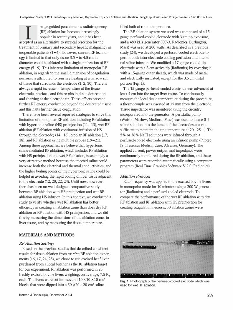
filled bath at room temperature.The RF ablation system we used was composed of a 15-
gauge perfused-cooled electrode with 3 cm tip exposure,and a 480 kHz generator (CC-3, Radionics, Burlington,Mass) was used at 200 watts. As described in a previousstudy (24), we developed a perfused-cooled electrode topermit both intra-electrode cooling perfusion and intersti-tial saline infusion. We modified a 17-gauge cooled-tipelectrode with a 3-cm active tip (Radionics) by covering itwith a 15-gauge outer sheath, which was made of metaland electrically insulated, except for the 3.5 cm distalportion (Fig. 1).
The 15-gauge perfused-cooled electrode was advanced atleast 4 cm into the target liver tissue. To continuouslymeasure the local tissue temperature during the procedure,a thermocouple was inserted at 15 mm from the electrode.Tissue impedance was monitored using the circuitryincorporated into the generator. A peristaltic pump(Watson-Marlow, Medford, Mass) was used to infuse 0 saline solution into the lumen of the electrodes at a ratesufficient to maintain the tip temperature at 20 25 . The5% or 36% NaCl solutions were infused through aperfused-cooled electrode using an infusion pump (PilotecIS; Fresenius Medical Care, Alzenau, Germany). Theapplied current, power output, and impedance werecontinuously monitored during the RF ablation, and theseparameters were recorded automatically using a computerprogram (Real Time Graphics Software V 2.0; Radionics).
Ablation ProtocolRadiofrequency was applied to the excised bovine livers
in monopolar mode for 10 minutes using a 200 W genera-tor (Radionics) and a perfused-cooled electrode. Tocompare the performance of the wet RF ablation with dryRF ablation and RF ablation with HS preinjection forcreating coagulation necrosis, 50 ablation zones were
Comparison Study of Wet Radiofrequency Ablation, Dry Radiofrequency Ablation and Ablation Using Hypertonic Saline Preinjection in Ex Vivo Bovine Liver
Korean J Radiol 5(4), December 2004 259
I
Fig. 1. Photograph of the perfused-cooled electrode which wasused for wet RF ablation.
created at five different regimens: group A; standard dry RFablation, group B; RF ablation with 11 cc of 5% NaClsolution preinjected, group C; wet RF ablation with infusionof 11cc of 5% NaCl solution at a rate of 1 mL/min, groupD; RF ablation with 6 cc of 36% NaCl solution preinjected,and group E; wet RF ablation with a infusion of 6 cc of 36%NaCl solution at a rate of 0.5 mL/min. In groups C and E,infusions of NaCl solutions were started before RF ablation(1 mL) and this was maintained during RF ablation atpredetermined infusion rates (wet RF ablation). The initialimpedance was set at 80 ohm with the electrode insertionby altering the distance between the electrodes and thedispersive metallic pad. Based on previous studies regardingsaline-mediated RF ablation, 5% and 36% NaCl solutionswere selected as the test solutions (12, 22, 26). Based on theresults of the previous study regarding the optimial concen-tration and volume of NaCl for RF ablation (12), a volumeof 6 mL saturated HS (36% NaCl) was used for comparisonbetween the two RF ablation techniques. To create thesame condition between the wet-RF ablation and the RFablation with preinjection of NaCl solutions, in group E(wet RF ablation), 1 mL of 36% NaCl was infused prior tothe RF ablation and 5 mL of the solution were injectedduring the RF ablation at a rate of 0.5 mL/min.Furthermore, based on other previous studies regarding wetRF ablation (17, 26), 5% NaCl at a rate of 1 mL/min wasselected to be infused. To create the same conditionbetween the two techniques, a volume of 11 mL wasselected to be preinjected.
The technical aspects of the RF ablation includingimpedance and wattage changes, tissue temperaturemeasured at 15 mm from the electrode, and thedimensions of the RF-coagulated area were compared foreach condition.
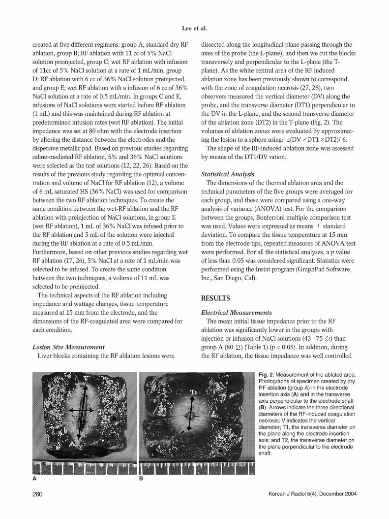
Lesion Size MeasurementLiver blocks containing the RF ablation lesions were
dissected along the longitudinal plane passing through theaxes of the probe (the L-plane), and then we cut the blockstransversely and perpendicular to the L-plane (the T-plane). As the white central area of the RF inducedablation zone has been previously shown to correspondwith the zone of coagulation necrosis (27, 28), twoobservers measured the vertical diameter (DV) along theprobe, and the transverse diameter (DT1) perpendicular tothe DV in the L-plane, and the second transverse diameterof the ablation zone (DT2) in the T-plane (Fig. 2). Thevolumes of ablation zones were evaluated by approximat-ing the lesion to a sphere using: (DV DT1 DT2)/ 6.
The shape of the RF-induced ablation zone was assessedby means of the DT1/DV ration.
Statistical AnalysisThe dimensions of the thermal ablation area and the
technical parameters of the five groups were averaged foreach group, and these were compared using a one-wayanalysis of variance (ANOVA) test. For the comparisonbetween the groups, Bonferroni multiple comparison testwas used. Values were expressed as means standarddeviation. To compare the tissue temperature at 15 mmfrom the electrode tips, repeated measures of ANOVA testwere performed. For all the statistical analyses, a p valueof less than 0.05 was considered significant. Statistics wereperformed using the Instat program (GraphPad Software,Inc., San Diego, Cal).
RESULTS
Electrical MeasurementsThe mean initial tissue impedance prior to the RF
ablation was significantly lower in the groups withinjection or infusion of NaCl solutions (43 75 ) thangroup A (80 ) (Table 1) (p < 0.05). In addition, duringthe RF ablation, the tissue impedance was well controlled
Lee et al.
260 Korean J Radiol 5(4), December 2004
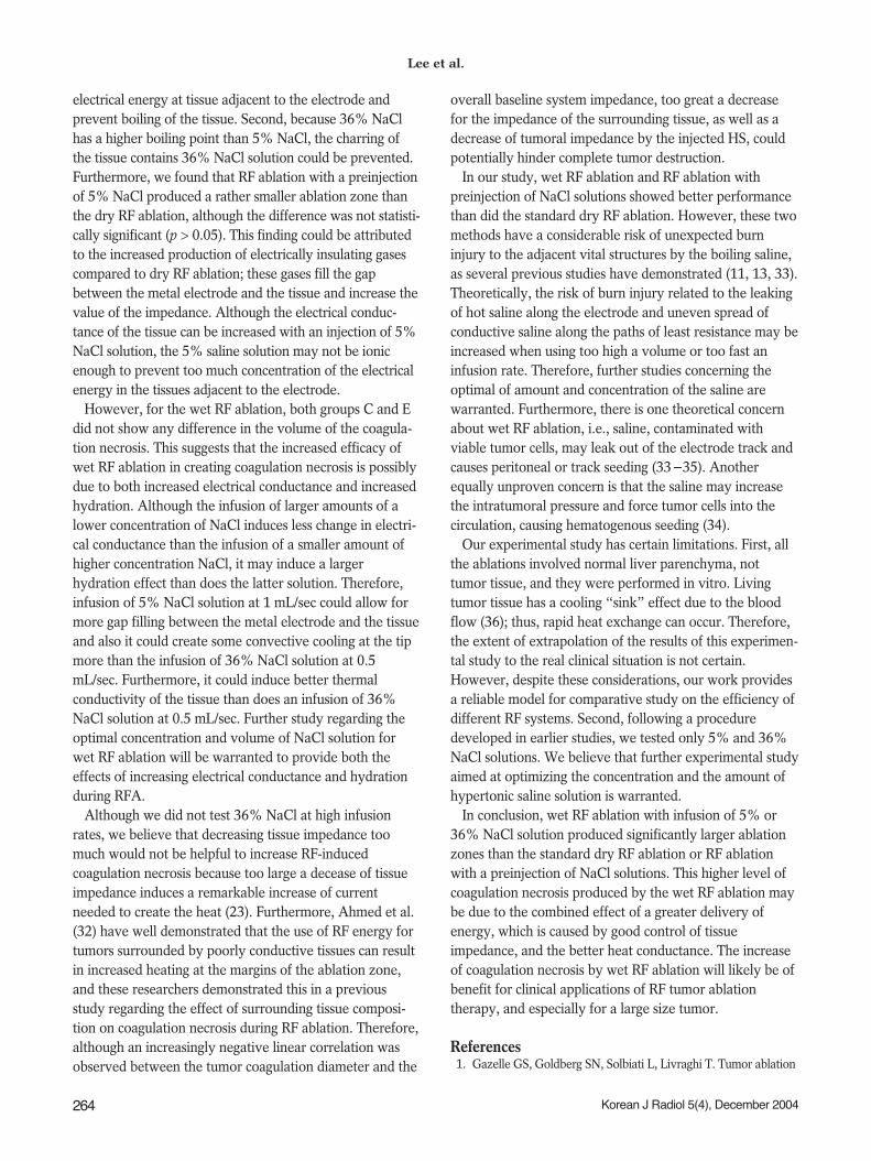
Fig. 2. Measurement of the ablated area.Photographs of specimen created by dryRF ablation (group A) in the electrodeinsertion axis (A) and in the transverseaxis perpendicular to the electrode shaft(B). Arrows indicate the three directionaldiameters of the RF-induced coagulationnecrosis: V indicates the verticaldiameter; T1, the transverse diameter onthe plane along the electrode insertionaxis; and T2, the transverse diameter onthe plane perpendicular to the electrodeshaft.
A B
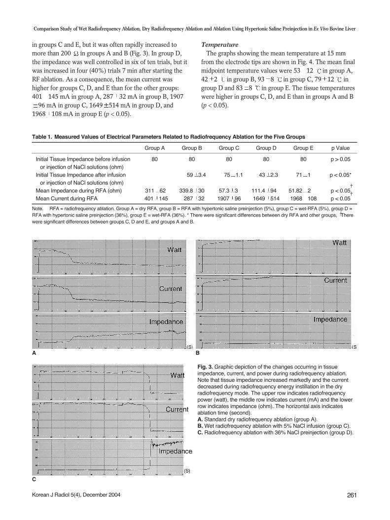
in groups C and E, but it was often rapidly increased tomore than 200 in groups A and B (Fig. 3). In group D,the impedance was well controlled in six of ten trials, but itwas increased in four (40%) trials 7 min after starting theRF ablation. As a consequence, the mean current washigher for groups C, D, and E than for the other groups:401 145 mA in group A, 287 32 mA in group B, 1907
96 mA in group C, 1649 514 mA in group D, and1968 108 mA in group E (p < 0.05).
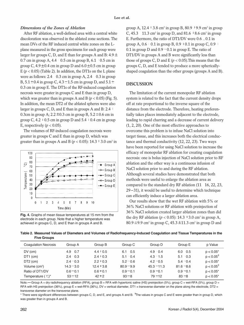
TemperatureThe graphs showing the mean temperature at 15 mm
from the electrode tips are shown in Fig. 4. The mean finalmidpoint temperature values were 53 12 in group A,42 2 in group B, 93 8 in group C, 79 12 ingroup D and 83 8 in group E. The tissue temperatureswere higher in groups C, D, and E than in groups A and B(p < 0.05).
Comparison Study of Wet Radiofrequency Ablation, Dry Radiofrequency Ablation and Ablation Using Hypertonic Saline Preinjection in Ex Vivo Bovine Liver
Korean J Radiol 5(4), December 2004 261
Table 1. Measured Values of Electrical Parameters Related to Radiofrequency Ablation for the Five Groups
Group A Group B Group C Group D Group E p Value
Initial Tissue Impedance before infusion 80 80 80 80 80 p > 0.05or injection of NaCl solutions (ohm)
Initial Tissue Impedance after infusion 00059 3.4 00075 1.1 0.43 2.3 71 1 p < 0.05*or injection of NaCl solutions (ohm)
Mean Impedance during RFA (ohm) .311 62 339.8 30 57.3 3. 111.4 940. 51.82 200. p < 0.05 Mean Current during RFA 0401 145 0.287 32 1907 96 1649 514 1968 108 p < 0.05
Note. RFA = radiofrequency ablation. Group A = dry RFA, group B = RFA with hypertonic saline preinjection (5%), group C = wet-RFA (5%), group D =RFA with hypertonic saline preinjection (36%), group E = wet-RFA (36%). * There were significant differences between dry RFA and other groups, Therewere significant differences between groups C, D and E, and groups A and B.
Fig. 3. Graphic depiction of the changes occurring in tissueimpedance, current, and power during radiofrequency ablation.Note that tissue impedance increased markedly and the currentdecreased during radiofrequency energy instillation in the dryradiofrequency mode. The upper row indicates radiofrequencypower (watt), the middle row indicates current (mA) and the lowerrow indicates impedance (ohm). The horizontal axis indicatesablation time (second).A. Standard dry radiofrequency ablation (group A).B. Wet radiofrequency ablation with 5% NaCl infusion (group C).C. Radiofrequency ablation with 36% NaCl preinjection (group D).
C
A B
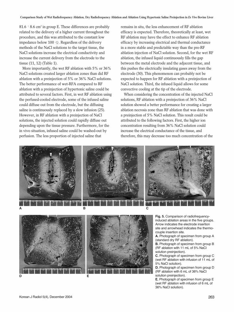
Dimensions of the Zones of AblationAfter RF ablation, a well-defined area with a central white
discoloration was observed in the ablated zone sections. Themean DVs of the RF induced central white zones on the L-plane measured in the gross specimens for each group werelarger for groups C, D, and E than for groups A and B: 4.90.7 cm in group A, 4.4 0.5 cm in group B, 6.1 0.5 cm ingroup C, 4.9 0.4 cm in group D and 6.0 0.5 cm in groupE (p < 0.05) (Table 2). In addition, the DT1s on the L planewere as follows: 2.4 0.3 cm in group A, 2.4 0.3 in groupB, 5.1 0.4 in group C, 4.3 1.5 cm in group D, and 5.10.3 cm in group E. The DT1s of the RF-induced coagulationnecrosis were greater in groups C and E than in group D,which was greater than in groups A and B (p < 0.05) (Fig. 5).In addition, the mean DT2 of the ablated spheres were alsolarger in groups C, D, and E than in groups A and B: 2.40.3cm in group A; 2.2 0.3 cm in group B, 5.2 0.6 cm ingroup C, 4.2 0.5 cm in group D and 5.4 0.4 cm in groupE, respectively (p < 0.05).
The volumes of RF-induced coagulation necrosis weregreater in groups C and E than in group D, which wasgreater than in groups A and B (p < 0.05): 14.3 3.0 cm3 in
group A, 12.4 3.8 cm3 in group B, 80.9 9.9 cm3 in groupC, 45.3 11.3 cm3 in group D, and 81.6 8.6 cm3 in groupE. Furthermore, the ratio of DT1/DV were 0.6 0.1 ingroup A, 0.6 0.1 in group B, 0.9 0.1 in group C, 0.90.1 in group D and 0.9 0.1 in group E. The ratio ofDT1/DV in groups A and B were significantly less thanthose of groups C, D and E (p < 0.05).This means that thegroups C, D, and E tended to produce a more spherically-shaped coagulation than the other groups (groups A and B).
DISCUSSION
The limitation of the current monopolar RF ablationsystem is related to the fact that the current density dropsoff at rate proportional to the inverse square of thedistance from the electrode. Therefore, heating preferen-tially takes places immediately adjacent to the electrode,leading to rapid charring and a decrease of current delivery(1, 2, 20). One of the most effective approaches toovercome this problem is to infuse NaCl solution intotarget tissue, and this increases both the electrical conduc-tance and thermal conductivity (12, 22, 23). Two wayshave been reported for using NaCl solution to increase theefficacy of monopolar RF ablation for creating coagulationnecrosis: one is bolus injection of NaCl solution prior to RFablation and the other way is a continuous infusion ofNaCl solution prior to and during the RF ablation.Although several studies have demonstrated that bothmethods were useful to enlarge the ablation area ascompared to the standard dry RF ablation (11 16, 22, 23,29 31), it would be useful to determine which techniquecan efficiently induce a larger ablation area.
Our results show that the wet RF ablation with 5% or36% NaCl solutions or RF ablation with preinjection of36% NaCl solution created larger ablation zones than didthe dry RF ablation (p < 0.05): 14.3 3.0 cm3 in group A,80.9 9.9 cm3 in group C, 45.3 11.3 cm3 in group D and
Lee et al.
262 Korean J Radiol 5(4), December 2004
Table 2. Measured Values of Diameters and Volumes of Radiofrequency-Induced Coagulation and Tissue Temperatures in theFive Groups
Coagulation Necrosis Group A Group B Group C Group D Group E p Value
DV (cm) 4.9 0.7 4.4 0.5 6.1 0.5 4.9 0.4 6.0 0.5 p < 0.05*DT1 (cm) 2.4 0.3 2.4 0.3 5.1 0.4 4.3 1.5 5.1 0.3 p < 0.05*DT2 (cm) 2.4 0.3 2.2 0.3 5.2 0.6 4.2 0.5 5.4 0.4 p < 0.05*Volume (cm3) 14.3 3.00 12.4 3.80 80.9 9.90 45.3 11.3 81.6 8.60 p < 0.05*Ratio of DT1/DV 0.6 0.1 0.6 0.1 0.9 0.1 0.9 0.1 0.9 0.1 p < 0.05*Temperature ( )* 53 12 42 20 93 80 79 12 83 80 p < 0.05*
Note. Group A = dry radiofrequency ablation (RFA), group B = RFA with hypertonic saline (HS) preinjection (5%), group C = wet-RFA (5%), group D =RFA with HS preinjection (36%), group E = wet-RFA (36%), DV = vertical diameter, DT1 = transverse diameter on the plane along the electrode, DT2 =transverse diameter on the transverse plane.* There were significant differences between groups C, D, and E, and groups A and B. The values in groups C and E were greater than in group D, whichwas greater than in groups A and B.
Fig. 4. Graphs of mean tissue temperatures at 15 mm from theelectrode in each group. Note that a higher temperature wasachieved in groups C, D, and E than in groups A and B.
81.6 8.6 cm3 in group E. These differences are probablyrelated to the delivery of a higher current throughout theprocedure, and this was attributed to the constant lowimpedance below 100 . Regardless of the deliverymethods of the NaCl solutions to the target tissue, theNaCl solutions increase the electrical conductivity andincrease the current delivery from the electrode to thetissue (11, 12) (Table 1).
More importantly, the wet RF ablation with 5% or 36%NaCl solutions created larger ablation zones than did RFablation with a preinjection of 5% or 36% NaCl solutions.The better performance of wet-RFA compared to RFablation with a preinjection of hypertonic saline could beattributed to several factors. First, in wet RF ablation usingthe perfused-cooled electrode, some of the infused salinecould diffuse out from the electrode, but the diffusingsaline is continuously replaced by a slow infusion (25).However, in RF ablation with a preinjection of NaClsolutions, the injected solution could rapidly diffuse outdepending upon the tissue pressure. Furthermore, for thein vivo situation, infused saline could be washed-out byperfusion. The less proportion of injected saline that
remains in situ, the less enhancement of RF ablationefficacy is expected. Therefore, theoretically at least, wetRF ablation may have the effect to enhance RF ablationefficacy by increasing electrical and thermal conductancein a more stable and predictable way than the pre-RFablation injection of NaCl solution. Second, for the wet RFablation, the infused liquid continuously fills the gapbetween the metal electrode and the adjacent tissue, andthis pushes the electrically insulating gases away from theelectrode (30). This phenomenon can probably not beexpected to happen for RF ablation with a preinjection ofNaCl solution. Third, the infused liquid allows for someconvective cooling at the tip of the electrode.
When considering the concentration of the injected NaClsolutions, RF ablation with a preinjection of 36% NaClsolution showed a better performance for creating a largerablation necrosis zone than RF ablation that was done witha preinjection of 5% NaCl solution. This result could beattributed to the following factors. First, the higher ionconcentration resulting from 36% NaCl solution couldincrease the electrical conductance of the tissue, andtherefore, this may decrease too much concentration of the
Comparison Study of Wet Radiofrequency Ablation, Dry Radiofrequency Ablation and Ablation Using Hypertonic Saline Preinjection in Ex Vivo Bovine Liver
Korean J Radiol 5(4), December 2004 263
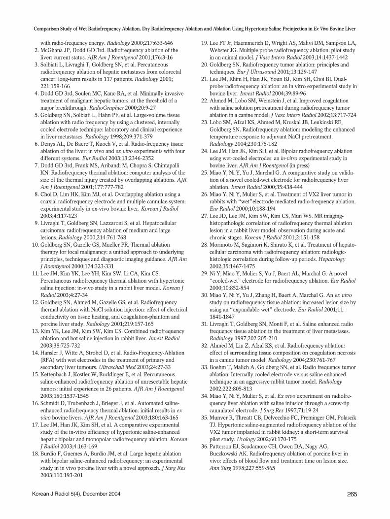
A B C
Fig. 5. Comparison of radiofrequency-induced ablation areas in the five groups.Arrow indicates the electrode insertionsite and arrowhead indicates the thermo-couple insertion site.A. Photograph of specimen from group A(standard dry RF ablation).B. Photograph of specimen from group B(RF ablation with 11 mL of 5% NaClsolution preinjection).C. Photograph of specimen from group C(wet RF ablation with infusion of 11 mL of5% NaCl solution).D. Photograph of specimen from group D(RF ablation with 6 mL of 36% NaClsolution preinjection).E. Photograph of specimen from group E(wet RF ablation with infusion of 6 mL of36% NaCl solution).
D E
electrical energy at tissue adjacent to the electrode andprevent boiling of the tissue. Second, because 36% NaClhas a higher boiling point than 5% NaCl, the charring ofthe tissue contains 36% NaCl solution could be prevented.Furthermore, we found that RF ablation with a preinjectionof 5% NaCl produced a rather smaller ablation zone thanthe dry RF ablation, although the difference was not statisti-cally significant (p > 0.05). This finding could be attributedto the increased production of electrically insulating gasescompared to dry RF ablation; these gases fill the gapbetween the metal electrode and the tissue and increase thevalue of the impedance. Although the electrical conduc-tance of the tissue can be increased with an injection of 5%NaCl solution, the 5% saline solution may not be ionicenough to prevent too much concentration of the electricalenergy in the tissues adjacent to the electrode.
However, for the wet RF ablation, both groups C and Edid not show any difference in the volume of the coagula-tion necrosis. This suggests that the increased efficacy ofwet RF ablation in creating coagulation necrosis is possiblydue to both increased electrical conductance and increasedhydration. Although the infusion of larger amounts of alower concentration of NaCl induces less change in electri-cal conductance than the infusion of a smaller amount ofhigher concentration NaCl, it may induce a largerhydration effect than does the latter solution. Therefore,infusion of 5% NaCl solution at 1 mL/sec could allow formore gap filling between the metal electrode and the tissueand also it could create some convective cooling at the tipmore than the infusion of 36% NaCl solution at 0.5mL/sec. Furthermore, it could induce better thermalconductivity of the tissue than does an infusion of 36%NaCl solution at 0.5 mL/sec. Further study regarding theoptimal concentration and volume of NaCl solution forwet RF ablation will be warranted to provide both theeffects of increasing electrical conductance and hydrationduring RFA.
Although we did not test 36% NaCl at high infusionrates, we believe that decreasing tissue impedance toomuch would not be helpful to increase RF-inducedcoagulation necrosis because too large a decease of tissueimpedance induces a remarkable increase of currentneeded to create the heat (23). Furthermore, Ahmed et al.(32) have well demonstrated that the use of RF energy fortumors surrounded by poorly conductive tissues can resultin increased heating at the margins of the ablation zone,and these researchers demonstrated this in a previousstudy regarding the effect of surrounding tissue composi-tion on coagulation necrosis during RF ablation. Therefore,although an increasingly negative linear correlation wasobserved between the tumor coagulation diameter and the
overall baseline system impedance, too great a decreasefor the impedance of the surrounding tissue, as well as adecrease of tumoral impedance by the injected HS, couldpotentially hinder complete tumor destruction.
In our study, wet RF ablation and RF ablation withpreinjection of NaCl solutions showed better performancethan did the standard dry RF ablation. However, these twomethods have a considerable risk of unexpected burninjury to the adjacent vital structures by the boiling saline,as several previous studies have demonstrated (11, 13, 33).Theoretically, the risk of burn injury related to the leakingof hot saline along the electrode and uneven spread ofconductive saline along the paths of least resistance may beincreased when using too high a volume or too fast aninfusion rate. Therefore, further studies concerning theoptimal of amount and concentration of the saline arewarranted. Furthermore, there is one theoretical concernabout wet RF ablation, i.e., saline, contaminated withviable tumor cells, may leak out of the electrode track andcauses peritoneal or track seeding (33 35). Anotherequally unproven concern is that the saline may increasethe intratumoral pressure and force tumor cells into thecirculation, causing hematogenous seeding (34).
Our experimental study has certain limitations. First, allthe ablations involved normal liver parenchyma, nottumor tissue, and they were performed in vitro. Livingtumor tissue has a cooling “sink” effect due to the bloodflow (36); thus, rapid heat exchange can occur. Therefore,the extent of extrapolation of the results of this experimen-tal study to the real clinical situation is not certain.However, despite these considerations, our work providesa reliable model for comparative study on the efficiency ofdifferent RF systems. Second, following a proceduredeveloped in earlier studies, we tested only 5% and 36%NaCl solutions. We believe that further experimental studyaimed at optimizing the concentration and the amount ofhypertonic saline solution is warranted.
In conclusion, wet RF ablation with infusion of 5% or36% NaCl solution produced significantly larger ablationzones than the standard dry RF ablation or RF ablationwith a preinjection of NaCl solutions. This higher level ofcoagulation necrosis produced by the wet RF ablation maybe due to the combined effect of a greater delivery ofenergy, which is caused by good control of tissueimpedance, and the better heat conductance. The increaseof coagulation necrosis by wet RF ablation will likely be ofbenefit for clinical applications of RF tumor ablationtherapy, and especially for a large size tumor.
References1. Gazelle GS, Goldberg SN, Solbiati L, Livraghi T. Tumor ablation
Lee et al.
264 Korean J Radiol 5(4), December 2004
with radio-frequency energy. Radiology 2000;217:633-6462. McGhana JP, Dodd GD 3rd. Radiofrequency ablation of the
liver: current status. AJR Am J Roentgenol 2001;176:3-163. Solbiati L, Livraghi T, Goldberg SN, et al. Percutaneous
radiofrequency ablation of hepatic metastases from colorectalcancer: long-term results in 117 patients. Radiology 2001;221:159-166
4. Dodd GD 3rd, Soulen MC, Kane RA, et al. Minimally invasivetreatment of malignant hepatic tumors: at the threshold of amajor breakthrough. RadioGraphics 2000;20:9-27
5. Goldberg SN, Solbiati L, Hahn PF, et al. Large-volume tissueablation with radio frequency by using a clustered, internallycooled electrode technique: laboratory and clinical experiencein liver metastases. Radiology 1998;209:371-379
6. Denys AL, De Baere T, Kuoch V, et al. Radio-frequency tissueablation of the liver: in vivo and ex vivo experiments with fourdifferent systems. Eur Radiol 2003;13:2346-2352
7. Dodd GD 3rd, Frank MS, Aribandi M, Chopra S, ChintapalliKN. Radiofrequency thermal ablation: computer analysis of thesize of the thermal injury created by overlapping ablations. AJRAm J Roentgenol 2001;177:777-782
8. Choi D, Lim HK, Kim MJ, et al. Overlapping ablation using acoaxial radiofrequency electrode and multiple cannulae system:experimental study in ex-vivo bovine liver. Korean J Radiol2003;4:117-123
9. Livraghi T, Goldberg SN, Lazzaroni S, et al. Hepatocellularcarcinoma: radiofrequency ablation of medium and largelesions. Radiology 2000;214:761-768
10. Goldberg SN, Gazelle GS, Mueller PR. Thermal ablationtherapy for focal malignancy: a unified approach to underlyingprinciples, techniques and diagnostic imaging guidance. AJR AmJ Roentgenol 2000;174:323-331
11. Lee JM, Kim YK, Lee YH, Kim SW, Li CA, Kim CS.Percutaneous radiofrequency thermal ablation with hypertonicsaline injection: in-vivo study in a rabbit liver model. Korean JRadiol 2003;4:27-34
12. Goldberg SN, Ahmed M, Gazelle GS, et al. Radiofrequencythermal ablation with NaCl solution injection: effect of electricalconductivity on tissue heating, and coagulation-phantom andporcine liver study. Radiology 2001;219:157-165
13. Kim YK, Lee JM, Kim SW, Kim CS. Combined radiofrequencyablation and hot saline injection in rabbit liver. Invest Radiol2003;38:725-732
14. Hansler J, Witte A, Strobel D, et al. Radio-Frequency-Ablation(RFA) with wet electrodes in the treatment of primary andsecondary liver tumours. Ultraschall Med 2003;24:27-33
15. Kettenbach J, Kostler W, Rucklinger E, et al. Percutaneoussaline-enhanced radiofrequency ablation of unresectable hepatictumors: initial experience in 26 patients. AJR Am J Roentgenol2003;180:1537-1545
16. Schmidt D, Trubenbach J, Brieger J, et al. Automated saline-enhanced radiofrequency thermal ablation: initial results in exvivo bovine livers. AJR Am J Roentgenol 2003;180:163-165
17. Lee JM, Han JK, Kim SH, et al. A comparative experimentalstudy of the in-vitro efficiency of hypertonic saline-enhancedhepatic bipolar and monopolar radiofrequency ablation. KoreanJ Radiol 2003;4:163-169
18. Burdio F, Guemes A, Burdio JM, et al. Large hepatic ablationwith bipolar saline-enhanced radiofrequency: an experimentalstudy in in vivo porcine liver with a novel approach. J Surg Res2003;110:193-201
19. Lee FT Jr, Haemmerich D, Wright AS, Mahvi DM, Sampson LA,Webster JG. Multiple probe radiofrequency ablation: pilot studyin an animal model. J Vasc Interv Radiol 2003;14:1437-1442
20. Goldberg SN. Radiofrequency tumor ablation: principles andtechniques. Eur J Ultrasound 2001;13:129-147
21. Lee JM, Rhim H, Han JK, Youn BJ, Kim SH, Choi BI. Dual-probe radiofrequency ablation: an in vitro experimental study inbovine liver. Invest Radiol 2004;39:89-96
22. Ahmed M, Lobo SM, Weinstein J, et al. Improved coagulationwith saline solution pretreatment during radiofrequency tumorablation in a canine model. J Vasc Interv Radiol 2002;13:717-724
23. Lobo SM, Afzal KS, Ahmed M, Kruskal JB, Lenkinski RE,Goldberg SN. Radiofrequency ablation: modeling the enhancedtemperature response to adjuvant NaCl pretreatment.Radiology 2004;230:175-182
24. Lee JM, Han JK, Kim SH, et al. Bipolar radiofrequency ablationusing wet-cooled electrodes: an in-vitro experimental study inbovine liver. AJR Am J Roentgenol (in press)
25. Miao Y, Ni Y, Yu J, Marchal G. A comparative study on valida-tion of a novel cooled-wet electrode for radiofrequency liverablation. Invest Radiol 2000;35:438-444
26. Miao Y, Ni Y, Mulier S, et al. Treatment of VX2 liver tumor inrabbits with “wet”electrode mediated radio-frequency ablation.Eur Radiol 2000;10:188-194
27. Lee JD, Lee JM, Kim SW, Kim CS, Mun WS. MR imaging-histopathologic correlation of radiofrequency thermal ablationlesion in a rabbit liver model: observation during acute andchronic stages. Korean J Radiol 2001;2:151-158
28. Morimoto M, Sugimori K, Shirato K, et al. Treatment of hepato-cellular carcinoma with radiofrequency ablation: radiologic-histologic correlation during follow-up periods. Hepatology2002;35:1467-1475
29. Ni Y, Miao Y, Mulier S, Yu J, Baert AL, Marchal G. A novel“cooled-wet” electrode for radiofrequency ablation. Eur Radiol2000;10:852-854
30. Miao Y, Ni Y, Yu J, Zhang H, Baert A, Marchal G. An ex vivostudy on radiofrequency tissue ablation: increased lesion size byusing an “expandable-wet” electrode. Eur Radiol 2001;11:1841-1847
31. Livraghi T, Goldberg SN, Monti F, et al. Saline enhanced radiofrequency tissue ablation in the treatment of liver metastases.Radiology 1997;202:205-210
32. Ahmed M, Liu Z, Afzal KS, et al. Radiofrequency ablation:effect of surrounding tissue composition on coagulation necrosisin a canine tumor model. Radiology 2004;230:761-767
33. Boehm T, Malich A, Goldberg SN, et al. Radio frequency tumorablation: Internally cooled electrode versus saline enhancedtechnique in an aggressive rabbit tumor model. Radiology2002;222:805-813
34. Miao Y, Ni Y, Mulier S, et al. Ex vivo experiment on radiofre-quency liver ablation with saline infusion through a screw-tipcannulated electrode. J Surg Res 1997;71:19-24
35. Munver R, Threatt CB, Delvecchio FC, Preminger GM, PolascikTJ. Hypertonic saline-augmented radiofrequency ablation of theVX2 tumor implanted in rabbit kidney: a short-term survivalpilot study. Urology 2002;60:170-175
36. Patterson EJ, Scudamore CH, Owen DA, Nagy AG,Buczkowski AK. Radiofrequency ablation of porcine liver invivo: effects of blood flow and treatment time on lesion size.Ann Surg 1998;227:559-565
Comparison Study of Wet Radiofrequency Ablation, Dry Radiofrequency Ablation and Ablation Using Hypertonic Saline Preinjection in Ex Vivo Bovine Liver
Korean J Radiol 5(4), December 2004 265