Embed Size (px)
Citation preview
Compartment Syndrome
Compartment SyndromeDefinition
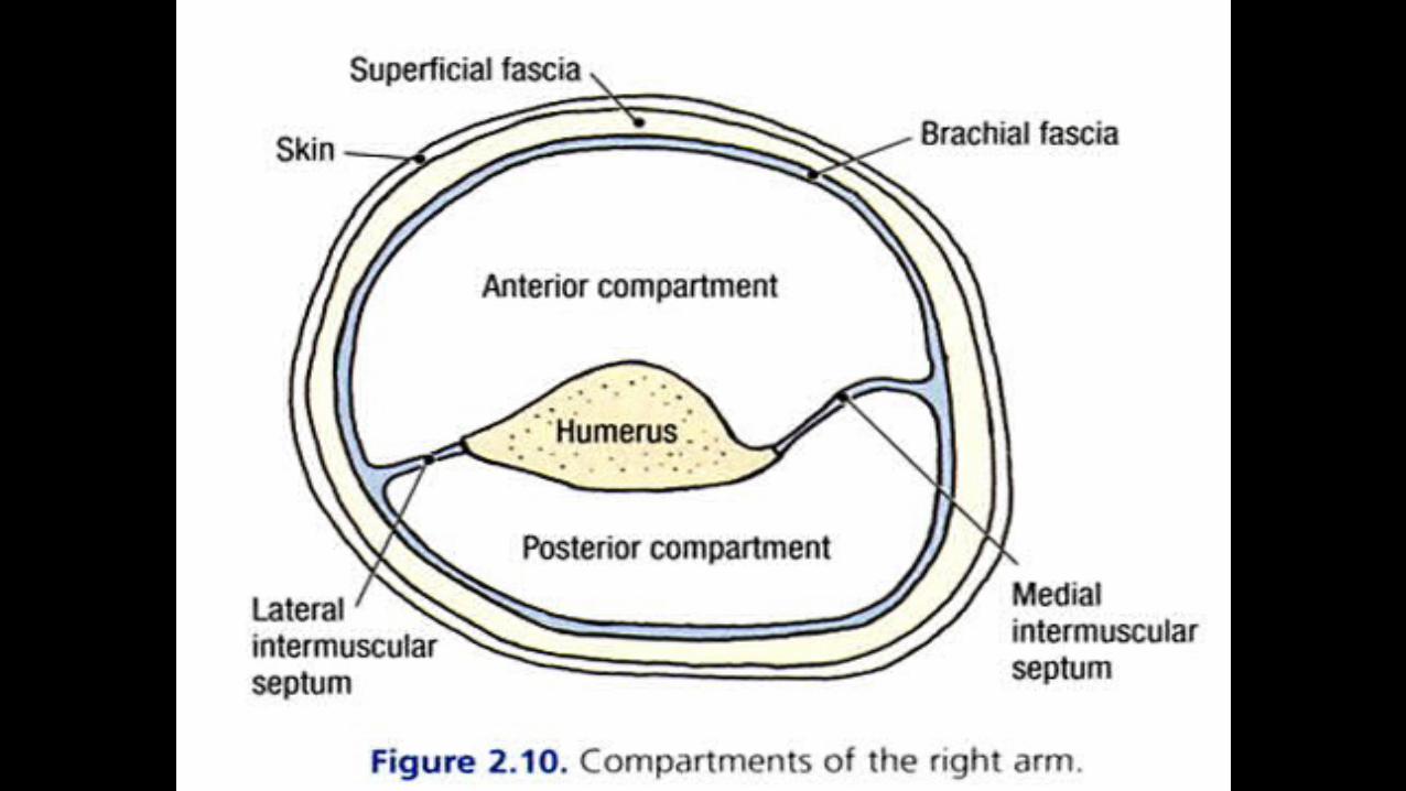
Elevated tissue pressure within a closed fascial space
Reduces tissue perfusion - ischemia Results in cell death - necrosis
True Orthopaedic Emergency
Acute Compartment Syndrome Of The Upper Arm
Compartment Syndrome
A condition in which increased pressure
within a limited space compromises the circulation and function of the tissues within that space.
Compartment Syndrome
when pressure within a closed muscle compartment exceeds the perfusion pressure it will results in muscle and nerve ischemia.
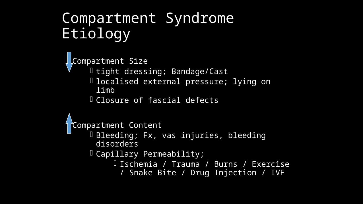
Compartment SyndromeEtiology
Compartment Size tight dressing; Bandage/Cast localised external pressure; lying on limb Closure of fascial defects
Compartment Content Bleeding; Fx, vas injuries, bleeding disorders Capillary Permeability;
Ischemia / Trauma / Burns / Exercise / Snake Bite / Drug Injection / IVF
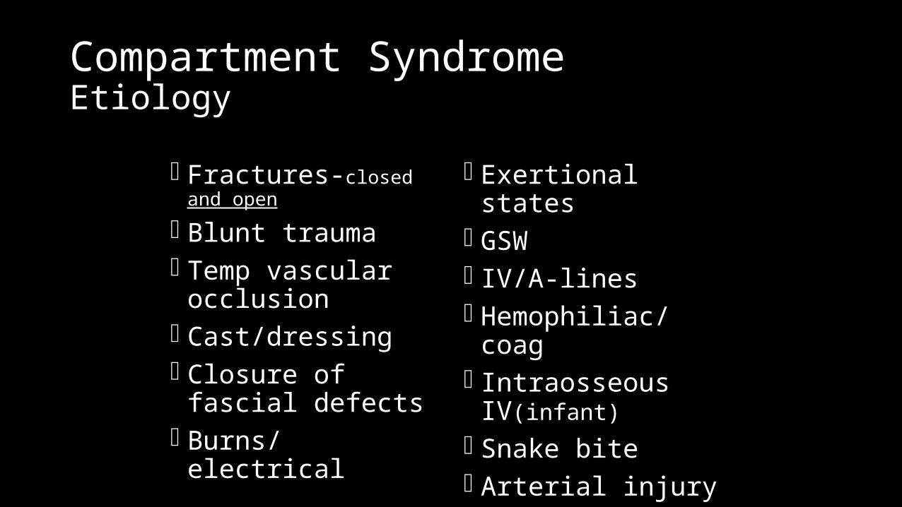
Compartment SyndromeEtiology
Fractures-closed and open
Blunt trauma Temp vascular
occlusion Cast/dressing Closure of fascial
defects Burns/electrical
Exertional states GSW IV/A-lines Hemophiliac/coag Intraosseous IV(infant) Snake bite Arterial injury
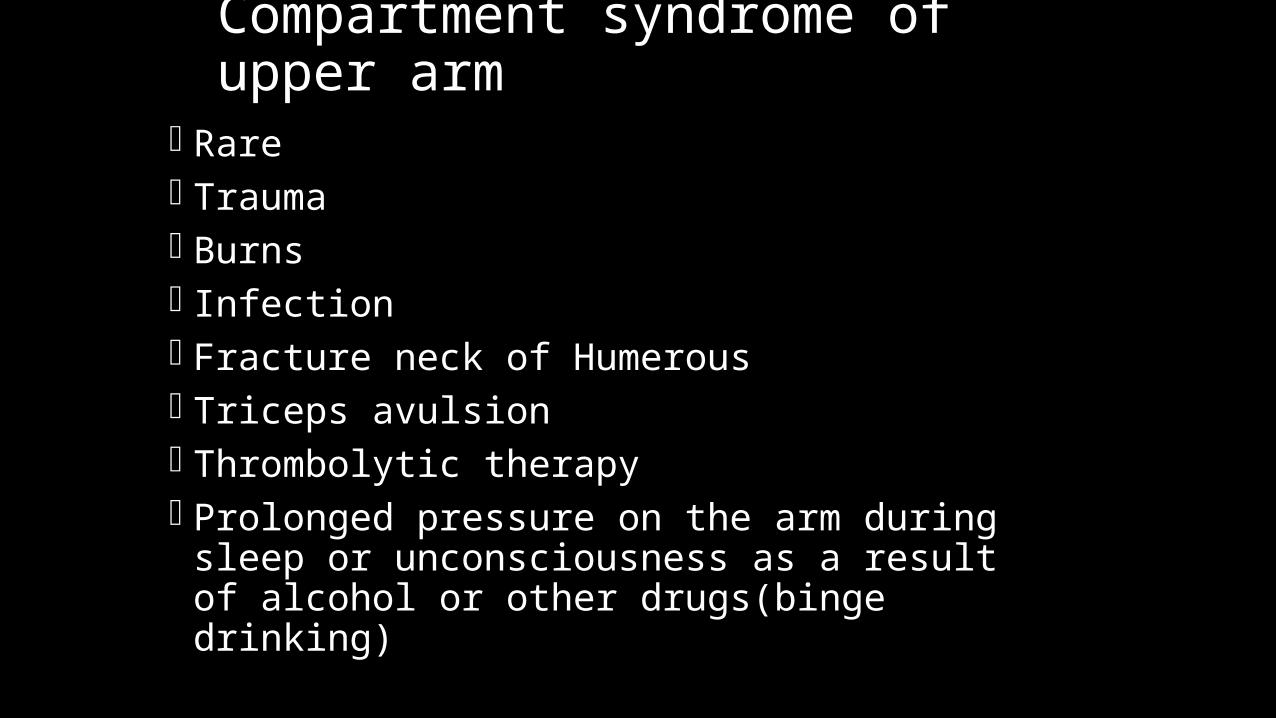
Compartment syndrome of upper arm
Rare Trauma Burns Infection Fracture neck of Humerous Triceps avulsion Thrombolytic therapy Prolonged pressure on the arm during sleep or
unconsciousness as a result of alcohol or other drugs(binge drinking)
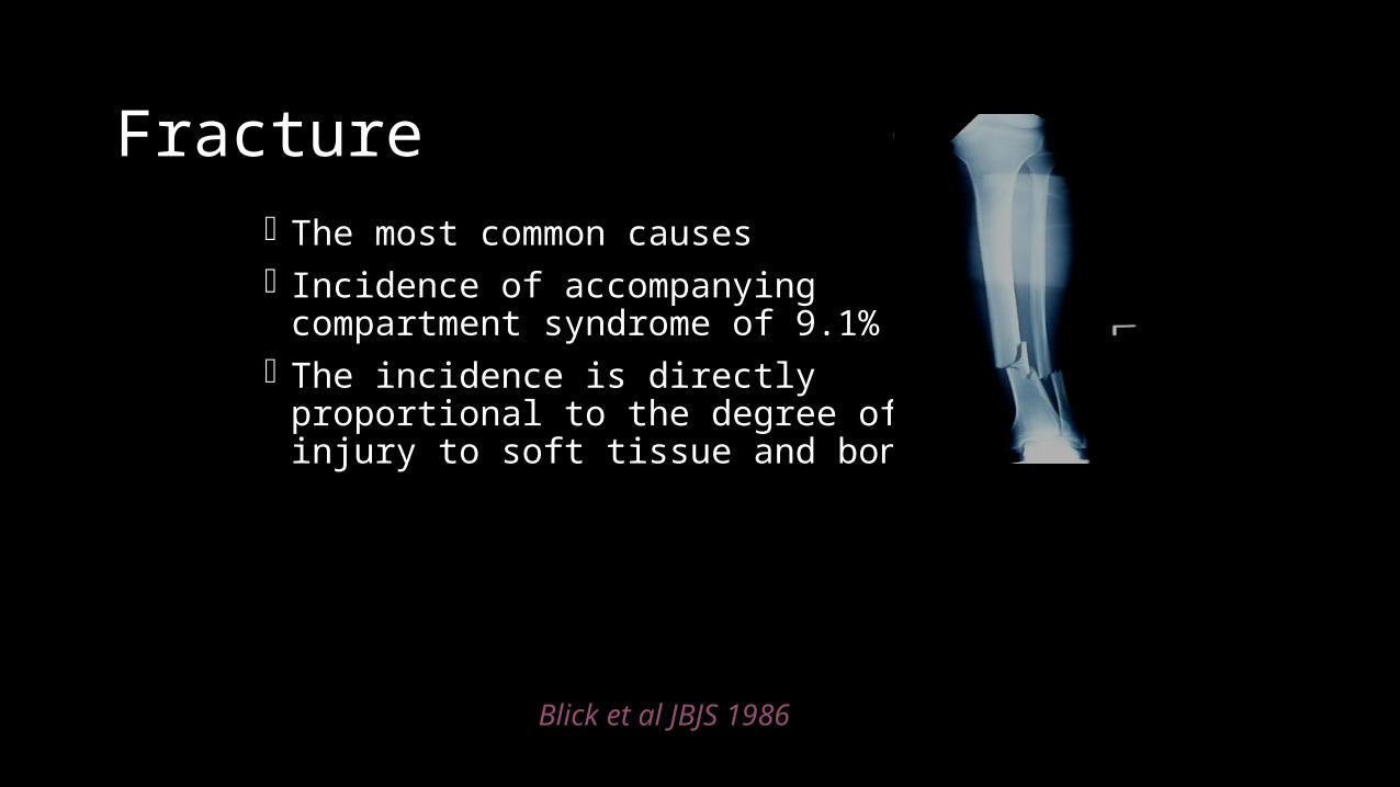
Fracture The most common causes Incidence of accompanying compartment
syndrome of 9.1% The incidence is directly proportional to the
degree of injury to soft tissue and bone
Blick et al JBJS 1986
Blunt Trauma
2nd most common cause About 23% of CS 25% due to direct blow
McQueen et al; JBJS Br 2000
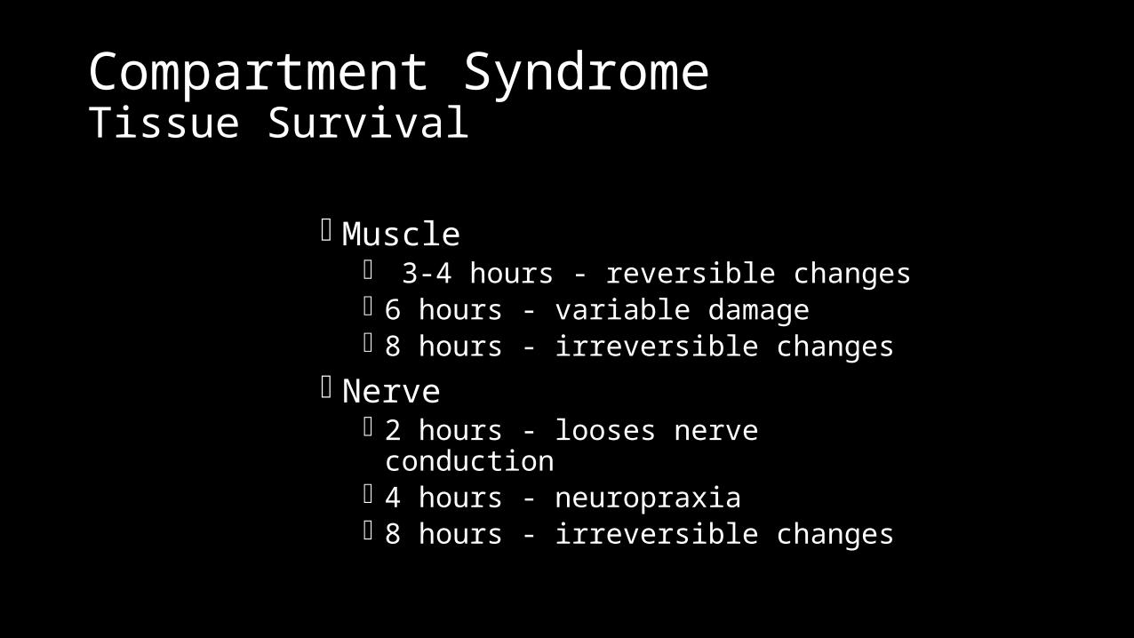
Compartment SyndromeTissue Survival
Muscle 3-4 hours - reversible changes 6 hours - variable damage 8 hours - irreversible changes
Nerve 2 hours - looses nerve conduction 4 hours - neuropraxia 8 hours - irreversible changes
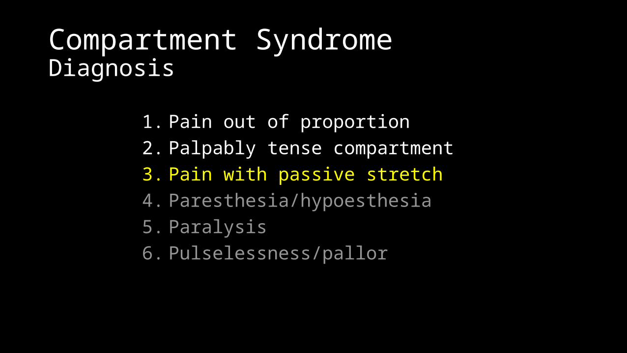
Compartment SyndromeDiagnosis
1. Pain out of proportion2. Palpably tense compartment3. Pain with passive stretch4. Paresthesia/hypoesthesia5. Paralysis6. Pulselessness/pallor
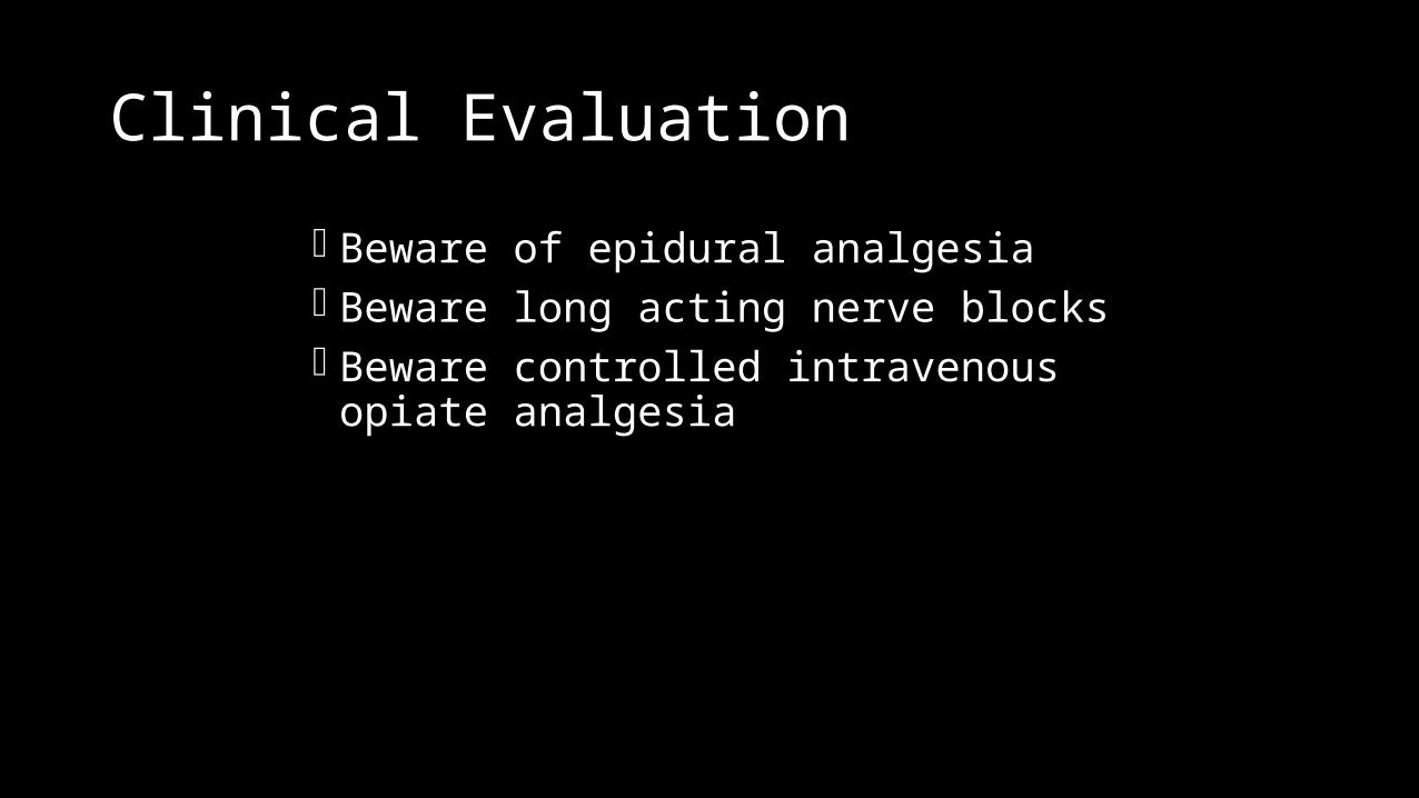
Clinical Evaluation
Beware of epidural analgesia Beware long acting nerve blocks Beware controlled intravenous opiate analgesia
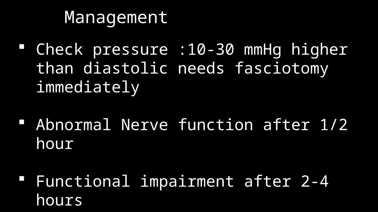
Management
Check pressure :10-30 mmHg higher than diastolic needs fasciotomy immediately
Abnormal Nerve function after 1/2 hour
Functional impairment after 2-4 hours Irreversible function loss after 4-12 hours Acute Renal Failure : Rhabdomyolysis
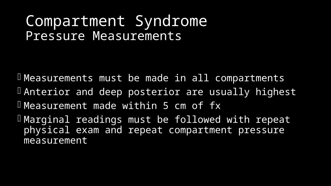
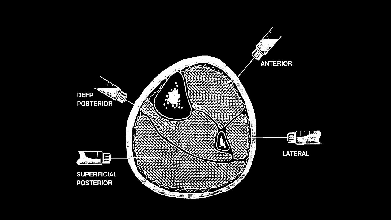
Compartment SyndromePressure Measurements
Measurements must be made in all compartments Anterior and deep posterior are usually highest Measurement made within 5 cm of fx Marginal readings must be followed with repeat physical exam and
repeat compartment pressure measurement
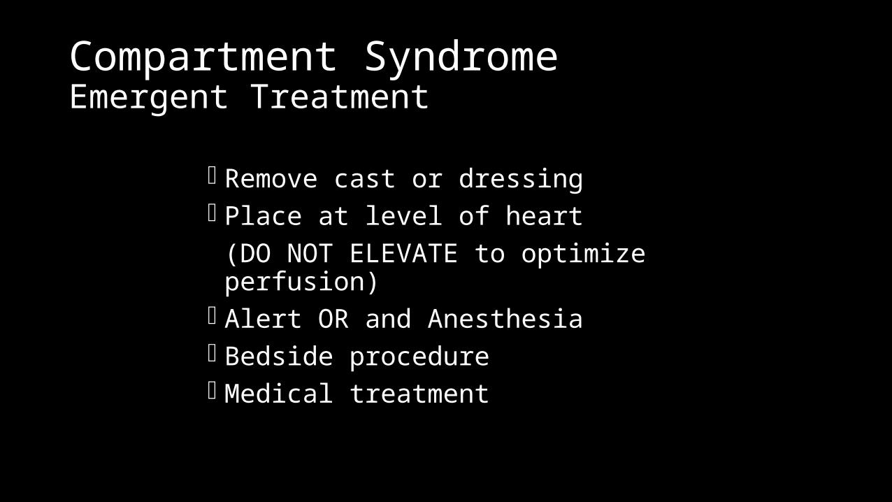
Compartment SyndromeEmergent Treatment
Remove cast or dressing Place at level of heart
(DO NOT ELEVATE to optimize perfusion) Alert OR and Anesthesia Bedside procedure Medical treatment
Compartment SyndromeDifferential Diagnosis
Arterial occlusion
Peripheral nerve injury
Muscle rupture
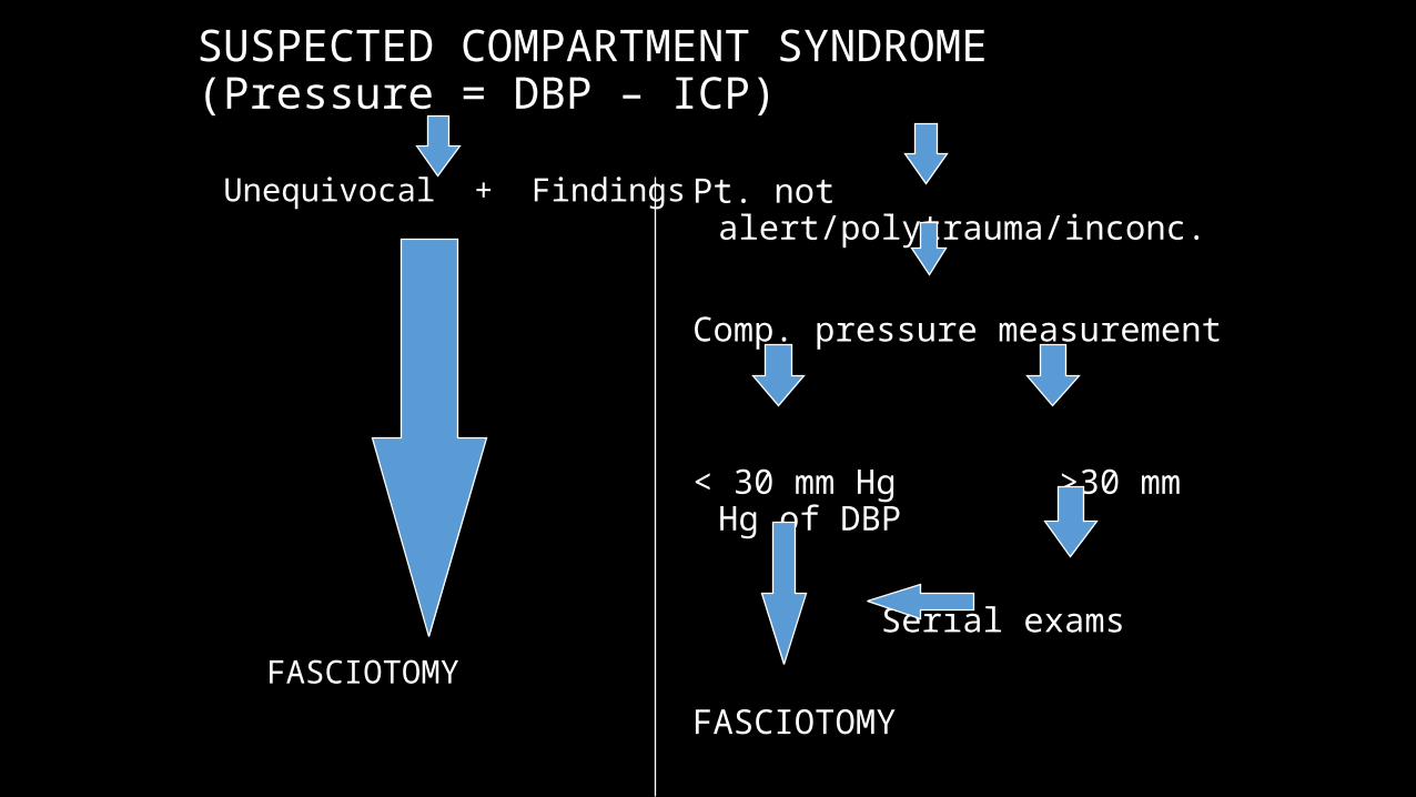
SUSPECTED COMPARTMENT SYNDROME (Pressure = DBP – ICP)
Unequivocal + Findings
FASCIOTOMY
Pt. not alert/polytrauma/inconc.
Comp. pressure measurement
< 30 mm Hg >30 mm Hg of DBP
Serial exams
FASCIOTOMY
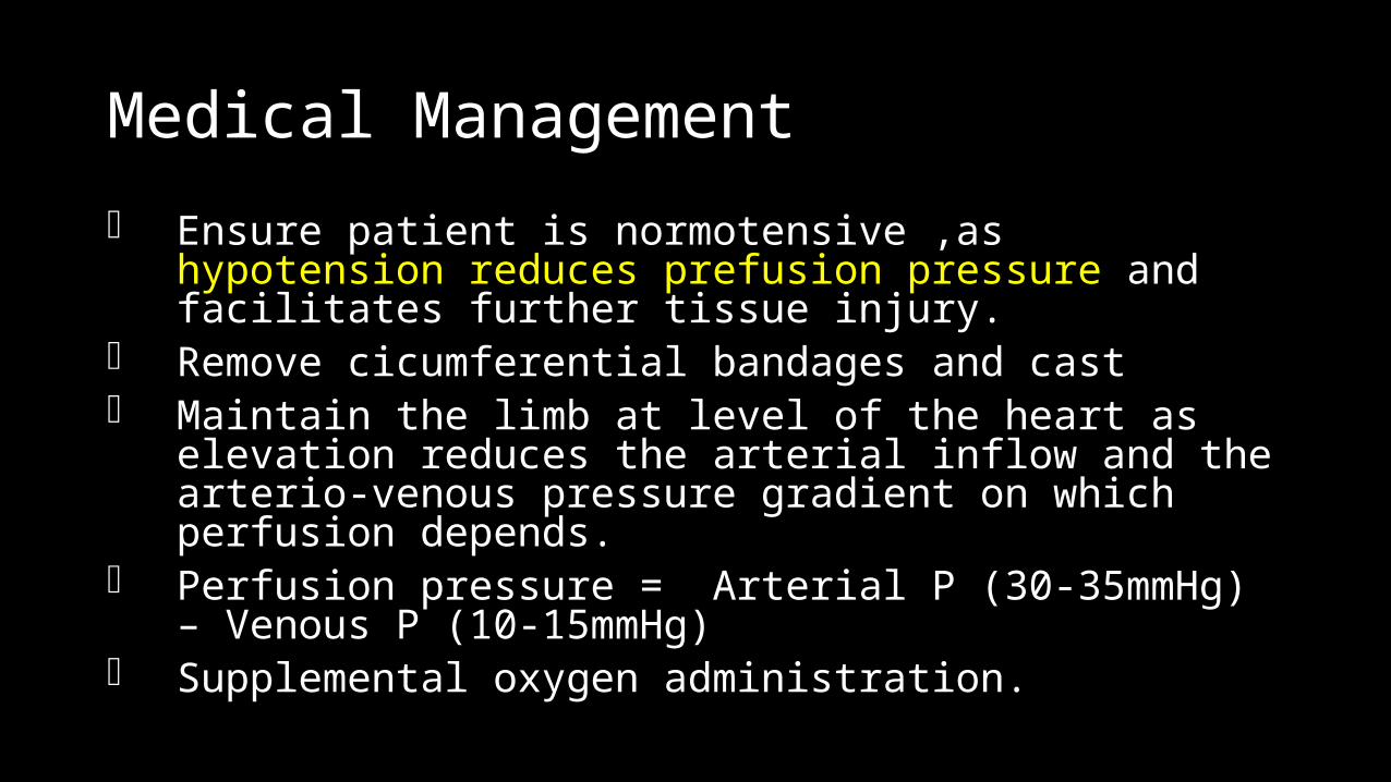
Medical Management
Ensure patient is normotensive ,as hypotension reduces prefusion pressure and facilitates further tissue injury.
Remove cicumferential bandages and cast Maintain the limb at level of the heart as elevation reduces the
arterial inflow and the arterio-venous pressure gradient on which perfusion depends.
Perfusion pressure = Arterial P (30-35mmHg) – Venous P (10-15mmHg)
Supplemental oxygen administration.
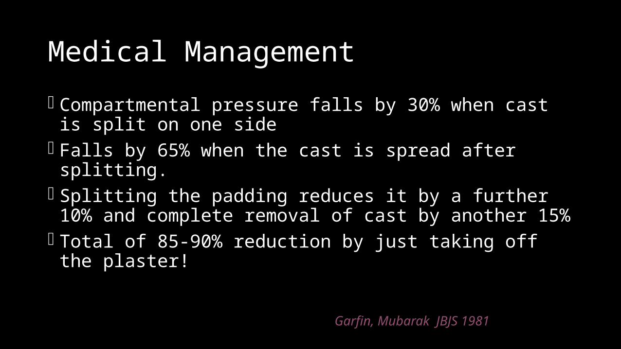
Medical Management
Compartmental pressure falls by 30% when cast is split on one side Falls by 65% when the cast is spread after splitting. Splitting the padding reduces it by a further 10% and complete
removal of cast by another 15% Total of 85-90% reduction by just taking off the plaster!
Garfin, Mubarak JBJS 1981
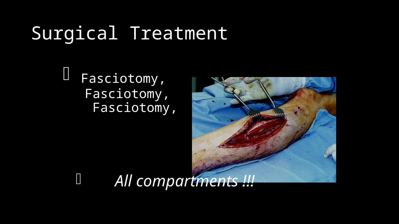
Surgical Treatment
Fasciotomy, Fasciotomy, Fasciotomy,
All compartments !!!
Fasciotomy Principles
Make early diagnosis Long extensile incisions Release all fascial compartments Preserve neurovascular structures Debride necrotic tissues Coverage within 7-10 days
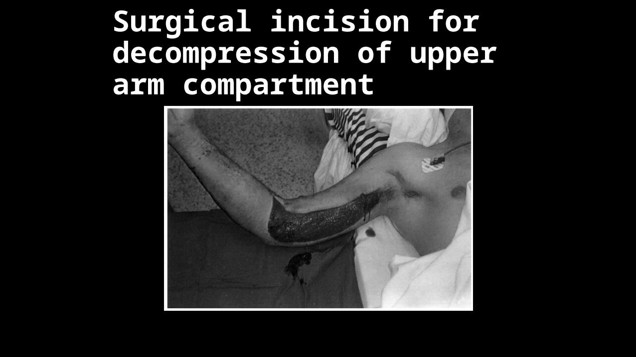
Surgical incision for decompression of upper arm compartment
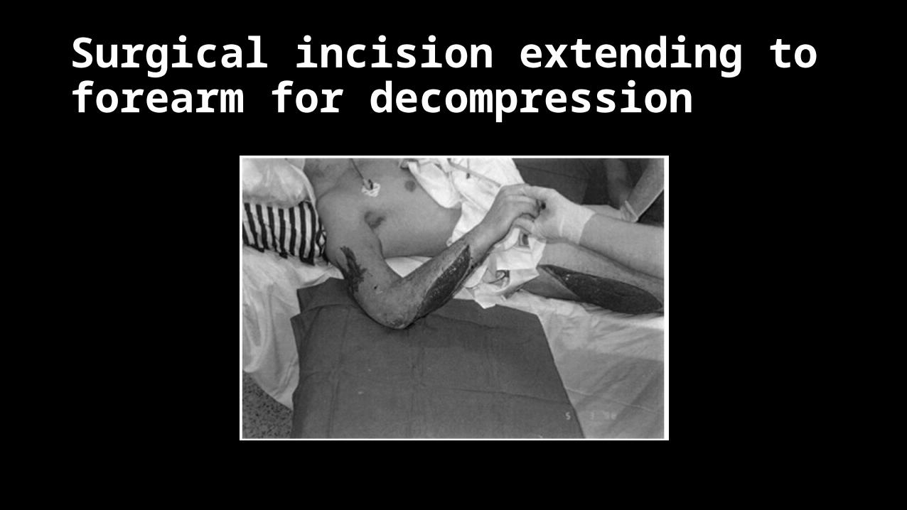
Surgical incision extending to forearm for decompression
Compartment SyndromeSurgical Treatment
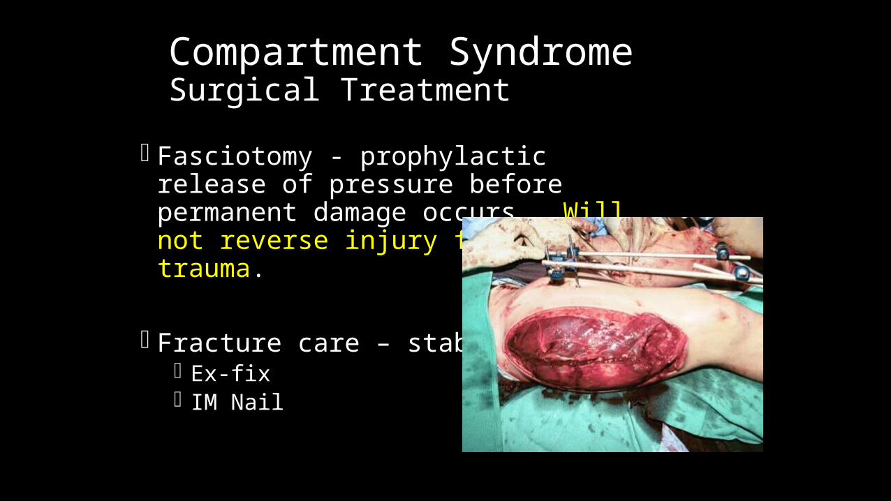
Fasciotomy - prophylactic release of pressure before permanent damage occurs. Will not reverse injury from trauma.
Fracture care – stabilization Ex-fix IM Nail

Compartment SyndromeIndications for Fasciotomy
Unequivocal clinical findings Rising tissue pressure Significant tissue injury or high risk patient Injury at high risk of compartment syndrome
CONTRAINDICATION - Missed compartment syndrome (>24-48 hrs)
Use a Generous Incision
Lengthening the skin incisions to an average of 16 cm decreases intracompartmental pressures significantly.
The skin envelope is a contributing factor in acute compartment syndromes of the leg and The use of generous skin incisions is supported
Compartment SyndromeLower Leg
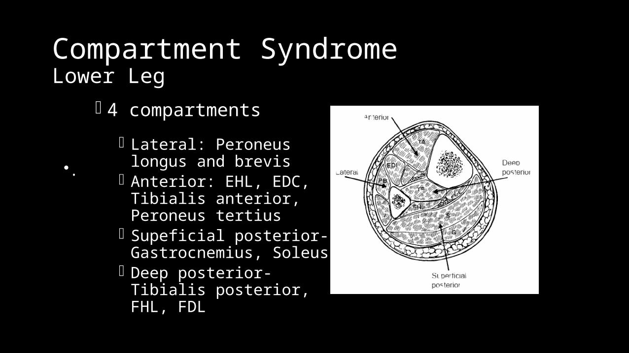
4 compartments
Lateral: Peroneus longus and brevis
Anterior: EHL, EDC, Tibialis anterior, Peroneus tertius
Supeficial posterior-Gastrocnemius, Soleus
Deep posterior-Tibialis posterior, FHL, FDL
•.
Compartment SyndromeHand
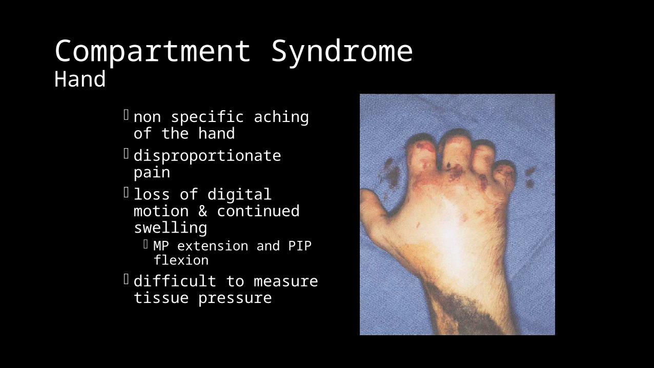
non specific aching of the hand
disproportionate pain loss of digital motion &
continued swelling MP extension and PIP
flexion difficult to measure
tissue pressure
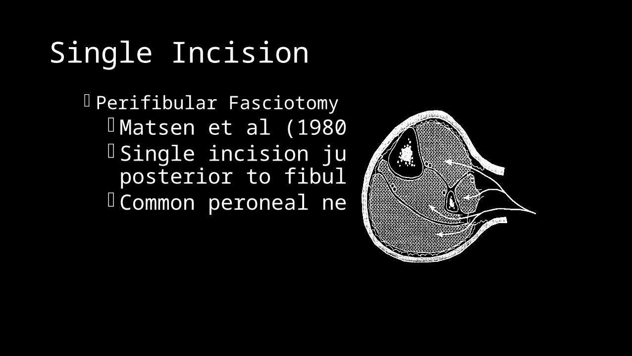
Single Incision
Perifibular Fasciotomy Matsen et al (1980) Single incision just
posterior to fibula Common peroneal nerve
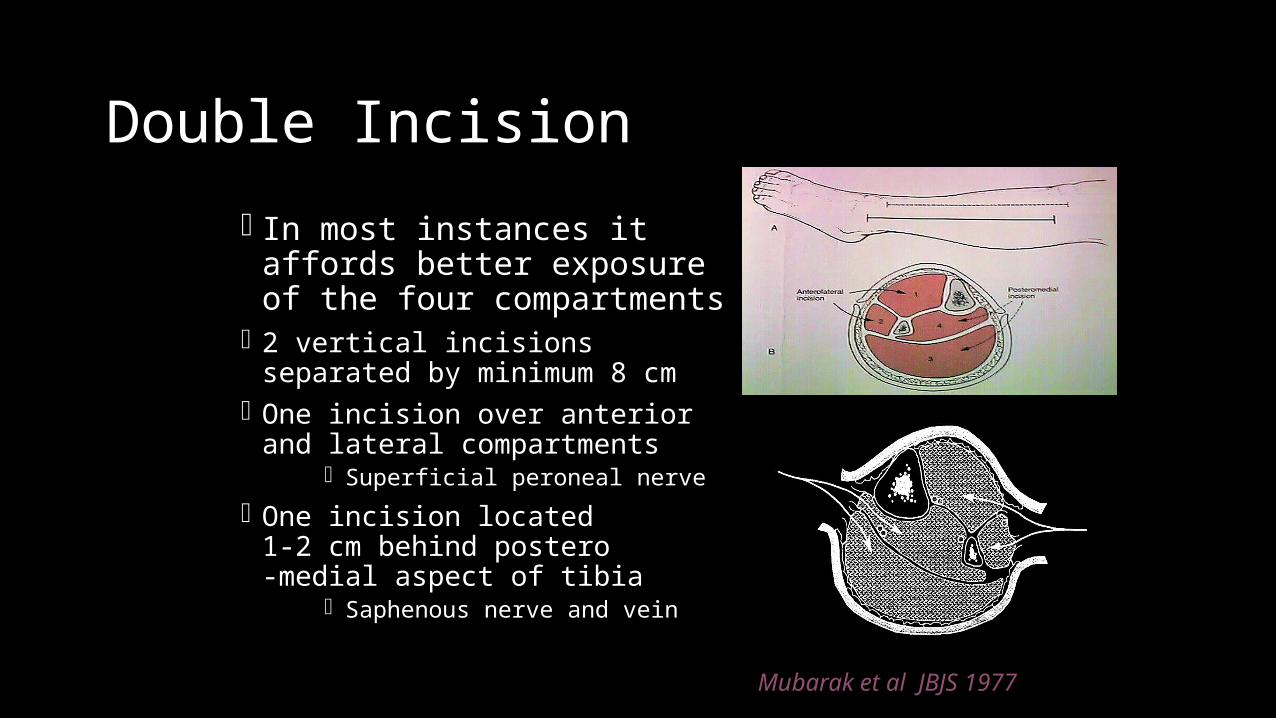
Double Incision
In most instances it affords better exposure of the four compartments
2 vertical incisions separated by minimum 8 cm
One incision over anterior and lateral compartments
Superficial peroneal nerve
One incision located 1-2 cm behind postero-medial aspect of tibia
Saphenous nerve and vein
Mubarak et al JBJS 1977
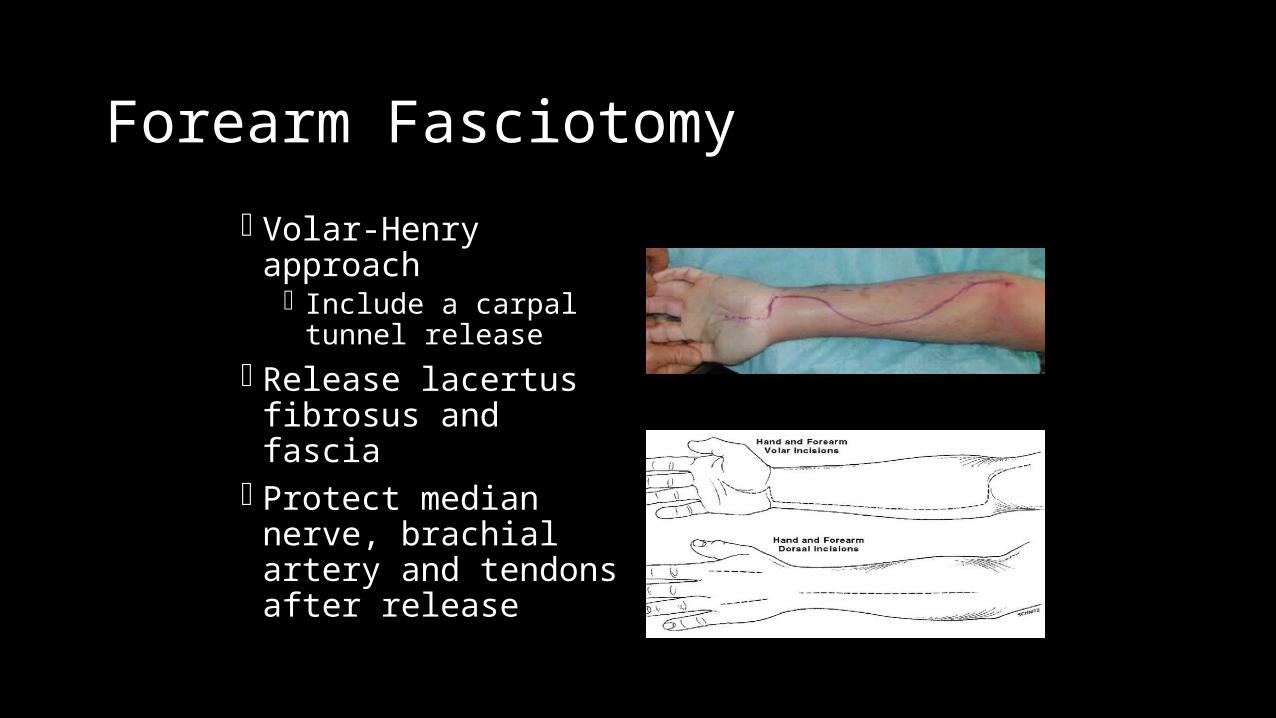
Forearm Fasciotomy
Volar-Henry approach Include a carpal tunnel
release Release lacertus
fibrosus and fascia Protect median nerve,
brachial artery and tendons after release
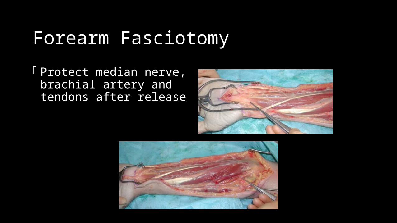
Forearm Fasciotomy
Protect median nerve, brachial artery and tendons after release
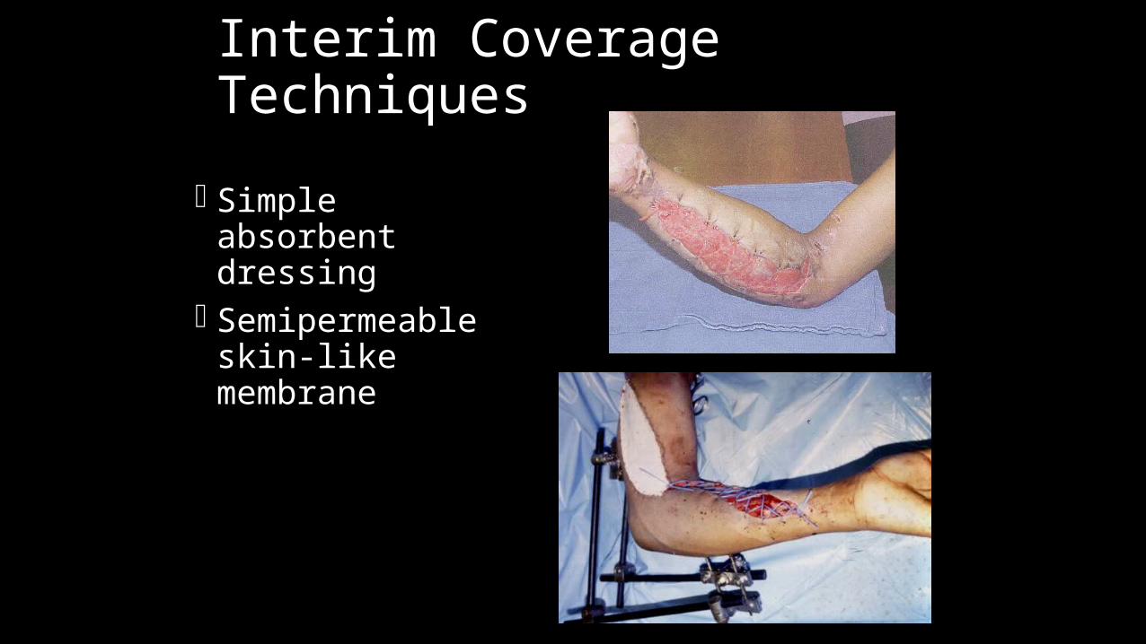
Interim Coverage Techniques
Simple absorbent dressing
Semipermeable skin-like membrane
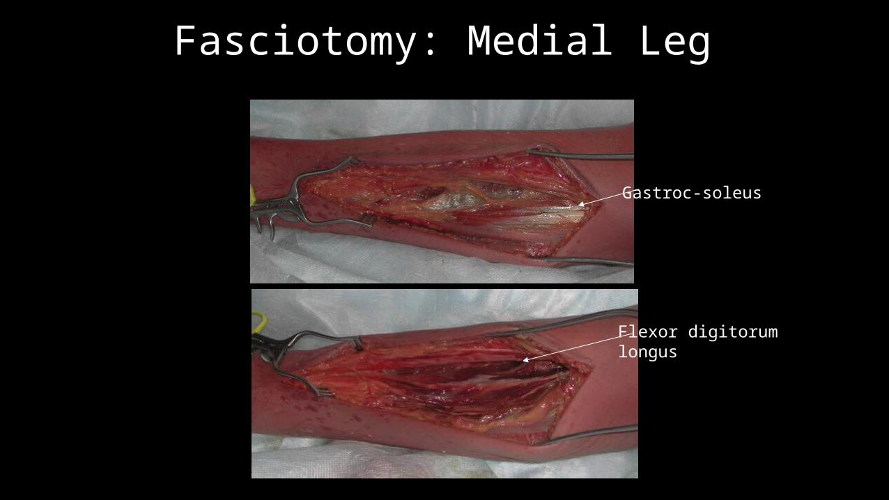
Fasciotomy: Medial Leg
Flexor digitorum longus
Gastroc-soleus
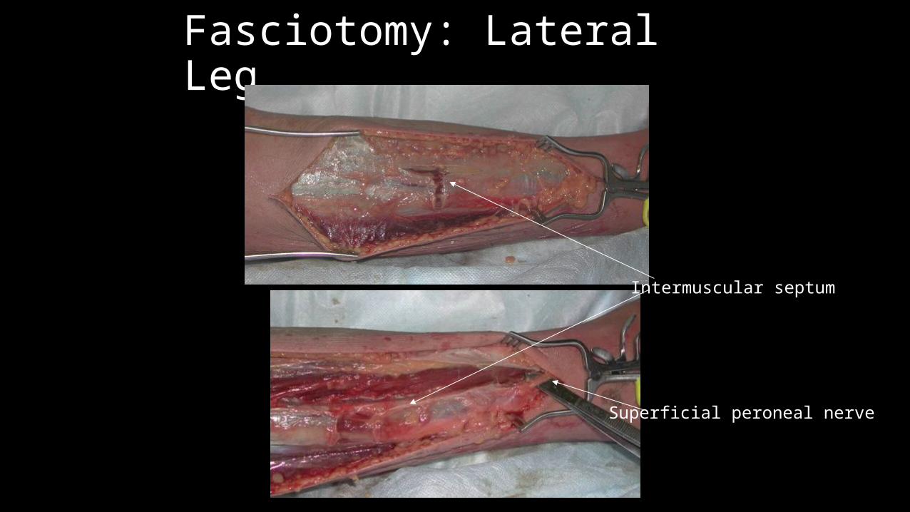
Fasciotomy: Lateral Leg
Superficial peroneal nerve
Intermuscular septum
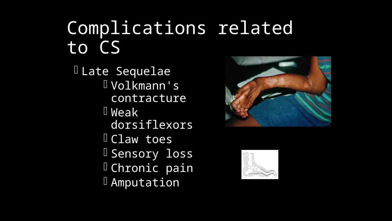
Complications related to CS
Late Sequelae Volkmann's
contracture Weak dorsiflexors Claw toes Sensory loss Chronic pain Amputation
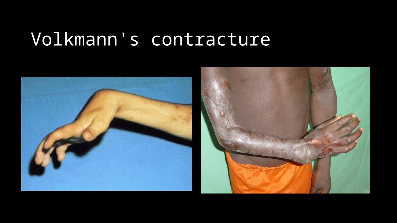
Volkmann's contracture
Wound Management
Wound is not closed at initial surgery Second look debridement with consideration for
coverage after 48-72 hrs Limb should not be at risk for further swelling Pt should be adequately stabilized Usually requires skin graft DPC possible if residual swelling is minimal Flap coverage needed if nerves, vessels, or bone exposed
Goal is to obtain definitive coverage within 7-10 days
Wound Management After the fasciotomy, a bulky compression dressing and a
splint are applied. “VAC” (Vacuum Assisted Closure) can be used Foot should be placed in neutral to prevent equinus
contracture. Incision for the fasciotomy usually can be closed after three
to five days
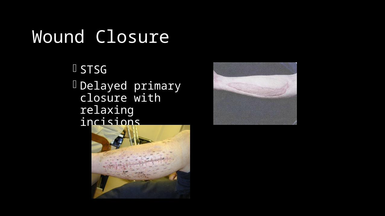
Wound Closure
STSG Delayed primary closure
with relaxing incisions
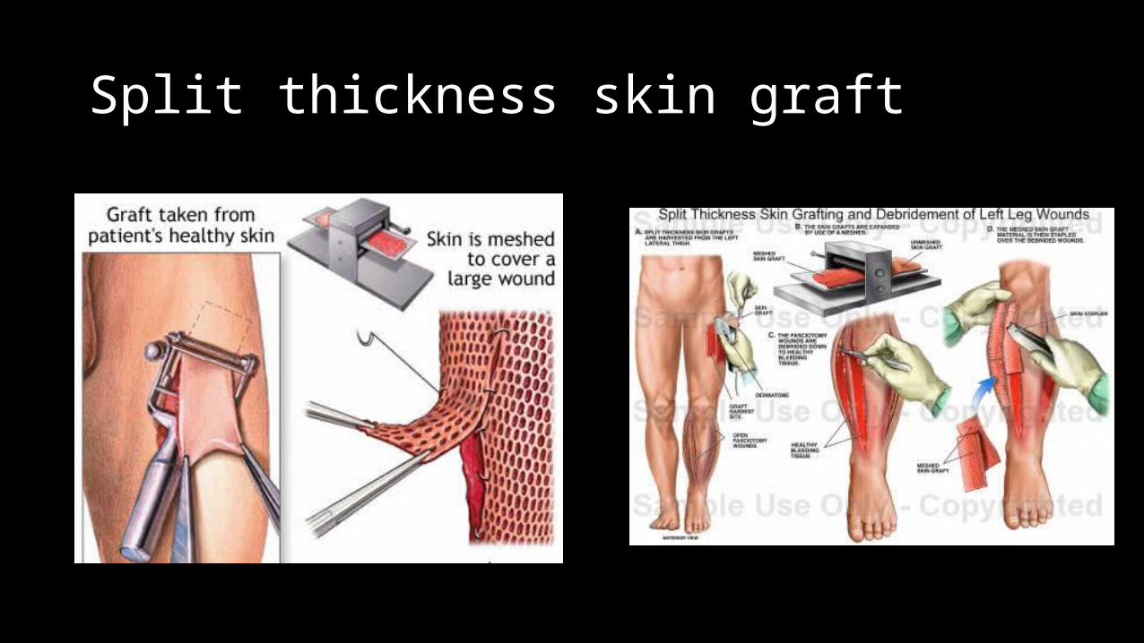
Split thickness skin graft
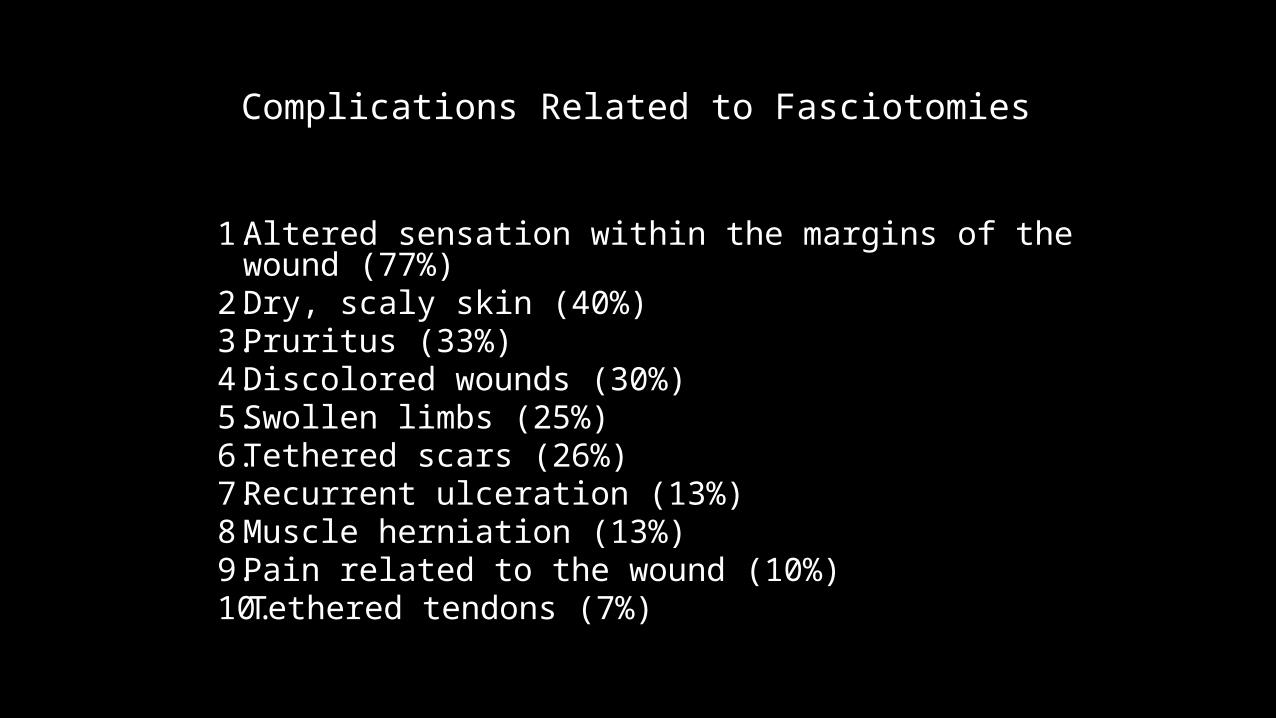
Complications Related to Fasciotomies
1. Altered sensation within the margins of the wound (77%) 2. Dry, scaly skin (40%) 3. Pruritus (33%) 4. Discolored wounds (30%) 5. Swollen limbs (25%) 6. Tethered scars (26%) 7. Recurrent ulceration (13%) 8. Muscle herniation (13%) 9. Pain related to the wound (10%) 10.Tethered tendons (7%)
Summary
Keep a high index of suspicion Treat as soon as you suspect CS If clinically evident, do not measure pressures Fasciotomy
Reliable, safe, and effective The only treatment for compartment syndrome, when performed in time