Embed Size (px)
Citation preview
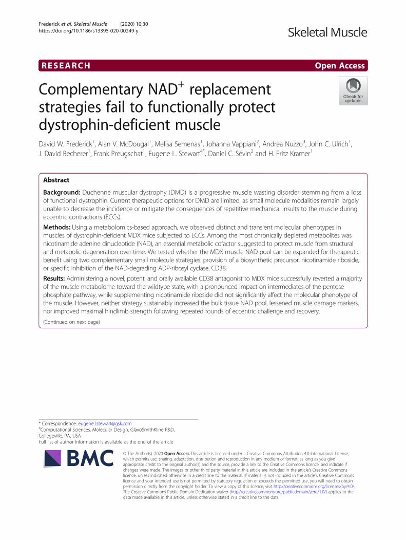
RESEARCH Open Access
Complementary NAD+ replacementstrategies fail to functionally protectdystrophin-deficient muscleDavid W. Frederick1, Alan V. McDougal1, Melisa Semenas1, Johanna Vappiani2, Andrea Nuzzo3, John C. Ulrich1,J. David Becherer1, Frank Preugschat1, Eugene L. Stewart4*, Daniel C. Sévin2 and H. Fritz Kramer1
Abstract
Background: Duchenne muscular dystrophy (DMD) is a progressive muscle wasting disorder stemming from a lossof functional dystrophin. Current therapeutic options for DMD are limited, as small molecule modalities remain largelyunable to decrease the incidence or mitigate the consequences of repetitive mechanical insults to the muscle duringeccentric contractions (ECCs).
Methods: Using a metabolomics-based approach, we observed distinct and transient molecular phenotypes inmuscles of dystrophin-deficient MDX mice subjected to ECCs. Among the most chronically depleted metabolites wasnicotinamide adenine dinucleotide (NAD), an essential metabolic cofactor suggested to protect muscle from structuraland metabolic degeneration over time. We tested whether the MDX muscle NAD pool can be expanded for therapeuticbenefit using two complementary small molecule strategies: provision of a biosynthetic precursor, nicotinamide riboside,or specific inhibition of the NAD-degrading ADP-ribosyl cyclase, CD38.
Results: Administering a novel, potent, and orally available CD38 antagonist to MDX mice successfully reverted a majorityof the muscle metabolome toward the wildtype state, with a pronounced impact on intermediates of the pentosephosphate pathway, while supplementing nicotinamide riboside did not significantly affect the molecular phenotype ofthe muscle. However, neither strategy sustainably increased the bulk tissue NAD pool, lessened muscle damage markers,nor improved maximal hindlimb strength following repeated rounds of eccentric challenge and recovery.
(Continued on next page)
© The Author(s). 2020 Open Access This article is licensed under a Creative Commons Attribution 4.0 International License,which permits use, sharing, adaptation, distribution and reproduction in any medium or format, as long as you giveappropriate credit to the original author(s) and the source, provide a link to the Creative Commons licence, and indicate ifchanges were made. The images or other third party material in this article are included in the article's Creative Commonslicence, unless indicated otherwise in a credit line to the material. If material is not included in the article's Creative Commonslicence and your intended use is not permitted by statutory regulation or exceeds the permitted use, you will need to obtainpermission directly from the copyright holder. To view a copy of this licence, visit http://creativecommons.org/licenses/by/4.0/.The Creative Commons Public Domain Dedication waiver (http://creativecommons.org/publicdomain/zero/1.0/) applies to thedata made available in this article, unless otherwise stated in a credit line to the data.
* Correspondence: [email protected] Sciences, Molecular Design, GlaxoSmithKline R&D,Collegeville, PA, USAFull list of author information is available at the end of the article
Frederick et al. Skeletal Muscle (2020) 10:30 https://doi.org/10.1186/s13395-020-00249-y
(Continued from previous page)
Conclusions: In the absence of dystrophin, eccentric injury contributes to chronic intramuscular NAD depletion withbroad pleiotropic effects on the molecular phenotype of the tissue. These molecular consequences can be moreeffectively overcome by inhibiting the enzymatic activity of CD38 than by supplementing nicotinamide riboside.However, we found no evidence that either small molecule strategy is sufficient to restore muscle contractile function orconfer protection from eccentric injury, undermining the modulation of NAD metabolism as a therapeutic approach forDMD.
Keywords: MDX, NAD+, CD38, NR, Eccentric, Injury, Metabolomics, Therapeutics
BackgroundDystrophinopathies are a class of diseases manifesting pri-marily in skeletal muscle and caused by a variety of muta-tions in the 2.4Mb dystrophin gene which render thedystrophin protein inactive. Dystrophin is a central force-transducing element of skeletal muscle, connecting theactin cytoskeleton to the extracellular matrix via thedystrophin-glycoprotein complex (DGC). In patients withDuchenne muscular dystrophy (DMD), the absence offunctional dystrophin leads to limb muscle weakness,followed by gradual muscle atrophy, cardiomyopathy, andpremature death. Dystrophin-deficient muscle is especiallysusceptible to damage following eccentric contractions, inwhich the muscle lengthens while generating opposingforce, as occurs during the act of sitting, descending stairs,and other activities of daily life [1]. Damaged muscle fibersthen undergo repeated cycles of clearance by the immunesystem and replacement by newly differentiated progeni-tor cells, in a process that spans weeks. Though progresshas been made in addressing the primary defects in dys-trophin via exon skipping or gene-replacement therapies[2], means of mitigating the effects of eccentric injuries tothe muscle of DMD patients have primarily been limitedto treatment with palliative anti-inflammatory drugs.The ability of muscle to harness chemical energy through
aerobic and anaerobic respiration is inherently linked to itsphysical structure. Accordingly, analytical techniques suchas NMR and mass spectrometry can detect chemical bio-markers of specific muscular dystrophies, which correlatewith the cause and severity of the disease, and can aid inidentifying points of therapeutic intervention. For example,it has long been appreciated that the muscle of adultdystrophin-deficient MDX mice contains lower levels ofenergy-storing and redox-active metabolites, such asphospho-creatine, ATP, and beta-hydroxybutyrate [3], yethigh levels of taurine and non-polar amino acids [4]. Morerecently, distinct metabolomes were also identified in popu-lations of the MDX muscle-resident cells involved in regen-eration, including satellite cells and adipose progenitors [5].Attempts to preserve muscle function by restoring specificmetabolic intermediates or co-factors in dystrophic musclehave shown hints of efficacy. For example, supplementingthe TCA cycle intermediate, alpha-ketoglutarate (aKG), to
the buffer of isolated MDX muscles was reported to im-prove fatigue resistance ex vivo [6]. In dystrophic mice har-boring mutations in the FKRP gene, aberrant glycosylationof the DGC component, alpha-dystroglycan, can be par-tially overcome by supplementing the drinking water with5% ribitol, the substrate of the mutated enzyme [7]. Thesestudies suggest that specific aspects of the pathophysiologyof muscular dystrophies arise from metabolic bottlenecksthat may be bypassed using exogenous small molecules.Nicotinamide adenine dinucleotide (NAD+ or NAD) is an
essential metabolic co-factor, which has been directly impli-cated in the maintenance of muscle mass and function dur-ing sarcopenia and other dystrophic states [8–11]. NAD hasalso been found to be depleted in the muscle of MDX mice[10, 12], suggesting potential for therapeutic intervention.At present, two complementary strategies exist for manipu-lating tissue NAD pools. The first, and most commonlystudied strategy, is to enhance NAD production by supple-menting biosynthetic precursors, such as nicotinamide ribo-side (NR) or nicotinamide mononucleotide, to cells with afunctional NAD salvage pathway [13]. The second strategyis to inhibit the enzymatic consumption of NAD by severalclasses of enzymes, including sirtuins, poly-ADP-ribosepolymerases (PARPs), and ADP-ribosyl cyclases (ARCs).CD38, the most widely expressed ARC, has been therapeut-ically targeted for indications ranging from multiple mye-loma to neurodegeneration [14]. Our group has previouslyshown that synthetic small molecule antagonists of CD38are capable of acutely increasing NAD in muscle and livertissue [15, 16], and others have reported that chronicallydosing one of these thiazoloquinolin(on)es, known in the lit-erature as compound 78c, mitigated structural remodelingand functional decline in the muscle of aged mice [11]. Des-pite these preliminary findings, the extent to which NADdepletion is a pathologically relevant or therapeutically tract-able feature of dystrophinopathies remains unclear.Recent reports have specifically investigated the poten-
tial for NAD supplementation to counteract the pathologyof DMD [10, 17]. Here, we have examined this topic fur-ther by characterizing the global metabolome of MDXmuscle following eccentric challenge and identifying dis-tinct stages of metabolic crisis and repair on the biochem-ical level. We found evidence that NAD is depleted both
Frederick et al. Skeletal Muscle (2020) 10:30 Page 2 of 14
acutely and chronically in the muscle of MDX mice withwide-ranging metabolic consequences. We furtherattempted to reverse the NAD depletion using a novel,highly potent imidazoquinoline inhibitor of CD38 andcompared its efficacy to nicotinamide riboside during re-peat bouts of eccentric challenge and recovery. Our resultsmay guide the development of NAD-targeting therapeu-tics for muscle diseases.
ResultsDystrophin deficiency alters the muscle NAD metabolomeand energy producing pathwaysTo assess the biochemical adaptations of muscle to dys-trophin deficiency, we analyzed the gastrocnemius mus-cles of MDX mice before and after eccentric challengeusing an untargeted metabolic profiling platform. Theplatform utilized a combination of GC/MS and LC/MS inpositive and negative ion mode to identify 762 chemicalentities, of which 552 were annotated as either polar
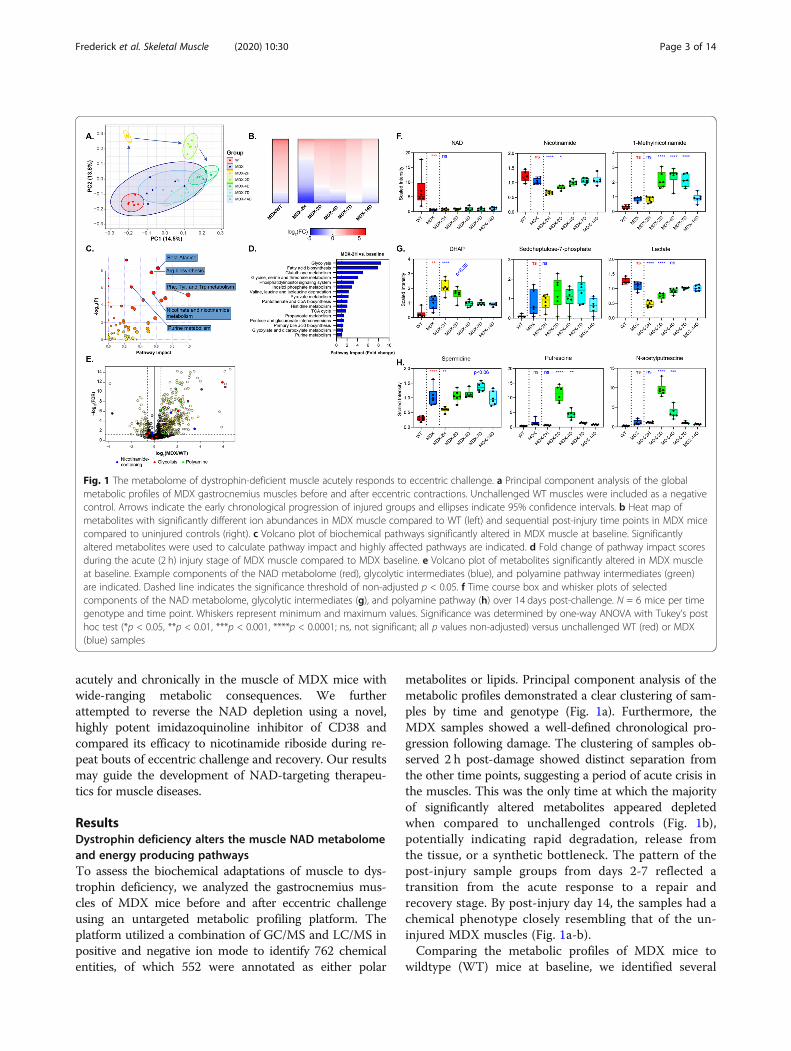
metabolites or lipids. Principal component analysis of themetabolic profiles demonstrated a clear clustering of sam-ples by time and genotype (Fig. 1a). Furthermore, theMDX samples showed a well-defined chronological pro-gression following damage. The clustering of samples ob-served 2 h post-damage showed distinct separation fromthe other time points, suggesting a period of acute crisis inthe muscles. This was the only time at which the majorityof significantly altered metabolites appeared depletedwhen compared to unchallenged controls (Fig. 1b),potentially indicating rapid degradation, release fromthe tissue, or a synthetic bottleneck. The pattern of thepost-injury sample groups from days 2-7 reflected atransition from the acute response to a repair andrecovery stage. By post-injury day 14, the samples had achemical phenotype closely resembling that of the un-injured MDX muscles (Fig. 1a-b).Comparing the metabolic profiles of MDX mice to
wildtype (WT) mice at baseline, we identified several
Fig. 1 The metabolome of dystrophin-deficient muscle acutely responds to eccentric challenge. a Principal component analysis of the globalmetabolic profiles of MDX gastrocnemius muscles before and after eccentric contractions. Unchallenged WT muscles were included as a negativecontrol. Arrows indicate the early chronological progression of injured groups and ellipses indicate 95% confidence intervals. b Heat map ofmetabolites with significantly different ion abundances in MDX muscle compared to WT (left) and sequential post-injury time points in MDX micecompared to uninjured controls (right). c Volcano plot of biochemical pathways significantly altered in MDX muscle at baseline. Significantlyaltered metabolites were used to calculate pathway impact and highly affected pathways are indicated. d Fold change of pathway impact scoresduring the acute (2 h) injury stage of MDX muscle compared to MDX baseline. e Volcano plot of metabolites significantly altered in MDX muscleat baseline. Example components of the NAD metabolome (red), glycolytic intermediates (blue), and polyamine pathway intermediates (green)are indicated. Dashed line indicates the significance threshold of non-adjusted p < 0.05. f Time course box and whisker plots of selectedcomponents of the NAD metabolome, glycolytic intermediates (g), and polyamine pathway (h) over 14 days post-challenge. N = 6 mice per timegenotype and time point. Whiskers represent minimum and maximum values. Significance was determined by one-way ANOVA with Tukey’s posthoc test (*p < 0.05, **p < 0.01, ***p < 0.001, ****p < 0.0001; ns, not significant; all p values non-adjusted) versus unchallenged WT (red) or MDX(blue) samples
Frederick et al. Skeletal Muscle (2020) 10:30 Page 3 of 14
biochemical pathways with a disproportionate impact onthe muscle. Among the highest confidence pathways (p< 10^ −5 and pathway impact> 0.5) were those relatingto the biosynthesis and metabolism of amino acids, suchas arginine, phenylalanine, tyrosine, and tryptophan, aswell as that of nicotinate and nicotinamide metabolism(Fig. 1c). Comparing the pathway impact scores fromthe acutely injured (2 h) to uninjured MDX muscle, wewere surprised to find glycolysis as the most responsivepathway (Fig. 1d). Additional pathways relating to energyproduction or storage, including those of fatty acid syn-thesis, pyruvate metabolism, and the TCA cycle, werealso injury responsive.Upon examining the specific metabolites altered at
baseline in the MDX muscle, we found a strikingly lowerabundance of NAD than almost any other metabolite(Fig. 1e, f). Consistent with the pathway analysis, severalother nicotinamide-containing metabolites and glycolyticintermediates also showed highly variable abundance.Primary NAD deficiency in mouse muscle and culturedmyotubes has been shown to restrict glycolytic flux at
the level of GAPDH, resulting in a characteristic buildupof intermediates in the pentose phosphate pathway [8,18]. Consistent with this model, we observed a signifi-cant increase in ion counts for glucose-6-phosphate(G6P), dihydroxyacetone phosphate (DHAP), ribose-5-phosphate, and a positive trend in sedoheptulose-7-phosphate (S7P) in MDX muscle (Fig. 1e, g). Addition-ally, we noted an increase in several poly-cationic speciesof polyamines, known to be derived from arginine (Fig.1e, h). Collectively, this pattern indicates a metabolicshift in MDX muscle at a steady state, partially stem-ming from the loss of the metabolic co-factor, NAD.The robust response of the global metabolome at 2 h
post-eccentric challenge led us to investigate NAD-relatedmetabolites at this and subsequent time points. As far as theplatform could resolve, NAD itself did not appear to re-spond to the challenge (Fig. 1f). However, Nam was acutelydepleted by more than one-third after 2 h, presumablyrestricting any residual activity of the NAD salvage pathway,and gradually returned to baseline after 4 days. Namhomeostasis was further altered by a doubling in the levels
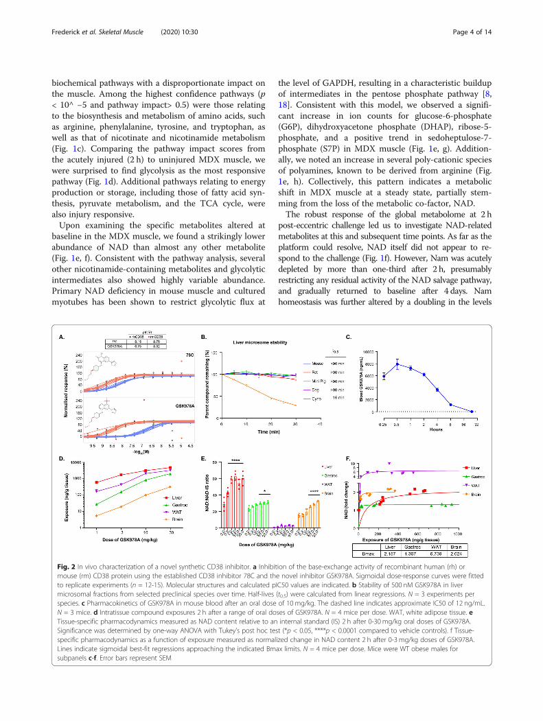
Fig. 2 In vivo characterization of a novel synthetic CD38 inhibitor. a Inhibition of the base-exchange activity of recombinant human (rh) ormouse (rm) CD38 protein using the established CD38 inhibitor 78C and the novel inhibitor GSK978A. Sigmoidal dose-response curves were fittedto replicate experiments (n = 12-15). Molecular structures and calculated pIC50 values are indicated. b Stability of 500 nM GSK978A in livermicrosomal fractions from selected preclinical species over time. Half-lives (t0.5) were calculated from linear regressions. N = 3 experiments perspecies. c Pharmacokinetics of GSK978A in mouse blood after an oral dose of 10 mg/kg. The dashed line indicates approximate IC50 of 12 ng/mL.N = 3 mice. d Intratissue compound exposures 2 h after a range of oral doses of GSK978A. N = 4 mice per dose. WAT, white adipose tissue. eTissue-specific pharmacodynamics measured as NAD content relative to an internal standard (IS) 2 h after 0-30 mg/kg oral doses of GSK978A.Significance was determined by one-way ANOVA with Tukey’s post hoc test (*p < 0.05, ****p < 0.0001 compared to vehicle controls). f Tissue-specific pharmacodynamics as a function of exposure measured as normalized change in NAD content 2 h after 0-3 mg/kg doses of GSK978A.Lines indicate sigmoidal best-fit regressions approaching the indicated Bmax limits. N = 4 mice per dose. Mice were WT obese males forsubpanels c-f. Error bars represent SEM
Frederick et al. Skeletal Muscle (2020) 10:30 Page 4 of 14
of 1-me-Nam in the 2 days following injury, which only nor-malized after 14 days. Evidence of a further constriction inglycolysis also emerged post-injury: levels of DHAP, imme-diately upstream of GAPDH, acutely increased in an oppos-ing pattern to that of lactate, a glycolytic end-product (Fig.1g). The most striking indicator of the repair phase was theappearance of polyamines, including spermine, spermidine,putrescine, and N-acetylputrescine, which were elevated inthe days following injury (Fig. 1h and Supplemental Table1). This class of biomolecules serves as a general marker ofcellular proliferation and is required for both myocyte differ-entiation and alternative macrophage activation [19, 20].Consistently, in the case of NAD-related metabolites, glyco-lytic intermediates, and polyamines, eccentric injury ampli-fied the disparities between MDX and WT muscle.
A novel synthetic CD38 antagonist increases NAD inmultiple tissuesOur group previously reported a series of novel chemicalentities (NCEs), which potently inhibit the constitutiveNAD-degrading enzyme, CD38. Related screening effortsyielded the imidazoquinoline dubbed GSK978A, which ex-hibited tenfold higher potency against mouse recombinantCD38 than the human enzyme (Fig. 2a). This potency isorders of magnitude greater than that of natural products,such as quercetin, and approximates that of 78c, the best-studied synthetic CD38 inhibitor to-date [11, 14]. Yet
GSK978A was more soluble and outperformed 78c in achromosomal stability test of genotoxicity, indicating im-proved suitability for long-term administration (data notshown). GSK978A was also predicted to have low intrinsicclearance in several small animal preclinical species, includ-ing mice and rats, but not larger cynomolgus monkeys (Fig.2b). As literature suggested a role for CD38 in the preven-tion of diet-induced obesity [21], the drug metabolism andpharmacokinetic characterization of the quinoline serieswas originally performed in obese WT mice. To confirmthe slow clearance kinetics in vivo, we administered a singleintermediate oral dose of 10mg/kg and found the com-pound still detectable in the blood after 24 h (Fig. 2c). Wenext performed a pharmacokinetic analysis of tissues sam-pled 2 h after oral doses from 1-30mg/kg. At these doses,the compound was identified within the liver, gastrocne-mius, adipose, and brain tissues at exposures that wellexceeded the IC50 of ~ 12 ng/mL (Fig. 2d). Accordingly, theNAD content was found to be significantly elevated in theliver, muscle, and brain when normalized to an internal ana-lytical standard (Fig. 2e). Despite high exposure, the NADrecovery from adipose was low and NAD changes were notsignificant. However, in most tissues, peak NAD elevationof at least 30% was achieved at a dose of 3mg/kg (Fig. 2f).To assess the potential pharmacodynamics of GSK978A
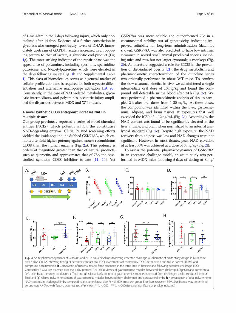
in an eccentric challenge model, an acute study was per-formed in MDX mice following 5 days of dosing at 3mg/
Fig. 3 Acute pharmacodynamics of GSK978A and NR in MDX hindlimbs following eccentric challenge. a Schematic of acute study design in MDX miceover 5 days (D1-D5) showing timing of eccentric contractions (ECC), assessments of contractility (CON), termination and tissue harvest (TERM), andcompound administration. b Comparison of maximal tetanic force produced in the same limb at baseline and following eccentric challenge (ECC).Contractility (CON) was assessed over the 5-day protocol (D1-D5). c Masses of gastrocnemius muscles harvested from challenged (right, R) and contralateral(left, L) limbs at the study conclusion. d Total and (e) relative NAD content of gastrocnemius muscles harvested from challenged and contralateral limbs. fTotal and (g) relative polyamine content of gastrocnemius muscles harvested from challenged and contralateral limbs. h Normalization of total polyamine toNAD contents in challenged limbs compared to the contralateral side. N = 8 MDX mice per group. Error bars represent SEM. Significance was determinedby one-way ANOVA with Tukey’s post hoc test (**p < 0.01, ***p < 0.001, ****p < 0.0001; ns, not significant or p value indicated)
Frederick et al. Skeletal Muscle (2020) 10:30 Page 5 of 14
kg. Dietary NR, which has been suggested to improve theperformance of MDX muscle [10], was included as a com-parator. Within 24 h of eccentric challenge, tetanic strengthwas lessened by > 50% in all treatment groups, despite pres-ervation of mass in the largest affected gastrocnemius mus-cles (Fig. 3a-c). Interestingly, the challenged muscles alsoshowed NAD depletion compared to the contralateral side,indicating that the muscle NAD pool does acutely respondto lengthening contractions (Fig. 3d). Mice treated withGSK978A, but not NR, showed a trend toward protectionfrom this effect, though it could not be attributed to specificNAD elevation in either limb (Fig. 3e). As a biomarker ofmuscle repair, total muscle polyamines showed clear eleva-tion in the injured limbs with a trend toward protection byGSK978A, especially when polyamines were normalized toNAD content (Fig. 3f-h). These results suggested that a lon-ger treatment regimen might be necessary to provide func-tional improvements to MDX mice.
Chronic NAD repletion does not provide functionalprotection from repetitive eccentric challengesWe next designed a long-term study with chronic ad-ministration of GSK978A or NR to longitudinally assess
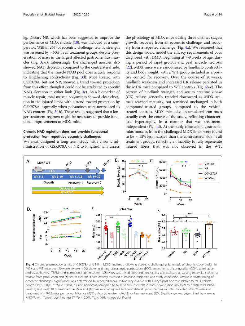
the physiology of MDX mice during three distinct stages:growth, recovery from an eccentric challenge, and recov-ery from a repeated challenge (Fig. 4a). We reasoned thatthis design would model the efficacy requirements of boysdiagnosed with DMD. Beginning at 7-9 weeks of age, dur-ing a period of rapid growth and peak muscle necrosis[22], MDX mice were randomized by hindlimb contractil-ity and body weight, with a WT group included as a posi-tive control for recovery. Over the course of 20 weeks,hindlimb weakness and increased CK release persisted inthe MDX mice compared to WT controls (Fig. 4b-c). Thepattern of hindlimb strength and serum creatine kinase(CK) release generally trended downward as MDX ani-mals reached maturity, but remained unchanged in bothcompound-treated groups, compared to the vehicle-treated controls. MDX mice also accumulated lean masssteadily over the course of the study, reflecting character-istic hypertrophy, in a manner that was treatment-independent (Fig. 4d). At the study conclusion, gastrocne-mius muscles from the challenged MDX limbs were foundto be ~ 15% less massive than the contralateral side in alltreatment groups, reflecting an inability to fully regenerateinjured fibers that was not observed in the WT.
Fig. 4 Chronic pharmacodynamics of GSK978A and NR in MDX hindlimbs following eccentric challenge. a Schematic of chronic study design inMDX and WT mice over 20 weeks (weeks 1-20) showing timing of eccentric contractions (ECC), assessments of contractility (CON), terminationand tissue harvest (TERM), and compound administration. GSK978A was dosed daily and contractility was assessed at varying intervals. b Maximaltetanic force production and (c) serum creatine kinase activity assessed at baseline, midpoint, and study conclusion. Arrows indicate timing ofeccentric challenges. Significance was determined by repeated measure two-way ANOVA with Tukey’s post hoc test relative to MDX vehiclecontrols (**p < 0.01, ****p < 0.0001; ns, not significant compared to MDX vehicle controls). d Body composition assessed by qNMR at baseline,week 8, and week 18 of treatment. e Mass and (f) mass ratio of injured and contralateral gastrocnemius muscles collected after 20 weeks oftreatment. N = 9-12 mice per group. Mice are MDX unless otherwise noted. Error bars represent SEM. Significance was determined by one-wayANOVA with Tukey’s post hoc test (****p < 0.001, **p < 0.01; ns, not significant)
Frederick et al. Skeletal Muscle (2020) 10:30 Page 6 of 14
Surprisingly, contralateral muscles tended to be largest inmice treated with GSK978A (Fig. 4e-f), which may be aconsequence of altered gait mechanics to favor the contra-lateral side, as it was not reflected in total lean mass.Hindlimb strength was also serially assessed following
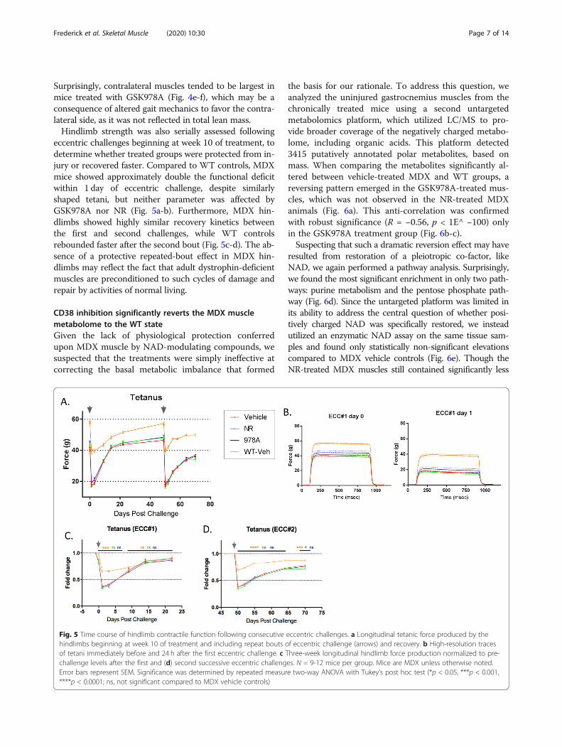
eccentric challenges beginning at week 10 of treatment, todetermine whether treated groups were protected from in-jury or recovered faster. Compared to WT controls, MDXmice showed approximately double the functional deficitwithin 1 day of eccentric challenge, despite similarlyshaped tetani, but neither parameter was affected byGSK978A nor NR (Fig. 5a-b). Furthermore, MDX hin-dlimbs showed highly similar recovery kinetics betweenthe first and second challenges, while WT controlsrebounded faster after the second bout (Fig. 5c-d). The ab-sence of a protective repeated-bout effect in MDX hin-dlimbs may reflect the fact that adult dystrophin-deficientmuscles are preconditioned to such cycles of damage andrepair by activities of normal living.
CD38 inhibition significantly reverts the MDX musclemetabolome to the WT stateGiven the lack of physiological protection conferredupon MDX muscle by NAD-modulating compounds, wesuspected that the treatments were simply ineffective atcorrecting the basal metabolic imbalance that formed
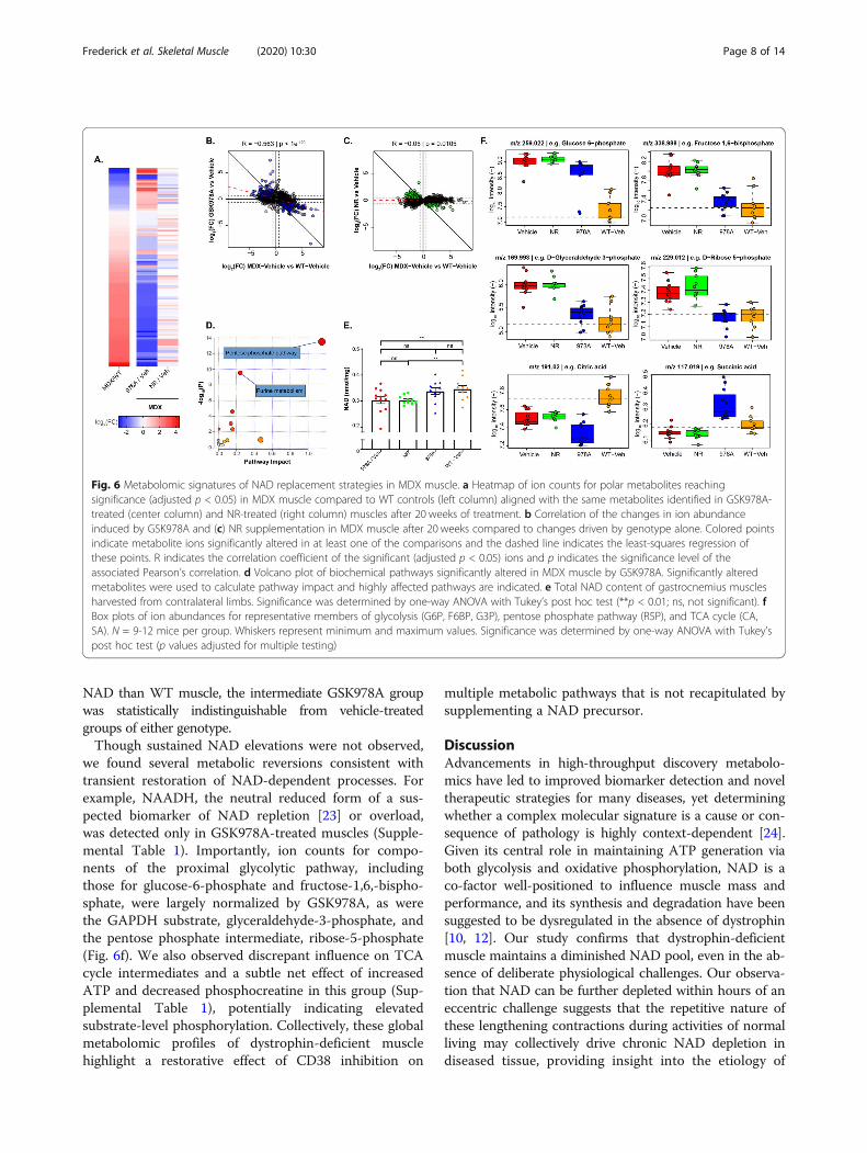
the basis for our rationale. To address this question, weanalyzed the uninjured gastrocnemius muscles from thechronically treated mice using a second untargetedmetabolomics platform, which utilized LC/MS to pro-vide broader coverage of the negatively charged metabo-lome, including organic acids. This platform detected3415 putatively annotated polar metabolites, based onmass. When comparing the metabolites significantly al-tered between vehicle-treated MDX and WT groups, areversing pattern emerged in the GSK978A-treated mus-cles, which was not observed in the NR-treated MDXanimals (Fig. 6a). This anti-correlation was confirmedwith robust significance (R = −0.56, p < 1E^ −100) onlyin the GSK978A treatment group (Fig. 6b-c).Suspecting that such a dramatic reversion effect may have
resulted from restoration of a pleiotropic co-factor, likeNAD, we again performed a pathway analysis. Surprisingly,we found the most significant enrichment in only two path-ways: purine metabolism and the pentose phosphate path-way (Fig. 6d). Since the untargeted platform was limited inits ability to address the central question of whether posi-tively charged NAD was specifically restored, we insteadutilized an enzymatic NAD assay on the same tissue sam-ples and found only statistically non-significant elevationscompared to MDX vehicle controls (Fig. 6e). Though theNR-treated MDX muscles still contained significantly less
Fig. 5 Time course of hindlimb contractile function following consecutive eccentric challenges. a Longitudinal tetanic force produced by thehindlimbs beginning at week 10 of treatment and including repeat bouts of eccentric challenge (arrows) and recovery. b High-resolution tracesof tetani immediately before and 24 h after the first eccentric challenge. c Three-week longitudinal hindlimb force production normalized to pre-challenge levels after the first and (d) second successive eccentric challenges. N = 9-12 mice per group. Mice are MDX unless otherwise noted.Error bars represent SEM. Significance was determined by repeated measure two-way ANOVA with Tukey’s post hoc test (*p < 0.05, ***p < 0.001,****p < 0.0001; ns, not significant compared to MDX vehicle controls)
Frederick et al. Skeletal Muscle (2020) 10:30 Page 7 of 14
NAD than WT muscle, the intermediate GSK978A groupwas statistically indistinguishable from vehicle-treatedgroups of either genotype.Though sustained NAD elevations were not observed,
we found several metabolic reversions consistent withtransient restoration of NAD-dependent processes. Forexample, NAADH, the neutral reduced form of a sus-pected biomarker of NAD repletion [23] or overload,was detected only in GSK978A-treated muscles (Supple-mental Table 1). Importantly, ion counts for compo-nents of the proximal glycolytic pathway, includingthose for glucose-6-phosphate and fructose-1,6,-bispho-sphate, were largely normalized by GSK978A, as werethe GAPDH substrate, glyceraldehyde-3-phosphate, andthe pentose phosphate intermediate, ribose-5-phosphate(Fig. 6f). We also observed discrepant influence on TCAcycle intermediates and a subtle net effect of increasedATP and decreased phosphocreatine in this group (Sup-plemental Table 1), potentially indicating elevatedsubstrate-level phosphorylation. Collectively, these globalmetabolomic profiles of dystrophin-deficient musclehighlight a restorative effect of CD38 inhibition on
multiple metabolic pathways that is not recapitulated bysupplementing a NAD precursor.
DiscussionAdvancements in high-throughput discovery metabolo-mics have led to improved biomarker detection and noveltherapeutic strategies for many diseases, yet determiningwhether a complex molecular signature is a cause or con-sequence of pathology is highly context-dependent [24].Given its central role in maintaining ATP generation viaboth glycolysis and oxidative phosphorylation, NAD is aco-factor well-positioned to influence muscle mass andperformance, and its synthesis and degradation have beensuggested to be dysregulated in the absence of dystrophin[10, 12]. Our study confirms that dystrophin-deficientmuscle maintains a diminished NAD pool, even in the ab-sence of deliberate physiological challenges. Our observa-tion that NAD can be further depleted within hours of aneccentric challenge suggests that the repetitive nature ofthese lengthening contractions during activities of normalliving may collectively drive chronic NAD depletion indiseased tissue, providing insight into the etiology of
Fig. 6 Metabolomic signatures of NAD replacement strategies in MDX muscle. a Heatmap of ion counts for polar metabolites reachingsignificance (adjusted p < 0.05) in MDX muscle compared to WT controls (left column) aligned with the same metabolites identified in GSK978A-treated (center column) and NR-treated (right column) muscles after 20 weeks of treatment. b Correlation of the changes in ion abundanceinduced by GSK978A and (c) NR supplementation in MDX muscle after 20 weeks compared to changes driven by genotype alone. Colored pointsindicate metabolite ions significantly altered in at least one of the comparisons and the dashed line indicates the least-squares regression ofthese points. R indicates the correlation coefficient of the significant (adjusted p < 0.05) ions and p indicates the significance level of theassociated Pearson’s correlation. d Volcano plot of biochemical pathways significantly altered in MDX muscle by GSK978A. Significantly alteredmetabolites were used to calculate pathway impact and highly affected pathways are indicated. e Total NAD content of gastrocnemius musclesharvested from contralateral limbs. Significance was determined by one-way ANOVA with Tukey’s post hoc test (**p < 0.01; ns, not significant). fBox plots of ion abundances for representative members of glycolysis (G6P, F6BP, G3P), pentose phosphate pathway (R5P), and TCA cycle (CA,SA). N = 9-12 mice per group. Whiskers represent minimum and maximum values. Significance was determined by one-way ANOVA with Tukey’spost hoc test (p values adjusted for multiple testing)
Frederick et al. Skeletal Muscle (2020) 10:30 Page 8 of 14
DMD. Though the degree to which wildtype dystrophinacutely mitigates, this process remains unresolved,associated therapeutic indications would be limited toless life-threatening conditions, such as exercise recov-ery or muscular trauma.The mechanism of acute muscle NAD depletion likely
reflects an imbalance in production and consumptionfluxes. One model suggests that calcium dysregulation,stemming from microtears in the sarcolemma, leads to aburst of genotoxic reactive oxygen species and hyperacti-vation of NAD-consuming PARPs [25, 26]. However,cleavage of NAD by PARPs would be expected to liber-ate nicotinamide, and our data clearly indicate the op-posite pattern. Rather, our finding that the methylatedwaste product, 1-me-Nam, is more abundant in the daysfollowing injury, suggests that the removal of nicotina-mide equivalents from the cytosol by the enzyme nico-tinamide N-methyltransferase (NNMT) may effectivelylimit the re-synthesis of NAD from nicotinamide via theNAD salvage pathway. The regulation of NNMT activityis still poorly understood [27], and bulk tissue analysis isunable to resolve whether infiltrating cell types are re-sponsible for the effect, but a consistent pattern of in-creased NNMT expression has been previously reportedin muscle biopsies from patients with a variety of dys-trophic conditions [10]. Furthermore, since 1-me-Namis prone to urinary secretion, it may be a useful indicatorof efficacy for oligonucleotide-based therapeutics, suchas those being tested in the FKRP mutant model oflimb-girdle muscular dystrophy [28]. We also found ele-vations in MDX muscle of several positive biomarkerspreviously identified in models of primary muscle NADdepletion [8, 18], including DHAP and S7P. Consistently,the large-scale metabolic imbalance secondary to NADdepletion appeared to be amplified in MDX muscle by abottleneck in glycolysis. The near-complete reversal ofthis imbalance by a small molecule CD38 antagonistprovided compelling evidence that depletion of one ormore of the pathway's cofactors is largely responsible forthe distinctive metabolomic fingerprint.CD38 is a uniquely complex pharmacological target
due to an unusual array of enzymatic activities andmodes of regulation. Additionally, the ability of theCD38 extracellular domain to function as a cell surfaceligand for CD31 represents a signaling mechanism thatmay be more effectively disrupted with monoclonal anti-bodies than small molecules and may contribute to thephenotype of CD38 knockout mice [21]. As an enzyme,CD38 can convert not only NAD but also NADP andnicotinic acid into calcium-mobilizing second messen-gers, such as cADPR and NAADP, in a mannerdependent on both membrane topology and local pH[29]. Thus, despite broad exposure, the specific pharma-codynamic effects of GSK978A might vary by cell type.
This is especially true when considering NAD synthesisand consumption fluxes, which vary widely betweenmouse tissues [30]. Indeed, the turnover of NAD inmouse muscle was recently found to be the slowest ofany tissue tested [30], and the degradative activity ofother enzymes, such as PARPs, may predominate [31]. Itis also possible that GSK978A primarily influences theglobal metabolic profile of muscle via ancillary effects oncalcium homeostasis, which is known to be dysregulatedin the absence of dystrophin [26], or infiltrating immunecells. Nonetheless, our conclusion that CD38 inhibitiondid not functionally protect dystrophin-deficient muscleis in line with that of Spaulding et al., who found thatlong-term administration of the CD38-inhibiting flavon-oid, quercetin, failed to protect isolated MDX musclesfrom contraction-induced injury [32].Though the muscle exposure of GSK978A was com-
parable to that of other tissues, the 30% NAD elevationthat we observed 2 h after dosing was modest by com-parison to the liver, brain, and adipose, which more thandoubled NAD content over the same period (Fig. 2). Wealso failed to detect significant changes in the muscleNAD pool following acute or chronic treatment. Thismay be an indication of several factors. First, becausemuscle makes up a large percentage of body mass,muscle-targeting drugs must have high volumes of dis-tribution. Limited solubility or excessive albumin bind-ing could effectively limit the interaction of quinolones,like GSK978A, with their intended target. Second, theaccuracy and variability of NAD quantitation is highlydependent on extraction conditions and analytical tech-niques. Our reliance on multiple mass spectrometry-based and enzymatic assays made it challenging to re-producibly measure subtle shifts in the NAD pool.Third, there may exist a biological upper limit to thesteady-state NAD content of muscle, as suggested byearlier transgenic models [33, 34]. The reversibility ofthe NMN adenyl transferase enzymes may effectivelylimit the expansion of the NAD pool in a tissue-specificmanner. Lastly, it is possible that CD38 is not a majorconsumer of NAD in muscle, or that the enzyme expres-sion is downregulated during pathology. Such transcrip-tional compensation has been observed previously inDMD muscle [10]. Nonetheless, a global assessment ofthe treated tissues was largely consistent with a transientor compartmentalized restoration of NAD-dependentpathways. This restoration did not manifest in the formof nicotinamide-containing metabolites, as predicted,but rather in a more stable impact on pentose phosphatepathway intermediates. The observed impact on purinemetabolism is likely to be directly linked vianormalization of ribose-5-phosphate, the pentosephosphate-derived nucleotide precursor (Fig. 6d, f).Interestingly, purine metabolism was identified in our
Frederick et al. Skeletal Muscle (2020) 10:30 Page 9 of 14
initial characterization of MDX muscle, but was not im-plicated in the acute response to eccentric injury (Fig.1c, d). Thus, GSK978A may be more effective at restor-ing chronic metabolic imbalances, rather than bufferingacute challenges.A central finding of our work is that specific antagon-
ism of CD38 is a more effective strategy than NR supple-mentation for restoring the metabolic imbalance ofMDX muscle. The low micromolar IC50 of GSK978Aachieves muscle NAD elevation similar to that of naturalproducts, such as NR [35], at less than 1% of the effect-ive dose. The relative inability of NR to affect the MDXmuscle metabolome likely stems from its poor bioavail-ability and short (< 3 min) half-life in the blood [8, 30],which is consistent with the absence of pharmaco-dynamics observed in several clinical trials [36–38].However, both NAD-modulating strategies employed inour study failed to improve muscle function. We werelargely unable to reproduce the results of the Auwerxgroup, who observed a significant reduction of plasmacreatine kinase and a nearly 50% protection from eccen-tric challenge in the same strain of MDX mice treatedwith NR for only 12 weeks [10]. The discrepancies mayderive from the fact that Ryu et al. assessed hindlimbtorque around the knee joint instead of the ankle, andadministered an NR-triflate salt to mice, instead of theNR chloride salt used in all neutraceutical formulations.Nonetheless, our finding that neither GSK978A nor NRhad any effect on the performance of MDX muscle overtime raises the question of whether biochemical imbal-ance is pathologically relevant in the absence of a centralstructural component like dystrophin. The favorablepharmacokinetics and brain penetrance of GSK978Asuggest that the compound may have better efficacy incertain neurodegenerative disorders, which feature NADdepletion, such as Cockayne syndrome or xeroderma pig-mentosa [39]. These and other indications for small mol-ecule NAD-modulators warrant further investigation.
ConclusionIn summary, MDX mice exhibit a chronic NAD deficitwith broad effects on the biochemical phenotype of thehindlimb muscle. The distinct global metabolome ofdystrophin-deficient muscle becomes acutely altered byeccentric injury and can be partly restored by inhibitionof CD38, though this intervention does not confer pro-tection against future injury. While primary NAD defi-ciency may suffice to drive both muscle weakness and atranscriptional profile resembling dystrophy over time,our current data strongly suggest that the characteristicmuscle weakness of MDX mice cannot be overcome byNAD replacement strategies alone and that such strat-egies would be unlikely to benefit patients with DMD.
MethodsAnimal care and useMale C57BL/10ScSn-Dmd<mdx>/J (MDX) and C57BL/10ScSn/J (WT) aged 7-9 weeks were individually housedwith ad libitum access to regular chow and water during a12: 12 h light: dark cycle under controlled temperatureand humidity. Pharmacokinetic studies were performed in5-month old C57BL6 mice fed a high fat diet (ResearchDiets D12492). GSK978A was custom synthesized anddissolved at 0.3 mg/mL in vehicle containing 0.5% hydro-xpropyl methyl cellulose and 0.1% polysorbate 80, pH 4.Ten milliliters per kilogram was administered daily in themorning by oral gavage. NR chloride was custom synthe-sized and dissolved in the drinking water at 12mM, sterilefiltered, and administered ad libitum in light protectedbottles, as described [8, 40]. All compounds were reformu-lated weekly. Body composition was monitored by quanti-tative NMR spectroscopy. All studies were conducted inaccordance with the GSK Policy on the Care, Welfare andTreatment of Laboratory Animals and were reviewed theInstitutional Animal Care and Use Committee either atGSK or by the ethical review process at the institutionwhere the work was performed.
Compound screeningNCEs were tested for inhibition of CD38 transglycosida-tion or base exchange activity by colorimetric assay basedon a published method [41] using recombinant mouseCD38 soluble domain protein purified from Pichia pas-toris. Briefly, 0.5 nM enzyme was incubated in buffer con-taining 50mM HEPES, pH 7.4, 1 mM CHAPS, 2 mMEDTA, 250 μM isonicotinaldehyde 2-pyridinylhydrazone,100 μM NAD, 1% DMSO, and 1-10,000 nM NCEs whileabsorbance was monitored at 405 nm. Inhibitor potencywas calculated with the following equation: y = A+((B-A)/(1 + (10^x/10^C)^D)), where A is the enzyme-free re-sponse, B is the inhibitor-free response, C is the log(IC50), and D is the hill slope.
Global metabolomics following eccentric challenge(external platform)Samples were prepared using the automated MicroLabSTAR system (Hamilton Company, Franklin MA). Recov-ery standards were added prior to the extraction processfor quality control purposes. Samples were lysed in ice-cold methanol and the resulting extract was divided intofour fractions: one each for analysis by reversed-phaseUPLC-MS/MS with positive and negative ion mode elec-trospray ionization, one for normal-phase UPLC-MS/MSplatform, and one for analysis by GC-MS. Samples werecentrifuged at 13,000×g for 10min and supernatants weredried under nitrogen. The MS system was a Thermo Sci-entific Q-Exactive high resolution/accurate mass orbitrapmass spectrometer operated at 35,000 mass resolution
Frederick et al. Skeletal Muscle (2020) 10:30 Page 10 of 14
which was interfaced with a heated electrospray ionization(HESI-II) source. Dried sample extracts were reconstitutedin solvents amenable to their respective method. One ali-quot was analyzed using acidic positive ion optimized con-ditions and another using basic negative ion optimizedconditions in two independent injections using separatededicated columns (Waters UPLC BEH C18-2.1 × 100mm, 1.7 μm). The extracts reconstituted in acidic condi-tions were gradient eluted using water and methanol con-taining 0.1% formic acid, while the basic extracts, whichalso used water/methanol, contained 6.5mM ammoniumbicarbonate. A third aliquot was analyzed via negativeionization following elution from a HILIC column (Wa-ters UPLC BEH Amide 2.1 × 150mm, 1.7 μm) using a gra-dient consisting of water and acetonitrile with 10mMammonium formate. The MS analyses alternated betweenMS and data-dependent MS2 scans using dynamic exclu-sion, and the scan range was from 80-1000m/z. The sam-ples designated for GC-MS analysis were derivatizedunder nitrogen using bistrimethyl-silyl-trifluoroacetamide(BSTFA). The GC column was a 20m × 0.18mm ID, with5% phenyl; 95% dimethylsilicone phase. Samples were ana-lyzed on a Thermo-Finnigan Trace DSQ fast-scanningsingle-quadrupole mass spectrometer using electronimpact ionization at unit mass resolution. Raw data wasextracted, peak-identified, and quality control processedusing Metabolon’s hardware and software. Peaks werequantified using area-under-the-curve. Compoundswere identified by comparison to library entries of puri-fied standards or recurrent unknown entities. Propri-etary visualization software was used to confirm theconsistency of peak identification among the samples.
Global metabolomics following chronic interventions(internal platform)Polar metabolites were extracted from frozen tissues fol-lowing lysis in a fivefold excess of ice cold 70% ethanolusing a bead homogenizer. Tissue lysates were furtherdiluted 1:20 in 70% ethanol, incubated at 75 °C for 3min, and centrifuged at 13,000×g for 10 min. Superna-tants were lyophilized, resuspended in 0.1 mL water, andsubjected to flow injection mass spectrometry. Non-targeted mass spectrometry of polar metabolites wasperformed as described [42]. Briefly, Q-exactive Plus(Thermo Scientific) in profile mode with scan range 50-1000 m/z was calibrated according to manufacturer pro-tocols. Resolution was set to 70,000 at 200 m/z withautomatic gain control target of 3E6 ions, 3.0 kV sprayvoltage, 120 ms maximum injection time, and 60 s acqui-sition time. Samples were injected in a randomized se-quence and analyzed in negative ion mode using amobile phase consisting of 60% isopropanol, 40% water,1 mM NH4F, 10 nM taurocholic acid, 20 nM homotaur-ine. Quality control was performed before each batch of
ten runs using a standard solution of 16 organic acids.Peak detection and global alignment of all scans wasperformed using a custom metabolomics data processingpipeline. Detected ion m/z values and isotope distribu-tions were matched against the human metabolomedatabase [43] assuming [M-H] and [M-2H] species andat most two 13C/12C exchanges to tentatively annotatemetabolites, with the method-inherent limitation of be-ing unable to distinguish between isomers.
Biochemical pathway analysisPathway analysis was performed on metabolites reachingan adjusted significance threshold of p < 0.05 for a givencomparison using the MetaboAnalyst 4.0. platform [44]and referencing the current KEGG pathway library formouse. Over-representation analysis was performedusing Fisher’s exact test and pathway topology analysiswas performed using relative-betweeness centrality.
Tissue pharmacokinetics and pharmacodynamicsTo determine the stability of new chemical entitiesin vitro, cryopreserved liver microsomes from several spe-cies (Sekisui Zenotech, Japan) were thawed and diluted to0.9 mg/mL in 50mM phosphate buffer, pH 7.4. NCEs inDMSO were added at 0.5 μM to the microsome suspen-sion and pre-incubated for 5min at 37 °C in a standardcell culture incubator with shaking at 80 RPM. Clearancereactions were started by the addition of 2mM NADPHand 5mM MgCl2 cofactors, then 100 μL of microsomesuspension was removed from the reaction at designatedtime points and mixed with 200 μL ice-cold stop solutioncontaining 80:20 methanol: acetonitrile containing 1%acetic acid. Microsome extracts were centrifuged at 10,000×g for 15min and supernatants were subjected to LC-MS/MS analysis (below). Metabolic stability expressed asa percentage of the parent compound remaining over timewas determined from the peak area ratios in order to cal-culate the turnover rate constant, k, by linear regressionand half-life according to the equation t0.5 = ln(2)/k. Forassessing NCE distribution and pharmacodynamicsin vivo, 10 μL of blood was harvested from the mouse tailvein, mixed with 50 μL of water and 40 μL of acetonitrile.Samples were sonicated for 5 min, vortexed for 5min, andcentrifuged at 2000×g for 20min. Supernatants were di-luted 1:5 in water and subjected to LC-MS/MS analysis.Tissues were bead homogenized for 2× 1min in a fourfoldexcess of ice-cold 80% acetonitrile and centrifuged at 13,000×g for 20min. Supernatants were diluted 1:10 in waterand subjected to LC-MS/MS analysis. As an internalstandard, 1.5 μmol of 18O-NAD was spiked into the tissuematrix. LC-MS/MS was performed on an Agilent 1290 In-finity system using a mobile phase of methanol containing0.1% formic acid and a Varian Polaris amide-C18 columncoupled to a Sciex API 4000 mass spectrometer. NAD
Frederick et al. Skeletal Muscle (2020) 10:30 Page 11 of 14
peaks were normalized to the internal standard and drugconcentrations were determined using a standard curvegenerated in the tissue matrix.
Hindlimb eccentric challenge and longitudinalcontractilityMice were anesthetized using isoflurane (3%/L O2) andplaced on a warming pad with their right hind limbs re-strained at the knee and foot affixed to a force trans-ducer with motor-arm (Aurora Scientific Instruments,Aurora, ON). Platinum sub-dermal electrodes wereinserted dorsally and ventrally to the femur to applyelectrical field stimulation (2.5 mA at 25 V) to the sciaticnerve and trigger contraction of the plantarflexor mus-cles of the lower limb. Muscles were stimulated isomet-rically at a single twitch (200 μs pulse) and tetanic (150Hz at 200 μs pulse for 0.8 s) frequencies to assess longi-tudinal force production over the course of the study.Eccentric injury was induced by subjecting hindlimbs toa series of 40 lengthening contractile stimuli, consistingof a sub-tetanic stimulation of 100 Hz at 200 μs pulse for0.4 s, while the motorized footplate applied an eccentricrotational torque. Animals were returned to holding en-closures and isometric titanic force was monitored to as-sess force deficit and recovery.
Acute pharmacodynamics following eccentric challengeFor acute eccentric challenge studies, male C57BL/10ScSn-Dmd<mdx>/J aged 24-26 weeks were individuallyhoused and treated for 5 days, as above. Baseline bodyweight and contractility were assessed 1 week before thestart of dosing and used for group randomization. On dayfour of treatment, right hindlimbs were subjected to theeccentric damage protocol 15min after oral dosing. Onday five, oral compounds were dosed 15min before con-tractility measurement and 60min before sacrifice. Mus-cles from both limbs were snap frozen and stored at−80 °C before analysis.
Creatine kinase measurementMice were anesthetized using 3% isoflurane and venouswhole blood was collected in a microcapillary from theretro-orbital sinus. Blood was allowed to clot at roomtemperature for 30 min, then centrifuged at 10,000×g for5 min. The resulting serum samples were diluted 1:3 inwater and subjected to automated enzymatic assay(Beckman Coulter, Brea CA).
NAD and polyamine measurementNAD was extracted from frozen muscles and measuredby enzymatic cycling assay, as described [34]. Briefly, 50mg of muscle was extracted in 0.5 mL 0.6M perchloricacid and diluted 1:100 in 100 mM phosphate buffer, pH8. Samples and NAD standards were further diluted 1:20
in a cycling mix containing 0.1% BSA, 2% ethanol,100 μg/ml alcohol dehydrogenase, 10 μg/ml diaphorase,20 μM resazurin, and 10 μM flavin mononucleotide in100 mM phosphate buffer. Enzymatic cycling at roomtemperature produced resorufin, the fluorescence ofwhich was monitored over time at ex/em 544/590 nm.Muscle total polyamines were measured using a fluoro-metric total polyamine assay kit (K475-100, Biovision)according to the manufacturer protocol. Briefly, frozenmuscles were ground under liquid nitrogen and a 100mg portion was further dounce homogenized in 0.5 mLof ice-cold homogenization buffer. Lysates were centri-fuged at 5000×g for 5 min and supernatants were furtherfiltered through 10 kD molecular weight cutoff spin col-umns. Extracts were assayed by fluorometric enzymaticassay and compared to a standard curve.
StatisticsData were compiled and analyzed using Microsoft Exceland graphed using Graphpad Prism. Statistical tests(Student’s 2-tailed t test, one-way ANOVA, repeatedmeasure two-way ANOVA, Tukey’s post hoc test, andleast-square correlation analysis) were calculated usingGraphpad Prism with a significance threshold of p <0.05, as indicated. For metabolomics data, p values wereadjusted for multiple hypothesis testing using eitherBenjamini’s and Hochberg’s method [45] (external plat-form) or Storey’s and Tibshirani’s method [46] (internalplatform data), and principal component analysis wasperformed on the first two of ten components using cus-tom R scripts.
Supplementary informationSupplementary information accompanies this paper at https://doi.org/10.1186/s13395-020-00249-y.
Additional file 1. Frederick et al Supplemental Table 1.
AbbreviationsNCE: Novel chemical entity; TCA: Tricarboxylic acid; DHAP: Dihydroxyacetonephosphate; ECCs: Eccentric contractions; NAD+ or NAD: Nicotinamideadenine dinucleotide; NADP: Nicotinamide adenine dinucleotide phosphate;NAADH: Nicotinic acid adenine dinucleotide (reduced); NAADP: Nicotinicacid adenine dinucleotide phosphate; DGC: Dystroglycan complex;PARP: Poly-ADP-ribose polymerase; ARC: ADP-ribosyl cyclase; aKG: Alpha-ketoglutarate; NR: Nicotinamide riboside; NNMT: Nicotinamide N-methyltransferase; Nam: Nicotinamide; S7P: Seduheptulose-7-phosphate;F6BP: Fructose-1,6-bisphosphate; CK: Creatine kinase
AcknowledgementsWe wish to thank C. Haffner for expertise in chemical synthesis, J. McNulty, K.Morasco, N. Milliken, and Metabolon, Inc. for providing technical expertise,and A. Hinken and H. Feldser for continued guidance and helpful revisionsof the text.
Authors’ contributionsDF, JB, JU, DS, FP, and HK designed experiments. DF, AM, MS, JV, JB, FP, andJU performed experiments. AN, ES, and DS analyzed and graphed in vitromolecule validation and metabolomics data. DF wrote the manuscript. DF,
Frederick et al. Skeletal Muscle (2020) 10:30 Page 12 of 14
AN, ES, FP, and HK edited the manuscript. The authors read and approvedthe final manuscript.
FundingThis work was funded in its entirety by GlaxoSmithKline, Inc. and facilitatedby postdoctoral fellowships awarded to DWF and AN.
Availability of data and materialsThe metabolomics datasets analyzed in the current study are available in theonline version of the article.The datasets analyzed in the current study are available from thecorresponding author upon request.
Ethics approval and consent to participateNot applicable.
Consent for publicationNot applicable.
Competing interestsThe authors declare that they have no competing interests.
Author details1Muscle Metabolism Unit, GlaxoSmithKline R&D, Research Triangle Park, NC,Collegeville, PA, USA. 2Cellzome, GlaxoSmithKline R&D, Heidelberg, Germany.3Target Sciences, Computational Biology, GlaxoSmithKline R&D, Collegeville,PA, USA. 4Computational Sciences, Molecular Design, GlaxoSmithKline R&D,Collegeville, PA, USA.
Received: 8 May 2020 Accepted: 22 September 2020
References1. Petrof BJ, Shrager JB, Stedman HH, Kelly AM, Sweeney HL. Dystrophin
protects the sarcolemma from stresses developed during musclecontraction. Proc Natl Acad Sci U S A. 1993 Apr 15;90(8):3710–4.
2. Chamberlain JR, Chamberlain JS. Progress toward gene therapy forDuchenne muscular dystrophy. Mol Ther J Am Soc Gene Ther. 2017;25(5):1125–31.
3. Dunn JF, Frostick S, Brown G, Radda GK. Energy status of cells lackingdystrophin: an in vivo/in vitro study of mdx mouse skeletal muscle. BiochimBiophys Acta BBA - Mol Basis Dis. 1991;1096(2):115–20.
4. Griffin J, Sang E, Evens T, Davies K, Clarke K. Metabolic profiles of dystrophinand utrophin expression in mouse models of Duchenne musculardystrophy. FEBS Lett. 2002;530(1-3):109–16.
5. Joseph J, Cho DS, Doles JD. Metabolomic analyses reveal extensive progenitor celldeficiencies in a mouse model of Duchenne muscular dystrophy. Metabolites.2018;8(4). https://doi.org/10.3390/metabo8040061. PMID 30282911.
6. Lindsay A, Chamberlain CM, Witthuhn BA, Lowe DA, Ervasti JM.Dystrophinopathy-associated dysfunction of Krebs cycle metabolism. HumMol Genet. 2019 Mar 15;28(6):942–51.
7. Cataldi MP, Lu P, Blaeser A, Lu QL. Ribitol restores functionally glycosylatedα-dystroglycan and improves muscle function in dystrophic FKRP-mutantmice. Nat Commun. 2018;9(1):3448.
8. Frederick DW, Loro E, Liu L, Davila A, Chellappa K, Silverman IM, et al. Lossof NAD homeostasis leads to progressive and reversible degeneration ofskeletal muscle. Cell Metab. 2016;24(2):269–82.
9. Gomes AP, Price NL, Ling AJY, Moslehi JJ, Montgomery MK, Rajman L,et al. Declining NAD(+) induces a pseudohypoxic state disruptingnuclear-mitochondrial communication during aging. Cell. 2013 Dec 19;155(7):1624–38.
10. Ryu D, Zhang H, Ropelle ER, Sorrentino V, Mázala DAG, Mouchiroud L, et al.NAD+ repletion improves muscle function in muscular dystrophy andcounters global PARylation. Sci Transl Med. 2016;8(361):361ra139.
11. Tarragó MG, Chini CCS, Kanamori KS, Warner GM, Caride A, de Oliveira GC,et al. A potent and specific CD38 inhibitor ameliorates age-relatedmetabolic dysfunction by reversing tissue NAD+ decline. Cell Metab. 2018;27(5):1081–1095.e10.
12. Chalkiadaki A, Igarashi M, Nasamu AS, Knezevic J, Guarente L. Muscle-specific SIRT1 gain-of-function increases slow-twitch fibers and ameliorates
pathophysiology in a mouse model of Duchenne muscular dystrophy. PLoSGenet. 2014;10(7):e1004490.
13. Yoshino J, Baur JA, Imai S-I. NAD+ intermediates: the biology andtherapeutic potential of NMN and NR. Cell Metab. 2018;27(3):513–28.
14. Hogan KA, Chini CCS, Chini EN. The multi-faceted ecto-enzyme CD38: rolesin immunomodulation, cancer, aging, and metabolic diseases. FrontImmunol. 2019;10:1187.
15. Becherer JD, Boros EE, Carpenter TY, Cowan DJ, Deaton DN, Haffner CD,et al. Discovery of 4-amino-8-quinoline carboxamides as novel,submicromolar inhibitors of NAD-hydrolyzing enzyme CD38. J Med Chem.2015;58(17):7021–56.
16. Haffner CD, Becherer JD, Boros EE, Cadilla R, Carpenter T, Cowan D, et al.Discovery, synthesis, and biological evaluation of thiazoloquin(az)olin(on)esas potent CD38 inhibitors. J Med Chem. 2015;58(8):3548–71.
17. Zhang H, Ryu D, Wu Y, Gariani K, Wang X, Luan P, et al. NAD+ repletionimproves mitochondrial and stem cell function and enhances life span inmice. Science. 2016;352(6292):1436–43. https://doi.org/10.1126/science.aaf2693.
18. Oakey LA, Fletcher RS, Elhassan YS, Cartwright DM, Doig CL, Garten A, et al.Metabolic tracing reveals novel adaptations to skeletal muscle cell energyproduction pathways in response to NAD+ depletion. Wellcome Open Res.2019;3:147.
19. Luchessi AD, Cambiaghi TD, Hirabara SM, Lambertucci RH, Silveira LR,Baptista IL, et al. Involvement of eukaryotic translation initiation factor 5A(eIF5A) in skeletal muscle stem cell differentiation. J Cell Physiol. 2009;218(3):480–9.
20. Puleston DJ, Buck MD, Klein Geltink RI, Kyle RL, Caputa G, O’Sullivan D, et al.Polyamines and eIF5A hypusination modulate mitochondrial respiration andmacrophage activation. Cell Metab. 2019;30(2):352–363.e8.
21. Barbosa MTP, Soares SM, Novak CM, Sinclair D, Levine JA, Aksoy P, et al. Theenzyme CD38 (a NAD glycohydrolase, EC 3.2.2.5) is necessary for thedevelopment of diet-induced obesity. FASEB J Off Publ Fed Am Soc ExpBiol. 2007;21(13):3629–39.
22. Dangain J, Vrbova G. Muscle development in mdx mutant mice. MuscleNerve. 1984;7(9):700–4.
23. Trammell SAJ, Schmidt MS, Weidemann BJ, Redpath P, Jaksch F, DellingerRW, et al. Nicotinamide riboside is uniquely and orally bioavailable in miceand humans. Nat Commun. 2016;7:12948.
24. Sévin DC, Kuehne A, Zamboni N, Sauer U. Biological insights throughnontargeted metabolomics. Curr Opin Biotechnol. 2015;34:1–8.
25. Pirinen E, Cantó C, Jo YS, Morato L, Zhang H, Menzies KJ, et al.Pharmacological inhibition of poly(ADP-ribose) polymerases improvesfitness and mitochondrial function in skeletal muscle. Cell Metab. 2014;19(6):1034–41.
26. Whitehead NP, Yeung EW, Allen DG. Muscle damage in mdx (dystrophic)mice: role of calcium and reactive oxygen species. Clin Exp PharmacolPhysiol. 2006;33(7):657–62.
27. Pissios P, Nicotinamide N. methyltransferase: more than a vitamin B3clearance enzyme. Trends Endocrinol Metab. 2017;28(5):340–53.
28. Vannoy CH, Leroy V, Broniowska K, Lu QL. Metabolomics analysis of skeletalmuscles from FKRP-deficient mice indicates improvement after genereplacement therapy. Sci Rep. 2019;9(1) [cited 2019 Jul 15]. Available from:http://www.nature.com/articles/s41598-019-46431-1.
29. Malavasi F, Deaglio S, Funaro A, Ferrero E, Horenstein AL, Ortolan E, et al.Evolution and function of the ADP ribosyl cyclase/CD38 gene family inphysiology and pathology. Physiol Rev. 2008;88(3):841–86.
30. Liu L, Su X, Quinn WJ, Hui S, Krukenberg K, Frederick DW, et al. Quantitativeanalysis of NAD synthesis-breakdown fluxes. Cell Metab. 2018;27(5):1067–1080.e5.
31. Bai P, Cantó C, Oudart H, Brunyánszki A, Cen Y, Thomas C, et al. PARP-1inhibition increases mitochondrial metabolism through SIRT1 activation. CellMetab. 2011;13(4):461–8.
32. Spaulding HR, Ballmann CG, Quindry JC, Selsby JT. Long-term quercetindietary enrichment partially protects dystrophic skeletal muscle. Kumar A,editor. PLoS One. 2016;11(12):e0168293.
33. Costford SR, Brouwers B, Hopf ME, Sparks LM, Dispagna M, Gomes AP, et al.Skeletal muscle overexpression of nicotinamide phosphoribosyl transferasein mice coupled with voluntary exercise augments exercise endurance. MolMetab. 2017; [cited 2017 Nov 20]; Available from: http://linkinghub.elsevier.com/retrieve/pii/S2212877817306816.
34. Frederick DW, Davis JG, Dávila A, Agarwal B, Michan S, Puchowicz MA, et al.Increasing NAD synthesis in muscle via nicotinamide
Frederick et al. Skeletal Muscle (2020) 10:30 Page 13 of 14
phosphoribosyltransferase is not sufficient to promote oxidativemetabolism. J Biol Chem. 2015;290(3):1546–58.
35. Cantó C, Houtkooper RH, Pirinen E, Youn DY, Oosterveer MH, Cen Y, et al.The NAD(+) precursor nicotinamide riboside enhances oxidativemetabolism and protects against high-fat diet-induced obesity. Cell Metab.2012;15(6):838–47.
36. Dollerup OL, Christensen B, Svart M, Schmidt MS, Sulek K, Ringgaard S, et al.A randomized placebo-controlled clinical trial of nicotinamide riboside inobese men: safety, insulin-sensitivity, and lipid-mobilizing effects. Am J ClinNutr. 2018;108(2):343–53.
37. Dollerup OL, Trammell SAJ, Hartmann B, Holst JJ, Christensen B, Møller N,et al. Effects of nicotinamide riboside on endocrine pancreatic function andincretin hormones in nondiabetic men with obesity. J Clin EndocrinolMetab. 2019;104(11):5703–14.
38. Dollerup OL, Chubanava S, Agerholm M, Søndergård SD, Altıntaş A, MøllerAB, et al. Nicotinamide riboside does not alter mitochondrial respiration,content or morphology in skeletal muscle from obese and insulin resistantmen. J Physiol. 2020;598(4):731–54. https://doi.org/10.1113/JP278752.
39. Lautrup S, Sinclair DA, Mattson MP, Fang EF. NAD+ in brain aging andneurodegenerative disorders. Cell Metab. 2019;30(4):630–55.
40. Fang EF, Scheibye-Knudsen M, Brace LE, Kassahun H, SenGupta T, Nilsen H,et al. Defective mitophagy in XPA via PARP-1 hyperactivation and NAD(+)/SIRT1 reduction. Cell. 2014;157(4):882–96.
41. Preugschat F, Tomberlin GH, Porter DJT. The base exchange reaction ofNAD+ glycohydrolase: identification of novel heterocyclic alternativesubstrates. Arch Biochem Biophys. 2008;479(2):114–20.
42. Fuhrer T, Heer D, Begemann B, Zamboni N. High-throughput, accurate massmetabolome profiling of cellular extracts by flow injection-time-of-flightmass spectrometry. Anal Chem. 2011;83(18):7074–80.
43. Wishart DS, Feunang YD, Marcu A, Guo AC, Liang K, Vázquez-Fresno R, et al.HMDB 4.0: the human metabolome database for 2018. Nucleic Acids Res.2018;46(D1):D608–17.
44. Chong J, Wishart DS, Xia J. Using MetaboAnalyst 4.0 for comprehensive andintegrative metabolomics data analysis. Curr Protoc Bioinformatics. 2019;68(1) [cited2020 Jun 26]. Available from: https://onlinelibrary.wiley.com/doi/abs/10.1002/cpbi.86.
45. Benjamini Y, Hochberg Y. Controlling the false discovery rate: a practicaland powerful approach to multiple testing. J R Stat Soc Ser B Methodol.1995;57(1):289–300.
46. Storey JD, Tibshirani R. Statistical significance for genomewide studies. ProcNatl Acad Sci U S A. 2003;100(16):9440–5.
Publisher’s NoteSpringer Nature remains neutral with regard to jurisdictional claims inpublished maps and institutional affiliations.
Frederick et al. Skeletal Muscle (2020) 10:30 Page 14 of 14