Embed Size (px)
Citation preview
doi:10.1136/jnnp.2007.120063 2007;7;145-157 Practical Neurology
G D Schott
Complex? Regional? Pain? Syndrome?
http://pn.bmj.com/cgi/content/full/7/3/145Updated information and services can be found at:
These include:
References
http://pn.bmj.com/cgi/content/full/7/3/145#BIBL
This article cites 35 articles, 8 of which can be accessed free at:
Rapid responses http://pn.bmj.com/cgi/eletter-submit/7/3/145
You can respond to this article at:
serviceEmail alerting
top right corner of the article Receive free email alerts when new articles cite this article - sign up in the box at the
Notes
http://journals.bmj.com/cgi/reprintformTo order reprints of this article go to:
http://journals.bmj.com/subscriptions/ go to: Practical NeurologyTo subscribe to
on 18 November 2008 pn.bmj.comDownloaded from
REVIEWPract Neurol 2007; 7: 145–157
Complex?Regional? Pain?Syndrome?G D Schott
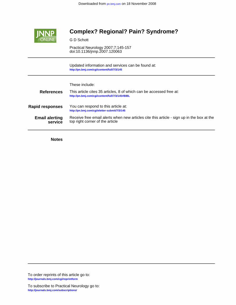
The story of complex regional pain
syndrome (CRPS) begins in 1864.
During the American Civil War, the
father of American neurology, Silas
Weir Mitchell (fig 1), together with
Morehouse and Keen, observed that soldiers
sustaining major nerve injuries affecting their
limbs sometimes experienced long-lasting
pain that was burning in quality, and ‘‘so
frequent and terrible as to demand from us
the fullest description’’.1 Soon afterwards he
termed the condition causalgia (Greek: kausos
(heat) + algos (pain)). Mitchell’s account, in
which he graphically describes many of the
associated features shown in table 1, is one of
the classics of neurology.
At the beginning of the 20th century, Paul
Sudeck made two important contributions.2
First, only five years after x rays had been
discovered, he identified the localised bone
atrophy (‘‘Knochenatrophie’’) that can develop
in the presence of acute, focal limb dis-
orders—and so, strictly speaking, the term
Sudeck’s atrophy should be reserved for the
radiological appearance of osteoporosis.
Second, he postulated an inflammatory
(‘‘entzundliche’’) cause—a concept now
thought increasingly plausible.
The next landmark contribution was the
paper from the famous French vascular
surgeon, Rene Leriche. Thinking the limb of
Figure 1Silas Weir Mitchell (1829–1914).
G D SchottConsultant Neurologist, The
National Hospital for Neurology
and Neurosurgery, Queen Square,
London WC1N 3BG, UK;
145Schott
www.practical-neurology.com
on 18 November 2008 pn.bmj.comDownloaded from
patients with causalgia resembled an ischae-
mic limb, and recalling that sympathectomy
was used to treat ischaemic limbs, in 1916 he
described how he had performed extensive
stripping of the peri-arterial nerve plexus
from the affected limb of a patient with
causalgia, and pain relief ensued.3 Stemming
from this pivotal report of a single case (fig 2),
the conceptual leap, whereby the sympathetic
nervous system became implicated in the
phenomenon of causalgia, resulted in the
100-year search for sympathetically mediated
mechanisms, and vast numbers of diverse
procedures being performed with the aim of
interrupting the sympathetic outflow in an
attempt to alleviate the pain.
Some decades after causalgia had been
described, others noted that sometimes a
milder syndrome could occur, but in the
absence of major nerve injury. Various terms
were introduced for this syndrome, includ-
ing minor causalgia, algodystrophy, and reflex
sympathetic dystrophy. Still much used
today, this last term was introduced in 1946
by Evans, because he postulated that trauma
that generated activity in afferents set up a
reflex in the spinal cord which stimulated
activity in sympathetic efferents, which in
turn resulted in dystrophic changes in the
periphery of the limb.4 Evans develop-
ed the prevailing theory of that time that
central changes in the spinal cord could
spread and even affect the brain—a remark-
ably prescient view in the light of current
research findings. However, the role of
the sympathetic nervous system and the
therapeutic benefit of interrupting it re-
main controversial;5–7 increasingly, attention
is now being paid to the contribution of
neurogenic pseudo-inflammation—returning
full circle back to Sudeck.
WRESTLING WITH DEFINITIONSAND CLASSIFICATIONUncertainties about delineating the major
from the minor forms of these disorders, and
about the involvement of the sympathetic
system, set the scene for nosological chaos. In
1986, the International Association for the
Study of Pain (IASP) simultaneously provided
two slightly different definitions of causalgia,
and sympathetic hyperactivity was included
in its definition of reflex sympathetic dystro-
phy. By 1994, the IASP had abandoned the
sympathetic component and had introduced
the new term complex regional pain syn-
drome, yet continued to divide the
syndrome into its two familiar subtypes,
reflex sympathetic dystrophy and causalgia,
but now designated Types I and II respectively
(table 2).8
The term CRPS, however, generates more
questions than answers. Why ‘‘complex’’,
when there is nothing more complex about
these pains than, for example, phantom pain
or anaesthesia dolorosa? And why ‘‘regional’’,
when, for example, pain in the hand after a
TABLE 1 The various accompanying features seen in complex regionalpain syndrome
Erythematous, cyanosed, pale or blotchy skinExcessive, reduced or absent sweatingInappropriate warmth or coldnessSwelling or atrophy of skinLoss of skin wrinkles, or glossinessExcess or loss of hairNails ridged, curved, thin, brittle or clubbedSubcutaneous atrophy or thickeningStiffness and restriction of passive limb movementsDupuytren’s and other contracturesOsteoporosis—spotty, localised or widespreadMuscle wasting, weakness, loss of dexterity, difficulty in initiatingmovements, ‘‘motor neglect’’Involuntary movements—tremor, unsteadiness, spasms, dystonia,myoclonic jerksVisuospatial and other perceptual disturbancesDetrusor and urinary sphincter dysfunction
Modified from Schott GD. Pain and the sympathetic nervous system. In:Mathias CJ, Bannister R, eds. Autonomic failure, 4th edition. Oxford: OxfordUniversity Press, 1999:520–26. (Reproduced with permission from OxfordUniversity Press.)
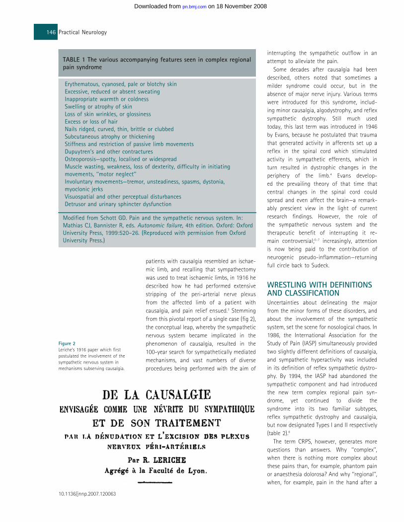
Figure 2Leriche’s 1916 paper which first
postulated the involvement of the
sympathetic nervous system in
mechanisms subserving causalgia.
146 Practical Neurology
10.1136/jnnp.2007.120063
on 18 November 2008 pn.bmj.comDownloaded from
fracture can spread to affect the whole arm,
or more widely? And what about the ‘‘pain’’,
which can vary from the trivial to the
overwhelming and, occasionally, can even be
absent?4, 9 And does ‘‘syndrome’’ refer to the
variable pain state, or the accompanying
features, and if so, to all of them or only
some? Just a few years after the term CRPS
was invented, it seems doubtful that the
diagnostic criteria will stand the test of time.
New criteria are under discussion in which
Types I and II are no longer distinguished
(table 3),10 and perhaps another new term will
be spawned. In the meanwhile, however, use
of the traditional terminology is dwindling,
and as CRPS is a term now used routinely by
pain specialists and increasingly so by
neurologists and in the neurological litera-
ture, it will be retained here.
WHAT ARE THESE DISORDERS?These extremely heterogeneous disorders are
characterised by pain, along with various
accompanying features (table 1).9, 11 The pain
itself is:
N spontaneous and characteristically burn-ing in quality but can be of almost anytype
N of proportion to the inciting cause
N often accompanied by various sensoryfeatures (table 4), including allodynia—theterm describing the phenomenon inwhich innocuous sensory stimuli are feltas pain.
Other accompanying neuropathic features
include the motor disorders, such as the
variable weakness and wasting, as well as the
wide range of involuntary movements—
although the contribution of psychological
factors remains controversial (see below).
Among the remarkably large number of
diverse and similarly variable associated
phenomena shown in table 1 are those with
pseudo-inflammatory, vascular, trophic or
musculoskeletal features. Even in the absence
of major nerve injury, many of these
associated features may yet be caused in
part by neurally-mediated mechanisms, blur-
ring the distinction between typical neuro-
pathic and non-neuropathic processes.
CRPS often shows considerable temporal
variation. This variation includes short-term,
hour-by-hour or diurnal changes, and far
TABLE 2 IASP classification of the complex regional pain syndrome(from Merskey and Bogduk, 19948)
Type I (reflex sympathetic dystrophy) Type II (causalgia)
Definition: A syndrome that developsafter an initiating noxious event, isnot limited to the distribution of asingle peripheral nerve, and isapparently disproportionate to theinciting event. It is associated atsome point with evidence ofoedema, changes in skin bloodflow, abnormal sudomotor activityin the region of the pain, orallodynia or hyperalgesia
Definition: Burning pain, allodynia,and hyperpathia usually in the handor foot after partial injury of a nerveor one of its major branches
Diagnostic criteria (2–4 mustbe satisfied):
Diagnostic criteria (all three mustbe satisfied):
1. The presence of aninitiating noxious event,or a cause ofimmobilisation
1. The presence of continuing pain,allodynia, or hyperalgesia after anerve injury, not necessarily limited tothe distribution of the injured nerve
2. Continuing pain, allodynia, orhyperalgesia with which the painis disproportionate to any incitingevent
2. Evidence at some time ofoedema, changes in skin blood flow,or abnormal sudomotor activity inthe region of the pain
3. Evidence at some time ofoedema, changes in skin bloodflow, or abnormal sudomotoractivity in the region of the pain
3. This diagnosis is excluded by theexistence of conditions that wouldotherwise account for the degree ofpain and dysfunction
4. This diagnosis is excluded bythe existence of conditions thatwould otherwise account forthe degree of pain anddysfunction
TABLE 3 Proposed modified research diagnostic criteria for complexregional pain syndrome (from Harden et al, 199910)
(1) Continuing pain disproportionate to any inciting event(2) At least one symptom in each of the four categories, and(3) One sign in two or more of the four categories. The four categories are:
– sensory– vasomotor– sudomotor/oedema– motor/trophic
and each category has several subcomponents*
*For details of subcomponents, see appendix C in Harden et al, 1999.11
147Schott
www.practical-neurology.com
on 18 November 2008 pn.bmj.comDownloaded from
longer changes extending over weeks, months
and years. Typically, during the first few
weeks the affected limb is warm compared
with the opposite limb; during the next few
months it can be warmer or cooler; and then
after many months or years it tends to be
cooler. The temporal changes are highly
variable in their degree and timing, and more
recent studies have questioned such tempo-
rally-determined staging.9, 12
In the light of these very heterogeneous
features, it becomes obvious that it is
extremely difficult to say what condition(s)
are being talked about, and the most honest,
albeit facetious, description is of ‘‘a ‘funny’
pain in a ‘funny-looking’ limb’’.5 Furthermore,
no single unifying explanation can account
for all the diverse features, and perhaps the
least uncertainty is that CRPS comprises a
spectrum of disorders, with the most severe
CRPS Type II (causalgia) at one end and the
more minor Type I (reflex sympathetic
dystrophy) at the other.
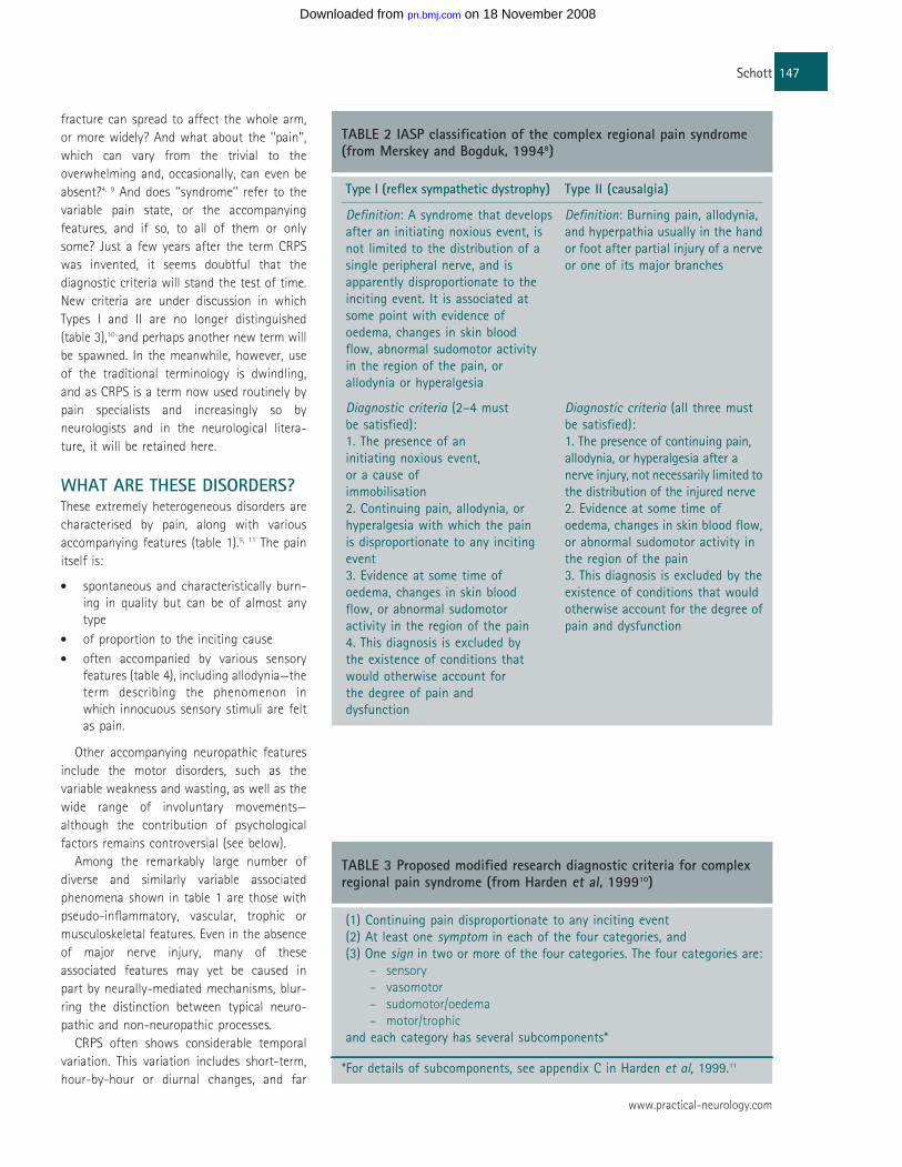
CRPS Type II (causalgia)It is unusual for a neurologist to see a patient
with this extremely severe condition, but once
seen, the patient is often unforgettable (fig 3).
Usually a devastating injury has occurred,
which by definition has caused a major nerve
injury (although ‘‘major’’ has never been
clarified). Often there is also significant
vascular damage. While the musket ball injury
may not feature much in district general
hospital practice, and bullet and knife wounds
are rarely seen, the commonest traumatic
cause is brachial plexus avulsion, often
following a motorcycle accident. The burning
pain is often of extreme severity and
dominates the patient’s life, and Weir
Mitchell’s remarkable description of the pain
and accompanying features has never been
bettered.1
CRPS Type I (reflex sympatheticdystrophy)Although CRPS Type I is far commoner than
Type II, it is nevertheless only infrequently
encountered in neurological practice. Some of
the accompanying features shown in table 1
are illustrated in figures 4–7. The most
common causes are shown in table 5, but in
about one quarter of cases no precipitating
cause can be found.9 Easily the commonest
peripheral cause, perhaps accounting for
about 50% of patients, is some form of
limb trauma. Such trauma is usually distal
TABLE 4 The characteristics of the pain in complex regional painsyndrome
l spontaneousl typically burningl unexpectedly severe considering any inciting causel mainly distal in the limb but spreadsl does not conform with peripheral nerve or root territoryl worse when limb dependentl accompanied by various sensory disturbances, eg numbness, hypo- and
hyperalgesia, hypo- and hyperpathia, allodynial worse with various stimuli
– touch– movement– temperature changes
Figure 3The terrible suffering caused by
causalgia. The obviously distressed
soldier immobilises the painful right
arm struck by a bullet. From Mayfield
FH, Devine JW. Causalgia. Surg GynecolObstet (now J Am Coll Surg)
1945;80:631–5. (Reproduced with
permission of the American College of
Surgeons.)
148 Practical Neurology
10.1136/jnnp.2007.120063
on 18 November 2008 pn.bmj.comDownloaded from
and can be mild or indeed trivial; it can be
accidental—causing, for example, an ankle
sprain, a crush injury to the hand, or scaphoid
fracture; or it can follow surgery—for
instance, for Dupuytren’s contracture, carpal
tunnel decompression or correction of hallux
valgus.
Limb trauma is often followed by self-
imposed immobility, and after surgery the
relevant part is usually immobilised in a
bandage or cast for days if not weeks. It is
now clear that such immobilisation, although
necessary, can also have drastic and even
unfortunate consequences. This view is strik-
ingly supported in studies of healthy volun-
teers undergoing immobilisation alone, in
whom prolonged casting causes features very
similar to CRPS: muscle atrophy, stiffness,
changes in skin colour, and trophic changes
affecting the skin, subcutaneous tissues, and
nails.13 Variable changes in skin temperature,
altered sensory thresholds and, after the cast
is removed, clumsiness similar to that found
in CRPS, have all been found, and pain, while
not a typical feature seen after immobility,
can occur.14 Thus immobility itself can induce
many features typically associated with CRPS,
and sometimes, as with a fracture needing
surgery and immobilisation, it is impossible to
know which is the specific trigger for the
ensuing CRPS—the initial injury, the operation
or the immobilisation.
Disorders of the central nervous system,
and systemic illness and other factors can
cause CRPS too. The commonest central
cause is stroke, but evaluation can be
complicated by variable degrees of weakness
and immobility, sensory loss and inattention,
other accompanying medical conditions such
as diabetes, and musculoskeletal factors
such as shoulder subluxation. Doubtless this
heterogeneity, and variation in definition,
account for the quoted post-stroke frequency
ranging from 1.5 to 61%.15
All of these ill-defined features of CRPS
result in several uncertainties:
N The occurrence of the syndrome isunpredictable, and it is unrelated to theseverity of the causative insult. It is alsounrelated to age, because individuals of
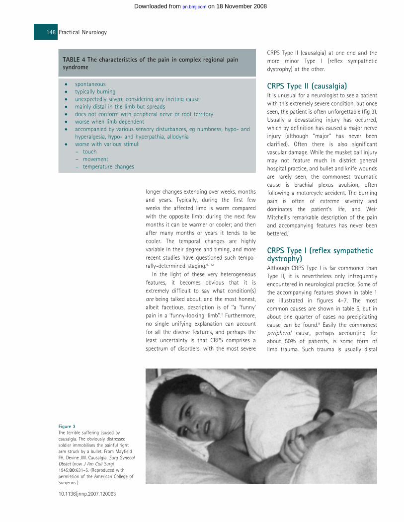
Figure 4The red and sweaty right hand in a
patient with CRPS Type I that followed
surgery, the scar from which is easily
visible above the wrist.
Figure 5The puffy left hand with an inability to
close the fist in a patient with CRPS
Type I. From Blumberg H, Hoffmann U,
Mohadjer M, et al. Clinical
phenomenology and mechanisms of
reflex sympathetic dystrophy: emphasis
on edema. In: Gebhart GF, Hammond
DL, Jensen TS, eds. Progress in painresearch and management, volume 2.Seattle, Washington: IASP Press,
1994:455–81. (Reproduced with
permission of the International
Association for the Study of Pain.)
Figure 7The swollen, dusky-red foot in a patient
with CRPS Type I affecting the right
foot.
Figure 6The shiny skin with loss of wrinkles,
curved and elongated nails, and tapered
fingers, in a patient with CRPS Type I
affecting the left hand.
149Schott
www.practical-neurology.com
on 18 November 2008 pn.bmj.comDownloaded from
any age, including children and adoles-cents, can be affected. Presumably, there-fore, it is some idiosyncratic response to,or the consequences of, the initiatingevent (rather than the event itself) thatgenerates the condition.
N It is unclear when pain becomes abnor-mal. A knee replacement may result insome long-term discomfort on walkingand this would be accepted as normal; aknee that after many months remainsextremely painful, particularly when thereare accompanying features such as swel-ling, warmth, and extreme sensitivity ofthe overlying tissues, is obviously abnor-mal. But the boundary between normaland abnormal is uncertain.
N The incidence of CRPS is very difficult togauge. Uncertainty about the frequencyfollowing stroke has been discussedabove. The most recent study suggestedan overall incidence of CRPS of over 26per 100,000 person years, and found thehighest incidence occurred after an upperlimb fracture in women in later life.16
However, the frequency of CRPS after
distal radial fractures has ranged from 1–2% when reported retrospectively, to upto 38% when reported prospectively.17 Inthe lower limb, knee replacement hasbeen used as a clinical model, andwhereas 20 years ago there were noreports of CRPS, it is now a very wellrecognised problem. In a recent prospec-tive series of 52 patients, at 6 monthsafter surgery 19% of patients met thecriteria for CRPS.17
PREDISPOSING FACTORS?Two factors may be relevant as to whether
an individual develops CRPS: their underlying
psychological predisposition, and their
genetic make-up.
What is the role of psychologicalfactors?Evaluating both the background and the
prevailing psychological and psychiatric
aspects relating to pain in these patients is
a subject fraught with difficulties. Further
problems arise because of various reported
perceptual disturbances, including neglect
phenomena and visuospatial distortions,
which are receiving increasing attention but
remain ill understood. Perhaps not surpris-
ingly, therefore, the role of psychological
factors has been the subject of vigorous
controversy.
One view is exemplified by Ochoa and
Verdugo, who consider most cases of CRPS as
‘‘A common clinical avenue for somatoform
expression’’,18 including the subset of patients
exhibiting abnormal movements and pos-
tures.19 A particularly difficult issue concerns
those patients who have post-traumatic fixed
dystonia and who fulfil the diagnostic criteria
for CRPS; in many patients a diagnosis of
psychogenic dystonia, a somatisation disor-
der, or both, can be made, and there appears
to be overlap between fixed dystonia and
CRPS.20
Whether antecedent psychological factors
predispose patients to developing CRPS
remains unclear. Two prospective but limited
studies have addressed this issue. One carried
out many years ago, which would be
considered inadequate by today’s standards,
indicated that prediction of outcome was
possible by a preoperative psychological
assessment;21 the other study did not find
TABLE 5 Causes of complex regional pain syndrome Type I (reflexsympathetic dystrophy)
Peripherall limb traumal electric shockMixed peripheral and centrall herpes zosterl brachial plexus avulsion and other injuriesCentrall strokel multiple sclerosisl spinal cord injuryl cerebral tumourl brain injuryDrugsl phenobarbitall isoniazidCardiopulmonary disordersl post-myocardial infarctionl post-cardiac surgeryl lung diseaseIdiopathic and other causesl occurrence in children (often affecting lower limbs)l immobilityl transient forms, eg pregnancyl flitting and recurrent forms
150 Practical Neurology
10.1136/jnnp.2007.120063
on 18 November 2008 pn.bmj.comDownloaded from
prediction possible,22 and at present satisfac-
tory data are not available to foretell which
individuals are likely to develop CRPS.23 My
personal impression is that while sufferers
may become seriously affected psychologi-
cally, and sometimes show features of major
depression (as expected in anyone who is in
constant pain, and who may have lost their
job and had their family and social life
shattered), they often seem to have led a
psychologically unremarkable life before the
condition developed.
Two additional factors are pertinent. First,
as trauma is so often the cause, litigation not
infrequently lurks in the background. Second,
very rare instances of malingering, as revealed
by covert video recordings, have also been
reported.19
Genetic factors?Patients with CRPS have been found more
likely than controls to have the HLA tissue
types HLA-DQ1, HLA-DR13 and HLA-DR2, and
other susceptibility loci for CRPS have also
been reported.24 The significance of these
observations, and the putative link between
any of these loci and the receptor for GABA,
remain unclear.
INVESTIGATIONSThese are rarely helpful in diagnosis, but are
usually necessary to exclude other disorders
ranging from tumours to arthritis, as well as
any underlying or associated neurological
causes. CRPS is not accompanied by abnorm-
alities on conventional haematological or
biochemical tests, and finding a raised ESR
or abnormal immunological or bone profile
studies means an alternative cause needs to
be sought. Occasionally neurophysiological
tests are helpful in CRPS Type I in excluding a
radiculopathy or peripheral nerve lesion.
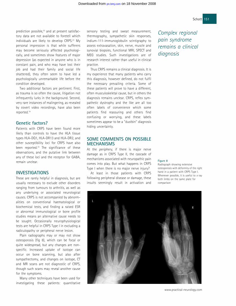
Plain radiographs may or may not show
osteoporosis (fig 8), which can be focal or
quite widespread, but any changes are non-
specific. Increased uptake of isotope can
occur on bone scanning, but also after
sympathectomy, and changes on isotope, CT
and MR scans are not diagnostic of CRPS,
though such scans may reveal another cause
for the symptoms.
Many other techniques have been used for
investigating these patients: quantitative
sensory testing and sweat measurement,
thermography, sympathetic skin responses,
indium-111-immunoglobulin scintigraphy to
assess extravasation, skin, nerve, muscle and
synovial biopsies, functional MRI, SPECT and
MEG studies. Such investigations are of
research interest rather than useful in clinical
practice.
Thus CRPS remains a clinical diagnosis. It is
my experience that many patients who carry
this diagnosis, however defined, do not fulfil
the necessary prevailing criteria. Some of
these patients will prove to have a different,
often musculoskeletal cause, but in others the
diagnosis remains unclear. CRPS, reflex sym-
pathetic dystrophy and the like are all too
often labels of convenience which some
patients find reassuring and others find
confusing or worrying, and these labels
sometimes appear to be a ‘‘dustbin’’ diagnosis
hiding uncertainty.
SOME COMMENTS ON POSSIBLEMECHANISMSAt the periphery, if there is major nerve
damage as in CRPS Type II, the cascade of
mechanisms associated with neuropathic pain
comes into play. But what happens in CRPS
Type I when there is no major nerve injury?
At least in those patients with CRPS
following peripheral disease or damage, these
insults seemingly result in activation and
Figure 8Radiograph showing extensive
osteoporosis with deformity of the right
hand in a patient with CRPS Type I.
Whenever possible, it is useful to x ray
both limbs on the same plate for
comparison
Complex regionalpain syndromeremains a clinicaldiagnosis
151Schott
www.practical-neurology.com
on 18 November 2008 pn.bmj.comDownloaded from
sensitisation of primary nociceptor afferents;
furthermore, a variety of neuropeptides and
neuromodulators, pro-inflammatory cyto-
kines, and other substances appear to be
released peripherally (and centrally) from
these afferents,9, 11 and perhaps from sympa-
thetic nerve endings, as well as from
damaged blood vessels. The resulting neuro-
genic pseudo-inflammation probably leads to
the afferent neuron developing abnormal
sensitivity to mechanical and thermal stimuli
together with adrenergic supersensitivity,
resulting in pain and other sensory features.
A number of factors arguing for and against
involvement of the sympathetic nervous
system are included in table 6, and further
details and references have been summarised
elsewhere.25 Of particular note is that, con-
trary to previous thinking, the sympathetic
outflow in these disorders is not hyperactive.
Bearing some resemblance to the pseudo-
inflammatory changes seen in diabetic and
non-diabetic Charcot joints,26 pseudo-inflam-
mation in CRPS may underlie the increased
blood flow and vascular permeability, skin
warming, hypervascularity of synovia and
muscle, immune infiltration of the skin, and
osteoporosis11—and hence the trophic fea-
tures of the syndrome. There may also be an
autoimmune component in some instances.27
Concerning vascular factors, a disorder
similar to CRPS has been produced experi-
mentally in rats, when reperfusion follows a
period of limb ischaemia caused by prolonged
tourniquet application.28 The clinical relevance
of this finding is that it recalls those patients
in whom CRPS is associated with limb
immobilisation in a cast, especially if applied
too tightly, when the consequences of
ischaemia may compound those due to
trauma and immobility and which were
discussed above.
These heterogeneous peripheral neural,
pseudo-inflammatory and vascular compo-
nents may explain the equally heterogeneous
clinical features seen among different
patients.
With regard to central mechanisms, not
only can CRPS result from central nervous
system lesions (table 5), but peripherally-
triggered CRPS often has features suggesting
the central nervous system has become
secondarily implicated (see below).
Conversely, a central nervous system lesion
such as stroke or tumour can produce the
peripheral features of CRPS, and so it is not
particularly helpful to distinguish rigidly
between peripheral and central causes when
considering the underlying mechanisms—one
can consider there to be functional neural
continuity. Yet there are several clinical and
experimental aspects which mean that the
central nervous system perhaps always
becomes involved:
N The distribution of the pain and otherfeatures which conforms to neither aperipheral nerve nor root territory, andcan show bilateral, mirror, quadrant orhemibody involvement.29
N Detailed neurovascular studies haveshown evidence of an abnormal unilateralreflex pattern of sympathetic vasocon-strictor neuronal activity in the affectedlimb of patients in the early stages ofCRPS Type I;30 this pattern, and in other
TABLE 6 Clinical features for and against involvement of thesympathetic nervous system in complex regional pain syndrome
For Against
l Some of the clinicalfeatures (eg, temperaturechanges, sweating) are orappear to be phenomenasubserved by sympatheticnerves
l Interrupting thesympathetic supply mayalleviate pain in anindividual patient
l Some of the clinical features (eg, warmth,swelling, redness) are mediated byvasoactive substances (substance P,calcium gene related peptide (CGRP),ATP, histamine, 5-HT, neurokinins, etc)released from small-diameter sensoryafferents, damaged blood vessels, etc
l Group studies have established thatinterrupting the sympathetic supply is nomore effective than placebo
l Pain and sensory featuresrelieved by sympatheticblock can be rekindled bylocal noradrenaline
l There is no relation between any pain reliefachieved and the typical effects followingsympathetic blockade, in respect of time ofonset, duration or degree
l Pain is increased by stressand cold, which increasesympathetic activity
l Pain that is apparentlysympathetically maintainedcan be increased when thepatient, excluding thethermally isolated limb, iscooled. This centralphenomenon is associatedwith increased activity incutaneous vasoconstrictornerves
l On microneurography, the peripheralsympathetic outflow is physiologicallynormal. There is reduced local venousnoradrenaline and 3,4-dihydroxyphenylethyleneglycol (DHPG),leading to possible denervationhypersensitivity
l The syndrome is not a feature ofexcessive (eg, thyrotoxicosis) or reduced(eg, autonomic failure) sympatheticactivity. CRPS has been described in asympathetically denervated limb
152 Practical Neurology
10.1136/jnnp.2007.120063
on 18 November 2008 pn.bmj.comDownloaded from
patients the presence of hyperhydrosis, isin keeping with central mechanisms.
N The PET scan changes which are seenwith immobility alone, and which arereminiscent of those seen in acute andcentral pain. In these conditions, the PETscan changes, among others, includeincreased blood flow in the cingulateand somatosensory cortices.14
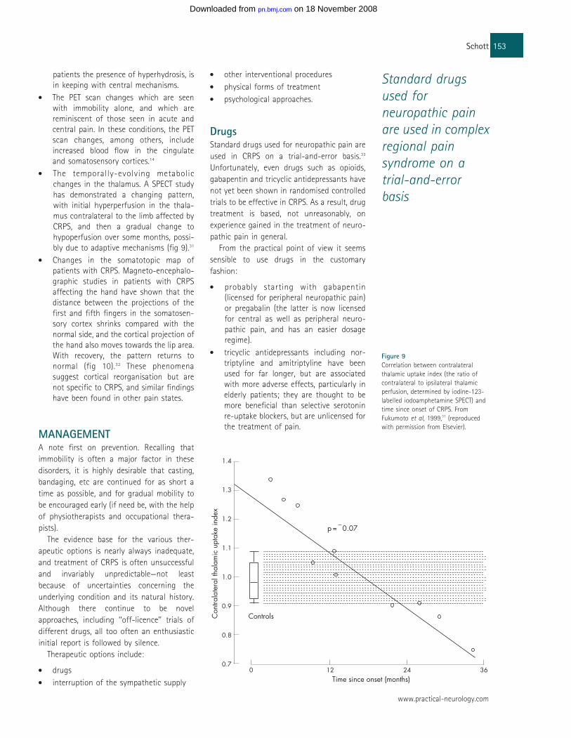
N The temporally-evolving metabolicchanges in the thalamus. A SPECT studyhas demonstrated a changing pattern,with initial hyperperfusion in the thala-mus contralateral to the limb affected byCRPS, and then a gradual change tohypoperfusion over some months, possi-bly due to adaptive mechanisms (fig 9).31
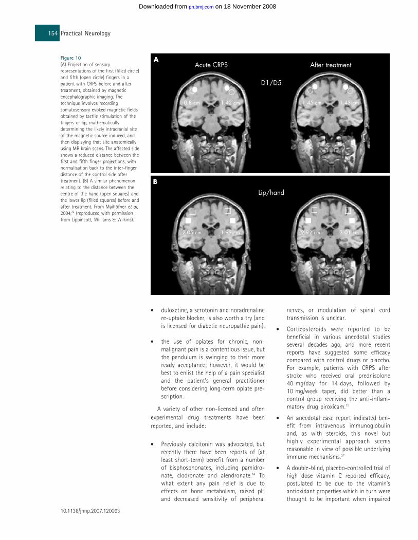
N Changes in the somatotopic map ofpatients with CRPS. Magneto-encephalo-graphic studies in patients with CRPSaffecting the hand have shown that thedistance between the projections of thefirst and fifth fingers in the somatosen-sory cortex shrinks compared with thenormal side, and the cortical projection ofthe hand also moves towards the lip area.With recovery, the pattern returns tonormal (fig 10).32 These phenomenasuggest cortical reorganisation but arenot specific to CRPS, and similar findingshave been found in other pain states.
MANAGEMENTA note first on prevention. Recalling that
immobility is often a major factor in these
disorders, it is highly desirable that casting,
bandaging, etc are continued for as short a
time as possible, and for gradual mobility to
be encouraged early (if need be, with the help
of physiotherapists and occupational thera-
pists).
The evidence base for the various ther-
apeutic options is nearly always inadequate,
and treatment of CRPS is often unsuccessful
and invariably unpredictable—not least
because of uncertainties concerning the
underlying condition and its natural history.
Although there continue to be novel
approaches, including ‘‘off-licence’’ trials of
different drugs, all too often an enthusiastic
initial report is followed by silence.
Therapeutic options include:
N drugs
N interruption of the sympathetic supply
N other interventional procedures
N physical forms of treatment
N psychological approaches.
DrugsStandard drugs used for neuropathic pain are
used in CRPS on a trial-and-error basis.33
Unfortunately, even drugs such as opioids,
gabapentin and tricyclic antidepressants have
not yet been shown in randomised controlled
trials to be effective in CRPS. As a result, drug
treatment is based, not unreasonably, on
experience gained in the treatment of neuro-
pathic pain in general.
From the practical point of view it seems
sensible to use drugs in the customary
fashion:
N probably starting with gabapentin(licensed for peripheral neuropathic pain)or pregabalin (the latter is now licensedfor central as well as peripheral neuro-pathic pain, and has an easier dosageregime).
N tricyclic antidepressants including nor-triptyline and amitriptyline have beenused for far longer, but are associatedwith more adverse effects, particularly inelderly patients; they are thought to bemore beneficial than selective serotoninre-uptake blockers, but are unlicensed forthe treatment of pain.
Figure 9Correlation between contralateral
thalamic uptake index (the ratio of
contralateral to ipsilateral thalamic
perfusion, determined by iodine-123-
labelled iodoamphetamine SPECT) and
time since onset of CRPS. From
Fukumoto et al, 1999,31 (reproduced
with permission from Elsevier).
Standard drugsused forneuropathic painare used in complexregional painsyndrome on atrial-and-errorbasis
153Schott
www.practical-neurology.com
on 18 November 2008 pn.bmj.comDownloaded from
N duloxetine, a serotonin and noradrenalinere-uptake blocker, is also worth a try (andis licensed for diabetic neuropathic pain).
N the use of opiates for chronic, non-malignant pain is a contentious issue, butthe pendulum is swinging to their moreready acceptance; however, it would bebest to enlist the help of a pain specialistand the patient’s general practitionerbefore considering long-term opiate pre-scription.
A variety of other non-licensed and often
experimental drug treatments have been
reported, and include:
N Previously calcitonin was advocated, butrecently there have been reports of (atleast short-term) benefit from a numberof bisphosphonates, including pamidro-nate, clodronate and alendronate.34 Towhat extent any pain relief is due toeffects on bone metabolism, raised pHand decreased sensitivity of peripheral
nerves, or modulation of spinal cordtransmission is unclear.
N Corticosteroids were reported to bebeneficial in various anecdotal studiesseveral decades ago, and more recentreports have suggested some efficacycompared with control drugs or placebo.For example, patients with CRPS afterstroke who received oral prednisolone40 mg/day for 14 days, followed by10 mg/week taper, did better than acontrol group receiving the anti-inflam-matory drug piroxicam.15
N An anecdotal case report indicated ben-efit from intravenous immunoglobulinand, as with steroids, this novel buthighly experimental approach seemsreasonable in view of possible underlyingimmune mechanisms.27
N A double-blind, placebo-controlled trial ofhigh dose vitamin C reported efficacy,postulated to be due to the vitamin’santioxidant properties which in turn werethought to be important when impaired
Figure 10(A) Projection of sensory
representations of the first (filled circle)
and fifth (open circle) fingers in a
patient with CRPS before and after
treatment, obtained by magnetic
encephalographic imaging. The
technique involves recording
somatosensory evoked magnetic fields
obtained by tactile stimulation of the
fingers or lip, mathematically
determining the likely intracranial site
of the magnetic source induced, and
then displaying that site anatomically
using MR brain scans. The affected side
shows a reduced distance between the
first and fifth finger projections, with
normalisation back to the inter-finger
distance of the control side after
treatment. (B) A similar phenomenon
relating to the distance between the
centre of the hand (open squares) and
the lower lip (filled squares) before and
after treatment. From Maihofner et al,2004,32 (reproduced with permission
from Lippincott, Williams & Wilkins).
154 Practical Neurology
10.1136/jnnp.2007.120063
on 18 November 2008 pn.bmj.comDownloaded from
blood flow and venous stasis arepresent.35
N Some improvement in pain was reportedin three of eight patients with dystoniaand CRPS treated with intrathecalbaclofen.36
Interrupting the sympatheticsupplyIn the light of the issues discussed above, this
therapeutic avenue deserves consideration in
its own right. Techniques of interrupting the
proximal cervical or lumbar sympathetic out-
flow have included surgical sympathectomies
of various types, sympathetic blocks with
local anaesthetics or destructive neurolytic
agents, and thermocoagulation. At the per-
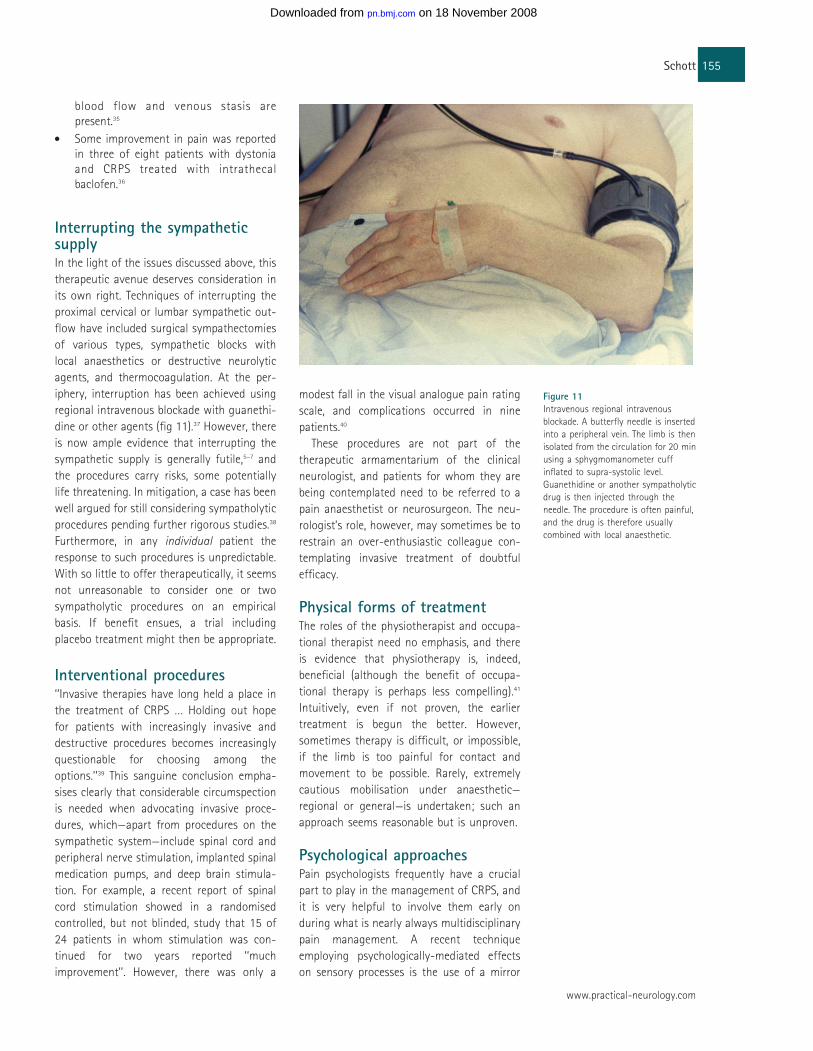
iphery, interruption has been achieved using
regional intravenous blockade with guanethi-
dine or other agents (fig 11).37 However, there
is now ample evidence that interrupting the
sympathetic supply is generally futile,5–7 and
the procedures carry risks, some potentially
life threatening. In mitigation, a case has been
well argued for still considering sympatholytic
procedures pending further rigorous studies.38
Furthermore, in any individual patient the
response to such procedures is unpredictable.
With so little to offer therapeutically, it seems
not unreasonable to consider one or two
sympatholytic procedures on an empirical
basis. If benefit ensues, a trial including
placebo treatment might then be appropriate.
Interventional procedures‘‘Invasive therapies have long held a place in
the treatment of CRPS … Holding out hope
for patients with increasingly invasive and
destructive procedures becomes increasingly
questionable for choosing among the
options.’’39 This sanguine conclusion empha-
sises clearly that considerable circumspection
is needed when advocating invasive proce-
dures, which—apart from procedures on the
sympathetic system—include spinal cord and
peripheral nerve stimulation, implanted spinal
medication pumps, and deep brain stimula-
tion. For example, a recent report of spinal
cord stimulation showed in a randomised
controlled, but not blinded, study that 15 of
24 patients in whom stimulation was con-
tinued for two years reported ‘‘much
improvement’’. However, there was only a
modest fall in the visual analogue pain rating
scale, and complications occurred in nine
patients.40
These procedures are not part of the
therapeutic armamentarium of the clinical
neurologist, and patients for whom they are
being contemplated need to be referred to a
pain anaesthetist or neurosurgeon. The neu-
rologist’s role, however, may sometimes be to
restrain an over-enthusiastic colleague con-
templating invasive treatment of doubtful
efficacy.
Physical forms of treatmentThe roles of the physiotherapist and occupa-
tional therapist need no emphasis, and there
is evidence that physiotherapy is, indeed,
beneficial (although the benefit of occupa-
tional therapy is perhaps less compelling).41
Intuitively, even if not proven, the earlier
treatment is begun the better. However,
sometimes therapy is difficult, or impossible,
if the limb is too painful for contact and
movement to be possible. Rarely, extremely
cautious mobilisation under anaesthetic—
regional or general—is undertaken; such an
approach seems reasonable but is unproven.
Psychological approachesPain psychologists frequently have a crucial
part to play in the management of CRPS, and
it is very helpful to involve them early on
during what is nearly always multidisciplinary
pain management. A recent technique
employing psychologically-mediated effects
on sensory processes is the use of a mirror
Figure 11Intravenous regional intravenous
blockade. A butterfly needle is inserted
into a peripheral vein. The limb is then
isolated from the circulation for 20 min
using a sphygmomanometer cuff
inflated to supra-systolic level.
Guanethidine or another sympatholytic
drug is then injected through the
needle. The procedure is often painful,
and the drug is therefore usually
combined with local anaesthetic.
155Schott
www.practical-neurology.com
on 18 November 2008 pn.bmj.comDownloaded from
box.42 More intriguing than useful, this
technique was originally developed for the
treatment of phantom pain and employs
visual feedback. Rather equivocal results have
been obtained in treating pain in CRPS, and
the mirror box has yet to show promise in the
long term.
PROGNOSISThe outcome for patients with CRPS is very
difficult to predict. Weir Mitchell reported
that ‘‘Many cases of burning pain last but a
few weeks’’,1 but he and others were all too
aware of patients whose pain continued
indefinitely. Some patients with CRPS con-
tinue to suffer for years, with weakness, sleep
disturbance and disability being prominent, in
addition to pain,43 but there are no adequate
long-term studies.
CONCLUSIONS‘‘Complex regional pain syndrome … remains
endlessly fascinating to all persons interested
in pain management. No other chronic pain
syndrome is as shrouded in confusion and
controversy.’’33 The unfortunate sufferer will
doubtless agree with the latter statement, but
not necessarily the former. There is a long
way to go in effectively treating what will
surely turn out to be several different
conditions.
ACKNOWLEDGEMENTThis paper was reviewed by Jon Stone,
Edinburgh, UK.
REFERENCES1. Mitchell SW, Morehouse GR, Keen WW. Gunshot
wounds and other injuries of nerves. Philadelphia:
JB Lippincott, 1864, 100–11. (Reprinted in ClinOrthop Relat Res, 1982;163:2–7).
2. Sudeck P. Uber die akute entzundliche
Knochenatrophie. Arch Klin Chir 1900;62:147–56.
3. Leriche R. De la causalgie envisagee comme une
nevrite du sympathique et de son traitement par la
denudation et l’excision des plexus nerveux peri-
arteriels. Presse Med 1916;24:178–80.
4. Evans JA. Reflex sympathetic dystrophy. SurgGynecol Obstet 1946;82:36–43.
5. Jadad AR, Carroll D, Glynn CJ, et al. Intravenous
regional sympathetic blockade for pain relief in
reflex sympathetic dystrophy: a systematic review
and a randomized, double-blind crossover study.
J Pain Symptom Manage 1995;10:13–20.
6. Kingery WS. A critical review of controlled clinical
trials for peripheral neuropathic pain and complex
regional pain syndromes. Pain 1997;73:123–39.
7. Mailis A, Furlan A. Sympathectomy for neuropathic
pain. Cochrane Database Syst Rev2003;CD002918.
8. Merskey H, Bogduk N, eds. Classification of chronic
pain. Descriptions of chronic pain syndromes anddefinitions of pain terms. Second edition. Seattle,
Washington: IASP Press, 1994:40–2.
9. Veldman PHJM, Reynen HM, Arntz IE, et al. Signs
and symptoms of reflex sympathetic dystrophy:
prospective study of 829 patients. Lancet1993;342:1012–16.
10. Harden RN, Bruehl S, Galer BS, et al. Complex
regional pain syndrome: are the IASP diagnostic
criteria valid and sufficiently comprehensive? Pain1999;83:211–19.
11. Birklein F. Complex regional pain syndrome. J Neurol2005;252:131–8.
12. Bruehl S, Harden RN, Galer BS, et al. Complex
regional pain syndrome: are there distinct subtypes
and sequential stages of the syndrome? Pain2002;95:119–24.
13. Butler SH, Nyman M, Gordh T. Immobility in
volunteers transiently produces signs and
symptoms of complex regional pain syndrome. In:
Devor M, Rowbotham MC, Wiesenfeld-Hallin Z, eds.
Progress in pain research and management, vol 16.
Seattle, Washington: IASP Press, 2000:657–60.
14. Butler SH. Disuse and CRPS. In: Harden RN, Baron R,
Janig W, eds. Complex regional pain syndrome.
Seattle, Washington: IASP Press, 2001:141–50.
PRACTICE POINTS
l Complex regional pain syndrome (CRPS) is an umbrella term introduced bythe International Association for the Study of Pain for conditions knownpreviously by different names—in particular causalgia (in which there ismajor nerve injury), and reflex sympathetic dystrophy (without major nerveinjury). Type I is synonymous with reflex sympathetic dystrophy, Type IIwith causalgia.
l These conditions have in common burning pain. CRPS typically affects alimb, mainly distally, and is variably accompanied by a host of otherfeatures which include sensory, thermal, sweating, motor and trophicphenomena.
l CRPS Type I occurs much more frequently than Type II and has manycauses, peripheral trauma being by far the commonest. Limb immobility isincreasingly recognised to be an important factor.
l Involvement of the sympathetic nervous system is no longer considered anessential component. Pseudo-inflammation in the periphery, andinvolvement of the central nervous system, are receiving increasingattention.
l The diagnosis remains clinical, and is sometimes inappropriately applied toany odd pain in a limb. Investigations are not useful in clinical practice,although are essential for excluding other causes.
l Treatment remains highly unsatisfactory, and no specific measure has beenestablished as reliably beneficial, although restoration of mobility seemscrucial. Various unlicensed drug treatments, interventional procedures,physical forms of treatment and psychological management are the usualapproaches, but benefit is always unpredictable. The natural history ofCRPS remains poorly understood.
l CRPS may well consist of a variety of different conditions subserved bydifferent mechanisms.
156 Practical Neurology
10.1136/jnnp.2007.120063
on 18 November 2008 pn.bmj.comDownloaded from
15. Kalita J, Vajpayee A, Misra UK. Comparison of
prednisolone with piroxicam in complex regional
pain syndrome following stroke: a randomized
controlled trial. Q J Med 2006;99:89–95.
16. de Mos M, de Bruijn AGJ, Huygen FJPM, et al. The
incidence of complex regional pain syndrome: a
population-based study. Pain 3 November 2006
[Epub ahead of print].
17. Stanos SP Jr, Harden RN, Wagner-Raphael L, et al.A prospective clinical model for investigating the
development of CRPS. In: Harden RN, Baron R,
Janig W, eds. Complex regional pain syndrome.
Seattle, Washington: IASP Press, 2001:151–64.
18. Ochoa JL, Verdugo RJ. Reflex sympathetic
dystrophy. A common clinical avenue for
somatoform expression. Neurol Clin1995;13:351–63.
19. Verdugo RJ, Ochoa JL. Abnormal movements in
complex regional pain syndrome: assessment of
their nature. Muscle Nerve 2000;23:198–205.
20. Schrag A, Trimble M, Quinn N, et al. The syndrome
of fixed dystonia: an evaluation of 103 patients.
Brain 2004;127:2360–72.
21. Zachariae L. Incidence and course of posttraumatic
dystrophy following operation for Dupuytren’s
contracture. Acta Chir Scand Suppl 1964;15(Suppl
336):1–51.
22. Field J, Gardner FV. Psychological distress with
algodystrophy. J Hand Surg [Br] 1997;22:100–01.
23. Bruehl S. Do psychological factors play a role in the
onset and maintenance of CRPS-I? In: Harden RN,
Baron R, Janig W, eds. Complex regional painsyndrome. Seattle, Washington: IASP Press,
2001:279–90.
24. van de Beek WJ, Roep BO, van der Slik AR, et al.Susceptibility loci for complex regional pain
syndrome. Pain 2003;103:93–7.
25. Schott GD. Reflex sympathetic dystrophy. J NeurolNeurosurg Psychiatry 2001;71:291–5.
26. Jeffcoate WJ, Game F, Cavanagh PR. The role of
proinflammatory cytokines in the cause of
neuropathic osteoarthropathy (acute Charcot foot)
in diabetes. Lancet 2005;366:2058–61.
27. Goebel A, Stock M, Deacon R, et al. Intravenous
immunoglobulin response and evidence for
pathogenic antibodies in a case of complex regional
pain syndrome 1. Ann Neurol 2005;57:463–64.
28. Coderre TJ, Xanthos DN, Francis L, et al. Chronic
post-ischemia pain (CPIP): a novel animal model of
complex regional pain syndrome-type I (CRPS-I;
reflex sympathetic dystrophy) produced by
prolonged hindpaw ischemia and reperfusion in the
rat. Pain 2004;112:94–105.
29. Schott GD. Mechanisms of causalgia and related
clinical conditions. The role of the central and of
the sympathetic nervous systems. Brain1986;109:717–38.
30. Wasner G, Heckmann K, Maier C, et al. Vascular
abnormalities in acute reflex sympathetic
dystrophy (CRPS I): complete inhibition of
sympathetic nerve activity with recovery. ArchNeurol 1999;56:613–20.
31. Fukumoto M, Ushida T, Zinchuk VS, et al.Contralateral thalamic perfusion in patients with
reflex sympathetic dystrophy syndrome. Lancet1999;354:1790–1.
32. Maihofner C, Handwerker HO, Neundorfer B, et al.Cortical reorganization during recovery from
complex regional pain syndrome. Neurology2004;63:693–701.
33. Rowbotham MC. Pharmacological management of
complex regional pain syndrome. Clin J Pain2006;22:425–9.
34. Berthelot J-M. Current management of reflex
sympathetic dystrophy syndrome (complex regional
pain syndrome type I). Joint Bone Spine2006;73:495–9.
35. Zollinger PE, Tuinebreijer WE, Kreis RW, et al. Effect
of vitamin C on frequency of reflex sympathetic
dystrophy in wrist fractures: a randomised trial.
Lancet 1999;354:2025–8.
36. van Hilten BJ, van de Beek WJ, Hoff JI, et al.Intrathecal baclofen for the treatment of dystonia
in patients with reflex sympathetic dystrophy.
N Engl J Med 2000;343:625–30.
37. Hannington-Kiff JG. Intravenous regional
sympathetic block with guanethidine. Lancet1974;i:1019–20.
38. Max MB, Gilron I. Sympathetically maintained pain:
has the emperor no clothes? Neurology1999;52:905–7.
39. Nelson DV, Stacey BR. Interventional therapies in
the management of complex regional pain
syndrome. Clin J Pain 2006;22:438–42.
40. Kemler MA, de Vet HCW, Barendse GAM, et al. The
effect of spinal cord stimulation in patients with
chronic reflex sympathetic dystrophy: two years’
follow-up of the randomized controlled trial. AnnNeurol 2004;55:13–18.
41. Oerlemans HM, Oostendorp RAB, de Boo T, et al.Pain and reduced mobility in complex regional
pain syndrome I: outcome of a prospective
randomised controlled clinical trial of adjuvant
physical therapy versus occupational therapy. Pain1999;83:77–83.
42. McCabe CS, Haigh RC, Ring EF, et al. A controlled
pilot study of the utility of mirror visual feedback in
the treatment of complex regional pain syndrome
(type 1). Rheumatology 2003;42:97–101.
43. Galer BS, Henderson J, Perander J, et al. Course of
symptoms and quality of life measurement in
Complex Regional Pain Syndrome: a pilot survey.
J Pain Symptom Manage 2000;20:286–92.
157Schott
www.practical-neurology.com
on 18 November 2008 pn.bmj.comDownloaded from


![Cronicon · neck pain, and pain of the shoulder region (cervicobrachial syndrome, rotator cuff syndrome), lumbar region (lumbar syndrome), chest pain and so on [2]. It is important](https://img.pdfslide.net/doc/110x75/5fa22c69706ace092c52fd11/cronicon-neck-pain-and-pain-of-the-shoulder-region-cervicobrachial-syndrome-rotator.jpg)