Embed Size (px)
Citation preview
Int J Anal Bio-Sci Vol. 8, No 4 (2020)
― 83 ―― 83 ―
1The Graduate School of Kagawa Prefectural
University of Health Sciences, 281-1 Hara Mure-cho,
Takamatsu, Kagawa 761-0123, Japan.2Department of Clinical Laboratory, Kinashi Obayashi
Hospital, 435-1 Fujii Kinashi-cho, Takamatsu City,
Kagawa 761-8024, Japan.3Department of Medical Technology, Faculty of Health
Sciences, Kagawa Prefectural University of Health
Sciences, 281-1 Hara Mure-cho, Takamatsu City,
Kagawa 761-0123, Japan.
*Corresponding author: Akemi Miyagawa, Department
of Clinical Laboratory, Kinashi Obayashi Hospital,
435-1 Fujii Kinashi-cho, Takamatsu City, Kagawa
761-8024, Japan.
Tel: +81-87-881-3631
Fax: +81-87-881-8022
E-mail: [email protected]
Received for publication: July 11, 2020
Accepted for publication: August 26, 2020
〈Research Article〉
Comprehensive assessment of oxidative stress degrees and anti-oxidant potential in dialysis patients
Akemi Miyagawa1,2,* and Kinya Tateishi3
Summary When the kidney is compromised, oxidative stress is aggravated by an increased rate of
reactive oxygen species generation and a decreased rate of substance elimination. In addition, dial-
ysis treatment itself is reported to be associated with oxidative stress in patients with terminal renal
insufficiency. This study was conducted to determine the optimum dialysis treatment. Diacron-
reactive oxygen metabolite and biological anti-oxidant potential tests were performed to assess
oxidative stress levels. Increased oxidative stress and decreased anti-oxidant potential were found
immediately after dialysis. A comparison of dialysis membranes revealed that ABH®-PA
membranes exhibited a significantly lower rate of decrease in anti-oxidant potential in terms of
oxidative stress compared with Maxiflux® membranes. Analysis of biochemical parameters identi-
fied significant strong positive correlations between diacron-reactive oxygen metabolites test
results and serum total protein, serum albumin, total cholesterol, high-density lipoprotein-choles-
terol, and low-density lipoprotein-cholesterol for the percentage decrease and between biological
anti-oxidant potential test results and inorganic phosphorus for pre-dialysis measurements.
ABH®-PA should be used as the dialysis membrane to minimize the increase in the degree of
oxidative stress caused by dialysis treatment. We recommend measuring serum total protein, serum
albumin, total cholesterol, and inorganic phosphorus before and after dialysis sessions to better
understand oxidative stress degrees and anti-oxidant potential.
Key words: d-ROMs, BAP, Dialysis treatment, Oxidative stress, Anti-oxidant potential
Int J Anal Bio-Sci Vol. 8, No 4 (2020)
― 84 ―― 84 ―
1. Introduction
The kidneys excrete waste products and are
involved in various other functions including regu-
lating electrolyte and pH balance, activating vitamin
D, and secreting hormones such as erythropoietin
and renin. These activities require a considerable
amount of oxygen and produce a large quantity of
reactive oxygen species (ROS) as a byproduct.
When the kidney is compromised, oxidative stress is
aggravated by an increased rate of ROS generation
and a decreased rate of substance elimination1-3.
Dialysis treatment itself is reported to increase
oxidative stress in patients with terminal renal insuf-
ficiency4-6. Within minutes of dialysis initiation, the
exposure of blood to dialyzer membranes and dialy-
sate triggers the activation of complement factors,
platelets, and polymorphonuclear white blood cells,
thereby inducing ROS production7. The increase in
oxidative stress with dialysis treatment is considered
to be related to several dialysis treatment-related
factors, such as the duration of the dialysis session
and the type of dialysis membrane, dialysate, antico-
agulant, and medications used8.
Currently, there are four methods for assessing
oxidative stress: measuring ROS occurring in the
body9-11, measuring systems that produce ROS12,
measuring substances produced by ROS13, and
measuring anti-oxidant substances induced and
generated in the body in response to ROS14,15. These
methods are based on several measurement systems,
each with their own advantages and disadvantages.
Two methods used to evaluate oxidative stress
are the diacron-reactive oxygen metabolites test
(d-ROMs)16,17, which is used to assess ROS genera-
tion; and the biological anti-oxidant potential test
(BAP)17,18, which is used to assess the levels of anti-
oxidant substances. In this study, we measured
oxidative stress before and after dialysis treatment
using d-ROMs and BAP and proposed a method for
comprehensively improving dialysis treatment.
2. Materials and Methods
Prior to conducting this study, we obtained
approval from the Ethics Committee of Kinashi
Obayashi Hospital (approval number 29-01) and the
Ethics Committee of Kagawa Prefectural University
of Health Sciences (approval number 252). Written
informed consent was also obtained from all
participants.
In total, 115 chronic dialysis patients agreed to
participate in the study (65 men and 50 women,
average age 69.9 [44–93] years). All participants
used sodium heparin as an anticoagulant during dial-
ysis sessions. The principal components of the
dialysate used included sodium chloride, glucose,
anhydrous sodium acetate, and sodium hydrogen
carbonate. The dialysis sessions lasted 4 h.
The participants comprised 42 patients with
diabetic nephropathy, 36 with chronic glomerulone-
phritis, 8 with nephrosclerosis, 5 with IgA nephritis,
4 with polycystic kidney disease, 3 with nephrotic
syndrome, 8 with known other conditions, and 9
with an uncertain diagnosis. Average dialysis history
was 10.8 years (0–47 years). Dialysis methods
comprised on-line hemodiafiltration (OHDF; n = 82)
and hemodialysis (HD; n = 33). Of the dialysis
membranes used by the 82 OHDF patients, 47 were
Maxiflux® (MFX; Nipro Corp., Osaka, Japan), 17
were Fineflux® (FIX; Nipro Corp.), 15 were the
ABH®-PA hollow-fiber type hemodiafiltration filter
(ABH-PA; Asahi Kasei Medical Co., Ltd., Tokyo,
Japan), and 3 were Toraysulfone® NV TDF-MV
(TDF-MV; Toray Medical Co., Ltd., Tokyo, Japan).
Of the dialysis membranes used by the 33 HD
patients, 19 were VitabranE® VPS®-HA (VPS-HA;
Asahi Kasei Medical Co., Ltd.), 5 were the KF-201
hollow fiber dialyzer (KF-201; Asahi Kasei Medical
Co., Ltd.), 5 were the APS®-EA hollow-fiber type
dialyzer (APS-EA; Asahi Kasei Medical Co., Ltd.),
3 were the Baxter Limited H12 hemodialyzer (Nipro
Corp.), and 1 was the FB-eco series triacetate
hollow-fiber dialyzer (FB; Nipro Corp.). In addition,
54 patients were taking iron preparations and 6 were
taking vitamin C.

Int J Anal Bio-Sci Vol. 8, No 4 (2020)
― 85 ―― 85 ―
Samples were collected in the dialysis room
immediately before and immediately after each dial-
ysis session. For sample preparation, blood was
allowed to stand at room temperature for 30 min and
serum was collected by centrifugation at 3,000 rpm
for 5 min. Samples were stored at −25°C until they
were assayed. The clinical features of the control
group are shown in Table 1. The control group
comprised 30 healthy individuals who were included
in our previous study19. Their renal function and
lipid biochemical test results were within the refer-
ence interval and their lipoprotein patterns were the
symmetric type on polyacrylamide gel disk electr-
ophoresis20.
d-ROMs and BAP were measured before the
dialysis session (pre-HD) and after the dialysis
session (post-HD) and their relationships with values
in the control group, underlying disease, current
medical history, age, gender, dialysis history, dial-
ysis methods, dialysis membranes used, and
biochemical parameters were examined. The BAP/
d-ROMs ratio was calculated as an index of compre-
hensive anti-oxidant potential, and its relationships
with each parameter were assessed. In addition, to
determine the influence of an individual dialysis
session on various measurements, the relationships
between the percentage decrease and each parameter
were also examined. Percentage decrease was calcu-
lated as [(pre-HD measurement values – post-HD
measurement values) / pre-HD measurement values
× 100] (Δ%). Dialysis membranes were compared
individually for two membranes.
The Free Carrio Duo free radical analyzer
(Diacron International, Grosseto, Italy) was used for
the d-ROMs and BAP tests (WISHERLL Co., Ltd.,
Tokyo, Japan), which were performed in accordance
with the manufacturer’s instructions. The d-ROMs
test, a method for comprehensively evaluating
oxidative stress in the body16,17, does not directly
measure active oxygen or free radicals, which cause
oxidative stress, but instead measures the resulting
active oxygen metabolites (mainly hydroperoxide).
BAP evaluates the power of reductive processes to
comprehensively inhibit oxidation reactions17,18.
Biochemical parameters were measured with a
7180 clinical analyzer (Hitachi High-Tech Corpo-
ration, Tokyo, Japan). The items measured were
blood urea nitrogen, creatinine, uric acid, calcium,
inorganic phosphorus (IP), serum iron, unsaturated
iron binding capacity (UIBC), magnesium,
β2-microglobulin, ferritin, α1-microglobulin,
sodium, potassium, chloride, serum total protein
(TP), serum albumin, total bilirubin (T-BIL), total
cholesterol (TC), triglycerides, high-density lipopro-
tein-cholesterol (HDL-C), low-density lipoprotein-
cholesterol (LDL-C), phospholipid, and C-reactive
protein (CRP). To measure changes in concentration
associated with the dialysis session, pre-HD and
post-HD hematocrit values (Ht) were measured as
an additional item using an exclusive reagent (only
in 18 patients) with an XT-4000i multi-item auto-
matic blood cell analyzer (Sysmex Corporation,
Kobe, Japan).
Sample size was confirmed using standard
deviation and 95% confidence interval and standard
error21. The Mann-Whitney U test, Wilcoxon signed-
rank test , and Spearman’s rank correlat ion
coefficient were used to determine significance22,
with P < 0.05 considered statistically significant.
Table 1 Clinical characteristics of the control group
Int J Anal Bio-Sci Vol. 8, No 4 (2020)
― 86 ―― 86 ―
3. Results
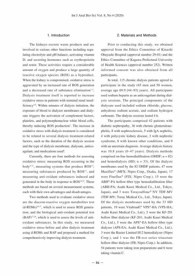
Fig. 1 shows the results of d-ROMs, BAP, and
the BAP/d-ROMs ratio for pre-HD, post-HD and the
control group. The post-HD d-ROMs results were
significantly higher. In contrast, post-HD BAP and
BAP/d-ROMs ratio were significantly lower (Fig. 1).
There was a significant but weak positive
correlation between age and Δ% of d-ROMs (r =
0.323, P < 0.001). Age and pre-HD and post-HD
BAP had significant but weak negative correlations
(pre-HD: r = −0.302, P < 0.01; post-HD: r = −0.295,
P < 0.01). Age and Δ% of the BAP/d-ROMs ratio
had a significant but weak negative correlation (r =
−0.220, P < 0.05). Δ% of BAP and Δ% of the BAP/
d-ROMs ratio had a significant but weak positive
correlation (BAP: r = 0.283, P < 0.01; BAP/d-ROMs
ratio: r = 0.231, P < 0.05) in relation to dialysis
history. There was no significant relationship of the
d-ROMs, BAP, and BAP/d-ROMs ratio results with
underlying disease, current medical history, gender,
iron preparations, and vitamin C use (data not
shown).
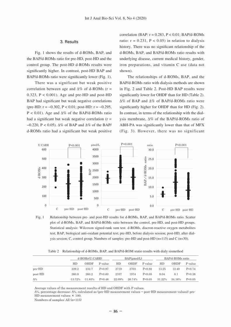
The relationships of d-ROMs, BAP, and the
BAP/d-ROMs ratio with dialysis methods are shown
in Fig. 2 and Table 2. Post-HD BAP results were
significantly lower for OHDF than for HD (Table 2).
Δ% of BAP and Δ% of BAP/d-ROMs ratio were
significantly higher for OHDF than for HD (Fig. 2).
In contrast, in terms of the relationship with the dial-
ysis membrane, Δ% of the BAP/d-ROMs ratio of
ABH-PA was significantly lower than that of MFX
(Fig. 3). However, there was no significant
Fig. 1 Relationship between pre- and post-HD results for d-ROMs, BAP, and BAP/d-ROMs ratio. Scatter
plot of d-ROMs, BAP, and BAP/d-ROMs ratio between the control, pre-HD, and post-HD groups.
Statistical analysis: Wilcoxon signed-rank sum test. d-ROMs, diacron-reactive oxygen metabolites
test; BAP, biological anti-oxidant potential test; pre-HD, before dialysis session; post-HD, after dial-
ysis session; C, control group. Numbers of samples: pre-HD and post-HD (n=115) and C (n=30).
Table 2 Relationship of d-ROMs, BAP, and BAP/d-ROM sratio results with dialy sismethod

Int J Anal Bio-Sci Vol. 8, No 4 (2020)
― 87 ―― 87 ―
difference in age or dialysis history between
ABH-PA users and MFX, FIX, and TDF-MV users
(data not shown). There was no significant differ-
ence between the HD dialysis membranes (data not
shown).
Table 3 shows the pre- and post-HD biochemical
parameters and Δ%. In relation to d-ROMs, Δ%
showed significant strong positive correlations with
TP, serum albumin, TC, HDL-C, and LDL-C (Fig.
4). IP showed a significant strong positive correla-
tion with pre-HD BAP and a significant positive
correlation with post-HD BAP (Fig. 5). On the other
Fig. 2 Relationship of d-ROMs (Δ%), BAP (Δ%), and BAP/d-ROMs ratio (Δ%) results with dialysis
method. Scatter plot of d-ROMs (Δ%), BAP (Δ%), and BAP/d-ROMs ratio (Δ%) between HD and
OHDF. HD, hemodialysis; OHDF, on-line hemodiafiltration. Numbers of samples: HD (n=33) and
OHDF (n=82).Δ%, percentage decrease; Δ%, calculated as (pre-HD measurement values − post-HD
measurement values)/ pre-HD measurement values × 100. Statistical analysis: Mann-Whitney test.
Δ% of BAP and Δ% of BAP/d-ROMs ratio were significantly higher for OHDF than for HD.
Fig. 3 Comparison among OHDF dialysis membranes for the BAP/d-ROMs
ratio Δ%. Numbers of samples: ABH-PA (n=15), MFX (n=47), FIX
(n=17), and TDF (n=3). *No significant difference between the BAP/
d-ROMs ratio Δ%. **Significant difference (P < 0.05) between the
BAP/d-ROMs ratio Δ%. Statistical analysis: Mann-Whitney test. The
smaller the Δ%, the smaller the influence of oxidative stress.

Int J Anal Bio-Sci Vol. 8, No 4 (2020)
― 88 ―― 88 ―
hand, the average change in Ht concentration of 18
patients calculated between pre- and post-HD was
108.36%. A significant strong positive correlation was
seen between the change in Ht concentration and
change in d-ROMs, which was calculated as (post-HD
measurement value / pre-HD measurement value) ×
100 (r = 0.82; P < 0.001; Fig. 6). Furthermore, Δ%
of five parameters-TP, serum albumin, TC, HDL-C,
Table 3 Biochemical results for the pre-and post-HD group sand Δ%
Fig. 4 Correlations in Δ% between d-ROMs and (A) serum total protein, (B) serum albumin, (C) total cholesterol, (D)
HDL-cholesterol, and (E) LDL-cholesterol. Axis shows Δ% of d-ROMs. Statistical analysis: Spearman’s rank
correlation coefficient test. Δ%, minus value means an increase.

Int J Anal Bio-Sci Vol. 8, No 4 (2020)
― 89 ―― 89 ―
and LDL-C-were significantly positively correlated
with d-ROMs. Although relationships with the
corrected results were identified based on the change
in Ht concentration, significant correlations were not
seen with any of the parameters (data not shown).
4. Discussion
One of the most notable results of this study is
that the dialysis session increases oxidative stress
and decreases anti-oxidant potential (Fig. 1). In
contrast, it has previously been reported23 that oxida-
tive stress is reduced post-HD by the purification
effect. This is due to decreased levels of oxidized
albumin and increased levels of mercaptalbumin
(reduced form). This difference may be attributable
to the comprehensive evaluation method of oxida-
tive stress and antioxidant activity used in this study.
The increase in oxidative stress is possibly due to
draining-related changes in the concentration of the
circulating blood because there was a significant
strong positive correlation between the change in Ht
concentration by dialysis session and the change in
d-ROMs (Fig. 6). The fall in anti-oxidant potential is
possibly due to the simultaneous removal of various
anti-oxidants along with waste products during dial-
ysis. A comparison of the results between the OHDF
and HD methods revealed that OHDF, which can
remove more waste products, was associated with a
significantly higher rate of decrease in anti-oxidant
potential post-HD (Fig. 2). These results suggest that
HD is better for mitigating the decrease in anti-
oxidant potential caused by the dialysis session.
However, OHDF24,25 has the advantage of removing
low-molecular-weight proteins and water in addition
to small- to medium-molecular-weight materials
compared with HD, which removes only small-
Fig. 5 Relationship between inorganic phosphorus pre-HD and BAP pre-HD and between inor-
ganic phosphorus post-HD and BAP post-HD. Statistical analysis: Spearman’s rank
correlation coefficient test. There were significant strong positive correlations between inor-
ganic phosphorus pre-HD and BAP pre-HD and between inorganic phosphorus post-HD
and BAP post-HD.
Fig. 6 Relationship between changes in Ht concentra-
tion by dialysis session and changes in d-ROMs.
Statistical analysis: Spearman’s rank correlation
coefficient test. Ht: hematocrit values. There was
a significant strong positive correlation between
changes in Ht concentration by dialysis session
and changes in d-ROMs.

Int J Anal Bio-Sci Vol. 8, No 4 (2020)
― 90 ―― 90 ―
molecular-weight materials and water. Recently, the
use of OHDF has been increasing26-28. Although
there was no significant difference, the reduction
rate of oxidative stress tended to be greater in OHDF
than in HD (Fig. 2). Taken together, the results
suggest the superiority of OHDF. Because the dialy-
sate used contains acetic acid but not citric acid, the
measurements were not affected; there are currently
no reports on the influence of acetic acid on dialysis.
The sample size used confirmed that the error calcu-
lated using each standard deviation and 95%
confidence interval was 6% or less of each average
value.
The second most noteworthy finding is that
ABH-PA was suggested to be an excellent dialysis
membrane. This is because a comparison of dialysis
membranes showed that ABH-PA users undergoing
OHDF dialysis had a significantly lower rate of
decrease in anti-oxidant potential in terms of oxida-
tive stress compared with MFX users (Fig. 3). Both
sample sizes were calculated from the standard devi-
ation, 95% confidence interval, and 5% error and
were confirmed to be statistically appropriate. There
was no significant relationship between underlying
disease, current medical history, gender, or the use
of iron preparations. Based on the findings of the
relationship between age and dialysis history, the
influence of oxidative stress due to dialysis decreases
with age, whereas the oxidative stress influence due
to dialysis increases with a longer dialysis history.
However, there was no significant difference in age
or dialysis history between ABH-PA dialysis
membrane users and MFX dialysis membrane users
(data not shown). The material of the ABH-PA
hollow-fiber membrane is polysulfone and polyvi-
nylpyrrolidone; that of the MFX hollow-fiber
membrane is polyethersulfone; and that of the FIX
hollow-fiber membrane is cellulose triacetate. Our
result is thus consistent with several studies
reporting that treatment with polysulfone membranes
is associated with reduced lipid peroxides, reduced
ROS production, and significantly higher serum
levels of anti-oxidants such as vitamins and cata-
lase8. In those reports, there was no mention of
vitamin E fixation for OHDF membranes.
The third most noteworthy result is that IP
measurements could be used to understand compre-
hensive anti-oxidant potential indicators. This is
because IP and BAP showed a significant strong
positive correlation pre-HD (r = 0.837) and a signifi-
cant positive correlation post-HD (r = 0.687; Fig. 5).
The excretion of IP by the kidney is affected by
renal failure; however, the excretion of other
substances affected by renal failure, such as creati-
nine and blood urea nitrogen, did not have as strong
a positive correlation as that of IP. Because IP is
found in phospholipids, which are constituents of
cell membranes, this relationship was investigated
further. However, no association with BAP was
found (data not shown). It is thus currently unclear
why BAP and IP have a significant strong positive
correlation.
The final noteworthy finding is that the rate of
change in TP, serum albumin, TC, HDL-C, and
LDL-C with dialysis session could be used to under-
stand the overall rate of increase in oxidative stress
due to the dialysis session. This is because the Δ%
values of TP, serum albumin, TC, HDL-C, and
LDL-C were significantly positively correlated with
the Δ% value of d-ROMs (Fig. 4; a minus Δ% value
indicates an increased rate). However, it was
suggested that changes in d-ROMs, TP, serum
albumin, TC, HDL-C, and LDL-C due to the dialysis
session were related to the change in concentration
of circulating blood induced by water removal
because there was a significant strong positive corre-
lation between the change in Ht concentration by
dialysis session and change in d-ROMs (Fig. 6). In
addition, TC had a considerably stronger correlation
(r = 0.90; P < 0.001) compared with the relationship
between HDL-C and LDL-C, and was similar to the
measured value (rate of increase) of d-ROMs (Fig.
4). The increase in TC may be due to only draining-
related changes in the concentration of the
circulating blood. The increases in HDL-C and
LDL-C, on the other hand, are possibly due to such
draining-related changes as well as the oxidative
stress and anti-oxidant potential associated with the
dialysis session, which may differ depending on the
lipoprotein patterns, and also possibly due to the

Int J Anal Bio-Sci Vol. 8, No 4 (2020)
― 91 ―― 91 ―
measurement principles for HDL-C and LDL-C
[Homogeneous method: MetaboLead® HDL-C
(Hitachi Chemical Diagnostics Systems Co. Ltd.),
MetaboLead® LDL-C (Hitachi Chemical Diagnostics
Systems Co. Ltd.)]29. Taken together, these results
suggest that the rate of change in TP, serum albumin,
and TC with dialysis session could be used to better
understand the overall rate of increase in oxidative
stress due to the dialysis session.
These results indicate that a single dialysis
session increases oxidative stress and decreases anti-
oxidant potential. However, the results do not show
significant differences due to underlying disease,
current medical history, oxidative stress, anti-oxidant
potential, or degree of oxidative stress. About 30,000
dialysis patients die every year in Japan26-28. The
leading cause of death is heart failure, followed by
infection and malignant tumors26-28. Several studies
have reported that heart failure and malignant tumors
are related to oxidative stress30-33. The above results
suggest that efficient dialysis treatment as well as
minimization of the level of oxidative stress caused
by the dialysis session are essential. In addition, it is
necessary to understand oxidative stress degrees and
anti-oxidant potential.
The results of this study show that ABH-PA is
the most appropriate OHDF dialysis membrane
because it minimizes the increase in oxidative stress
caused by dialysis treatment. In addition, we recom-
mend measuring pre- and post-HD TP, serum
albumin, TC, and IP to better understand oxidative
stress degrees and anti-oxidant potential.
Conflict of interest
The authors declare that there are no conflicts
of interest.
References
1. Small DM, Coombes JS, Bennett N, Johnson DW,
and Gobe GC: Oxidative stress, anti-oxidant therapies
and chronic kidney disease. Nephrology, 17: 311-321,
2012.
2. Locatelli F, Canaud B, Eckardt KU, Stenvinkel P,
Wanner C, and Zoccali C: Oxidative stress in end-
stage renal disease: An emerging threat to patient
outcome. Nephrology Dialysis Transplantatin, 18:
1272-1280, 2003.
3. Rutkowski P, Slominska EM, Szolkiewicz M,
Aleksandrowicz E, Smolenski RT, Wolyniec W,
Renke M, Wisterowicz K, Swierczynski J, and
Rutkowski B : Relationship between uremic toxins
and oxidative stress in patients with chronic renal
failure. Scandinavian Journal of Urology and
Nephrology, 41:243-248, 2007.
4. Kuragano T, and Nakanishi T: Oxidative stress caused
by renal failure and dialysis therapy [Jpn]. Journal of
Japanese Society for Dialysis Therapy, 43:260-263,
2010.
5. McDonald CI, Fraser JF, Coombes JS, and Fung YL:
Oxidative stress during extracorporeal circulation.
European Journal of Cardio-Thoracic Surgery,
46:937-943, 2014.
6. Morena M, Delbosc S, Dupuy AM, Canaud B, and
Cristol JP: Overproduction of reactive oxygen species
in end-stage renal disease patients: a potential compo-
nent of hemodialysis-asso ciated inflammation.
Hemodialysis Internationl, 9:37-46, 2005.
7. Liakopoulos V, Roumeliotis S, Gorny X, Dounousi E,
and Mertens PR: Oxidative stress in hemodialysis
patients: a review of the literature. Oxidative
Medicine and Cellular Longevity, 2017: 3081856,
2017.
8. Liakopoulos V, Roumeliotis S, Zarogiannis S,
Eleftheriadis T, and Mertens PR: Oxidative stress in
hemodialysis: Causative mechanisms,clinical implica-
tions, and possible therapeutic interventions. Seminars
in Dialysis, 32:58-57, 2019.
9. Khan AU, and Kasha M: Direct spectroscopic obser-
vation of singlet oxygen emission at 1268 nm excited
by sensitizing dyes of biological interest in liquid
solution. Proceedings of the National Academy of
Sciences of the United States of America, 76
:6047–6049,1979.
10. Roubaud V, Sankarapandi S, Kuppusamy P, Tordo P,
and Zweier JL: Quantitative measurement of super-
o x i d e g e n e r a t i o n u s i n g t h e s p i n t r a p
5-(diethoxyphosphoryl)-5-methyl-1-pyrroline-N-
oxide. Analytical Biochemistry, 247:404-411, 1997.
11. Boveris A, Oshino N, and Chance B: The cellular
production of hydrogen peroxide. Biochemical
Journal, 128:617-630, 1972.
12. Ohta Y, Akiyama K, and Tokunaga K: Study of
oxidative damage by myeloperoxidase derived

Int J Anal Bio-Sci Vol. 8, No 4 (2020)
― 92 ―― 92 ―
oxygen [Jpn]. Journal of Analytical Bio-Science,
35:133-139, 2012.
13. Higuchi T, Mano Y, Ishikawa Y, et al.: Links between
plasma 8-hydroxy-2’-deoxyguanosine (8-OHdG) and
various parameters in hemodialysis patients [Jpn].
Journal of Japanese Society for Dialysis Therapy,
45:1035-1043, 2012.
14. Tukamoto T, Sakurai N, Shimazu T, Sato T, and
Maeba T: Effect of Vitamin E-coated Dialyzer on
Glutathione Metabolism and Membrane Fluidity in
Red Blood Cells from Patients with Chronic Renal
Failure under Hemodialysis Therapy [Jpn]. The St.
Marianna medical journal, 29:681-689, 2001.
15. Tajbakhsh R, Qorbani M, Mehrpour G, Rahimzadeh
M, Azimzadeh MM, and Mirmiranpour H: Effect of
hemodialysis on oxidants and antioxidant factors in
chronic renal failure. Saudi Journal of Kidney
Diseases and Transplantation, 28: 507-516, 2017.
16. Seki.Y: Evaluation of total oxidative stress by
d-ROMs testing [Jpn]. Journal of Analytical
Bio-Science, 32:301-306, 2009.
17. Kawakami T, Yura A, Ryu K, Sanbe T, Ogawa K,
Inagaki M, Oguchi K, Kochidaira H, and Iwaki S:
The correlation between oxidative stress and lipids in
blood [Jpn]. Journal of the Showa University Society,
74:403-412, 2014.
18. Dohi.K, Satoh.K, Ohtaki H, Shioda S, Miyake Y,
Shido M, and Aruga T: Elevated Plasma Levels of
Bilirubin in Patients With Neurotrauma Reflect its
Pathophysiological Role in Free Radical Scavenging.
in vivo, 19:855-860, 2005.
19. Tuyuguchi Y, Tateishi K, Miyagawa A. and
Nakagawa T: The relationship between lipoprotein
patterns by polyacrylamide gel disc electrophoresis
and oxidative stress or anti-oxidant potential [Jpn].
The Kagawa Journal of Medical technology, 32:52-
58, 2018.
20. Yoshida A, Kodama M, Nomura H, and Naito M:
Classification of lipoprotein profile by polyacrylamide
gel disc electrophoresis. Internal Medicine, 42:
244-249, 2003.
21. Edited by Nagata Y. How to determine sample size,
15th ed. Asakura Publishing Co., Ltd., Tokyo, (2014)
22. Edited by Yanai H. The Useful Addin Forms on
Excel, 4th ed. OMS Publishing, Tokyo, (2016)
23. Terawaki H, Nakayama K, Matsuyama Y, Nakayama
M, Sato T, Hosoya T, Era S, and Ito S: Dialyzable
uremic solutes contribute to enhanced oxidation of
serum albumin in regular hemodialysis patients.
Blood Purif, 25: 274-279, 2007.
24. Chapter I: Edited by Suzuki S. Dialysis Therapy
Manual, 7th ed.54-56, Japan medical center Co,
Tokyo, (2010)
25. Hideki K: On-line hemodiafiltration (HDF) [Jpn]. The
Japanese Journal of Nephrology, 55: 523-528, 2013.
26. Masakane I, Taniguchi M, Nakai S, et al.: Annual
dialysis data report 2015, JSDT Registry [Jpn].
Journal of Japanese Society for Dialysis Therapy,
50:1-62, 2017.
27. Masakane I, Taniguchi M, Nakai S, et al.:2016
Annual Dialysis Data Report, JSDT Renal Date
Registry [Jpn]. Journal of Japanese Society for
Dialysis Therapy, 51:1-51, 2018.
28. Nitta K, Masakane I, Hanafusa N, et al.:2017 Annual
Dialysis Data Report, JSDT Renal Date Registry
[Jpn]. Journal of Japanese Society for Dialysis
Therapy, 51:699-766, 2018.
29. Kayamori Y, Nakamura M, Sakurabayashi I, and
Kang D: Evaluation method of a homogeneous assay
for measuring serum HDL-cholesterol and LDL-
cholesterol [Jpn]. Journal of Analytical Bio-Science,
31: 263-270, 2008.
30. Kobayashi S: Cardiovascular events in hemodialysis
patients: challenging against vascular calcification.
Annals of Vascular Diseases, 10: 1–7, 2017.
31. Ide T, Tsutsui H, Kinugawa S, Suematsu N,
Hayashidani S, Ichikawa K, Utsumi H, Machida Y,
Egashira K, and Takeshita A: Direct evidence for
increased hydroxyl radicals originating from super-
oxide in the failing myocardium. Circulation
Research, 86:152-157, 2000.
32. Seteesh R, Rao Bitla AR, Budugu SR, Mutheeswariah
Y, Narendra H, Phaneedra BV, and Lakshmi AY:
Oxidative stress in relation to obesity in breast cancer.
Indian Journal of CANCER, 56:41-44, 2019.
33. Trifanescu O, Gruia MI, Gales L, Trifanscu R, and
Anghel R: Tumor is an Oxidative Stress Factor in
Ovarian Cancer Patients. Chirurgia(Bucur), 113:
687-694, 2018.