Embed Size (px)
Citation preview

MOLECULAR MEDICINE REPORTS 18: 639-655, 2018
Abstract. Alzheimer's disease (AD) is a complex and multi-factorial disease. In order to understand the genetic influence in the progression of AD, and to identify novel pharmaceutical agents and their associated targets, the present study discusses computational modeling and biomarker evaluation approaches. Based on mechanistic signaling pathway approaches, various computational models, including biochemical and morpho-logical models, are discussed to explore the strategies that may be used to target AD treatment. Different biomarkers are inter-preted on the basis of morphological and functional features of amyloid β plaques and unstable microtubule-associated tau protein, which is involved in neurodegeneration. Furthermore, imaging and cerebrospinal fluids are also considered to be key methods in the identification of novel markers for AD. In
conclusion, the present study reviews various biochemical and morphological computational models and biomarkers to inter-pret novel targets and agonists for the treatment of AD. This review also highlights several therapeutic targets and their associated signaling pathways in AD, which may have poten-tial to be used in the development of novel pharmacological agents for the treatment of patients with AD. Computational modeling approaches may aid the quest for the development of AD treatments with enhanced therapeutic efficacy and reduced toxicity.
Contents
1. Introduction2. Neuropathology and disease mechanisms3. Neuronal receptors and their associations with AD via
downstream signaling pathways4. The use of receptor-based pharmaceutical agents to treat
AD5. Computational modeling and simulations of AD6. AD biomarkers7. Conclusions and future directions
1. Introduction
Memory loss is naturally associated with old age (1). Specifically, dementia is the clinical condition whereby the severity of the symptoms, such as memory loss, begins to affect normal functioning and social life (2,3). Alzheimer's disease (AD) is a slow but progressive and lethal neurodegen-erative disorder (4), and the risk of developing AD increases in individuals >65 years of age (5). However, cases of early-onset AD (EOAD) have been reported in individuals between 40 and 50 years of age. EOAD occurs less frequently and is classified as presenile dementia of the Alzheimer type, whereas the late‑onset form of AD (LOAD) is classified as senile dementia
Computational modeling and biomarker studies of pharmacological treatment of Alzheimer's disease (Review)
MUBASHIR HASSAN1,2, QAMAR ABBAS3, SUNG-YUM SEO1, SABA SHAHZADI2,4, HANY AL ASHWAL5, NAZAR ZAKI5, ZEESHAN IQBAL2 and AHMED A. MOUSTAFA6,7
1Department of Biology, College of Natural Sciences, Kongju National University, Gongju, Chungcheongnam 32588, Republic of Korea; 2Institute of Molecular Science and Bioinformatics, Dyal Singh Trust Library, Lahore 54000;
3Department of Physiology, University of Sindh, Jamshoro 76080; 4Department of Bioinformatics, Virtual University Davis Road Campus, Lahore 54000, Pakistan; 5College of Information Technology, United Arab
Emirates University, Al-Ain 15551, United Arab Emirates; 6School of Social Sciences and Psychology; 7MARCS Institute for Brain, Behavior and Development, Western Sydney University, Sydney, NSW 2751, Australia
Received May 8, 2017; Accepted July 5, 2017
DOI: 10.3892/mmr.2018.9044
Correspondence to: Dr Hany Al Ashwal, College of Information Technology, United Arab Emirates University, Al-Ain 15551, United Arab EmiratesE-mail: [email protected]
Abbreviations: AChE, acetylcholinesterase; AD, Alzheimer's disease; APOE, apolipoprotein E; APP, amyloid precursor protein; Aβ, amyloid β; BACE, β-secretase; BChE, butyrylcholinesterase; CaMKII/IV, calcium/calmodulin-dependent protein kinase II/IV; CREB, cAMP response element-binding protein; CSF, cerebrospinal fluid; DG, dentate gyrus; EC, entorhinal cortex; EOAD, early onset AD; ERK, extracellular signal-regulated kinase; GABA, γ-aminobutyric acid; LOAD, late onset AD; LTP, long-term potentiation; MAPK, mitogen-activated protein kinase; nAChRs, nicotinic acetylcholine receptors; NFT, neurofibrillary tangles; NMDA, N-methyl-D-aspartate; MR, muscarinic receptors; PKA, protein kinase A; RSM, runaway synaptic modification
Key words: Alzheimer's disease, genetics, computational modeling, simulation, biomarkers

HASSAN et al: COMPUTATIONAL MODELING AND BIOMARKERS FOR ALZHEIMER'S DISEASE640
of the Alzheimer type and affects 7% of individuals >65 and 40% of individuals >80 years of age (6).
AD is a complex and multifactorial disorder (7), the progression of AD is influenced by genetic, environmental and dietary factors. The genetic vulnerability is also associated with AD in autosomal dominant linkage and is considered to be early onset familial AD (8). Autosomal dominant familial AD is primarily attributed to mutations in the following three protein-encoded genes: Amyloid precursor protein (APP); and presenilin (PSEN) 1 and 2 (9). Mutations in APP and PSEN genes lead to increased production of amyloid β (Aβ)42, a small protein that is the primary component of senile plaques (10). It has been observed that environmental and genetic differences may also be risk factors for and govern sporadic AD, without following the autosomal-dominant inheritance pattern. The ε4 apolipoprotein E (APOE) allele is considered to be a genetic risk factor for AD (11,12). Genome-wide association studies have demonstrated that mutations in genetic material are frequently associated with AD, and mutations in specific genes are considered to be risk factors for the development of AD (13). The genomic location and functional characteristics of these AD-associated genes are provided in Table I, whereas a graphical depiction of AD-mediated genes, i.e., genes that are altered during AD, is presented in Fig. 1. In addition, previous reports have demonstrated that environmental and dietary factors, including toxic metals, air pollutants, pesticides and diet, are also risk factors for AD (14). Various toxic compounds, including iron (Fe), zinc (Zn), copper (Cu), aluminum (Al) and lead (Pb), have been reported to alter APP expression and Aβ aggregation, and a high cholesterol diet is reported to be impli-cated in plaque formation and subsequent (15).
2. Neuropathology and disease mechanisms
Neurobiological data have demonstrated that AD is charac-terized by the degeneration of neurons and disturbances in neuronal synapsis within cortical and subcortical areas (16). Amyloid plaques and neurofibrillary tangle (NFT) accu-mulations have been reported to be governing mechanisms of AD in humans (17). Plaques are characterized by dense deposition of Aβ, while NFTs are clumps of microtubules associated with tau protein. Aβ consists of 39-43 amino acids, which are also found in APPs. Proteomic studies have demonstrated that APP is a transmembrane protein that aids neuron growth and post-injury repair (18,19). In AD, β- and γ-secretase are proteolytic enzymes that cleave APP into smaller fragments, which accumulate outside the neurons to form senile plaques (20,21). The basic mechanistic pathway of AD is presented in Fig 2A.
Glycogen synthase kinase 3 (GSK-3) is also associated with neuronal loss and is potentially implicated in AD, as GSK-3 forms associations with Aβ and NFTs, which is considered to be a major hallmark of AD (22). GSK-3 controls various metabolic processes, including phosphorylation, protein complex formation and subcellular distribution (23). Additionally, GSK-3 is considered to increase the production of Aβ and NFTs by hyperphosphorylation of tau proteins (24). Furthermore, disturbance in hippocampal volume, inflam-mation and oxidative stress may also be implicated in the pathology of AD.
3. Neuronal receptors and their associations with AD via downstream signaling pathways
Acetylcholine (ACh) receptors and AD. ACh receptors are the most important target proteins that specifically bind to ACh neurotransmitters. Based on the affinities and speci-ficities with neurotransmitters, ACh receptors are divided into nicotinic Ach receptors (nAChRs) and muscarinic receptors (MRs). nAChRs are localized to skeletal neuromuscular junctions and autonomic ganglia, whilst MRs are present in the brain and parasympathetic effector organs (25), and are associated with cognition in AD (26,27). Tsang et al (28) iden-tified that M1/G‑protein coupling significantly decreased with the progression of AD, whereas the density of M1 receptors was not reduced. Furthermore, another in vitro study reported that an M1 receptor agonist, TBPB, reduced Aβ production, which indicates that the M1 receptor may be used as a novel therapeutic target for the treatment of AD (29). Furthermore, in a knockout mouse study, the M3 receptor was reported to be associated with fear learning and memory conditions, which is relevant to AD symptoms (30).
Another type of receptor that has been extensively inves-tigated is the nicotinic Ach receptor (nAChR), which consists of two subtypes, α7 and α4β2 (31). Previous studies have reported that the expression of these receptors is reduced with the progression of AD (25,32). Young et al (33) also investigated the role of α7-nAChR in knockout mice and demonstrated an impairment in the attention of knockout mice compared with wild type mice (33). However, another study reported conflicting results in α7-nAChR knockout mice by demonstrating neuroprotective effects compared with normal groups (34). However, additional studies have reported that α7-nAChR agonists have led to improvements in cognitive deficits (35-37).
ACh receptors and signaling pathways in AD. It has been observed that Ach receptors are associated with improvements in cognitive deficits in patients with AD (38). For example, ACh receptors govern calcium signaling, which has been demon-strated to improve learning and memory in aging (38,39). Upon activation, ACh receptors trigger increases in calcium levels, which induces various intracellular processes that mediate learning and memory (40). Calcium signaling medi-ates three different types of effects, which include rapid, short and long-term effects. Short and long-term effects are the result of signaling cascades and changes in gene expression, respectively (41).
Specifically, following activation of calcium influx, long-term effects involve the activation of calcium/calmod-ulin-dependent protein kinase II/IV (CaMKII/IV), extracellular signal-regulated kinase/mitogen-activated protein kinase (ERK/MAPK) and cAMP response element-binding protein (CREB). As a result, the activated enzymatic cascades alter the gene expression and may govern cognition symptoms via long-term potentiation (LTP) (42-45). An antagonistic association was observed between Aβ peptides and cholin-ergic systems. The binding of Aβ to nAChRs is also a factor in the activation of calcium, and may induce certain down-stream signaling pathways that lead to a decline in cognition (Fig. 2B) (46,47).
MOLECULAR MEDICINE REPORTS 18: 639-655, 2018 641
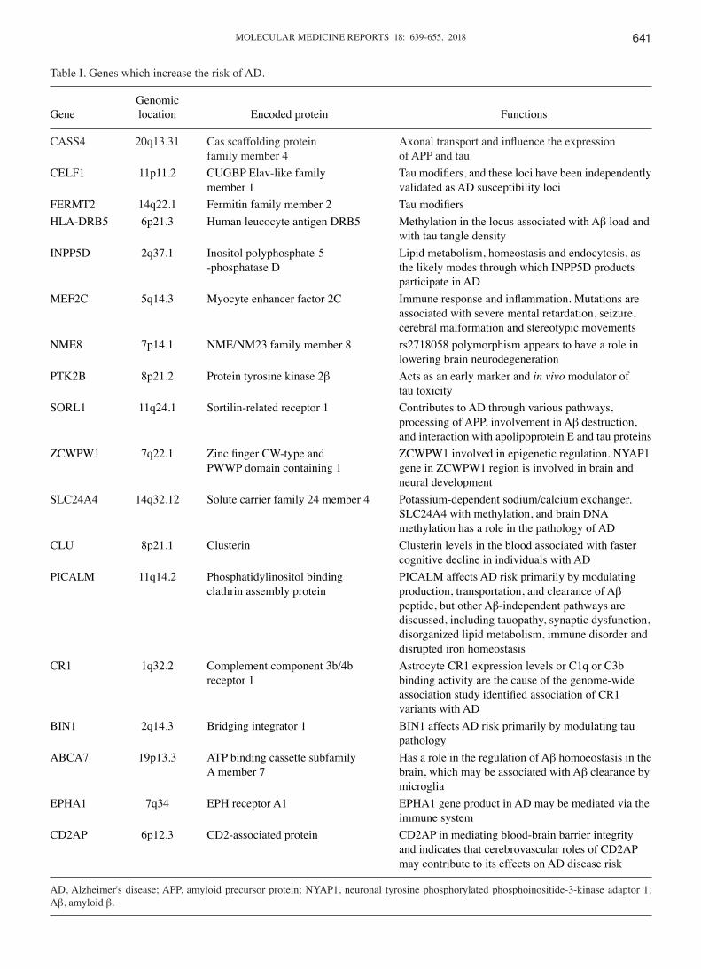
Table I. Genes which increase the risk of AD.
Genomic Gene location Encoded protein Functions
CASS4 20q13.31 Cas scaffolding protein Axonal transport and influence the expression family member 4 of APP and tauCELF1 11p11.2 CUGBP Elav‑like family Tau modifiers, and these loci have been independently member 1 validated as AD susceptibility lociFERMT2 14q22.1 Fermitin family member 2 Tau modifiersHLA-DRB5 6p21.3 Human leucocyte antigen DRB5 Methylation in the locus associated with Aβ load and with tau tangle densityINPP5D 2q37.1 Inositol polyphosphate-5 Lipid metabolism, homeostasis and endocytosis, as -phosphatase D the likely modes through which INPP5D products participate in ADMEF2C 5q14.3 Myocyte enhancer factor 2C Immune response and inflammation. Mutations are associated with severe mental retardation, seizure, cerebral malformation and stereotypic movementsNME8 7p14.1 NME/NM23 family member 8 rs2718058 polymorphism appears to have a role in lowering brain neurodegenerationPTK2B 8p21.2 Protein tyrosine kinase 2β Acts as an early marker and in vivo modulator of tau toxicitySORL1 11q24.1 Sortilin-related receptor 1 Contributes to AD through various pathways, processing of APP, involvement in Aβ destruction, and interaction with apolipoprotein E and tau proteinsZCWPW1 7q22.1 Zinc finger CW‑type and ZCWPW1 involved in epigenetic regulation. NYAP1 PWWP domain containing 1 gene in ZCWPW1 region is involved in brain and neural developmentSLC24A4 14q32.12 Solute carrier family 24 member 4 Potassium-dependent sodium/calcium exchanger. SLC24A4 with methylation, and brain DNA methylation has a role in the pathology of ADCLU 8p21.1 Clusterin Clusterin levels in the blood associated with faster cognitive decline in individuals with ADPICALM 11q14.2 Phosphatidylinositol binding PICALM affects AD risk primarily by modulating clathrin assembly protein production, transportation, and clearance of Aβ peptide, but other Aβ-independent pathways are discussed, including tauopathy, synaptic dysfunction, disorganized lipid metabolism, immune disorder and disrupted iron homeostasisCR1 1q32.2 Complement component 3b/4b Astrocyte CR1 expression levels or C1q or C3b receptor 1 binding activity are the cause of the genome-wide association study identified association of CR1 variants with ADBIN1 2q14.3 Bridging integrator 1 BIN1 affects AD risk primarily by modulating tau pathologyABCA7 19p13.3 ATP binding cassette subfamily Has a role in the regulation of Aβ homoeostasis in the A member 7 brain, which may be associated with Aβ clearance by microgliaEPHA1 7q34 EPH receptor A1 EPHA1 gene product in AD may be mediated via the immune systemCD2AP 6p12.3 CD2-associated protein CD2AP in mediating blood-brain barrier integrity and indicates that cerebrovascular roles of CD2AP may contribute to its effects on AD disease risk
AD, Alzheimer's disease; APP, amyloid precursor protein; NYAP1, neuronal tyrosine phosphorylated phosphoinositide-3-kinase adaptor 1; Aβ, amyloid β.

HASSAN et al: COMPUTATIONAL MODELING AND BIOMARKERS FOR ALZHEIMER'S DISEASE642
Serotonin receptors and AD. An increased serotonin (5-hydroxytryptamine) concentration in the synaptic cleft has been reported to be a potential therapeutic strategy to slow the progression of AD (48,49). Serotonin targets specific receptors at postsynaptic neurons and mediates downstream signaling pathways that control cognition. It has been reported that ≥16 different types of serotonin receptors exist, which are catego-rized into 7 subfamilies (5-HT1-5-HT7) (50). All serotonin receptors are G-protein-coupled receptors (GPCRs), excluding the 5-HT3 receptor (50). The activation of these receptors stimulates downstream signal transduction pathways that govern certain intracellular responses. The protein kinase A (PKA) signaling cascade is responsible for the inhibition and stimulation of phospholipase C/protein kinase C, which regu-lates the ERK/MAPK pathways (51,52). Subsequently, these activations affect cognitive impairment in neurodegenerative diseases.
Results from animal and clinical experiments have also demonstrated the importance of 5-HT in cognitive dysfunc-tion and memory deficits (53). Increases in the expression of 5-HT1A receptors were reported to be associated with cognitive impairment, and these receptors are therefore considered to be potential targets for the treatment of AD (54). Furthermore, Garcia-Alloza et al (55) demonstrated that
5-HT1B/1D receptors were associated with cognitive dysfunc-tion in AD. It was also observed that the density of the 5-HT2A receptor was significantly reduced in the frontal and temporal cortical neurons in patients with AD compared with healthy participants. Furthermore, various studies have reported an important association between 5-HT2 receptors and cognitive decline in AD (56-58).
The serotonin receptor 5-HT6 has an important role in various mechanistic pathways within the brain (59). It is primarily expressed in the striatal, hippocampal and cortical areas (60). Notably, it was previously reported that inhibition of the 5-HT6 receptor improved learning and memory (61,62). Another animal study also demonstrated the importance of 5-HT6, as the agonist SB-271046 improved age-associated deficits and spatial recognition memory in aged mice (63). It was also reported that, another agonist, WAY-181187, may also be used to modulate synaptic plasticity via attenuation of LTP (64).
Serotonin receptors and downstream signaling pathways in AD. 5-HT receptor-mediated signaling pathways are associ-ated with improvements incognitive defects (65). The 5-HT6 receptor stimulates G-proteins, which results in cAMP production via adenylyl cyclase activation (66,67). cAMP subsequently triggers PKA, which, via phosphorylation, activates CREB (67). A number of studies have indicated that the 5-HT6 receptor modulates various neurotransmit-ters, including glutamate and Ach, to aid memory processes (Fig. 2C) (68,69).
Adrenergic receptors and AD. Adrenergic receptors are metabotropic GPCRs, which are divided into two major groups, α and β. Adrenergic receptors are typically sensitized for norepinephrine and epinephrine neurotransmitters. A number of studies have reported that adrenergic receptors (α and β) are closely associated with cognitive decline in AD (70,71). An expression study by Kalaria and Harik (72) demonstrated that β2 levels were increased in the cortex and hippocampus of patients with AD. In addition, a behavioral study reported that certain structural changes in adrenergic receptors were associated with the presence or absence of aggressive behavior in AD patients (73).
Dopamine receptors and AD. Dopamine receptors exhibit important roles in various human functions, including cogni-tion and learning (50). Dopamine receptors are divided into two different classes, D1- and D2-like receptors, which consist of five subtypes. D1‑type receptors include D1 and D5 receptors, whereas D2-type receptors include D2, D3 and D4 receptors (74). Functionally, D1- and D2-type receptors function in synaptic plasticity and cognition by stimulating the protein signaling cascade of cAMP/PKA and CREB modulation (43,44). However, another study demonstrated that dopamine receptors were directly associated with AD and Parkinson's disease (50).
N‑methyl‑D‑aspartate (NMDA) receptors and its pathway in AD. NMDA/glutamate receptors are have been extensively studied, and are abundantly expressed in the cerebral cortex, hippocampus, nucleus accumbens and striatum (75,76).
Figure 1. AD-mediated genes which are altered during AD. AD, Alzheimer's disease; APOE, apolipoprotein E; NME8, NME/NM23 family member 8; MADD, MAP kinase-activating death domain; CASS4, cas scaffolding protein family member 4; SORL1, sortilin-related receptor 1; CD2AP, CD2-associated protein; PSEN, presenilin; INPP5D, inositol polyphos-phate-5-phosphatase D; FERMT2, fermitin family member 2; HLA-DRB5, human leucocyte antigen DRB5; MEF2C, myocyte enhancer factor 2C; RIN3, Ras and Rab interactor 3; TREM2, triggering receptor expressed on myeloid cells 2; CLU, clusterin; NYAP1, neuronal tyrosine phosphorylated phosphoinositide-3-kinase adaptor 1; EPHA1, EPH receptor A1; PLD3, phos-pholipase D family member 3; PTK2B, protein tyrosine kinase 2β; PICALM, phosphatidylinositol binding clathrin assembly protein; CR1, complement component 3b/4b receptor 1; ZCWPW1, zinc finger CW‑type and PWWP domain containing 1; SLC24A4, solute carrier family 24 member 4; CELF1, CUGBP Elav-like family member 1; BIN1, bridging integrator 1. ADAM10, a disintegrin and metalloproteinase domain-containing protein 10; AAP, amyloid precursor protein; DGS2, DiGeorge syndrome/velocardiofacial syndrome complex 2.

MOLECULAR MEDICINE REPORTS 18: 639-655, 2018 643
Variations in glutamatergic receptors are implicated in the pathogenesis of neurodegenerative diseases, such as AD, as they are associated with neuronal death (77). A reduced expression study on NMDA, NMDA receptor subunit 1 and subunit 2B proteins in rat models reported that there is a close association between NMDA receptors and cognitive deficits (78). Neuronal loss induced by amyloid plaques are a consequence of NMDA receptor modulation. Amyloid plaques activate NMDA receptors, which results in higher calcium
influx into neurons, ERK1/2 activation and mediation of respective downstream enzymes (79-82). Therefore, NMDA signaling pathways have a potential role in the pathogenesis of cognitive dysfunctions (Fig. 2D).
Acetylcholinesterase (AChE) as an AD drug target. AChE is a type of hydrolase, and exhibits key functions in cholinergic neurotransmission in the autonomic and somatic nervous systems (83). AChE interacts with Ach, converting it into
Figure 2. Mechanistic overview of AD with neuronal signaling pathways. (A) General mechanism of tau-mediated AD is presented. A double membrane is highlighted in silver containing an embedded complex of β-secretase and APP (80). β-secretase and APP protein are indicated by maroon and purple colors, respectively. The γ-secretase enzyme is acting to cleave APP into Aβ40 and Aβ42 subunits. The clump of Aβ40, termed amyloid plaques, are generated by a process termed oligomerization and interactions with other two enzymes, APoE and neprilysin IDE. The aggregated plaques lead to neuronal loss and synaptic dysfunctionality, which ultimately results in cognition deficits. (B) In the acetylcholine signaling pathway, acetylcholine stimulates calcium influx after interacting with its respective receptor at the synaptic complex. This calcium flux activates a series of signaling proteins, including CaMKII/IV, ERK/MAPK and CREB. As a result, the activated enzymatic cascade leads to altered gene expression and may govern cognition symptoms via LTP (40-43). (C) In the serotonin signaling pathway, activation of the 5-HT6 receptor stimulates G-proteins, which results in increased cAMP production via AC activation. This cAMP triggers PKA activation, which phosphorylates and regulates the CREB transcription factor, which subsequently leads to cognition dysfunction (65). (D) In the glutamic acid signaling pathway, activation of the NMDA receptor by glutamic acid mediates calcium signaling from presynaptic to postsynaptic neurons. CaM and ERK1/2 protein cascades are activated, which ultimately leads to CREB activation and cognition dysfunction (77-79). AD, Alzheimer's disease; APP, amyloid precursor protein; Aβ, amyloid β; APoE, apolipoprotein E; IDE, insulin-degrading enzyme; CaMK, calcium/calmodulin-dependent protein kinase; ERK, extracellular signal-regulated kinase; MAPK, mitogen-activated protein kinase; CREB, cAMP response element-binding protein; LTP, long-term potentiation; AC, adenylyl cyclase; PKA, protein kinase A; NMDA, N-methyl-D-aspartate; nAChRs, nicotinic acetylcholine receptors; VDCCs, voltage-dependent calcium channels; ER, endoplasmic reticulum; CaM, calmodulin; CaMKK; calcium/calmodulin-dependent protein kinase kinase; GPCR, G-protein coupled receptor.

HASSAN et al: COMPUTATIONAL MODELING AND BIOMARKERS FOR ALZHEIMER'S DISEASE644
choline and acetic acid (82). Expression studies have demon-strated that AChE is frequently present in motor neurons and certain other types of conducting tissue, including nerve and muscle, motor and sensory fibers, and cholinergic and non‑cholinergic fibers (84,85). In AD, cholinergic neurons mediate memory deficits and cognitive decline by reducing the level of ACh (86,87). Therefore, AChE may be considered as a novel target to reverse AD symptoms. Furthermore, butyrylcholinesterase (BChE) is also considered to be a minor player in the regulation of synaptic ACh levels (88). Therefore, inhibition of BChE may also be considered a valid approach to restore cholinergic function in AD (89,90).
4. The use of receptor‑based pharmaceutical agents to treat AD
AChE‑based inhibitors. The majority of neuromuscular problems are treated by AChE inhibitors, which are also considered to be first‑generation drugs for the treatment of AD. There are four established inhibitors (donepezil, galantamine, rivastigmine and tacrine) that are commonly used to improve cognition (91). However, tacrine is not as reputable due to poor tolerability (92,93). Donepezil demonstrated its neuroprotec-tive effects by diminishing the excitotoxicity of glutamate by reducing Aβ load and cell toxicity, as well as increasing cell life span (94,95). Rivastigmine is a cholinergic agent that targets AChE and BChE. Clinical trials have indicated that rivastig-mine improves cognition, with few side effects in patients with AD (96). Tacrine is another inhibitor that increases ACh levels from cholinergic nerve endings. Tacrine inhibits the activity of certain enzymes, including monoamine oxidase, and suppresses γ-aminobutyric acid (GABA) signaling, which results in the release of dopamine, noradrenaline and serotonin from nerve endings, and improves memory in patients with AD (97).
Recently, novel inhibitors have been synthesized from natural and synthetic sources for patients with AD. Huperzine A (Hup A) is an AChE inhibitor that is primarily used in the treatment of memory disorders. Hup A is highly potent and has a higher bioavailability compared with donepezil and tacrine, but is less effective compared with BChE inhibitors for treating AD symptoms (98). Recent attempts have demon-strated that derivatives of Hup A, with aromatic rings, exhibit potential therapeutic effects for AD symptoms (99). However, further studies are required to assess the potential benefits of Hup A for treating AD (100). Camps et al (101) synthesized hybrids of innovative tacrine and Hup A as a cholinesterase agonists to treat AD. This designed agonist exhibits different functional moieties at basic nuclei of chemical compounds, and provided good results at various positions. The halogen moiety had a higher activity and increased therapeutic effec-tiveness of treating AD compared with tacrine. However, it also exhibits limited inhibition of BChE. Furthermore, the agonist designed by Camps et al (101) also has the propensity to cross the blood-brain barrier.
Huperzine B (Hup B) is also considered to be an AChE inhibitor with reversible and effective properties. Hup B is less potent compared with Hup A, and is also used as a template structure to synthesize novel compounds that inhibit AChE (102). Another potent derivative is bis-Hup B, which
consists of two Hup B molecules connected to a carbon-nitrogen chain by an amine group. The bis-Hup B compound has exhib-ited higher inhibitory potential against AChE compared with against BChE (103).
Berberine is another candidate compound with multiple biological activities, including the potential to cross the blood brain barrier and target the central nervous system (CNS). Berberine acts as an inhibitor of AChE (104,105) and also performs a neuroprotective function by reducing NMDA-induced excitotoxicity (105).
β‑secretase (BACE) as a therapeutic target for AD. A number of BACE1 inhibitors are being synthesized for the treatment of AD (105). OM99-2 is a peptide inhibitor of BACE1 that exhibits strong hydrogen binding within the active binding region of target proteins (106-108). KMI-429 is also considered to be an effective BACE1 inhibitor, with a 50% inhibition concentration (IC50) of 3.9 nM. In a mouse study, Asai et al (109) demonstrated that Aβ production was reduced in APP transgenic and normal mice following KMI-429 treatment. Another mouse study was performed by Luo and Yan (110) using the GSK188909 agonist (non-peptide) against BACE1. The results indicated potential therapeutic effects of GSK188909-induced inhibition of BACE1, via reduction of Aβ levels in the brain. These results demonstrate that GSK188909 may be considered a beneficial inhibitor in the treatment of AD (110,111). In addition, another orally administered inhibitor, 4-phenoxypyrrolidine, is also considered to be a potent agent against BACE1, and it has similar functions and pharmacokinetic/pharmacodynamic properties to GSK188909 (110,112). Furthermore, GRL-8234 is also a potent inhibitor of BACE, with an inhibitory constant (Ki) value of 1.8 (107). Chang et al (113), in a study on trans-genic mice, demonstrated the prominent effects of GRL-8234 on cerebrospinal fluid (CSF) and Aβ production. CTS-21166 has also been reported to reduce Aβ levels in the brain by >35-40%, and was the only inhibitor to pass phase I clinical trials (114).
γ‑ and α‑secretase‑based drugs for AD. The designing of novel γ-secretase agonists remains a challenging approach due to its non-amyloid behavior and interaction with meta-bolic processes. Various undesired effects are also generated, including gastrointestinal lethality, hematological toxicity and skin reactions (114).
The first compound that was synthesized as a γ-secretase agonist was DAPT (115,116). A pharmacokinetic evalua-tion study demonstrated that DAPT overdose is required to inhibit Aβ production in the neuronal cells of APP transgenic mice (117). LY-450139 (semagacestat) is also an inhibitor of APP cleavage (118). However, certain side effects are associated with LY-450139, including thymus atrophy and a reduction in the number of lymphocytes (118). BMS-708163 is another inhibitor that is reported to reduce Aβ40 levels in CSF without causing adverse effects (119). PF-3084014, a non-competitive compound, has been investigated in mice and humans (118). Begacestat (GSI-953) is also an effective agonist against γ-secretase, which controls Aβ production. Furthermore, in Tg2576 transgenic mice studies, high doses of GSI-953 reduced Aβ41 levels in the brain (120,121). A clinical study was performed using GSI-953 for AD treatment (119,122), and the results demonstrated that

MOLECULAR MEDICINE REPORTS 18: 639-655, 2018 645
GSI-953 does not exhibit a positive effect on the reduction of Aβ40 levels in the CSF of patients with AD (120).
In addition, a number of candidate molecules have been synthesized by considering α-secretase as a target molecule (123). Of these, etazolate (EHT-0202) activates neuronal α-secretase and, as a result, enhances soluble APP production (124). Etazolate was investigated in a phase II study in patients with mild-to-moderate AD. Results revealed that etazolate exhibited good clinical efficacy in patients with AD (124). Bryostatin-1 and exebryl-1 are potent inhibitors of α‑secretase, which significantly affect Aβ production and improve memory (125).
GSK‑3 inhibitors in the treatment of AD. Hu et al (126) demon-strated the importance of GSK-3 as a receptor molecule in the prevalence of AD. GSK-3 agonists may have positive thera-peutic effects on patients with AD. The investigated compound SB216763-a (a GSK-3 inhibitor) was synthesized for potential use in the treatment of AD. Functionally, SB216763 reduced phospho-glycogen synthase by 39% and increased glycogen levels by 44%, which demonstrates its potent inhibition of receptor activity (126).
It is difficult to fully understand all of the receptor‑based mechanistic signaling pathways and the interactions of neurotransmitters with drugs by experimentation. Therefore, computational modeling and simulation approaches are considered to be important for targeting and investigating the neurodegeneration disorders. The present review will highlight a number of computational modeling attempts and biomarker interpretations to improve the understanding of the pathogenesis and symptoms of AD.
5. Computational modeling and simulations of AD
To interpret the basic mechanism of AD, computational models have been designed on the basis of amyloid plaques, NFTs and hippocampus functions. Furthermore, additional models are based on neuronal functionality and the synaptic transmission of neurotransmitters.
Biochemical and morphological modeling. Aβ is considered to be a major hallmark and pathological feature of AD (127). Based on Aβ aggregation factors, kinetics, mechanistic path-ways and its morphological appearance, Aβ is considered to be a key feature in the design of computational models for AD. Experimental and theoretical investigations on Aβ have investigated the kinetics, mechanistic pathways and fibril-logenesis (128-138). Various computational models have been proposed on the basis of Aβ kinetics, which include fibril elongation and Aβ self-association (130,137). Through computational modeling, the monomers of the oligomers of β‑peptides in elongated fibrils were arranged into compact aggregations of complexes of pro-peptides in irregular symmetry (132,133,136). The key factors regarding oligo-meric β-peptides in previous models include the exclusion of filaments and fibril discrimination, and the use of non‑physio-logical (pH ~1) experimental conditions (139).
Pallitto and Murphy (140) designed a mathematical model on the basis of the kinetics of Aβ aggregation. The core feature of this model was identifying that Aβ is partitioned between
two pathways. The first pathway produces a stable structure of monomers and dimers, and the second pathway produces an unstable β-sheet, containing intermediate-aggregated oligomers (141). A model by Kim et al (131) further explained the involvement of Aβ oligomers in the fibrillogenic pathway by evaluating the effect of urea on aggregation kinetics, size distribution and aggregate morphology. An enhanced urea concentration has a direct effect on β-sheet contents, including a decrease in the aggregation size and changes in the morphology of aggregates. The computational model results supported the hypothesis that the amyloidogenesis pathway and the globular aggregates were involved as intermediates rather than an alternative aggregated species.
Plaque‑based computational modeling. Amyloid plaque formation is also considered a key biochemical concept to design models (142,143). In addition, the kinetics of APP processing and downstream intracellular interactions of calcium and Aβ were observed in the AD brain (144-146). The secretases (α, β and γ) function as cleaving agents of APP. It has been observed that secretase agonists target APP and minimize the Aβ production, and may slow the progression of AD (147). Based on intracellular calcium and Aβ interactions, a computational model was built to account for established characteristics of AD, which include its irreversibility, acute to chronic pathology and inherent random characteristics of sporadic AD.
Anastasio (148) developed a computational model of AD on the basis of an amyloid hypothesis. The regulatory pathway in the model was justified by interrelated equations. Furthermore, the molecular conditions were symbolized by arbitrary integer values in the equations, and a set of rules were employed to justify the changes in model elements, which change the levels of other elements. The model explained the disruption of Aβ regulation through the interconnection of various diseases and pathological processes, including cerebrovascular disease (CVD), inflammation and oxidative stress. More precisely, it was reported that CVD contributes to the progression of AD. Additionally, multiple target therapies were more effec-tive compared with single target treatments. In addition, Anastasio (149) designed an additional model based on the knowledge of previous model evaluations and incorporated factors such as the role of estrogen in the regulation of Aβ to predict effective AD therapy. The predicted model results demonstrated that, by administering non‑steroidal anti‑inflam-matory drug therapy, estrogen levels decrease, which leads to marked reductions in Aβ. Furthermore, Anastasio (150) extended this work to further understand the synaptic plas-ticity dysregulation of Aβ. The predicted results indicated a normalization of nAChRs. Neuronal proteins responded to the neurotransmitter ACh, which addresses the effects of Aβ on synaptic plasticity. These results contributed substantially to the understanding of how combinations of drugs may be used in the pathogenesis of synaptic diseases (150).
Craft et al (151) investigated the association between AD treatment effects and Aβ levels in different parts of the body. The study investigated fluctuations in Aβ levels in the brain, CSF and plasma prior to and following medication states. This was achieved by employing an infinite set of nonlinear differ-ential equations. Based on the polymerization ratio calculation,

HASSAN et al: COMPUTATIONAL MODELING AND BIOMARKERS FOR ALZHEIMER'S DISEASE646
results indicated that, when values were >1, Aβ amalgamation increased indefinitely. Whereas the Aβ levels in the CSF and plasma remained in a steady state. However, polymerization ratio calculations <1 demonstrated a steady-state of Aβ levels throughout the body.
GSK‑3b, p53, Aβ and tau‑based modeling. In intracellular signaling, multiple proteins are interconnected through specific receptor‑mediated pathways. GSK‑3b, p53, Aβ and tau proteins are observed in computational modeling to investigate the mechanistic pathways that mediate AD (152). A multi-compartment model for GSK-3b, p53, Aβ and tau proteins was designed to determine the associations among these proteins (152). The predicted results demonstrated that abrupt changes in DNA damage the p53 and Mdm2 complex. As a result, GSK-3b/p53 complexes are formed, which enhance the transcriptional activity of p53 and GSK-3b. Consequently, there are increases in the production of Aβ, Mdm2, mRNA and tau phosphorylation. Computational model results indi-cate that, in normal states, Aβ is degraded in cells and, upon dephosphorylation, degradation become optimized within cells. However, under conditions of stress, Aβ production and tau phosphorylation increase. Therefore, adjusting the DNA damage parameter may clear Aβ and stop the phosphorylation of the tau protein. Additionally, plaque and tangle formation were independent, even with GSK-3b overactivity (152).
In another computational model based on Aβ functionality, which was developed by Diem et al (153), the results indicated that the deposition of Aβ in human artery walls reflect the lymphatic drainage pathway with the progression of AD. Initially, the diffusion of Aβ occurs from the brain to basement membranes in capillaries and arteries via extracellular spaces of gray matter in the brain. However, the exact mechanism of perivascular elimination of Aβ remains under consideration. Based on this mechanistic approach, a computational model was designed to explain the process of periarterial drainage with regards to diffusion in the brain, and demonstrated that periarterial drainage along basement membranes is rapid compared with diffusion. The predicted results indicated that failure of periarterial drainage is a mechanism underlying the pathogenesis of AD, in addition to complications associated with its immunotherapy (153).
Immunity‑based modeling. Proctor et al (154) investigated the passive and active immunization effects against Aβ, plaques, phosphorylated-tau and tangles. In their extended model, Aβ clearance was elicited by the administration of antibodies, which were modeled by the addition of species termed ‘anti-Aβ’ and ‘Glia’ (with an additional species to represent microglia). Both additions (antibodies and microglia) were run by predetermined time points during simulation. The predicted model results demonstrated that immunization helped to clear the plaques. However, immunization only exhibited limited effects on soluble Aβ, tau or tangles. The results of this model demonstrated that immunotherapy against Aβ is more effec-tive during early stages of AD.
An additional network interaction model of Aβ, neuroin-flammation, mitochondrial dysfunction and lipid metabolism dysregulation was designed by Kyrtsos and Baras (155). The basic purpose of this computational model was to investigate
the short and moderate level effects of inflammation, and mutational effects on the ApoE allele. Their model was based on cellular and molecular levels. In cellular levels, four different types of cells, which included neurons, astrocytes, microglia and brain endothelial cells, were used to interact with each other. While at the molecular level, each cellular downstream metabolic network was addressed to mediate the metabolic responses of particular cell types. Modeling of chemical species for each cell type was performed by average distribution. The simulation results indicated that the ApoE4 allele ultimately led to an increase in Aβ. This increase causes ATP to collapse and an elevation of glutamate levels, which is the major cause of neuronal loss in a local region. The computational model results demonstrated that inflammation may be considered as a key component in the pathogenesis of AD. Furthermore, inflammation strength and duration are also important factors in AD progression (155).
Single cell‑based models for AD. To interpret the Aβ function-ality more adequately against AD, single-cell-based models were employed. The different cell-based-models act as a single framework, which investigates the different properties of individual cells. Chen (156) revealed that a higher expression of Aβ in cells leads to intrinsic disruption of electrical proper-ties in the dendrites of the hippocampus. In the dendrites of pyramidal cells, Aβ bocks the A-type potassium channels, which results in enhanced membrane excitability and calcium influx (156). Hyperexcitability of dendrites gradually leads to degenerative changes or neuronal cell death (157). The effect of Aβ was modeled by decreasing the maximal conductance in transient A-type potassium channels. The simulation results for this experimental study demonstrated that, when Aβ affected the potassium current, there was increased invasion of back-propagated action potentials (bAPs) from the cell body into the apical dendritic trunk of CA1 pyramidal neurons. In another study by Hoffman et al (158), similar results were observed following the administration of pharmacological agents that blocked the A-type potassium current (158). The simulation results indicated that the disturbance of normal dendritic elec-trical activity caused by an intra-articular blockade produces significant differences between the depolarizations of Aβ and normal cases in the distal oblique branches compared with the dendritic trunk. Furthermore, a number of studies have reported that modified synaptic membrane properties disturb the firing properties of CA1 pyramidal neurons under current and voltage clamp conditions by the amalgamation of Aβ (159,160).
In addition, Abramov et al (161) identified that increases in endogenous Aβ enhances the initial release probability (p0) at the CA3-CA1 synapses of the hippocampus, without altering the intrinsic neuronal excitability and postsynaptic function. The increased level of p0 is also directly involved in the destruction of vesicles by increasing Aβ production (162). Furthermore, a hippocampal CA1 pyramidal neuron model demonstrated that the enrichment in p0 affects the synaptic short‑term plasticity of the synapse and the firing probability of the CA1 output neuron (163).
Neural network and drug‑based models for AD. A neural network model of corticohippocampus formation was

MOLECULAR MEDICINE REPORTS 18: 639-655, 2018 647
designed to investigate the effects of scopolamine, a drug that blocks cellular effects of ACh, on the encoding and retrieval of memories in paired associate tasks (163). Four modules were present in this model by Hasselmo and Wyble, which included the entorhinal cortex (EC), dentate gyrus (DG), region CA3 and region CA1 (164). In each module, ‘memory’ was represented as a pattern of neural activation. The information following the patterns among the four modules were represented as EC to DG to CA3 to CA1. The represented items, such as individual words, in CA3 neurons exhibited weaker recurrent connec-tions compared with contextual information. Their detailed model simulation demonstrated that scopolamine impaired the encoding of new input patterns, but had no effect on previously learnt recalled patterns. Results indicated that impairment is selective in free recall, upon the recognition of items that are not already encoded. This model was the first attempt to simulate the effects of a drug on human memory. The experi-ment investigated and quantified the physiological effects at a cellular level. To design novel drugs against neurodegenerative diseases, modeling attempts to interlink the behavior, physi-ology and molecular biological aspects in a single constrained model for human memory functions.
An additional computational modeling approach was established to investigate the modulation and control storage, and AD dynamics within the hippocampal CA3 network on the basis of subcortical cholinergic and GABAergic inputs (165). To build upon Meschnik and Finkel's model (165), Buzsaki developed a ‘two-stage’ memory model and highlighted the importance of interneurons, basket cells and chandelier cells in memory (166-168). Furthermore, Lisman et al (168) designed a computational model on the basis of embedded γ cycles within the θ cycles. Their results demonstrated that attractor‑based auto-associative memory may be implemented by the synchro-nization of γ-frequency ranges. Each newly arrived input pattern at the commencement of θ cycle with embedded 5‑10 γ-cycles generated a network activity to congregate various γ-cycles as a steady attractor, which characterizes the stored memory. Their predicted results support the hypothesis that CA3 pyramidal cells generate distinct behavioral functions by bursting and spiking patterns. In addition, the change between behavioral states associated with the online processing and recall of information is regulated by cholinergic input in the hippocampus. A deficiency of cholinergic neurons is associ-ated with a reduction of γ frequency. The reduction of γ-cycles within the θ rhythm results in memory loss and cognition, which is associated with AD (169).
Roberts et al (170) created a cortical circuitry computational model using preclinical data available on pharmacological receptors for cholinergic and catecholamine neurotransmit-ters (170). Working memory was identified as a measure of cognitive functions. The pathology of AD includes neuronal and synaptic loss, and decreases in cholinergic tone. The model explains the differential effects of an NMDA agonist, memantin, in EOAD and LOAD conditions. In addition, the model also explains the inhibition of the NMDA receptor NR2C/NR2D subunits, which are present on inhibitory inter-neurons; the NMDA receptor is inhibited to compensate for the higher excitatory decay detected with pathology.
Bianchi et al (171) developed a memory encoding and retrieval model in the brain based on a previous computational
study by Cutsuridis et al (172). Their model explains that by enhancing CREB activity, hippocampal CA1 pyramidal neuron properties change, which may contribute to improve-ments in memory storage. The CREB effects were modeled with a decrease in the conductance peaks of medium after-hyperpolarization (52%) and small after-hyperpolariza-tion (64%), and increased conductance peak of AMPA (266%) currents. The results demonstrated that, by improving CREB function in AD-like conditions, the stored pattern in network recall quality increased significantly. The results confirmed that CREB-based agonists may be used as a novel approach for the treatment of AD.
The synaptic deletion and compensation model. The initia-tion of synaptic deletion and compensation model was initially proposed by Horn et al (173) and further developed by Ruppin and Reggia (174). The artificial neural progression model for AD (173) deviates from the excitotoxicity, which does not account for cognitive impairment. It has been observed that a 50% loss of synaptic connections is considered a primary factor for cognitive deficits. In earlier stages of AD, the loss of connections is compensated by strengthening the remaining connections. Horn's model demonstrated that synaptic connec-tions are associated with memory loss and disturbances in learning patterns. The rate of memory deterioration may be minimized by enhancing the remaining connection weight of a constant multiplication factor (173).
The synaptic runway model was based on associative memory and memory storage as a pattern of neuronal spatio-temporal activation (175,176). Memory storage activates different analog patterns that interact with previous associa-tions. For example, if there is an overlap between patterns or if the memory capacity is exhausted. The results demonstrated that the significant increase in the number of associations stored by the network may govern pathological increases in the strength of synaptic connections. As a result, such synaptic connections give rise to an increase in neuronal activity, high metabolic demand and may eventually cause excitotoxicity. The synaptic runway model investigated two basic mechanistic approaches to memory, encoding and retrieval, to attempt to reduce neuronal cell death (176). In normal conditions, neuro-modulation is satisfactory to preclude the variations in runaway synaptic modification (RSM). Whereas, in manifestations of disease conditions such as AD, the RSM neuromodulation is inevitable. However, the threshold levels for RSM in AD are lower compared with controls (177).
Bhattacharya et al (178) designed a computational model to determine the association between active synapses and α-band frequency amongst individuals that are healthy, exhibit mild cognitive impairments and patients with AD. An additional aim was to duplicate the dysfunctional electroencephalogram experimental data in patients with AD. Their model simulated neurological regions composed of various multilayer neurons, three for the thalamus and four for the cortex module. However, this model was limited as it did not simulate the association between α-band frequencies and ACh.
Neurocomputational model at system level. Computational exploration of AD has reported changes in hippocampal func-tionality and behavioral performance (178,179). For example,

HASSAN et al: COMPUTATIONAL MODELING AND BIOMARKERS FOR ALZHEIMER'S DISEASE648
the ability to learn and adapt learning to novel situations is impaired in AD. Moustafa et al (180) also identified that simu-lated learning occurred through an interaction between the hippocampal region and basal ganglia (180). Hebbian learning and temporal difference algorithms were used to train the model, and the results indicated that hippocampal damage leads to impaired learning performance.
Additionally, a computational model by McAuley et al (181) investigated the functional association between cortisol and the hippocampus in aged individuals and patients with AD (180). The results of this model demonstrated the existence of an antagonistic association; as cortisol levels increase, there is a decline in hippocampal functions and cognitive perfor-mance. An estimation approach of this model depicts that, in 90-year-old individuals, increases in cortisol levels lead to a 30% reduction in hippocampal functions. A limitation of this model was that it only considered the effect of cortisol recep-tors in the hippocampus; the effects of cortisol on other brain regions were not considered.
6. AD Biomarkers
Neurological biomarkers. Generally, a biomarker is a param-eter of physiological, biochemical or anatomical domains that indicates normal biological and pathological processes or reactions to a therapeutic intervention (182). Biomarkers are currently considered to be important factors in the diag-nosis of neurodegeneration (183). AD biomarkers, which include Aβ plaques, and tau‑associated and fluid biomarkers, have been validated in clinical trials (183), and are currently being used within therapeutic trials (184). There are two major categories of AD markers, which are Aβ plaques and tau-associated neurodegeneration. Furthermore, certain types of AD models based on imaging measurements and CSF analytes exist (185-189). Various targeted proteins and receptors may also be used as markers by inhibiting their downstream signaling pathways by using antagonists. Aβ and tau proteins are employed as early markers in the treatment of certain cognitive disorders, including LOAD, lewy body dementia, mild cognitive impairment, vascular dementia and frontotemporal lobar degeneration (190,191).
Neuronal death occurs due to a loss of neuronal synapses (192,193), which results in structural and functional changes (which may be used as markers) in brain regions associated with memory, which include frontal, temporal and parietal lobes (194). The strength of these markers is dependent upon the time scale of disease, whether it is early or late-stage AD. The disturbance of a single neuron or neurotransmitter could not be considered as a sole factor for the prevalence of neurodegenerative diseases; the risk of developing neurodegen-erative diseases may be due to the disruption of interconnected signaling pathways across multiple neurological regions (195). For example, damage to neuronal cells or neurotransmitters have been reported to govern atrophy of structures in frontal, temporal and parietal lobes (196-200).
Studies concerning AD have demonstrated that damage may occur at various regions of the brain, including the neocortex, EC, hippocampus, amygdala, nucleus basalis, anterior thalamus and the corpus callosum within these lobes (201-204). Neuronal damage results in atrophy of
structures in the frontal, temporal and parietal lobes. Consistent with the heterogeneous symptomatology of AD, damage may be localized to numerous sites within these lobes. An abnormal paleness of the ceruleus locus, which contains neuromelanin neurons, is also considered to be a key feature of AD (205). The neuropathological structures, NFTs and senile plaques, within affected brain regions of AD are also considered to be markers. The accumulation of NFTs in the affected regions following neuronal death causes abnormalities in structure of the cytoskeleton, which is important for preserving the cell structure as well as for transportation (206). In addition, the hyperphosphorylation of tau interrupts axonal transport, which leads to disturbances in various molecular movements and results in neuron death (202,207).
BACE1 and amyloid plaque‑based biomarkers. The secre-tases (β- and γ-secretase) cleave APP into various types of Aβ protein. It has been reported that an elevated level of BACE1 activity may contribute to the amyloidgenic process in AD (207,208). Therefore, BACE1 is considered to be a biomarker for monitoring amyloidogenic APP metabolism in the CNS (209).
Aβ is the fundamental element of senile plaques, which is considered to be a common biomarker for AD (210). Depending on the structure of senile plaques, they are classi-fied as either neuritic or diffuse plaques (201). Neuritic plaques have spherical morphology with a periphery of neurites, which may include axons, astrocytes and microglia, with neigh-boring dense amyloid proteins (206). Diffuse plaques have an amorphous morphological appearance without neurites. Diffuse plaques may be present in normal aging brain tissue (211). However, a number of studies have also reported that diffuse plaques may or may not be ancestors of neuritic plaques (201,212). Amyloid angiopathy is a generic term for blood vessel (arteries, veins and capillaries) disease. Amyloid angiopathy is also considered to be a marker for AD, as it involves the accumulation of amyloid protein in the cerebral blood vessels of patients with AD (213-215).
Glucose metabolism and oxidative free radicals as biomarkers. Additional reported biomarkers for the pathogenesis of neuro-degenerative diseases include glucose metabolism, oxidative
Table II. Biomarkers based on clinical and exploratory research.
Clinical ExploratoryAD biomarkers research research
Neurological - YesBACE1 Yes -Amyloid plaque-based Yes -Glucose metabolism Yes -LOAD Yes -Blood-based Preclinical
‘-’ indicates that no data is available at present. AD, Alzheimer's disease; BACE, β-secretase; LOAD, late-onset AD.
MOLECULAR MEDICINE REPORTS 18: 639-655, 2018 649
free radical damage to mitochondrial DNA, neuroreceptors and neurotransmitter functional activity (216,217). It has been reported that decreases in glucose metabolism (218,219) and augmented oxidative free radical damage (220-222) are responsible for neuronal death in the temporal and temporoparietal regions of the brain. Furthermore, changes in the neurotransmitter activity may govern abnormal types of neuroreceptor responses. Data mining revealed that a reduced density of nAChR, and sero-tonin and α2-epinephrine receptors (217), reduces the binding of neurotransmitters and may disturb synaptic efficiency. Any modulator of neurotransmitters, including ACh, serotonin and GABA, may be considered beneficial in the improvement of cognition in AD. In addition, other neurochemical markers, including N-acetylaspartate and myoinositol, have also been reported as potential treatments for AD (223).
LOAD biomarkers. Modeling AD biomarkers becomes more important in the elderly state, due to the neurodegenerative nature of AD. A previous study using autopsy demonstrated that the medial temporal tauopathy may be decreased by two-thirds after the age of 50, and is present in the majority of individuals >70 years of age (224). Furthermore, a number of studies have reported that tauopathy precedes LOAD (224-226). In Aβ deposition, CSF levels of Aβ42 and amyloid PET scans are highly effective parameters for biomarkers to accurately identify EOAD (227).
It has been observed that magnetic resonance imaging (MRI) studies may be considered as quantitative biomarker measures for AD, on the basis of calculating the values of AD-signature regions (228). The summation calculation is primarily performed by an anatomic atlas that is spatially registered to the subject's imaging study (228). Additional potential AD biomarkers that may be employed to investigate the pathology of AD include visinin-like protein 1, a CSF analyte (229), diffusion and perfusion MRI (230) and agonist of tau PET imaging (231). These biomarkers are reported as novel suggestions and limited experimental data exists currently.
Blood‑based biomarkers. Certain blood-based proteomic biomarkers are also being used for AD treatment (232). However, there are disadvantages associated with the complex nature of blood-based biomarkers. One of the most prominent hindrances is the presence of multiple dynamic ranges of proteins in the blood (233). The blood-brain barrier is inter-rupted in aging patients with AD. This results in enhanced permeability, which is considered to be the first indicator of cognitive impairment in AD (234). The association between the blood and the brain is strengthened by blood-brain barrier disruption. This association may aid with the detection of protein-based biomarkers during the earlier stages of AD (235). However, blood-based biomarker associations with AD are lower compared with the CSF, due to the reduced peptide (Aβ) concentration in the CSF (235). AD biomarkers at preliminary exploratory stages, and biomarkers that are currently being tested in clinical studies are presented in Table II.
7. Conclusions and future directions
AD is a slow neurodegenerative disorder in which patho-physiological irregularities lead to obvious symptoms such
as severe memory loss. This review has demonstrated that mechanistic gene/receptor-mediated signaling pathways may be used as novel therapeutic targets to treat cognitive symp-toms. To interpret such receptors and their effects on Aβ, various computational modeling and simulation approaches have been employed to identify novel targets for AD. Furthermore, identification of potential biomarkers may also be considered an important approach prior to the implementa-tion of in vitro and in vivo experiments. Therefore, the design of interventional approaches (modeling and simulations) that target the appropriate molecular pathways in developmental stages of AD depends upon specific AD biomarkers. This may improve treatment by allowing individual patients to receive the most appropriate drug for them in the shortest amount of time (236-240). However, current AD models have limitations, which include not explaining the effects of mechanistic path-ways and cytotoxicity. Furthermore, there is no comprehensive explanation of the ACh neuronal transmission that leads to AD and other neurodegenerative diseases. Future models should aim to investigate and explain the molecular mechanisms underlying the implication of ACh in the development of AD in the human hippocampus. In addition, drug simulations should also be addressed to determine their effects on other brain compartments. Notably a model has already been suggested to explain the dysfunction of ACh in AD (164). Finally, drug models may be more helpful if they considered key knowledge regarding dosage form, targeted receptors and their associ-ated downstream signaling pathways. Detailed computational modeling and simulation approaches are essential to under-standing what chemical compounds may be synthesized in order treat or cure AD.
Acknowledgements
HA and NZ received financial support from the United Arab Emirates University (grant no. CIT 31T085).
References
1. Hedden T and Gabrieli JD: Insights into the ageing mind: A view from cognitive neuroscience. Nat Rev Neurosci 5: 87-96, 2004.
2. Ganguli M: Depression, cognitive impairment and dementia: Why should clinicians care about the web of causation?. Indian J Psychiatry 51 (Suppl 1): S29-S34, 2009.
3. Tarawneh R and Holtzman DM: The clinical problem of symp-tomatic Alzheimer disease and mild cognitive impairment. Cold Spring Harb Perspect Med 2: a006148, 2012.
4. Burns A and Iliffe S: Alzheimer's disease. BMJ 338: b158. 2009. 5. Mendez MF: Early-onset alzheimer's disease: Nonamnestic
subtypes and type 2 AD. Arch Med Res 43: 677-685, 2012. 6. Amaducci LA, Fratiglioni L, Rocca WA, Fieschi C, Livrea P,
Pedone D, Bracco L, Lippi A, Gandolfo C, Bino G, et al: Risk factors for clinically diagnosed Alzheimer's disease: A case-control study of an Italian population. Neurology 36: 922-931, 1986.
7. Mayeux R: Understanding Alzheimer's disease: Expect more genes and other things. Ann Neurol 39: 689-690, 1996.
8. Blennow K, de Leon MJ and Zetterberg H: Alzheimer's disease. Lancet 368: 387-403, 2006.
9. Waring SC and Osenberg RN: Genome-wide association studies in Alzheimer disease. Arch Neurol 65: 329-334, 2008.
10. Selkoe DJ: Translating cell biology into therapeutic advances in Alzheimer's disease. Nature 399 (6738 Suppl): S23-S31, 2008.
11. Mahley RW, Weisgraber KH and Huang Y: Apolipoprotein E4: A causative factor and therapeutic target in neuropathology, including Alzheimer's disease. Proc Natl Acad Sci USA 103: 5644-5651, 2006.

HASSAN et al: COMPUTATIONAL MODELING AND BIOMARKERS FOR ALZHEIMER'S DISEASE650
12. Strittmatter WJ, Saunders AM, Schmechel D, Pericak-Vance M, Enghild J, Salvesen GS and Roses AD: Apolipoprotein E: High-avidity binding to beta-amyloid and increased frequency of type 4 allele in late-onset familial Alzheimer disease. Proc Natl Acad Sci USA 90: 1977-1981, 1993.
13. Bertram L and Tanzi ER: Genome-wide association studies in Alzheimer's disease. Hum Mol Genet 18: R137-R145, 2009.
14. Killin LO, Starr JM, Shiue IJ and Russ TC: Environmental risk factors for dementia: A systematic review. BMC Geriatr 16: 175, 2016.
15. Dosunmu R, Wu J, Basha MR and Zawia NH: Environmental and dietary risk factors in Alzheimer's disease. Expert Rev Neurother 7: 887-900, 2007.
16. Wenk GL: Neuropathologic changes in Alzheimer's disease. J Clin Psychiatry 64 (Suppl 9): S7-S10, 2003.
17. Tiraboschi P, Sabbagh MN, Hansen LA, Salmon DP, Merdes A, Gamst A, Masliah E, Alford M, Thal LJ and Corey-Bloom J: Alzheimer disease without neocortical neurofibrillary tangles: ‘A second look’. Neurology 62: 1141-1147, 2004.
18. Priller C, Bauer T, Mitteregger G, Krebs B, Kretzschmar HA and Herms J: Synapse formation and function is modulated by the amyloid precursor protein. J Neurosci 26: 7212-7221, 2006.
19. Turner PR, O'Connor K, Tate WP and Abraham WC: Roles of amyloid precursor protein and its fragments in regulating neural activity, plasticity and memory. Prog Neurobiol 70: 1-32, 2003.
20. Hooper NM: Roles of proteolysis and lipid rafts in the processing of the amyloid precursor protein and prion protein. Biochem Soc Trans 33: 335-338, 2005.
21. Ohnishi S and Takano K: Amyloid fibrils from the viewpoint of protein folding. Cell Mol Life Sci 61: 511-524, 2004.
22. Jope RS and Johnson GV: The glamour and gloom of glycogen synthase kinase-3. Trends Biochem Sci 29: 95-102, 2004.
23. Hooper C, Killick R and Lovestone S: The GSK3 hypothesis of Alzheimer's disease. J Neurochem 104: 1433-1439, 2008.
24. Jope RS, Yuskaitis CJ and Beurel E: Glycogen synthase kinase-3 (GSK3): Inflammation, diseases and therapeutics. Neurochem Res 32: 577-595, 2007.
25. Paterson D and Nordberg A: Neuronal nicotinic receptors in the human brain. Prog Neurobiol 61: 75-111, 2000.
26. Clader JW and Wang Y: Muscarinic receptor agonists and antagonists in the treatment of Alzheimer's disease. Curr Pharm Des 11: 3353-3361, 2005.
27. Jiang S, Li Y, Zhang C, Zhao Y, Bu G, Xu H and Zhang YW: M1 muscarinic acetylcholine receptor in Alzheimer's disease. Neurosci Bull 30: 295-307, 2014.
28. Tsang SW, Lai MK, Kirvell S, Francis PT, Esiri MM, Hope T, Chen CP and Wong PT: Impaired coupling of muscarinic M1 receptors to G-proteins in the neocortex is associated with severity of dementia in Alzheimer's disease. Neurobiol Aging 27: 1216-1223, 2006.
29. Jones CK, Brady AE, Davis AA, Xiang Z, Bubser M, Tantawy MN, Kane AS, Br idges TM, Kennedy JP, Bradley SR, et al: Novel selective allosteric activator of the M1 muscarinic acetylcholine receptor regulates amyloid processing and produces antipsychotic-like activity in rats. J Neurosci 28: 10422-10433, 2008.
30. Poulin B, Butcher A, McWilliams P, Bourgognon JM, Pawlak R, Kong KC, Bottrill A, Mistry S, Wess J, Rosethorne EM, et al: The M3-muscarinic receptor regulates learning and memory in a receptor phosphorylation/arrestin-dependent manner. Proc Natl Acad Sci USA 107: 9440-9445, 2010.
31. Wevers A and Schröder H: Nicotinic acetylcholine receptors in Alzheimer's disease. J Alzheimers Dis 1: 207-219, 1999.
32. Rinne JO, Myllykylä T, Lönnberg P and Marjamäki P: A Postmortem study of brain nicotinic receptors in Parkinson's and Alzheimer's disease. Brain Res 547: 167-170, 1991.
33. Young JW, Meves JM, Tarantino IS, Caldwell S and Geyer MA: Delayed procedural learning in α7-nicotinic acetylcholine receptor knockout mice. Genes Brain Behav 10: 720-733, 2011.
34. Dziewczapolski G, Glogowski CM, Masliah E and Heinemann SF: Deletion of the alpha 7 nicotinic acetylcholine receptor gene improves cognitive deficits and synaptic pathology in a mouse model of Alzheimer's disease. J Neurosci 29: 8805-8815, 2009.
35. Chen L, Wang H, Zhang Z, Li Z, He D, Sokabe M and Chen L: DMXB (GTS-21) ameliorates the cognitive deficits in beta amyloid (25-35(-) ) injected mice through preventing the dysfunc-tion of alpha7 nicotinic receptor. J Neurosci Res 88: 1784-1794, 2010.
36. Faghih R, Gfesser GA and Gopalakrishnan M: Advances in the discovery of novel positive allosteric modulators of the alpha7 nicotinic acetylcholine receptor. Recent Pat CNS Drug Discov 2: 99-106, 2007.
37. Roncarati R, Scali C, Comery TA, Grauer SM, Aschmi S, Bothmann H, Jow B, Kowal D, Gianfriddo M, Kelley C, et al: Procognitive and neuroprotective activity of a novel alpha7 nicotinic acetylcholine receptor agonist for treatment of neuro-degenerative and cognitive disorders. J Pharmacol Exp Ther 329: 459-468, 2009.
38. Gubbins EJ, Gopalakrishnan M and Li J: Alpha7 nAChR-medi-ated activation of MAP kinase pathways in PC12 cells. Brain Res 1328: 1-11, 2010.
39. Miwa JM, Stevens TR, King SL, Caldarone BJ, Ibanez-Tallon I, Xiao C, Fitzsimonds RM, Pavlides C, Lester HA, Picciotto MR and Heintz N: The prototoxin lynx1 acts on nicotinic acetylcho-line receptors to balance neuronal activity and survival in vivo. Neuron 51: 587-600, 2006.
40. Turner TJ: Nicotine enhancement of dopamine release by a calcium-dependent increase in the size of the readily releasable pool of synaptic vesicles. J Neurosci 24: 11328-11336, 2004.
41. Shen JX and Yakel JL: Nicotinic acetylcholine receptor-mediated calcium signaling in the nervous system. Acta Pharmacol Sin 30: 673-680, 2009.
42. Bitner RS, Bunnelle WH, Anderson DJ, Briggs CA, Buccafusco J, Curzon P, Decker MW, Frost JM, Gronlien JH, Gubbins E, et al: Broad‑spectrum efficacy across cognitive domains by alpha7 nicotinic acetylcholine receptor agonism correlates with activation of ERK1/2 and CREB phosphorylation pathways. J Neurosci 27: 10578-10587, 2007.
43. Chang KT and Berg DK: Voltage-gated channels block nicotinic regulation of CREB phosphorylation and gene expression in neurons. Neuron 32: 855-865, 2001.
44. Hu M, Liu QS, Chang KT and Berg DK: Nicotinic regulation of CREB activation in hippocampal neurons by glutamatergic and nonglutamatergic pathways. Mol Cell Neurosci 21: 616-625, 2002.
45. Ji D, Lape R and Dani JA: Timing and location of nicotinic activity enhances or depresses hippocampal synaptic plasticity. Neuron 31: 131-141, 2001.
46. Auld DS, Kornecook TJ, Bastianetto S and Quirion R: Alzheimer's disease and the basal forebrain cholinergic system: Relations to beta-amyloid peptides, cognition, and treatment strategies. Prog Neurobiol 68: 209-245, 2002.
47. Lilja AM, Porras O, Storelli E, Nordberg A and Marutle A: Functional interactions of fibrillar and oligomeric amyloid‑β with alpha7 nicotinic receptors in Alzheimer's disease. J Alzheimers Dis 23: 335-347, 2011.
48. Claeysen S, Bockaert J and Giannoni P: Serotonin: A new hope in alzheimer's disease? ACS Chem Neurosci 6: 940-943, 2015.
49. Geldenhuys WJ and Van der Schyf CJ: Role of serotonin in Alzheimer's disease: A new therapeutic target? CNS Drugs 25: 765-781, 2011.
50. Xu Y, Yan J, Zhou P, Li J, Gao H, Xia Y and Wang Q: Neurotransmitter receptors and cognitive dysfunction in Alzheimer's disease and Parkinson's disease. Prog Neurobiol 97: 1-13, 2012.
51. Li Y, Huang XF, Deng C, Meyer B, Wu A, Yu Y, Ying W, Yang GY, Yenari MA and Wang Q: Alterations in 5-HT2A receptor binding in various brain regions among 6-hydroxydopa-mine-induced Parkinsonian rats. Synapse 3: 224-230, 2010.
52. Polter AM and Li X: 5-HT1A receptor-regulated signal transduc-tion pathways in brain. Cell Signal 22: 1406-1412, 2010.
53. Sumiyoshi T, Park S, Jayathilake K, Roy A, Ertugrul A and Meltzer HY: Effect of buspirone, a serotonin1A partial agonist, on cognitive function in schizophrenia: A randomized, double-blind, placebo-controlled study. Schizophr Res 95: 158-168, 2007.
54. Lai MK, Tsang SW, Francis PT, Keene J, Hope T, Esiri MM, Spence I and Chen CP: Postmortem serotoninergic correlates of cognitive decline in Alzheimer's disease. Neuroreport 13: 1175-1178, 2002.
55. Garcia-Alloza M, Hirst WD, Chen CP, Lasheras B, Francis PT and Ramírez MJ: Differential involvement of 5-HT(1B/1D) and 5-HT6 receptors in cognitive and non-cognitive symptoms in Alzheimer's disease. Neuropsychopharmacology 29: 410-416, 2004.
56. Blin J, Baron JC, Dubois B, Crouzel C, Fiorelli M, Attar-Lévy D, Pillon B, Fournier D, Vidailhet M and Agid Y: Loss of brain 5-HT2 receptors in Alzheimer's disease. In vivo assessment with positron emission tomography and [18F]setoperone. Brain 116: 497-510, 1993.
MOLECULAR MEDICINE REPORTS 18: 639-655, 2018 651
57. Hasselbalch SG, Madsen K, Svarer C, Pinborg LH, Holm S, Paulson OB, Waldemar G and Knudsen GM: Reduced 5-HT2A receptor binding in patients with mild cognitive impairment. Neurobiol Aging 29: 1830-1838, 2008.
58. Lai MK, Tsang SW, Alder JT, Keene J, Hope T, Esiri MM, Francis PT and Chen CP: Loss of serotonin 5HT2A receptors in the postmortem temporal cortex correlates with rate of cognitive decline in Alzheimer's disease. Psychopharmacology (Berl) 179: 673-677, 2005.
59. Ramírez MJ: 5-HT6 receptors and Alzheimer's disease. Alzheimers Res Ther 5: 15, 2013.
60. Ruat M, Traiffort E, Arrang JM, Tardivel-Lacombe J, Diaz J, Leurs R and Schwartz JC: A novel rat serotonin (5-HT6) receptor: Molecular cloning, localization and stimulation of cAMP accumulation. Biochem Biophys Res Commun 193: 268-276, 1993.
61. Mitchell ES and Neumaier JF: 5-HT6 receptors: A novel target for cognitive enhancement. Pharmacol Ther 108: 320-333, 2005.
62. Perez-García G and Meneses A: Oral administration of the 5-HT6 receptor antagonists SB-357134 and SB-399885 improves memory formation in an autoshaping learning task. Phar Biochem Behav 81: 673-682, 2005.
63. Da Silva Costa V, Duchatelle P, Boulouard M and Dauphin F: Selective 5-HT6 receptor blockade improves spatial recognition memory and reverses age‑related deficits in spatial recognition memory in the mouse. Neuropsychopharmacology 34: 488-500, 2009.
64. West PJ, Marcy VR, Marino MJ and Schaffhauser H: Activation of the 5-HT(6) receptor attenuates longterm potentiation and facilitates GABAergic neurotransmission in rat hippocampus. Neuroscience 164: 692-701, 2009.
65. Zhang G and Stackman RW Jr: The role of serotonin 5-HT2A receptors in memory and cognition. Front Pharmacol 6: 225, 2015.
66. Yun HM and Rhim H: The serotonin-6 receptor as a novel thera-peutic target. Exp Neurobiol 20: 159-168, 2011.
67. Nichols DE and Nichols CD: Serotonin receptors. Chem Rev 108: 1614-1641, 2008.
68. Hirst WD, Stean TO, Rogers DC, Sunter D, Pugh P, Moss SF, Bromidge SM, Riley G, Smith DR, Bartlett S, et al: SB-399885 is a potent, selective 5-HT6 receptor antagonist with cognitive enhancing properties in aged rat water maze and novel object recognition models. Eur J Pharmacol 553: 109-119, 2006.
69. Schechter LE, Lin Q, Smith DL, Zhang G, Shan Q, Platt B, Brandt MR, Dawson LA, Cole D, Bernotas R, et al: Neuropharmacological profile of novel and selective 5-HT6 receptor agonists: WAY-181187 and WAY-208466. Neuropsychopharmacology 33: 1323-1335, 2008.
70. Laureys G, Clinckers R, Gerlo S, Spooren A, Wilczak N, Kooijman R, Smolders I, Michotte Y and De Keyser J: Astrocytic beta(2)-adrenergic receptors: From physiology to pathology. Prog Neurobiol 91: 189-199, 2010.
71. Shimohama S, Taniguchi T, Fujiwara M and Kameyama M: Biochemical characterization of alphaadrenergic receptors in human brain and changes in Alzheimer-type dementia. J Neurochem 47: 1295-1301, 1986.
72. Kalaria RN and Harik SI: Increased alpha 2- and beta 2-adren-ergic receptors in cerebral microvessels in Alzheimer disease. Neurosci Lett 106: 233-238, 1989.
73. Russo-Neustadt A and Cotman CW: Adrenergic receptors in Alzheimer's disease brain: Selective increases in the cerebella of aggressive patients. J Neurosci 17: 5573-5580, 1997.
74. Contreras F, Fouillioux C, Bolívar A, Simonovis N, Hernández-Hernández R, Armas-Hernandez MJ and Velasco M: Dopamine, hypertension and obesity. J Hum Hypertens 16 (Suppl 1): S13-S17, 2002.
75. Nilsson A, Eriksson M, Muly EC, Akesson E, Samuelsson EB, Bogdanovic N, Benedikz E and Sundström E: Analysis of NR3A receptor subunits in human native NMDA receptors. Brain Res 1186: 102-112, 2007.
76. Janssen WG, Vissavajjhala P, Andrews G, Moran T, Hof PR and Morrison JH: Cellular and synaptic distribution of NR2A and NR2B in macaque monkey and rat hippocampus as visualized with subunit‑specific monoclonal antibodies. Exp Neurol 191 (Suppl 1): S28-S44, 2005.
77. Proctor DT, Coulson EJ and Dodd PR: Post-synaptic scaf-folding protein interactions with glutamate receptors in synaptic dysfunction and Alzheimer's disease. Prog Neurobiol 93: 509-521, 2011.
78. Sun H, Zhang J, Zhang L, Liu H, Zhu H and Yang Y: Environmental enrichment influences BDNF and NR1 levels in the hippocampus and restores cognitive impairment in chronic cerebral hypoperfused rats. Curr Neurovasc Res 7: 268-280, 2010.
79. Amadoro G, Ciotti MT, Costanzi M, Cestari V, Calissano P and Canu N: NMDA receptor mediates tau-induced neurotoxicity by calpain and ERK/MAPK activation. Proc Natl Acad Sci USA 103: 2892-2897, 2006.
80. Fortin DA, Davare MA, Srivastava T, Brady JD, Nygaard S, Derkach VA and Soderling TR: Long-term potentiation-depen-dent spine enlargement requires synaptic Ca2+-permeable AMPA receptors recruited by CaM-kinase I. J Neurosci 30: 11565-11575, 2010.
81. Guetg N, Abdel Aziz S, Holbro N, Turecek R, Rose T, Seddik R, Gassmann M, Moes S, Jenoe P, Oertner TG, et al: NMDA receptor-dependent GABAB receptor internalization via CaMKII phosphorylation of serine 867 in GABAB1. Proc Natl Acad Sci USA 107: 13924-13929, 2010.
82. Silva T, Reis J, Teixeira J and Borges F: Alzheimer's disease, enzyme targets and drug discovery struggles: From natural products to drug prototypes. Ageing Res Rev 15: 116-145, 2014.
83. Colović MB, Krstić DZ, Lazarević‑Pašti TD, Bondžić AM and Vasić VM: Acetylcholinesterase Inhibitors: Pharmacology and Toxicology. Curr Neuropharmacol 11: 315-335, 2013.
84. de Almeida JP and Saldanha C: Nonneuronal cholinergic system in human erythrocytes: Biological role and clinical relevance. J Membr Biol 234: 227-234, 2010.
85. Massoulié J, Pezzementi L, Bon S, Krejci E and Vallette FM: Molecular and cellular biology of cholinesterases. Prog Neurobiol 41: 31-91, 1993.
86. Schliebs R and Arendt T: The significance of the cholinergic system in the brain during aging and in Alzheimer's disease. J Neural Transm (Vienna) 113: 1625-1644, 2006.
87. Schliebs R and Arendt T: The cholinergic system in aging and neuronal degeneration. Behav Brain Res 221: 555-563, 2011.
88. Greig NH, Lahiri DK and Sambamurti K: Butyrylcholinesterase: An important new target in Alzheimer's disease therapy. Int Psychogeriatr 14 (Suppl 1): S77-S91, 2002.
89. Lane RM, Kivipelto M and Greig NH: Acetylcholinesterase and its inhibition in Alzheimer disease. Clin Neuropharmacol 27: 141-149, 2004.
90. Lane RM, Potkin SG and Enz A: Targeting acetylcho-linesterase and butyrylcholinesterase in dementia. Int J Neuropsychopharmacol 9: 101-124, 2006.
91. Grossberg GT: Cholinesterase Inhibitors for the treatment of alzheimer's disease: Getting on and staying on. Curr Ther Res Clin Exp 64: 216-235, 2003.
92. Francis PT, Palmer AM, Snape M and Wilcock GK: The cholin-ergic hypothesis of Alzheimer's disease: A review of progress. J Neurol Neurosurg Psychiatry 66: 137-147, 1999.
93. Prakash A, Kalra J, Mani V, Ramasamy K and Majeed AB: Pharmacological approaches for Alzheimer's disease: Neurotransmitter as drug targets. Expert Rev Neurother 15: 53-71, 2015.
94. Akasofu S, Kimura M, Kosasa T, Sawada K and Ogura H: Study of neuroprotection of donepezil, a therapy for Alzheimer's disease. Chem Biol Interact 175: 222-226, 2008.
95. Takada Y, Yonezawa A, Kume T, Katsuki H, Kaneko S, Sugimoto H and Akaike A: Nicotinic acetycholine receptor-mediated neuroprotection by donepezil against gluta-mate neurotoxicity in rat cortical neurons. J Pharmacol Exp Ther 306: 772-777, 2003.
96. Farlow M, Veloso F, Moline M, Yardley J, Brand-Schieber E, Bibbiani F, Zou H, Hsu T and Satlin A: Safety and tolerability of donepezil 23 mg in moderate to severe Alzheimer's disease. BMC Neurol 11: 57, 2011.
97. Ba ra r FSK: Essent ia ls of Pha r macotherapeut ics, Antiparkinsonian drugs. 4th edition. S. Chand and Company Ltd., New Delhi, p169, 2007.
98. Bai DL, Tang XC and He XC: Huperzine A, a potential thera-peutic agent for treatment of Alzheimer's disease. Curr Med Chem 7: 355-374, 2000.
99. Yan J, Sun L, Wu G, Yi P, Yang F, Zhou L, Zhang X, Li Z, Yang X, Luo H and Qiu M: Rational design and synthesis of highly potent antiacetylcholinesterase activity huperzine A derivatives. Bioorg Med Chem 17: 6937-6941, 2009.
100. Li J, Wu HM, Zhou RL, Liu GJ and Dong BR: Huperzine A for Alzheimer's disease. Cochrane Database Syst Rev 16: CD005592, 2008

HASSAN et al: COMPUTATIONAL MODELING AND BIOMARKERS FOR ALZHEIMER'S DISEASE652
101. Camps PE, El Achab R, Morral J, Muñoz-Torrero D, Badia A, Baños JE, Vivas NM, Barril X, Orozco M and Luque FJ: New tacrine-huperzine A hybrids (huprines): Highly potent tight-binding acetylcholinesterase inhibitors of interest for the treatment of Alzheimer's disease. J Med Chem 43: 4657-4666, 2000.
102. Mehta M, Adem A and Sabbagh M: New Acetylcholinesterase Inhibitors for Alzheimer's Disease. Int J Alzheimers Dis 2012: 728983, 2012.
103. Feng S, Wang Z, He X, Zheng S, Xia Y, Jiang H, Tang X and Bai D: Bis-huperzine B: Highly potent and selective acetylcho-linesterase inhibitors. J Med Chem 48: 655-657, 2005.
104. Jung HA, Min BS, Yokozawa T, Lee JH, Kim YS and Choi JS: Anti-Alzheimer and antioxidant activities of Coptidis Rhizoma alkaloids. Biol Pharm Bull 32: 1433-1438, 2009.
105. Kulkarni SK and Dhir A: Berberine: A plant alkaloid with thera-peutic potential for central nervous system disorders. Phytother Res 24: 317-324, 2010.
106. Turner AJ, Fisk L and Nal ivaeva NN: Target ing amyloid-degrading enzymes as therapeutic strategies in neuro-degeneration. Ann N Y Acad Sci 1035: 1-20, 2004.
107. Ghosh AK, Gemma S and Tang J: Beta-Secretase as a thera-peutic target for Alzheimer's disease. Neurotherapeutics 5: 399-408, 2008.
108. Sathya M, Premkumar P, Karthick C, Moorthi P, Jayachandran KS and Anusuyadevi M: BACE1 in Alzheimer's disease. Clin Chim Acta 414: 171-178, 2012.
109. Asai M, Hattori C, Iwata N, Saido TC, Sasagawa N, Szabó B, Hashimoto Y, Maruyama K, Tanuma S, Kiso Y and Ishiura S: The novel beta-secretase inhibitor KMI-429 reduces amyloid beta peptide production in amyloid precursor protein transgenic and wild-type mice. J Neurochem 96: 533-540, 2006.
110. Luo X and Yan R: Inhibition of BACE1 for therapeutic use in Alzheimer's disease. Int J Clin Exp Pathol 3: 618-628, 2010.
111. Hussain I, Hawkins J, Harrison D, Hille C, Wayne G, Cutler L, Buck T, Walter D, Demont E, Howes C, et al: Oral administration of a potent and selective non peptidic BACE-1 inhibitor decreases beta-cleavage of amyloid precursor protein and amyloid-beta production in vivo. J Neurochem 100: 802-809, 2007.
112. Iserloh U, Pan J, Stamford AW, Kennedy ME, Zhang Q, Zhang L, Parker EM, McHugh NA, Favreau L, Strickland C and Voigt J: Discovery of an orally efficaceous 4‑phenoxypyr-rolidine-based BACE-1 inhibitor. Bioorg Med Chem Lett 18: 418-422, 2008.
113. Chang WP, Huang X, Downs D, Cirrito JR, Koelsch G, Holtzman DM, Ghosh AK and Tang J: Beta-secretase inhibitor GRL-8234 rescues age-related cognitive decline in APP trans-genic mice. FASEB J 25: 775-784, 2011.
114. Mangialasche F, Solomon A, Winblad B, Mecocci P and Kivipelto M: Alzheimer's disease: Clinical trials and drug development. Lancet Neurol 9: 702-716, 2010.
115. Dovey HF, John V, Anderson JP, Chen LZ, de Saint Andrieu P, Fang LY, Freedman SB, Folmer B, Goldbach E, Holsztynska EJ, et al: Functional gamma-secretase inhibitors reduce beta-amyloid peptide levels in brain. J Neurochem 76: 173-181, 2001.
116. Wolfe MS: Inhibition and modulation of gamma-secretase for Alzheimer's disease. Neurotherapeutics 5: 391-398, 2008.
117. Lanz TA, Himes CS, Pallante G, Adams L, Yamazaki S, Amore B and Merchant KM: The gamma-secretase inhibitor N-[N-(3,5-difluorophenacetyl)-L-alanyl]-S-phenylglycine t-butyl ester reduces A beta levels in vivo in plasma and cerebrospinal fluid in young (plaque-free) and aged (plaque-bearing) Tg2576 mice. J Pharmacol Exp Ther 305: 864-871, 2003.
118. Imbimbo BP: Alzheimer's disease: γ-secretase inhibitors. Drug Discov Today 5: 169-175, 2008.
119. De Strooper B, Vassar R and Golde T: The secretases: Enzymes with therapeutic potential in Alzheimer disease. Nat Rev Neurol 6: 99-107, 2010.
120. Panza F, Frisardi V, Imbimbo BP, Capurso C, Logroscino G, Sancarlo D, Seripa D, Vendemiale G, Pilotto A and Solfrizzi V: REVIEW: γ -Secretase inhibitors for the treatment of alzheim-er's disease: The current state. CNS Neurosci Ther 16: 272-284, 2010.
121. Martone RL, Zho H, Atchison K, Comery T, Xu JZ, Huang X, Gong X, Jin M, Kreft A, Harrison B, et al: Begacestat (GSI-953): A novel, selective thiophene sulphonamide inhibitor of amyloid precursor protein gamma-secretase for the treatment of Alzheimer's disease. J Pharmacol Exp Ther 331: 598-608, 2009.
122. Hopkins CR: ACS chemical neuroscience molecule spotlight on begacestat (GSI-953). ACS Chem Neurosci 3: 3-4, 2012.
123. Han SH and Mook-Jung I: Diverse molecular targets for tera-peutic strategies in alzheimer's disease. J Korean Med Sci 29: 893-902, 2014.
124. Desire L, Marcade M, Peillon H, Drouin D, Sol O and Pando M: Clinical trials of EHT 0202, a neuroprotective and procognitive alpha-secretase stimulator for Alzheimer's disease. Alzheimers Dement 5: P255-P256, 2009.
125. Snow AD, Cummings J, Lake T, Hu Q, Esposito L, Cam J, Hudson M, Smith E and Runnels S: Exebryl-1: A novel small molecule currently in human clinical trials as a disease-modi-fying drug for the treatment of Alzheimer's disease. Alzheimer's Dement 5: P418, 2009.
126. Hu S, Begum AN, Jones MR, Oh MS, Beech WK, Beech BH, Yang F, Chen P, Ubeda OJ, Kim PC, et al: GSK3 inhibitors show benefits in an Alzheimer's disease (AD) model of neurodegen-eration but adverse effects in control animals. Neurobiol Dis 33: 193-206, 2009.
127. Serrano-Pozo A, Frosch MP, Masliah E and Hyman BT: Neuropathological alterations in alzheimer disease. Cold Spring Harb Perspect Med 1: a006189, 2011.
128. Harper JD and Lansbury PT Jr: Models of amyloid seeding in Alzheimer's disease and scrapie: Mechanistic truths and physiological consequences of the time-dependent solubility of amyloid proteins. Annu Rev Biochem 66: 385-407, 1997.
129. Inouye H and Kirschner DA: A beta fibrillogenesis: Kinetic parameters for fibril formation from congo red binding. J Struct Biol 130: 123-129, 2000.
130. Jarrett JT, Berger EP and Lansbury PT: The carboxy terminus of the beta amyloid protein is critical for the seeding of amyloid formation: Implications for the pathogenesis of Alzheimer's disease. Biochemistry 32: 4693-4697, 1993.
131. Kim JR, Muresan Lee KY and Murphy RM: Urea modulation of beta‑amyloid fibril growth: Experimental studies and kinetic models. Protein Sci 13: 2888-2898, 2004.
132. Lomakin A, Chung DS, Benedek GB, Kirschner DA and Teplow DB: On the nucleation and growth of amyloid beta‑protein fibrils: Detection of nuclei and quantitation of rate constants. Proc Natl Acad Sci USA 93: 1125-1129, 1996.
133. Lomakin A, Teplow DB, Kirschner DA and Benedek GB: Kinetic theory of fibrillogenesis of amyloid beta-protein. Proc Natl Acad Sci USA 94: 7942-7947, 1997.
134. McLaurin J, Franklin T, Zhang X, Deng J and Fraser PE: Interactions of Alzheimer amyloid-beta peptides with glycosaminoglycans effects on fibril nucleation and growth. Eur J Biochem 266: 1101-1110, 1999.
135. Murphy RM and Pallitto MM: Probing the kinetics of beta-amyloid self-association. J Struct Biol 130: 109-122, 2000.
136. Naiki H and Nakakuki K: First-order kinetic model of Alzheimer's beta‑amyloid fibril extension in vitro. Lab Invest 74: 374-383, 1996.
137. Tomski SJ and Murphy RM: Kinetics of aggregation of synthetic beta-amyloid peptide. Arch Biochem Biophys 294: 630-638, 1992.
138. Walsh DM, Lomakin A, Benedek GB, Condron MM and Teplow DB: Amyloid beta‑protein fibrillogenesis: Detection of a protofibrillar intermediate. J Biol Chem 272: 22364-22372, 1997.
139. Harper JD, Wong SS, Lieber CM and Lansbury PT Jr: Assembly of A beta amyloid protofibrils: An in vitro model for a possible early event in Alzheimer's disease. Biochemistry 38: 8972-8980, 1999.
140. Pallitto MM and Murphy RM: A mathematical model of the kinetics of beta-amyloid fibril growth from the denaturated state. Biophys J 81: 1805-1822, 2001.
141. Barrow CJ, Yasuda A, Kenny PT and Zagorski MG: Solution conformations and aggregational properties of synthetic amyloid beta peptides of Alzheimer's disease. analysis of circular dichroism spectra. J Mol Biol 225: 1075-1093, 1992.
142. Cruz L, Urbanc B, Buldyrev SV, Christie R, Gómez-Isla T, Havlin S, McNamara M, Stanley HE and Hyman BT: Aggregation and disaggregation of senile plaques in Alzheimer disease. Proc Natl Acad Sci USA 94: 7612-7616, 1997.
143. Urbanc B, Cruz L, Buldyrev SV, Havlin S, Hyman BT and Stanley HE: Dynamic feedback in an aggregation-disaggre-gation model. Phys Rev E Stat Phys Plasmas Fluids Relat Interdiscip Topics 60: 2120-2126, 1999.
144. De Caluwé J and Dupont G: The progression towards Alzheimer's disease described as a bistable switch arising from the positive loop between amyloids and Ca(2+). J Theor Biol 331: 12-18, 2013.
MOLECULAR MEDICINE REPORTS 18: 639-655, 2018 653
145. Ortega F, Stott J, Visser S and Bendtsen C: Interplay between α-, β-, and γ-secretases determines biphasic amyloid-β protein level in the presence of γ-secretases inhibitor. J Biol Chem 288: 785-792, 2013.
146. Schmidt V, Baum K, Lao A, Rateitschak K, Schmitz Y, Teichmann A, Wiesner B, Petersen CM, Nykjaer A, Wolf J, et al: Quantative modelling of amyloidogenic processing and its influ-ence by SORLA in Alzheimer's disease. EMBO J 31: 187-200, 2012.
147. Guardia-Laguarta C and Pera M and Lleó A: Gamma-Secretase as a therapeutic target in Alzheimer's disease. Curr Drug Targets 11: 506-517, 2010.
148. Anastasio TJ: Data driven modelling of Alzheimer's disease pathogenesis. J Theor Biol 290: 60-72, 2011.
149. Anastasio TJ: Exploring the contribution of estrogen to amyloid-beta regulation: A novel multifactorial computational modelling approach. Front Pharmacol 4: 16, 2013.
150. Anastasio TJ: Computational identification of potential multitarget treatments for ameliorating the adverse effects of amyloid-β on synaptic plasticity. Front Pharmacol 5: 85, 2014.
151. Craft DL, Wein LM and Selkoe DJ: A mathematical model of the impact of novel treatments on the A beta burden in the Alzheimer's brain, CSF and plasma. Bull Math Biol 64: 1011-1031, 2002.
152. Proctor CJ and Gray DA: GSK3 and p53-is there a link in Alzheimer's disease? Mol Neurodegener 5: 7, 2010.
153. Diem AK, Tan M, Bressloff NW, Hawkes C, Morris AW, Weller RO and Carare RO: A simulation model of periarterial clearance of amyloid-β from the brain. Front Aging Neurosci 8: 18, 2016.
154. Proctor CJ, Boche D, Gray DA and Nicoll JA: Investigating interventions in alzheimer's disease with computer simulation models. PLoS ONE 8: e73631, 2013.
155. Kyrtsos CR and Baras JS: Studying the role of ApoE in Alzheimer's disease pathogenesis using a systems biology model. J Bioinform Comput Biol 11: 1342003, 2013.
156. Chen C: beta-Amyloid increases dendritic Ca2+ influx by inhibiting the A-type K+ current in hippocampal CA1 pyra-midal neurons. Biochem Biophys Res Commun 338: 1913-1919, 2005.
157. Good TA and Murphy RM: Effect of beta-amyloid block of the fast-inactivating K+ channel on intracellular Ca2+ and excitability in a modeled neuron. Proc Natl Acad Sci USA 93: 15130-15135, 1996.
158. Hoffman DA, Magee JC, Colbert CM and Johnston D: K+ channel regulation of signal propagation in dendrites of hippo-campal pyramidal neurons. Nature 387: 869-875. 1997.
159. Culmone V and Migliore M: Progressive effect of beta amyloid peptides accumulation on CA1 pyramidal neurons: A model study suggesting possible treatments. Front Comp Neurosci 6: 52, 2012.
160. Wilson RS, Boyle PA, Yu L, Barnes LL, Schneider JA and Bennett DA: Life-span cognitive activity, neuropathologic burden, and cognitive aging. Neurology 81: 314-321, 2013.
161. Abramov E, Dolev I, Fogel H, Ciccotosto GD, Ruff E and Slutsky I: Amyloid-beta as a positive endogenous regulator of release probability at hippocampal synapses. Nat Neurosci 12: 1567-1576, 2009.
162. Parodi J, Sepulveda FJ, Roa J, Opazo C, Inestrosa NC and Aguayo LG: Beta-amyloid causes depletion of synaptic vesicles leading to neurotransmission failure. J Biol Chem 285: 2506-2514, 2010.
163. Romani A, Marchetti C, Bianchi D, Leinekugel X, Poirazi P, Migliore M and Marie H: Computational modeling of the effects of amyloid-beta on release probability at hippocampal synapses. Front Comp Neurosci 7: 1, 2013.
164. Hasselmo ME and Wyble BP: Free recall and recognition in a network model of the hippocampus: Simulating effects of scopolamine on human memory function. Behav Brain Res 89: 1-34, 1997.
165. Menschik ED and Finkel LH: Neuromodulatory control hippo-campal function: Towards a model of Alzheimer's disease. Artif Intell Med 13: 99-121, 1998.
166. Buzsáki G: Two-stage model of memory trace formation: A role for 'noisy' brain states. Neuroscience 31: 551-570, 1989.
167. Buzsáki G and Chrobak JJ: Temporal structure in spatially orga-nized neuronal ensembles: A role for interneuronal networks. Curr Opin Neurobiol 5: 504-510, 1995.
168. Lisman JE and Idiart MA: Storage of 7 +/- 2 short-term memo-ries in oscillatory subcycles. Science 267: 1512-1515, 1995.
169. Lisman J: The theta/gamma discrete phase code occurring during the hippocampal phase precession may be a more general brain coding scheme. Hippocampus 15: 913-922, 2005.
170. Roberts PD, Spiros A and Geerts H: Simulations of symptom-atic treatments for Alzheimer's disease: Computational analysis of pathology and mechanisms of drug action. Alzheimers Res Ther 4: 50, 2012.
171. Bianchi D, De Michele P, Marchetti C, Tirozzi B, Cuomo S, Marie H and Migliore M: Effects of increasing CREB-dependent transcription on the storage and recall processes in a hippo-campal CA1 microcircuit. Hippocampus 24: 165-177, 2014.
172. Cutsuridis V, Cobb S and Graham BP: Encoding and retrieval in the hippocampal CA1 microcircuit model. Hippocampus 20: 423-446, 2010.
173. Horn D, Ruppin E, Usher M and Hermann M: Neural network modeling of memory deterioration in Alzheimer's disease. Neural Comput 5: 736-749, 1993.
174. Ruppin E and Reggia JA: A neural model of memory impairment in diffuse cerebral atrophy. Br J Psychiatry 166: 19-28, 1995.
175. Hasselmo ME: Runaway synaptic modification in models of cortex: Implications for Alzheimer's disease. Neural Netw 7: 13-40, 1994.
176. Hasselmo ME: A computational model of the progression of Alzheimer's disease. MD Comput 14: 181-191, 1997.
177. Siegle GJ and Hasselmo ME: Using connectionist models to guide assessment of psychological disorder. Psychol Assess 14: 263-278, 2002.
178. Bhattacharya BS, Coyle D and Maguire LP: A thal-amo-cortico-thalamic neural mass model to study alpha rhythms in Alzheimer's Disease. Neural Netw 24: 631-645, 2011.
179. Gluck MA, Myers CE, Nicolle MM and Johnson S: Computational models of the hippocampal region: Implications for prediction of risk for Alzheimer's disease in non-demented elderly. Curr Alzheimer Res 3: 247-257, 2006.
180. Moustafa AA, Keri S, Herzallah MM, Myers CE and Gluck MA: A neural model of hippocampal-striatal interactions in associa-tive learning and transfer generalization in various neurological and psychiatric patients. Brain Cogn 74: 132-144, 2010.
181. McAuley MT, Kenny RA, Kirkwood TB, Wilkinson DJ, Jones JJ and Miller VM: A mathematical model of aging-related and cortisol induced hippocampal dysfunction. BMC Neurosci 10: 26, 2009.
182. Jack CR Jr and Holtzman DM: Biomarker modeling of Alzheimer's disease. Neuron 80: 1347-1358, 2013.
183. Mayeux R: Biomarkers: Potential uses and limitations. NeuroRx 1: 182-188, 2004.
184. Blennow K, Hampel H and Zetterberg H: Biomarkers in amyloid-β immunotherapy trials in Alzheimer's disease. Neuropsychopharmacology 39: 189-201, 2014.
185. Blennow K, Zetterberg H and Fagan AM: Fluid biomarkers in alzheimer disease. Cold Spring Harb Perspect Med 2: a006221, 2012.
186. Albert MS, DeKosky ST, Dickson D, Dubois B, Feldman HH, Fox NC, Gamst A, Holtzman DM, Jagust WJ, Petersen RC, et al: The diagnosis of mild cognitive impairment due to Alzheimer's disease: Recommendations from the national institute on aging-alzheimer's association workgroups on diagnostic guide-lines for alzheimer's disease. Alzheimers Dement 7: 270-279, 2011.
187. Dubois B, Feldman HH, Jacova C, Cummings JL, Dekosky ST, Barberger-Gateau P, Delacourte A, Frisoni G, Fox NC, Galasko D, et al: Revising the definition of Alzheimer's disease: A new lexicon. Lancet Neurol 9: 1118-1127, 2010.
188. Jack CR Jr, Albert MS, Knopman DS, McKhann GM, Sperling RA, Carrillo MC, Thies B and Phelps CH: Introduction to the recommendations from the national institute on aging-alzheimer's association workgroups on diagnostic guide-lines for alzheimer's disease. Alzheimers Dement 7: 257-262, 2011.
189. McKhann GM, Knopman DS, Chertkow H, Hyman BT, Jack CR Jr, Kawas CH, Klunk WE, Koroshetz WJ, Manly JJ, Mayeux R, et al: The diagnosis of dementia due to Alzheimer's disease: Recommendations from the national institute on aging-alzheimer's association workgroups on diagnostic guide-lines for alzheimer's disease. Alzheimers Dement 7: 263-269, 2011.
190. Castro-Chavira SA, Fernandez T, Nicolini H, Diaz-Cintra S and Prado-Alcala RA: Genetic markers in biological fluids for aging-related major neurocognitive disorder. Curr Alzheimer Res 12: 200-209, 2015.
HASSAN et al: COMPUTATIONAL MODELING AND BIOMARKERS FOR ALZHEIMER'S DISEASE654
191. Sonnen JA, Montine KS, Quinn JF, Kaye JA, Breitner JCS and Montine TJ: Biomarkers for cognitive impairment and dementia in elderly people. Lancet Neurol 7: 704-714, 2008.
192. Dekkers MP, Nikoletopoulou V and Barde YA: Cell biology in neuroscience: Death of developing neurons: New insights and implications for connectivity. J Cell Biol 203: 385-393, 2013.
193. Terry RD: Basis of structural Alzheimer disease and some pathogenic concepts. In: Alzheimer's disease: From molecular biology to therapy. Becker P and Giacobini F (eds). Birkhäuser Publishing Ltd., Cambridge, MA, pp19-23, 1996.
194. Burggren A and Brown J: Imaging markers of structural and functional brain changes that precede cognitive symptoms in risk for Alzheimer's disease. Brain Imaging Behav 8: 251-261, 2014.
195. Zlokovic BV: Neurovascular pathways to neurodegeneration in Alzheimer's disease and other disorders. Nat Rev Neurosci 12: 723-738, 2011.
196. Desikan RS, Cabral HJ, Hess CP, Dillon WP, Glastonbury CM, Weiner MW, Schmansky NJ, Greve DN, Salat DH, Buckner RL, et al: Automated MRI measures identify individ-uals with mild cognitive impairment and Alzheimer's disease. Brain 132: 2048-2057, 2009.
197. Dickerson BC and Wolk DA; Alzheimer's Disease Neuroimaging Initiative: MRI cortical thickness biomarker predicts AD-like CSF and cognitive decline in normal adults. Neurology 78: 84-90, 2012.
198. Hua X, Leow AD, Lee S, Klunder AD, Toga AW, Lepore N, Chou YY, Brun C, Chiang MC, Barysheva M, et al: 3D characterization of brain atrophy in Alzheimer's disease and mild cognitive impairment using tensor-based morphometry. Neuroimage 41: 19-34, 2008.
199. Morra JH, Tu Z, Apostolova LG, Green AE, Avedissian C, Madsen SK, Parikshak N, Hua X, Toga AW, Jack CR Jr, et al: Validation of a fully automated 3D hippocampal segmenta-tion method using subjects with Alzheimer's disease mild cognitive impairment, and elderly controls. Neuroimage 43: 59-68, 2008.
200. Morra JH, Tu Z, Apostolova LG, Green AE, Avedissian C, Madsen SK, Parikshak N, Toga AW, Jack CR Jr, Schuff N, et al: Automated mapping of hippocampal atrophy in 1-year repeat MRI data from 490 subjects with Alzheimer's disease, mild cognitive impairment and elderly controls. Neuroimage 45 (1 Suppl): S3-S15, 2009.
201. Mirra SS and Markesbery WR: The Neuropathology of Alzheimer's Disease. In: Alzheimer's Disease: Cause (s), Diagnosis, Treatment and Care. Khachaturian ZS and Radebaugh TS (eds). CRC press, New York, NY, pp111-123, 1996.
202. Price DL: Aging of the brain and dementia of the Alzheimer type. In: Principles of Neural Sciences. Kandel ER and Jessel TM (eds) McGraw-Hill, New York, NY, pp1149-1168, 2000.
203. Teipel SJ, Bayer W, Alexander GE, Bokde AL, Zebuhr Y, Teichberg D, Müller-Spahn F, Schapiro MB, Möller HJ, Rapoport SI and Hampel H: Regional pattern of hippocampus and corpus callosum atrophy in Alzheimer's disease in relation to dementia severity: Evidence for early neocortical degenera-tion. Neurobiol Aging 24: 85-94, 2003.
204. Xanthakos S, Krishnan KR, Kim DM and Charles HC: Magnetic resonance imaging of Alzheimer's disease. Prog Neuropsychopharmacol Biol Psychiatry 20: 597-626, 1996.
205. Moon WJ, Park JY, Yun WS, Jeon JY, Moon YS, Kim H, Kwak KC, Lee JM and Han SH: A comparison of substantia nigra T1 hyperintensity in parkinson's disease dementia, alzheimer's disease and age-matched controls: Volumetric analysis of neuromelanin imaging. Korean J Radiol 17: 633-640, 2016.
206. Serrano-Pozo A, Frosch MP, Masliah E and Hyman BT: Neuropathological alterations in alzheimer disease. Cold Spring Harb Perspect Med 1: a006189, 2011.
207. Avila J, Lim F, Moreno F, Belmonte C and Cuello AC: Tau func-tion and dysfunction in neurons: Its role in neurodegenerative disorders. Mol Neurobiol 25: 213-231, 2002.
208. Cole SL and Vassar R: The Alzheimer's disease beta-secretase enzyme, BACE1. Mol Neurodegener 2: 22, 2007.
209. Zet terberg H, Andreasson U, Hansson O, Wu G, Sankaranarayanan S, Andersson ME, Buchhave P, Londos E, Umek RM, Minthon L, et al: Elevated cerebrospinal fluid BACE1 activity in incipient Alzheimer disease. Arch Neurol 65: 1102-1107, 2008.
210. Selkoe DJ: Cell biology of protein misfolding: The examples of Alzheimer's and Parkinson's diseases. Nat Cell Biol 6: 1054-1061, 2004.
211. Cairns NJ, Ikonomovic MD, Benzinger T, Storandt M, Fagan AM, Shah AR, Reinwald LT, Carter D, Felton A, Holtzman DM, et al: Absence of Pittsburgh compound B detec-tion of cerebral amyloid beta in a patient with clinical, cognitive, and cerebrospinal fluid markers of Alzheimer disease: A case report. Arch Neurol 66: 1557-1562, 2009.
212. Selkoe DJ: Alzheimer's disease: A central role for amyloid. J Neuropathol Exp Neurol 53: 438-447, 1994.
213. de Courten-Myers GM: Cerebral amyloid angiopathy and Alzheimer's disease. Neurobiol Ageing 25: 603-604, 2004.
214. Haglund M, Sjöbeck M and Englund E: Severe cerebral amyloid angiopathy characterizes an underestimated variant of vascular dementia. Dement Geriatr Cogn Disord 18: 132-137, 2004.
215. Tian J, Shi J, Bailey K and Mann DM: Negative association between amyloid plaques and cerebral amyloid angiopathy in Alzheimer's disease. Neurosci Lett 352: 137-140, 2003.
216. Hassan M, Sehgel SA and Sajid R: Regulatory cascade of neuronal loss and glucose metabolism. CNS Neurol Disord Drug Targets 13: 1232-1245, 2014.
217. Budinger TF: Neuroimaging Applications for the Study of Alzheimer's Disease. In: Alzheimer’s Disease: Cause (s), Diagnosis, Treatment and Care. Khachaturian ZS and Radebaugh TS (eds). CRC press, New York, NY, pp146-169, 1996.
218. Hoyer S: Oxidative metabolism deficiencies in brains of patients with Alzheimer's disease. Acta Neurol Scand 94: 18-24, 1996.
219. Planel E, Miyasaka T, Launey T, Chui DH, Tanemura K, Sato S, Murayama O, Ishiguro K, Tatebayashi Y and Takashima A: Alterations in glucose metabolism induce hypothermia leading to tau hyperphosphorylation through differential inhibition of kinase and phosphatase activities: Implications for alzheimer's disease. J Neurosci 24: 2401-2411, 2004.
220. Evans PH: Free radicals in brain metabolism and pathology. Br Med Bull 49: 577-587, 1993.
221. Eckert A, Keil U, Kressmann S, Schindowski K, Leutner S, Leutz S and Müller WE: Effects of EGb 761 Ginkgo biloba extract on mitochondrial function and oxidative stress. Pharmacopsychiatry 1 (Suppl 1): S15-S23, 2003.
222. Baloyannis SJ, Costa V and Michmizos D: Mitochondrial alterations in Alzheimer's disease. Am J Alzheimers Dis Other Demen 19: 89-93, 2004.
223. Choi JK, Carreras I, Aytan N, Jenkins-Sahlin E, Dedeoglu A and Jenkins BG: The effects of aging, housing and ibuprofen treat-ment on brain neurochemistry in a triple transgene Alzheimer's disease mouse model using magnetic resonance spectroscopy and imaging. Brain Res 1590: 85-96, 2014.
224. Braak H and Braak E: Frequency of stages of Alzheimer-related lesions in different age categories. Neurobiol Aging 18: 351-357, 1997.
225. Haroutunian V, Purohit DP, Perl DP, Marin D, Khan K, Lantz M, Davis KL and Mohs RC: Neurofibrillary tangles in nondemented elderly subjects and mild Alzheimer disease. Arch Neurol 56: 713-718, 1999.
226. Price JL and Morris JC: Tangles and plaques in nondemented aging and ‘preclinical’ Alzheimer's disease. Ann Neurol 45: 358-368, 1999.
227. Jack CR Jr, Dickson DW, Parisi JE, Xu YC, Cha RH, O'Brien PC, Edland SD, Smith GE, Boeve BF, Tangalos EG, et al: Antemortem MRI findings correlate with hippocampal neuropathology in typical aging and dementia. Neurology 58: 750-757, 2002.
228. Senjem ML, Gunter JL, Shiung MM, Petersen RC and Jack CR Jr: Comparison of different methodological implementations of voxel-based morphometry in neurodegenerative disease. Neuroimage 26: 600-608, 2005.
229. Tarawneh R, D'Angelo G, Macy E, Xiong C, Carter D, Cairns NJ, Fagan AM, Head D, Mintun MA, Ladenson JH, et al: Visinin like protein-1: Diagnostic and prognostic biomarker in Alzheimer disease. Ann Neurol 70: 274-285, 2011.
230. Struyfs H, Van Hecke W, Veraart J, Sijbers J, Slaets S, De Belder M, Wuyts L, Peters B, Sleegers K, Robberecht C, et al: Diffusion kurtosis imaging: A possible MRI biomarker for AD diagnosis?. J Alzheimers Dis 48: 937-948, 2015.
231. James OG, Doraiswamy PM and Borges-Neto S: PET Imaging of tau pathology in alzheimer's disease and tauopathies. Front Neurol 6: 38, 2015.
232. Baird AL, Westwood S and Lovestone S: Blood-based proteomic biomarkers of alzheimer's disease pathology. Front Neurol 6: 236, 2015.
233. Anderso NL and Anderson NG: The human plasma proteome: History, character, and diagnostic prospects. Mol Cell Proteomics 1: 845-867, 2002.
MOLECULAR MEDICINE REPORTS 18: 639-655, 2018 655
234. Montagne A, Barnes SR, Sweeney MD, Halliday MR, Sagare AP, Zhao Z, Toga AW, Jacobs RE, Liu CY, Amezcua L, et al: Blood-brain barrier breakdown in the aging human hippo-campus. Neuron 85: 296-302, 2015.
235. Lewczuk P, Esselmann H, Bibl M, Paul S, Svitek J, Miertschischk J, Meyrer R, Smirnov A, Maler JM, Klein C, et al: Electrophoretic separation of amyloid beta peptides in plasma. Electrophoresis 25: 3336-3343, 2004.
236. Fox NC, Black RS, Gilman S, Rossor MN, Griffith SG, Jenkins L and Koller M; AN1792(QS-21)-201 Study: Effects of Abeta immunization (AN1792) on MRI measures of cerebral volume in Alzheimer disease. Neurology 64: 1563-1572, 2005.
237. Frisoni GB and Delacourte A: Neuroimaging outcomes in clinical trials in Alzheimer's disease. J Nutr Health Aging 13: 209-212, 2009.
238. Rinne JO, Brooks DJ, Rossor MN, Fox NC, Bullock R, Klunk WE, Mathis CA, Blennow K, Barakos J, Okello AA, et al: 11C‑PiB PET assessment of change in fibrillar amyloid‑beta load in patients with Alzheimer's disease treated with bapineuzumab: A phase 2, double-blind, placebo-controlled, ascending-dose study. Lancet Neurol 9: 363-372, 2010.
239. Sperling RA, Jack CR Jr and Aisen PS: Testing the right target and right drug at the right stage. Sci Transl Med 3: 111cm33, 2011.
240. Geerts H, Dacks PA, Devanarayan V, Haas M, Khachaturian ZS, Gordon MF, Maudsley S, Romero K and Stephenson D; Brain Health Modeling Initiative (BHMI): Big data to smart data in Alzheimer's disease: The brain health modeling initiative to foster actionable knowledge. Alzheimers Dement 12: 1014-1021, 2016.
This work is licensed under a Creative Commons Attribution-NonCommercial-NoDerivatives 4.0 International (CC BY-NC-ND 4.0) License.