Embed Size (px)
Citation preview
Slide 1
Computed Tomography (CT):
A “simplified” Topographical review
of the Brain
Jon Wheiler, ACNP-BC
___________________________________
___________________________________
___________________________________
___________________________________
___________________________________
___________________________________
___________________________________
Slide 2 Tomography vs Topography
Tomography:
• A technique for displaying a
representation of a cross
section through a human
body or other solid object
using X-rays or ultrasound:
Topography:
• A detailed description or
representation on a map of
the natural and artificial
features of an area:
___________________________________
___________________________________
___________________________________
___________________________________
___________________________________
___________________________________
___________________________________
Slide 3 Learning Objective
To reinforce and expand your knowledge base
regarding computed tomography and brain
anatomy whereby enhancing your function as a
Healthcare provider.
___________________________________
___________________________________
___________________________________
___________________________________
___________________________________
___________________________________
___________________________________
Slide 4
Disclosure:
I am not a Radiologist, Neuroradiologist,
Neurologist, Neurointensivist, or
Neurosurgeon:
I have no financial affiliation with any
commercial supporters of this conference:
___________________________________
___________________________________
___________________________________
___________________________________
___________________________________
___________________________________
___________________________________
Slide 5 Outline
Computed Tomography Design and Function
Neuroanatomy
Head CT Mapping
___________________________________
___________________________________
___________________________________
___________________________________
___________________________________
___________________________________
___________________________________
Slide 6
Computed Tomography Basics
___________________________________
___________________________________
___________________________________
___________________________________
___________________________________
___________________________________
___________________________________
Slide 7 Computed Tomography (CT) Physics
Physics of CT simplified:
– CT is comprised of a rotating Xray source and detector
– The X-rays are absorbed in varying degrees by different tissues
– A computer assigns numerical numbers based on tissue density
– Hounsfield Units (HUs) : Sir Godfrey Newbold Hounsfield
HU Table:
– Air -1000
– Water 0
– Bone +1000
– Anatomy
– Windowing:– Center – HU value
– Width - +/-
– Numerical Values based on density
Substance HUs
Air -1000
Fat -120
Water 0
Muscle +40
Bone +400
___________________________________
___________________________________
___________________________________
___________________________________
___________________________________
___________________________________
___________________________________
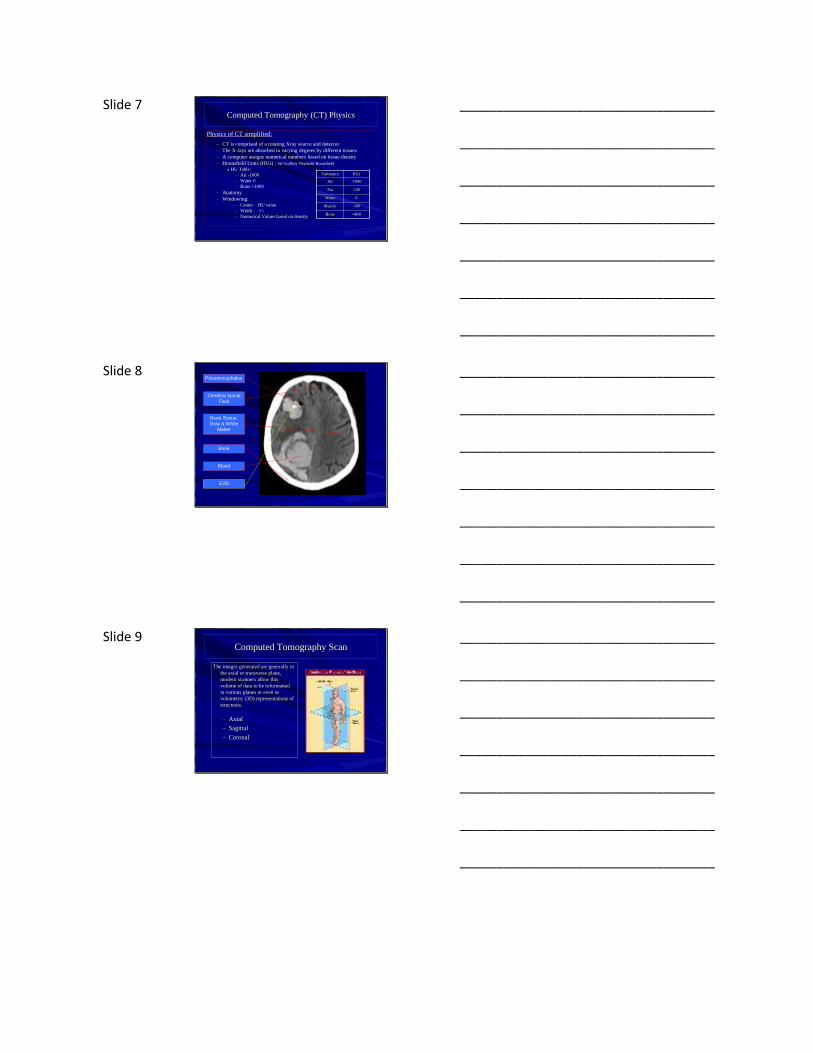
Slide 8 Pneumocephalus
Cerebral Spinal
Fluid
Brain Tissue:
Gray & White
Matter
Bone
Blood
EVD
___________________________________
___________________________________
___________________________________
___________________________________
___________________________________
___________________________________
___________________________________
Slide 9 Computed Tomography Scan
The images generated are generally in
the axial or transverse plane,
modern scanners allow this
volume of data to be reformatted
in various planes or even as
volumetric (3D) representations of
structures.
– Axial
– Sagittal
– Coronal
___________________________________
___________________________________
___________________________________
___________________________________
___________________________________
___________________________________
___________________________________
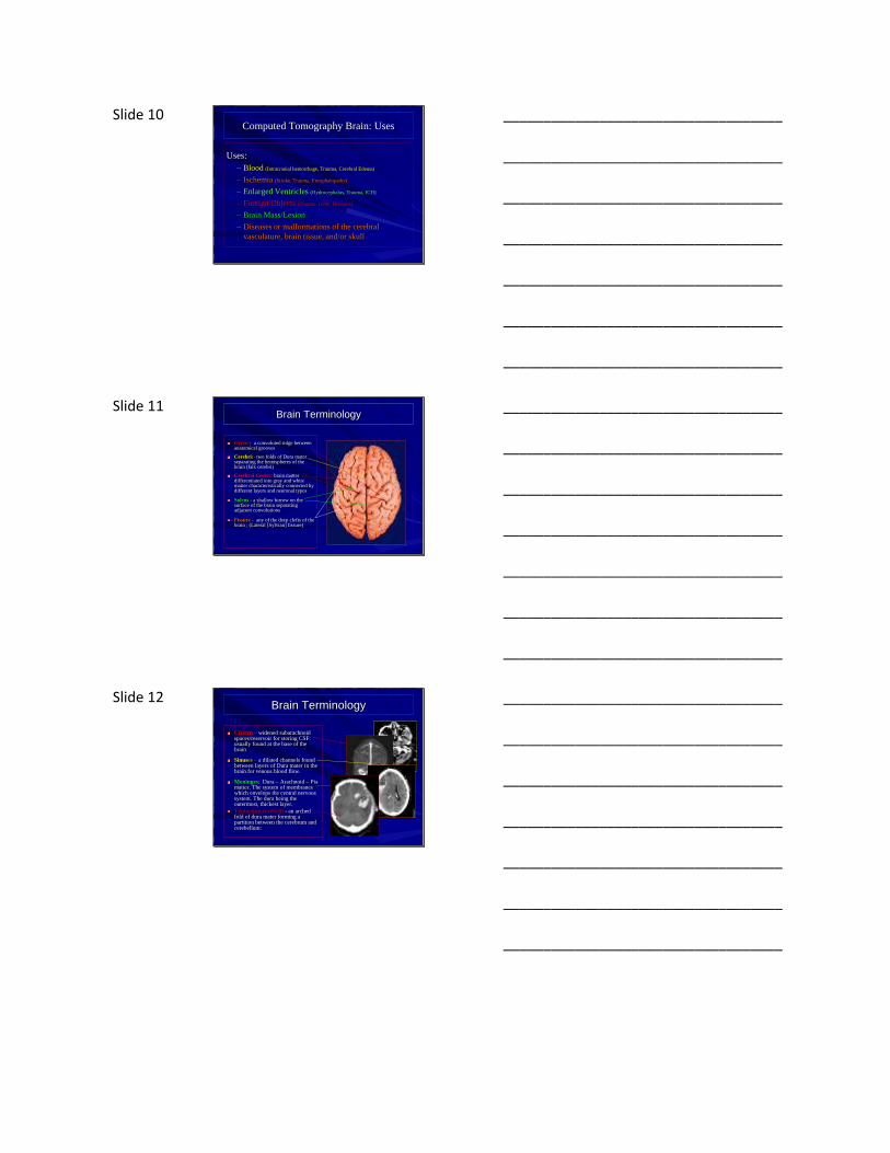
Slide 10 Computed Tomography Brain: Uses
Uses:
– Blood (Intracranial hemorrhage, Trauma, Cerebral Edema)
– Ischemia (Stroke, Trauma, Encephalopathy)
– Enlarged Ventricles (Hydrocephalus, Trauma, ICH)
– Foreign Objects (Trauma, GSW, Diseases)
– Brain Mass/Lesion
– Diseases or malformations of the cerebral
vasculature, brain tissue, and/or skull
___________________________________
___________________________________
___________________________________
___________________________________
___________________________________
___________________________________
___________________________________
Slide 11 Brain Terminology
Gyrus - a convoluted ridge between anatomical grooves
Cerebri:- two folds of Dura mater separating the hemispheres of the brain (falx cerebri)
Cerebral Cortex: brain matter differentiated into gray and white matter characteristically connected by different layers and neuronal types
Sulcus - a shallow furrow on the surface of the brain separating adjacent convolutions
Fissure - any of the deep clefts of the brain ; (Lateral [Sylvian] fissure)
___________________________________
___________________________________
___________________________________
___________________________________
___________________________________
___________________________________
___________________________________
Slide 12 Brain Terminology
Cistern – widened subarachnoid spaces/reservoir for storing CSF: usually found at the base of the brain
Sinuses – a dilated channels found between layers of Dura mater in the brain for venous blood flow.
Meninges: Dura – Arachnoid – Pia matter. The system of membranes which envelops the central nervous system. The dura being the outermost, thickest layer.
Tentorium cerebelli - an arched fold of dura mater forming a partition between the cerebrum and cerebellum:
___________________________________
___________________________________
___________________________________
___________________________________
___________________________________
___________________________________
___________________________________

Slide 13 Brain Terminology
Choroid Plexus - is the area on the ventricles of the brain where cerebrospinal fluid is produced by modified ependymal cells.
Ventricular System - A system of communicating cavities in the brain that are continuous with the central canal of the spinal cord providing a pathway for CSF
Ganglia: the "basal ganglia" is a group of nuclei interconnected with the cerebral cortex, thalamus and brainstem
___________________________________
___________________________________
___________________________________
___________________________________
___________________________________
___________________________________
___________________________________
Slide 14 Brain Terminology
Insula –(Insular Cortex) considered the 5th lobe of the cortex which separates the temporal lobe the parietal lobe dorsally.
Hemisphere – left and right: Dominant vs Nondominant
Lobes – purely anatomical classification of areas of the brain related to different brain functions. Frontal lobe, Parietal lobe, Temporal lobe, Occipital lobe, Insula, and Cerebellum.
Regions – Functional and cognitive areas of the brain as related to function: Sensorimotor Area, Hindbrain, Midbrain, Forebrain, & Neural pathways.
___________________________________
___________________________________
___________________________________
___________________________________
___________________________________
___________________________________
___________________________________
Slide 15 Anatomical Terminology
Posterior – Toward the back – situated behind; dorsal.
Anterior – Before or in front of – the abdominal side of the body.
Dorsal – going from the belly to the spinal column.
Ventral – going from the spinal column to the belly.
Caudal – Inferior in position – towards the tail.
Cephalad – Superior in position –towards the head/top.
Inferior – Beneath – below – the undersurface.
Superior – Higher than – Situated above.
Medial – Pertaining to the middle – Nearer the medial plane.
Lateral – Pertaining to the side – away from the mid-plane.
Proximal/Distal - from tip of an appendage (distal) to where it joins the body (proximal).
___________________________________
___________________________________
___________________________________
___________________________________
___________________________________
___________________________________
___________________________________
Slide 16 The Homunculus
___________________________________
___________________________________
___________________________________
___________________________________
___________________________________
___________________________________
___________________________________
Slide 17 The Homunculus
___________________________________
___________________________________
___________________________________
___________________________________
___________________________________
___________________________________
___________________________________
Slide 18
___________________________________
___________________________________
___________________________________
___________________________________
___________________________________
___________________________________
___________________________________
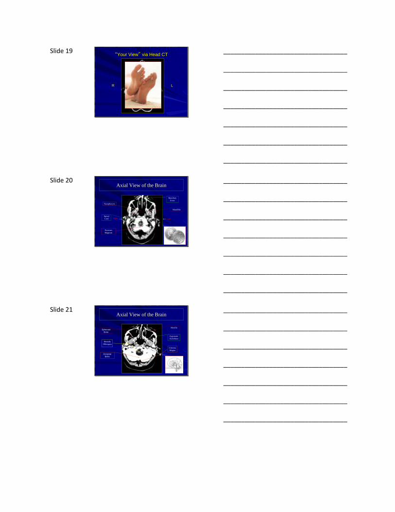
Slide 19 “Your View” via Head CT
LR
___________________________________
___________________________________
___________________________________
___________________________________
___________________________________
___________________________________
___________________________________
Slide 20 Axial View of the Brain
Right Left
Foramen
Magnum
Spinal
Cord
Nasopharynx
Maxillary
Sinus
Mandible
___________________________________
___________________________________
___________________________________
___________________________________
___________________________________
___________________________________
___________________________________
Slide 21 Axial View of the Brain
Occipital
Bone
Sphenoid
Bone
Zygomatic
Arch/Bone
Cisterna
Magna
Medulla
Oblongata
Maxilla
___________________________________
___________________________________
___________________________________
___________________________________
___________________________________
___________________________________
___________________________________

Slide 22 Axial View of the Brain
(R) Vertebral
Artery
Cerebellum
4th
Ventricle
External
Acoustic Meatus
Nasal
Septum
Cerebellar
Tonsil
___________________________________
___________________________________
___________________________________
___________________________________
___________________________________
___________________________________
___________________________________
Slide 23 Axial View of the Brain
Fourth
Ventricle
Medulla
oblongata Basilar
Artery
Posterior
Inferior lobe
cerebellum
Mastoid Air
Cells
___________________________________
___________________________________
___________________________________
___________________________________
___________________________________
___________________________________
___________________________________
Slide 24 Axial View of the Brain
Cerebellum
Pons/Cerebral
Peduncles
Cerebellopontine
cistern
Basilar
Artery
Cerebellar
VermisExternal
Auditory
Canal
Cerebellar
Tonsil
___________________________________
___________________________________
___________________________________
___________________________________
___________________________________
___________________________________
___________________________________

Slide 25 Axial View of the Brain
Occipital Bone
Pons
(Metencephalon)
Global
Socket
Cerebellar
Hemisphere
___________________________________
___________________________________
___________________________________
___________________________________
___________________________________
___________________________________
___________________________________
Slide 26 Axial View of the Brain
Frontal Tip
Temporal
Lobe
4th
Ventricle
Suprasellar
CisternTemporal
Bone
Ear
Tentorium
Cerebelli
Eye Orbits
___________________________________
___________________________________
___________________________________
___________________________________
___________________________________
___________________________________
___________________________________
Slide 27 Axial View of the Brain
Basilar
Artery
Temporal
Lobe
Cerebellopontine
Cistern
Occipital Lobe
Globes
Cerebellum
___________________________________
___________________________________
___________________________________
___________________________________
___________________________________
___________________________________
___________________________________

Slide 28 Axial View of the Brain
Tentorium
Cerebelli
Pons
Temporal
Lobe
Eyeball
Lateral Rectus
Muscle
Lens
___________________________________
___________________________________
___________________________________
___________________________________
___________________________________
___________________________________
___________________________________
Slide 29 Axial View of the Brain
Temporal Pole
Lateral ventricle
Basilar
Artery Medial Temporal
Lobes
Occipital
Lobe
Cerebral
Peduncles
___________________________________
___________________________________
___________________________________
___________________________________
___________________________________
___________________________________
___________________________________
Slide 30 Axial View of the Brain
Hippocampus
Temporal Pole of
Lateral Ventricle
Frontal
Sinus
Optic Tract
Sylvian
Fissure
Uncus
___________________________________
___________________________________
___________________________________
___________________________________
___________________________________
___________________________________
___________________________________
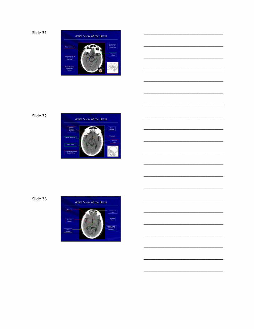
Slide 31 Axial View of the Brain
Pons/ Cerebral
Peduncles
Midbrain
Cingulate
GyrusTemporal Horns of
the Lateral
Ventricles
Hippocampus
Gyrus rectus:
Base of the
Frontal Lobe
___________________________________
___________________________________
___________________________________
___________________________________
___________________________________
___________________________________
___________________________________
Slide 32 Axial View of the Brain
Transverse/Superior
Sagittal Sinus
Lateral Ventricles
Third
Ventricle
Amygdala
Lateral
Fissure
(Sylvian)
Temporal
Lobe
Falx Cerebri
___________________________________
___________________________________
___________________________________
___________________________________
___________________________________
___________________________________
___________________________________
Slide 33 Axial View of the Brain
General area of Tentorium of Cerebellum
Thalamic
Region
Third
Ventricle
Putamen
Choroid
Plexus
Orbitofrontal
Cortex
___________________________________
___________________________________
___________________________________
___________________________________
___________________________________
___________________________________
___________________________________
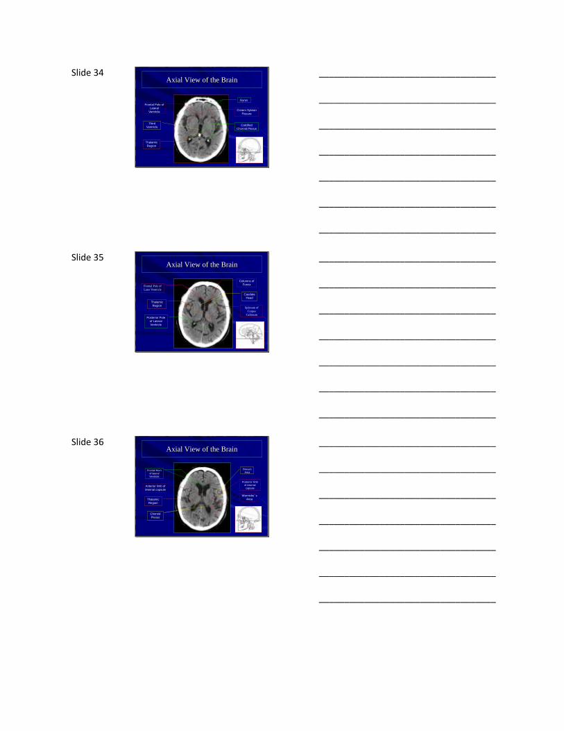
Slide 34 Axial View of the Brain
Calcified
Choroid Plexus
Gyrus
Thalamic
Region
Third
Ventricle
Frontal Pole of
Lateral
VentricleCistern Sylvian
Fissure
___________________________________
___________________________________
___________________________________
___________________________________
___________________________________
___________________________________
___________________________________
Slide 35 Axial View of the Brain
Thalamic
Region
Columns of
Fornix
Posterior Pole
of Lateral
Ventricle
Caudate
Head
Frontal Pole of
Later Ventricle
Splenum of
Corpus
Callosum
___________________________________
___________________________________
___________________________________
___________________________________
___________________________________
___________________________________
___________________________________
Slide 36 Axial View of the Brain
Broca's
Area
Choroid
Plexus
Thalamic
Region
Anterior limb of
internal capsule
Frontal Horn
of lateral
Ventricle
Posterior limb
of internal
capsule
Wernicke’s
Area
___________________________________
___________________________________
___________________________________
___________________________________
___________________________________
___________________________________
___________________________________
Slide 37 Axial View of the Brain
Superior
Sagittal
Sinus
Septum
Parietal
Lobe
Body of
Lateral
Ventricles
Frontal
Lobe
Insular
Cortex
Occipital
Lobe
___________________________________
___________________________________
___________________________________
___________________________________
___________________________________
___________________________________
___________________________________
Slide 38 Axial View of the Brain
Falx
Cerebri
Corpus
callosum
Precentral
Gyrus
Central
Sulcus
Postcentral
Gyrus
___________________________________
___________________________________
___________________________________
___________________________________
___________________________________
___________________________________
___________________________________
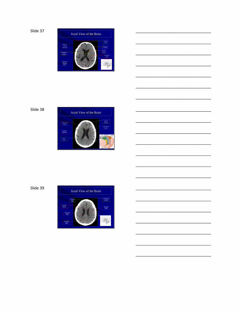
Slide 39 Axial View of the Brain
Frontal lobes
Parietal Lobe
Temporal lobe
Occipital Lobe
Central
sulcus
Cingulate
sulcus
___________________________________
___________________________________
___________________________________
___________________________________
___________________________________
___________________________________
___________________________________

Slide 40 Axial View of the Brain
Central
Sulcus
Postcentral
Gyrus
Occipital
Gyrus
Precentral Gyrus
Occipital Bone
Superior
Sagittal Sinus
Lateral
Ventricle body
___________________________________
___________________________________
___________________________________
___________________________________
___________________________________
___________________________________
___________________________________
Slide 41 Axial View of the Brain
Central
Sulcus
Superior sagittal sinus
Superior Frontal Gyrus
Post central
Gyrus
Precentral
GyrusIntraparietal
Sulcus
___________________________________
___________________________________
___________________________________
___________________________________
___________________________________
___________________________________
___________________________________
Slide 42 Axial View of the Brain
___________________________________
___________________________________
___________________________________
___________________________________
___________________________________
___________________________________
___________________________________
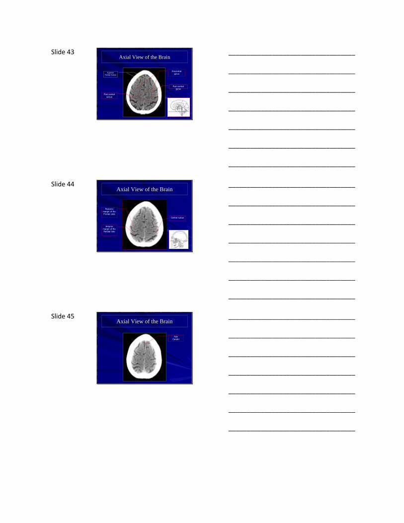
Slide 43 Axial View of the Brain
Post central sulcus
Superior Frontal Sulcus
Precentral gyrus
Post central gyrus
___________________________________
___________________________________
___________________________________
___________________________________
___________________________________
___________________________________
___________________________________
Slide 44 Axial View of the Brain
Central sulcus
Posterior margin of the Frontal Lobe
Anterior margin of the Parietal lobe
___________________________________
___________________________________
___________________________________
___________________________________
___________________________________
___________________________________
___________________________________
Slide 45 Axial View of the Brain
Falx Cerebri
___________________________________
___________________________________
___________________________________
___________________________________
___________________________________
___________________________________
___________________________________
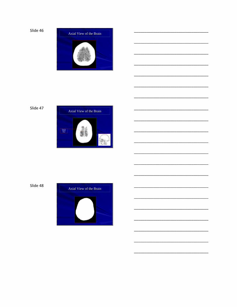
Slide 46 Axial View of the Brain
___________________________________
___________________________________
___________________________________
___________________________________
___________________________________
___________________________________
___________________________________
Slide 47 Axial View of the Brain
Superior Sagittal Sinus
___________________________________
___________________________________
___________________________________
___________________________________
___________________________________
___________________________________
___________________________________
Slide 48 Axial View of the Brain
___________________________________
___________________________________
___________________________________
___________________________________
___________________________________
___________________________________
___________________________________
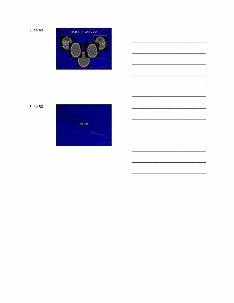
Slide 49 Head CT: Bone View
___________________________________
___________________________________
___________________________________
___________________________________
___________________________________
___________________________________
___________________________________
Slide 50
The End
___________________________________
___________________________________
___________________________________
___________________________________
___________________________________
___________________________________
___________________________________